Human Papillomavirus Testing in Head and Neck …...cell carcinomas (SCCs) in these patients tend to...
39
CAP Laboratory Improvement Programs Human Papillomavirus Testing in Head and Neck Carcinomas Guideline From the College of American Pathologists James S. Lewis Jr, MD; Beth Beadle, MD, PhD; Justin A. Bishop, MD; Rebecca D. Chernock, MD; Carol Colasacco, MLIS, SCT(ASCP); Christina Lacchetti, MHSc; Joel Todd Moncur, MD, PhD; James W. Rocco, MD, PhD; Mary R. Schwartz, MD; Raja R. Seethala, MD; Nicole E. Thomas, MPH, CT(ASCP) CM ; William H. Westra, MD; William C. Faquin, MD, PhD Context.—Human papillomavirus (HPV) is a major cause of oropharyngeal squamous cell carcinomas, and HPV (and/or surrogate marker p16) status has emerged as a prognostic marker that significantly impacts clinical management. There is no current consensus on when to test oropharyngeal squamous cell carcinomas for HPV/p16 or on which tests to choose. Objective.—To develop evidence-based recommenda- tions for the testing, application, interpretation, and reporting of HPV and surrogate marker tests in head and neck carcinomas. Design.—The College of American Pathologists con- vened a panel of experts in head and neck and molecular pathology, as well as surgical, medical, and radiation oncology, to develop recommendations. A systematic review of the literature was conducted to address 6 key questions. Final recommendations were derived from strength of evidence, open comment period feedback, and expert panel consensus. Results.—The major recommendations include (1) test- ing newly diagnosed oropharyngeal squamous cell carci- noma patients for high-risk HPV, either from the primary tumor or from cervical nodal metastases, using p16 immunohistochemistry with a 70% nuclear and cytoplas- mic staining cutoff, and (2) not routinely testing non- squamous oropharyngeal carcinomas or nonoropharyngeal carcinomas for HPV. Pathologists are to report tumors as HPV positive or p16 positive. Guidelines are provided for testing cytologic samples and handling of locoregional and distant recurrence specimens. Conclusions.—Based on the systematic review and on expert panel consensus, high-risk HPV testing is recom- mended for all new oropharyngeal squamous cell carcino- ma patients, but not routinely recommended for other head and neck carcinomas. (Arch Pathol Lab Med. 2018;142:559–597; doi: 10.5858/ arpa.2017-0286-CP) T ranscriptionally active human papillomavirus (HPV) has been identified as an important cause of oropharyngeal carcinoma. 1–5 Human papillomavirus–positive oropharyn- geal squamous cell carcinoma (OPSCC) has shown a significant increase in incidence during the past several decades, in contrast to conventional smoking- and alcohol- related head and neck squamous cell carcinoma (HNSCC), which has decreasing incidence. 1,5 The Centers for Disease Control and Prevention estimates that there are more than 16 000 cases of HPV-positive OPSCC per year in the United States. 6 These represent between 60% and 80% of all OPSCCs in the United States and Canada. Rates in many northern European countries also seem to be high, whereas rates in other parts of Europe are closer to 15% to 30%. Human papillomavirus–positive OPSCC rates are more variable in other continents but also appear to be substantially lower than for North America. For example, in India, rates may be less than 5%, and another large study found HPV-positive OPSCC rates of 16% across Europe, 36% in Central and South America, and 17% in Asia. 7–10 Patients with HPV-positive OPSCC tend to be younger, former- or nonsmokers, and male, with risk factors for exposure to high-risk HPV (HR-HPV). 2,11–13 The squamous Accepted for publication October 23, 2017. Published as an Early Online Release December 18, 2017. Supplemental digital content is available for this article at www. archivesofpathology.org in the May 2018 table of contents. From the Department of Pathology, Microbiology, and Immunol- ogy, Vanderbilt University Medical Center, Nashville, Tennessee (Dr Lewis); the Department of Radiation Oncology, Stanford University Medical Center, Palo Alto, California (Dr Beadle); the Department of Pathology, Johns Hopkins Hospital, Baltimore, Maryland (Drs Bishop and Westra); the Department of Pathology and Immunology, Washington University School of Medicine, Saint Louis, Missouri (Dr Chernock); Surveys, the College of American Pathologists, Northfield, Illinois (Mss Colasacco and Thomas); Policy and Advocacy, American Society of Clinical Oncology, Alexandria, Virginia (Ms Lacchetti); the Department of Pathology, Walter Reed National Military Medical Center, Bethesda, Maryland (Dr Moncur); the Department of Otolaryngology–Head and Neck Surgery, Ohio State University Wexler Medical Center, Columbus (Dr Rocco); the Department of Pathology and Genomic Medicine, Houston Meth- odist Hospital, Houston, Texas (Dr Schwartz); the Department of Pathology, University of Pittsburgh Medical Center, Pittsburgh, Pennsylvania (Dr Seethala); and the Department of Pathology, Massachusetts General Hospital, Boston (Dr Faquin). Authors’ disclosures of potential conflicts of interest and author contributions are found in the Appendix at the end of this article. Reprints: James S. Lewis Jr, MD, Department of Pathology, Microbi- ology, and Immunology, Vanderbilt University Medical Center, Room 3020D, Vanderbilt University Hospital, 1211 Medical Center Dr, Nashville, TN 3723-0233 (email: [email protected]). Arch Pathol Lab Med—Vol 142, May 2018 HPV Testing Head & Neck Carcinomas: CAP Guideline—Lewis et al 559
Transcript of Human Papillomavirus Testing in Head and Neck …...cell carcinomas (SCCs) in these patients tend to...

CAP Laboratory Improvement Programs
Human Papillomavirus Testing in Head and NeckCarcinomas
Guideline From the College of American Pathologists
James S. Lewis Jr, MD; Beth Beadle, MD, PhD; Justin A. Bishop, MD; Rebecca D. Chernock, MD; Carol Colasacco, MLIS, SCT(ASCP);Christina Lacchetti, MHSc; Joel Todd Moncur, MD, PhD; James W. Rocco, MD, PhD; Mary R. Schwartz, MD; Raja R. Seethala, MD;
Nicole E. Thomas, MPH, CT(ASCP)CM; William H. Westra, MD; William C. Faquin, MD, PhD
� Context.—Human papillomavirus (HPV) is a major causeof oropharyngeal squamous cell carcinomas, and HPV(and/or surrogate marker p16) status has emerged as aprognostic marker that significantly impacts clinicalmanagement. There is no current consensus on when totest oropharyngeal squamous cell carcinomas for HPV/p16or on which tests to choose.
Objective.—To develop evidence-based recommenda-tions for the testing, application, interpretation, andreporting of HPV and surrogate marker tests in head andneck carcinomas.
Design.—The College of American Pathologists con-vened a panel of experts in head and neck and molecularpathology, as well as surgical, medical, and radiationoncology, to develop recommendations. A systematicreview of the literature was conducted to address 6 keyquestions. Final recommendations were derived from
strength of evidence, open comment period feedback,and expert panel consensus.
Results.—The major recommendations include (1) test-ing newly diagnosed oropharyngeal squamous cell carci-noma patients for high-risk HPV, either from the primarytumor or from cervical nodal metastases, using p16immunohistochemistry with a 70% nuclear and cytoplas-mic staining cutoff, and (2) not routinely testing non-squamous oropharyngeal carcinomas or nonoropharyngealcarcinomas for HPV. Pathologists are to report tumors asHPV positive or p16 positive. Guidelines are provided fortesting cytologic samples and handling of locoregional anddistant recurrence specimens.
Conclusions.—Based on the systematic review and onexpert panel consensus, high-risk HPV testing is recom-mended for all new oropharyngeal squamous cell carcino-ma patients, but not routinely recommended for otherhead and neck carcinomas.
(Arch Pathol Lab Med. 2018;142:559–597; doi: 10.5858/arpa.2017-0286-CP)
Transcriptionally active human papillomavirus (HPV) hasbeen identified as an important cause of oropharyngeal
carcinoma.1–5 Human papillomavirus–positive oropharyn-geal squamous cell carcinoma (OPSCC) has shown asignificant increase in incidence during the past severaldecades, in contrast to conventional smoking- and alcohol-related head and neck squamous cell carcinoma (HNSCC),which has decreasing incidence.1,5 The Centers for DiseaseControl and Prevention estimates that there are more than16 000 cases of HPV-positive OPSCC per year in the UnitedStates.6 These represent between 60% and 80% of allOPSCCs in the United States and Canada. Rates in manynorthern European countries also seem to be high, whereasrates in other parts of Europe are closer to 15% to 30%.Human papillomavirus–positive OPSCC rates are morevariable in other continents but also appear to besubstantially lower than for North America. For example,in India, rates may be less than 5%, and another large studyfound HPV-positive OPSCC rates of 16% across Europe,36% in Central and South America, and 17% in Asia.7–10
Patients with HPV-positive OPSCC tend to be younger,former- or nonsmokers, and male, with risk factors forexposure to high-risk HPV (HR-HPV).2,11–13 The squamous
Accepted for publication October 23, 2017.Published as an Early Online Release December 18, 2017.Supplemental digital content is available for this article at www.
archivesofpathology.org in the May 2018 table of contents.From the Department of Pathology, Microbiology, and Immunol-
ogy, Vanderbilt University Medical Center, Nashville, Tennessee (DrLewis); the Department of Radiation Oncology, Stanford UniversityMedical Center, Palo Alto, California (Dr Beadle); the Department ofPathology, Johns Hopkins Hospital, Baltimore, Maryland (Drs Bishopand Westra); the Department of Pathology and Immunology,Washington University School of Medicine, Saint Louis, Missouri(Dr Chernock); Surveys, the College of American Pathologists,Northfield, Illinois (Mss Colasacco and Thomas); Policy andAdvocacy, American Society of Clinical Oncology, Alexandria,Virginia (Ms Lacchetti); the Department of Pathology, Walter ReedNational Military Medical Center, Bethesda, Maryland (Dr Moncur);the Department of Otolaryngology–Head and Neck Surgery, OhioState University Wexler Medical Center, Columbus (Dr Rocco); theDepartment of Pathology and Genomic Medicine, Houston Meth-odist Hospital, Houston, Texas (Dr Schwartz); the Department ofPathology, University of Pittsburgh Medical Center, Pittsburgh,Pennsylvania (Dr Seethala); and the Department of Pathology,Massachusetts General Hospital, Boston (Dr Faquin).
Authors’ disclosures of potential conflicts of interest and authorcontributions are found in the Appendix at the end of this article.
Reprints: James S. Lewis Jr, MD, Department of Pathology, Microbi-ology, and Immunology, Vanderbilt University Medical Center, Room3020D, Vanderbilt University Hospital, 1211 Medical Center Dr,Nashville, TN 3723-0233 (email: [email protected]).
Arch Pathol Lab Med—Vol 142, May 2018 HPV Testing Head & Neck Carcinomas: CAP Guideline—Lewis et al 559

cell carcinomas (SCCs) in these patients tend to havesmaller primary tumors, but present with early nodalmetastases.1,3 As a group, patients with HPV-positiveOPSCC have improved clinical outcomes compared withpatients with conventional, HPV-negative HNSCC whenmanaged by similar modalities.2,11–13
Testing for HR-HPV in HNSCC has become increasinglyimportant during the past decade. Determining that anOPSCC is positive for HR-HPV (by strictly defined testing inthe correct clinical and pathologic contexts) has significantimplications for patient prognosis, and it is now integratedinto the recently updated American Joint Committee onCancer (AJCC) staging manual14; furthermore, HPV statusdetermines patient eligibility for clinical trials investigatingnew treatment regimens and modalities.15,16 In addition,determining that a metastatic SCC of unknown primary to acervical lymph node is HPV positive strongly points to theoropharynx as the site of origin, with consequences forsubsequent clinical management and treatment deci-sions.15–19 For these reasons, several organizations, includ-ing the College of American Pathologists (CAP), the RoyalCollege of Pathologists, and Cancer Care Ontario, havesupported the establishment of evidence-based guidelinesfor HR-HPV testing in HNSCC.17,20
There are many important questions about HR-HPVtesting that remain to be answered by evidence-basedguidelines, including which anatomic sites and subtypes ofHNSCC warrant HPV testing, when and how to test tissuespecimens, and what should be done with fine-needleaspiration (FNA) samples.21–26 In 2013, the CAP appointedan 11-person expert panel (EP) and a 9-person advisorypanel to address these and other related questions toformulate a comprehensive set of recommendations.
METHODS
This evidence-based guideline was developed following thestandards endorsed by the National Academy of Medicine,formerly the Institute of Medicine.27 A detailed description of themethods and a systematic review (including the quality assessmentand complete analysis of the evidence) used to create this guidelinecan be found in the supplemental digital content at www.archivesofpathology.org in the May 2018 table of contents.
Panel Composition
The CAP convened an EP consisting of members with expertise inhead and neck and molecular pathology and surgical, medical, andradiation oncology to develop the guideline. In addition, a researchmethodologist consultant served on the EP for the systematic reviewof the evidence. An advisory panel consisting of 2 patient advocates, 4pathologists, 1 medical oncologist/molecular epidemiologist, 1 radi-ation oncologist, and 1 methodologist assisted the EP. The followingorganizations provided official panel representation: the AmericanAcademy of Otolaryngology—Head and Neck Surgery Foundation,the American Society of Clinical Oncology (ASCO), and the AmericanSociety of Cytopathology.
In addition, the guideline was submitted to ASCO’s Head andNeck Guideline Advisory Group and ASCO’s Clinical PracticeGuideline Committee for review of the final manuscript. Nosuggestions for revisions were proposed, and it was agreed that theguideline should be considered for endorsement by ASCO.
Conflict of Interest Policy
In accordance with the CAP conflict of interest policy (in effectApril 2010), members of the EP disclosed all financial interests from12 months prior to appointment throughout the development ofthis guideline. Individuals were instructed to disclose anyrelationship that could be interpreted as constituting an actual,
potential, or apparent conflict. Complete disclosures of the EPmembers are listed in the Appendix. Disclosures of interest judgedby the oversight group to be conflicts are as follows: R.R.S, researchgrants, National Institutes of Health (Bethesda, Maryland);W.H.W., consultancy, Merck & Co, (Kenilworth, New Jersey).The majority of EP members (9 of 11) were assessed as having norelevant conflicts of interest. The CAP provided funding for theadministration of the project; no industry funds were used in thedevelopment of the guideline. All panel members volunteered theirtime and were not compensated for their involvement, except forthe contracted methodologist. Please see the supplemental digitalcontent for full details on the conflict of interest policy.
Objective
The scope of the panel was to develop evidence-basedrecommendations for the various methodologies and applicationsof HR-HPV testing in head and neck carcinomas. The keyquestions are listed as follows:
1. Should patients with newly diagnosed OPSCC, nonoropharyngealSCC (non-OPSCC), oropharyngeal non-SCC, nonoropharyngealnon-SCC, and cervical nodal metastatic carcinomas of unknownand/or known primary be routinely tested for HR-HPV?
2. Do relevant clinical outcomes of specific tests or testingalgorithms for HR-HPV differ based on items such as specimensize, type and length of tissue fixation, or the criteria/definitionfor a positive p16 immunohistochemistry (IHC) or in situhybridization (ISH) test?
3. For patients with OPSCC, non-OPSCC, and cervical nodalmetastatic SCC, what is the optimal method of reporting HPVtest results to best inform patients and clinicians about theclinical significance of the results (including considerationsabout uncertainty)?
4. Should patients with recurrent/persistent OPSCC, non-OPSCC,and cervical nodal metastatic SCC be routinely tested for HR-HPV?
5. Should patients with locally and/or regionally recurrentOPSCC, non-OPSCC, and cervical nodal metastatic SCC beroutinely tested for HR-HPV?
6. Should patients with distant disease be tested for HR-HPV?
Refer to the supplemental digital content for all of thesubquestions under these main key questions.
Literature Search and Collection
A comprehensive search for literature was initially performed inMEDLINE using the OvidSP interface on March 3, 2014,encompassing the publication dates of January 1, 1995, to March3, 2014. A supplemental literature search was completed inPubMed (March 26, 2014) encompassing the publication dates ofJanuary 1, 1995, to March 26, 2014. An additional literature searchwas performed using Scopus (March 29, 2014) to identify relevantarticles published between January 1, 1995, and March 29, 2014, injournals not indexed in MEDLINE. The literature search of theelectronic databases was conducted in 2 arms: the first combinedmedical subject headings and keywords to address the conceptshead and neck neoplasms, human papillomavirus (HPV), andlaboratory testing, and the second combined medical subjectheadings and keywords for the concepts head and neck neoplasms,human papillomavirus (HPV), and outcomes. The results of both armsof the search were combined and deduplicated. Limits were set forhuman studies published in English, and a publication filter wasapplied to exclude lower levels of evidence such as letters,commentaries, editorials, and case reports.
A search for gray (unindexed) literature included a review ofguideline and systematic review repository sites (eg, GuidelinesInternational Network, National Guideline Clearinghouse, Co-chrane Library, Prospero, Centre for Reviews and Dissemination),and relevant medical organizations’ Web sites to identify guide-lines, protocols, and standards. A review of meeting abstracts
560 Arch Pathol Lab Med—Vol 142, May 2018 HPV Testing Head & Neck Carcinomas: CAP Guideline—Lewis et al

published in the years 2012–2014 from pathology and oncologyorganizations and EP recommendations completed the systematicliterature review. The Ovid search was rerun on July 11, 2016, toidentify articles published since March 1, 2014, that providedinformation that would alter the recommendations in any way.
Detailed information regarding the literature search strategy canbe found in the supplemental digital content.
Inclusion and Exclusion Criteria
Practice guidelines, consensus documents, systematic reviews,meta-analyses, randomized controlled trials (RCTs), comparativestudies, reviews, case-controlled studies, case series, and evaluationstudies were eligible for inclusion.
Published studies were selected for full-text review if they meteach of the following criteria:
1. Patients with tissue or cytology aspiration material taken fromthe workup of� Oropharyngeal primaries� Cervical nodal metastasis of unknown primary� Regional or distant metastasis from known or suspected
oropharyngeal primary� Other head and neck sites (eg, sinonasal)� All carcinomas in the head and neck
2. Human studies3. Patients of all ages and either sex4. Studies published in English5. The study compared, prospectively or retrospectively, laboratory
testing methodologies or potential testing algorithms for HPVtesting
6. The study addressed one of the key questions7. The study included measurable data such as the negative
predictive value or positive predictive value, if testing method-ologies used to determine HPV status, alone and in combina-tion; negative and positive concordance across the platforms;sensitivity and specificity of individual tests; and accuracy indetermining HPV status.
Articles were excluded from the systematic review if they werenoncomparative or qualitative studies, including editorials, com-mentaries, or letters; animal studies; full-text articles not availablein English; studies that included patients with other tumor typesnot specified in the inclusion criteria; studies that did not includerelevant measurable data; and studies that did not address at leastone of the key questions.
Detailed information about the inclusion and exclusion criteria isavailable in the supplemental digital content.
Quality Assessment
An assessment of study quality was performed by a researchmethodologist for all fully published studies meeting inclusioncriteria. Studies only available in abstract form did not undergoformal quality assessment. Formal quality assessment involveddetermining the risk of bias by assessing key indicators, based onstudy design and methodologic rigor. Refer to the supplementaldigital content for the definitions of ratings for strength of evidence(Supplemental Table 1) and for the quality assessment results.
Assessing the Strength of Recommendations
Development of recommendations required that the panelreview the identified evidence and make a series of key judgments.Grades for strength of recommendations were developed by theCAP Pathology and Laboratory Quality Center and are described inTable 1.
Guideline Revision
This guideline will be reviewed every 4 years, or earlier in theevent of publication of substantive and high-quality evidence thatcould potentially alter the original guideline recommendations. If
necessary, the entire panel will reconvene to discuss potentialchanges. When appropriate, the panel will recommend revision ofthe guideline to the CAP for review and approval.
Disclaimer
The CAP developed the Pathology and Laboratory QualityCenter as a forum to create and maintain evidence-based practiceguidelines and consensus statements. Practice guidelines andconsensus statements reflect the best available evidence and expertconsensus supported in practice. They are intended to assistphysicians and patients in clinical decision making and to identifyquestions and settings for further research. With the rapid flow ofscientific information, new evidence may emerge between the timea practice guideline or consensus statement is developed and whenit is published or read. Guidelines and statements are notcontinually updated and may not reflect the most recent evidence.Guidelines and statements address only the topics specificallyidentified therein and are not applicable to other interventions,diseases, or stages of diseases. Furthermore, guidelines andstatements cannot account for individual variation among patientsand cannot be considered inclusive of all proper methods of care orexclusive of other treatments. It is the responsibility of the treatingphysician or other health care provider, relying on independentexperience and knowledge, to determine the best course oftreatment for the patient. Accordingly, adherence to any practiceguideline or consensus statement is voluntary, with the ultimatedetermination regarding its application to be made by the physicianin light of each patient’s individual circumstances and preferences.The CAP makes no warranty, express or implied, regardingguidelines and statements and specifically excludes any warrantiesof merchantability and fitness for a particular use or purpose. TheCAP assumes no responsibility for any injury or damage to personsor property arising out of or related to any use of this statement orfor any errors or omissions.
The views expressed in this document do not reflect the officialpolicy of the Department of the Army, the Department of the Navy,the Department of the Air Force, the Department of Defense, or theUS government. The identification of specific products or scientificinstruments does not constitute endorsement or implied endorse-ment on the part of the Department of Defense or any componentagency.
RESULTS
Of the 2803 unique studies identified in the systematicreview, 503 met inclusion criteria and underwent dataextraction. One hundred fifty-seven of these studies madeup the evidentiary base and informed the guidelinestatements (recommendations). The vast majority werepublished, peer-reviewed articles, but 31 studies werepublished only in abstract form. All 157 underwent dataextraction and qualitative analysis. Abstracts included in the157 studies reported at least partial data. Those that did notreport data for any of the outcomes of interest wereexcluded. Data from abstracts were used only in concertwith peer-reviewed data, as they added support forrecommendation statements. Abstract data alone were notused in the formulation of recommendations.
The EP met 16 times through Web-based meeting forumsfrom November 22, 2013, through September 21, 2016.Additional work was completed via electronic mail. The EPmet in person February 8 and 9, 2014, to formally initiate theproject, and again April 9, 2016, to review the evidence todate and draft recommendations.
A public comment period was held from July 18 to August8, 2016, on the CAP Web site. Fourteen draft recommen-dations, 2 demographic questions, and 3 questions aboutfeasibility were posted for feedback.
Arch Pathol Lab Med—Vol 142, May 2018 HPV Testing Head & Neck Carcinomas: CAP Guideline—Lewis et al 561

‘‘Agree’’ and ‘‘disagree’’ responses were captured forevery proposed recommendation. In addition, 269 writtencomments were captured. Seven draft recommendationsachieved more than 90% agreement, 6 achieved between80% and 90% agreement, and 1 received 57% agreement.Each EP member was assigned 1 or 2 draft recommenda-tions for which to review the public comments and topresent to the panel for group discussion. After consider-ation of the comments, 6 draft recommendations weremaintained with the original language and 7 were revised.Resolution of all changes was obtained by unanimousconsensus of the panel members using nominal grouptechnique (rounds of subsequent teleconference webinarsand email discussion). Final EP recommendations wereapproved by a formal vote. The panel considered laboratoryefficiency and feasibility throughout the entire process,although neither cost nor cost-effectiveness analyses wereperformed. A description of the benefits and harms ofimplementing the guideline statements is included in thesupplemental digital content.
An independent review panel, masked to the EP andvetted through the conflict of interest process, provided finalapproval on behalf of the CAP Council on Scientific Affairs.In addition, the guideline was submitted to ASCO’s Headand Neck Guideline Advisory Group and ASCO’s ClinicalPractice Guideline Committee for review of the finalmanuscript. No suggestions for revisions were proposedand it was agreed that the guideline should be consideredfor endorsement by ASCO. The final recommendations aresummarized in Table 2, and an algorithmic approach for theworkup of patient specimens is provided in Figure 1.
GUIDELINE STATEMENTS
Statement 1.—Strong Recommendation.—Pathologistsshould perform HR-HPV testing on all patients with newlydiagnosed OPSCC, including all histologic subtypes. Thistesting may be performed on the primary tumor or on aregional lymph node metastasis when the clinical findingsare consistent with an oropharyngeal primary.
The strength of evidence is convincing to support thisguideline statement.
The evidentiary base supporting this recommendationcomprised 110 studies, of which 1 was a meta-analysis,28 8were subgroup analyses from RCTs,11,29–35 77 were obser-vational studies,1,10,36–111 and 24 were studies only publishedin abstract form.112–135 The meta-analysis received a qualityscore of 7 out of a possible 11 points, and all supporting RCTdata had a risk of bias determined to be moderate. The
supporting observational studies ranged from low tomoderate risk of bias, with the exception of one study thatwas assessed to have a high risk of bias.1 This study was aretrospective cohort with retrospective data collection andindustry sponsorship. None of the other studies were foundto have methodologic flaws that would raise concerns aboutthe findings. Refer to Supplemental Table 2 in thesupplemental digital content for the quality assessmentresults for studies included in the statement 1 evidentiarybase.
Breaking from a tradition that has broadly grouped allcarcinomas arising from the oral and oropharyngeal subsitesas oral cancer, these guidelines maintain a sharp distinctionbetween those carcinomas arising in the oropharynx andthose arising in the oral cavity proper. Testing for thepresence of HPV must be guided by a familiarity with headand neck anatomy, including those structures that define theoral cavity as separate from the oropharynx (Figure 2). Theoral cavity proper comprises the lips, gingiva, retromolartrigone, hard palate, buccal mucosa, mobile tongue, andfloor of the mouth, whereas the oropharynx comprises thepalatine tonsils, soft palate, base of tongue (posterior to thecircumvallate papillae), and lateral and posterior pharyngealwalls. Oropharyngeal tonsillar structures (ie, lingual andpalatine tonsils), particular hot spots for HPV-relatedcarcinogenesis, are present in the oropharynx, but not inthe oral cavity.
Oropharyngeal SCCs with transcriptionally active HR-HPV represent a unique type of HNSCC. These HPV-positive OPSCCs have risk-factor, demographic, morpho-logic, molecular, and clinical profiles that stand apart fromother HNSCCs.
Human papillomavirus status of a primary or metastaticOPSCC may have diagnostic, staging, and even therapeuticimplications. Currently, however, the call for routine HPVtesting reflects its standing as a powerful prognosticindicator for patients with OPSCC. The literature over-whelmingly supports the conclusion that HPV status is animportant and independent predictor of overall and disease-specific survival for patients with OPSCC. The survivalbenefit of HPV-positive OPSCC is maintained across nearlyall studies, despite significant heterogeneity in patientpopulations, sample size, methods of HPV detection, tumorstage, tumor treatment, comorbidity, and inclusion ofvarious other prognostic factors in the analysis. In largeprospective studies where patient populations with OPSCCare uniformly staged and treated, significant reduction inrisk of progression and disease-related death is confirmedfor HPV-positive tumors.11,12,31,32,35
Table 1. Grades for Strength of Recommendationsa
Designation Recommendation Rationale
Strong recommendation Recommend for or against aparticular practice (Can include‘‘must’’ or ‘‘should’’)
Supported by convincing (high) or adequate (intermediate) qualityof evidence and clear benefit that outweighs any harms
Recommendation Recommend for or against aparticular practice (Can include‘‘should’’ or ‘‘may’’)
Some limitations in quality of evidence (adequate [intermediate]or inadequate [low]), balance of benefits and harms, values, orcosts but panel concludes that there is sufficient evidence and/or benefit to inform a recommendation
Expert consensus opinion Recommend for or against aparticular practice (Can include‘‘should’’ or ‘‘may’’)
Serious limitations in quality of evidence (inadequate [low] orinsufficient), balance of benefits and harms, values, or costs, butpanel consensus is that a statement is necessary
No recommendation No recommendation for or againsta practice
Insufficient evidence or agreement of the balance of benefits andharms, values, or costs to provide a recommendation
a Derived from Andrews et al.262
562 Arch Pathol Lab Med—Vol 142, May 2018 HPV Testing Head & Neck Carcinomas: CAP Guideline—Lewis et al

Figure 1. High-risk human papillomavirus (HR-HPV) testing in head and neck squamous cell carcinomas (SCCs). Abbreviations: IHC,immunohistochemistry; OP, oropharyngeal. 1Consider HR-HPV–specific testing for equivocal p16 results (50%–70% nuclear and cytoplasmicstaining). 2May also be reported as p16 negative with a comment specifying that the tumor is very likely HPV negative. 3May also be reported as p16positive with a comment specifying that the tumor is very likely HPV positive. 4HR-HPV may be indicated in patients where the clinical suspicion foran HPV-positive SCC is high. 5Consider Epstein-Barr encoding region (EBER) in situ hybridization for Epstein-Barr virus for the rare metastaticnonkeratinizing squamous cell carcinoma that is HR-HPV negative. 6Include comment, ‘‘Likely oropharyngeal primary.’’
Arch Pathol Lab Med—Vol 142, May 2018 HPV Testing Head & Neck Carcinomas: CAP Guideline—Lewis et al 563

Testing for HR-HPV should be performed on all OPSCCsregardless of histologic type. The morphologic spectrum ofHPV-positive OPSCC includes variants with papillary,adenosquamous, lymphoepithelioma-like, sarcomatoid,and basaloid features. Morphologic variation does not seemto influence clinical behavior, and it does not abrogate theneed for HPV testing.136–138
In the open comment period, of the 168 respondents,93.45% (n ¼ 157) agreed with the recommendation and5.36% (n ¼ 9) disagreed. There were 22 written comments.Most of these comments were directed at the method ofHPV testing and were, accordingly, taken into considerationin the final drafting of statement 2. Others reflectedconfusion about the anatomic definition of the oropharynxand its distinction from the oral cavity. To highlight thisimportant distinction, a brief description of the anatomy ofthe oropharynx and oral cavity has been provided.
Statement 2.—Recommendation.—For oropharyngeal tis-sue specimens (ie, noncytology), pathologists shouldperform HR-HPV testing by surrogate marker p16 IHC.Additional HPV-specific testing may be done at thediscretion of the pathologist and/or treating clinician, or inthe context of a clinical trial.
The strength of evidence is adequate.Sixty-seven studies (5 RCTs,11,29,32,34,35 48 observational
studies,* and 14 studies published in abstract form†) makeup the evidentiary base for statement 2. Of these studies, 31reported on laboratory outcomes of interest (Table 3) and 51on clinical outcomes (Table 4). The risk of bias assessment ofthe majority of the included studies ranged from low to
Table 2. Summary of Guideline Statements
Guideline StatementStrength of
Recommendation
1. Pathologists should perform HR-HPV testing on all patients with newly diagnosed OPSCC,including all histologic subtypes. This testing may be performed on the primary tumor or on aregional lymph node metastasis when the clinical findings are consistent with an oropharyngealprimary.
Strong recommendation
2. For oropharyngeal tissue specimens (ie, noncytology), pathologists should perform HR-HPVtesting by surrogate marker p16 IHC. Additional HPV-specific testing may be done at thediscretion of the pathologist and/or treating clinician, or in the context of a clinical trial.
Recommendation
3. Pathologists should not routinely perform HR-HPV testing on patients with nonsquamouscarcinomas of the oropharynx.
Expert consensus opinion
4. Pathologists should not routinely perform HR-HPV testing on patients with nonoropharyngealprimary tumors of the head and neck.
Recommendation
5. Pathologists should routinely perform HR-HPV testing on patients with metastatic SCC ofunknown primary in a cervical upper or mid jugular chain lymph node. An explanatory note onthe significance of a positive HPV result is recommended.
Recommendation
6. For tissue specimens (ie, noncytology) from patients presenting with metastatic SCC of unknownprimary in a cervical upper– or mid–jugular chain lymph node, pathologists should perform p16IHC.
Expert consensus opinion
Note: Additional HR-HPV testing on p16-positive cases should be performed for tumorslocated outside of level II or III (nonroutine testing) in the neck and/or for tumors withkeratinizing morphology.
7. Pathologists should perform HR-HPV testing on head and neck FNA SCC samples from allpatients with known OPSCC not previously tested for HR-HPV, with suspected OPSCC, or withmetastatic SCC of unknown primary.
Expert consensus opinion
Note: No recommendation is made for or against any specific testing methodology for HR-HPV testing in FNA samples. If the result of HR-HPV testing on the FNA sample is negative,testing should be performed on tissue if it becomes available. If pathologists use cytologysamples for p16 IHC testing, they should validate the criteria (ie, cutoff) for a positive result.
8. Pathologists should report p16 IHC positivity as a surrogate for HR-HPV in tissue specimens (ie,noncytology) when there is at least 70% nuclear and cytoplasmic expression with at leastmoderate to strong intensity.
Expert consensus opinion
9. Pathologists should not routinely perform low-risk HPV testing on patients with head and neckcarcinomas.
Expert consensus opinion
10. Pathologists should not repeat HPV testing on patients with locally recurrent, regionallyrecurrent, or persistent tumor if primary tumor HR-HPV status has already been established. Ifinitial HR-HPV status was never assessed or results are unknown, testing is recommended. HPVtesting may be performed on a case-by-case basis for diagnostic purposes if there is uncertaintyregarding whether the tumor in question is a recurrence or a new primary SCC.
Expert consensus opinion
11. Pathologists should not routinely perform HR-HPV testing on patients with distant metastases ifprimary tumor HR-HPV status has been established. HPV testing may be performed on a case-by-case basis for diagnostic purposes if there is uncertainty regarding whether the tumor inquestion is a metastasis or a new primary SCC.
Expert consensus opinion
12. Pathologists should report primary OPSCCs that test positive for HR-HPV or its surrogate markerp16 as HPV positive and/or p16 positive.
Expert consensus opinion
13. Pathologists should not provide a tumor grade or differentiation status for HPV-positive/p16-positive OPSCCs.
Expert consensus opinion
14. Pathologists should not alter HR-HPV testing strategy based on patient smoking history. Expert consensus opinion
Abbreviations: FNA, fine-needle aspiration; HPV, human papillomavirus; HR-HPV, high-risk HPV; IHC, immunohistochemistry; OPSCC,oropharyngeal SCC; SCC, squamous cell carcinoma.
* References 1, 36, 38, 42–44, 47–54, 56, 57, 59, 64, 65, 67, 69–78,80, 81, 83, 84, 87, 89, 91, 92, 96–101, 104, 106, 108, 110, 111.
† References 112, 116, 118–120, 122, 125–127, 129–131, 134, 135.
564 Arch Pathol Lab Med—Vol 142, May 2018 HPV Testing Head & Neck Carcinomas: CAP Guideline—Lewis et al

moderate, with only 2 studies assessed as high1,68 because ofindustry funding. Although the vast majority of includedstudies were retrospective cohorts, and the inherentlimitations of retrospective designs should be taken intoconsideration, the collection of data did occur prospectivelyin all but 2 studies.1,38 None of the other studies were foundto have methodologic flaws that would call into question thestudy findings. Refer to Supplemental Table 3 in thesupplemental digital content for the quality assessmentresults for all studies included in the statement 2 evidentiarybase.
Because the literature strongly supports that HR-HPVstatus is independently prognostic in OPSCC and that itshould be routinely assessed, there is a need for consistencyin clinical practice. Which test or combination of HR-HPVtests to perform is one of the more controversial issues inhead and neck pathology. There are numerous HPV-specifictests, as well as the surrogate markers p16 IHC andhematoxylin-eosin morphology to consider. Although it isideal to have a reference or standard criterion test, thecurrent literature does not clearly support one. The testshould be the one that best stratifies patient survivaloutcomes while also being practical and inexpensive. p16IHC is the test the EP considers to best fit that role. p16 ismarkedly overexpressed in tumor cells with transcriptionallyactive HR-HPV because the viral E7 oncoprotein destabi-lizes pRb, functionally removing suppression of p16expression and allowing tumor cells with high p16 levelsto bypass pRb-dependent cell cycle arrest. The result ismarked overexpression of p16, making it an excellentsurrogate marker of viral infection in the correct con-text.139,140 Based on abundant literature on p16 IHC as anindependent predictor of improved patient prognosis inOPSCC,‡ and on its widespread availability, ease andreproducibility of interpretation,53 and excellent perfor-
mance on small specimen samples such as small biopsiesand tissue microarray punches,11,89,142 the EP recommendsthat p16 testing be performed. Many consider the detectionof HR-HPV E6 and E7 messenger RNA (mRNA) by ISH asthe gold standard.89,143,144 Although this is an excellent test,and perhaps even the ideal test from a purely scientificperspective, it isn’t widely available for clinical use, is muchmore expensive than p16, and is more technically challeng-ing to perform, and the data do not show statistically betterperformance than p16 IHC alone in OPSCC. Because p16 isonly a surrogate marker for HR-HPV, and its overexpressionis not always associated with the presence of HR-HPV,53,75,81,89 as practice changes and HPV-specific testssuch as RNA ISH become more widely available clinically,the latter may become the recommended test in the future.
For studies analyzing p16 IHC alone as a prognosticmarker, the majority found it to be a marker of favorableoutcome in multivariate analysis with univariate hazardratios for death between 0.2 and 0.5§ for overall, disease-free, and/or disease-specific survival compared with p16-negative patients. Several prospective and randomizedcontrolled studies showed p16 IHC to be strongly prognos-tic alone as well.11,12,32,145 In many of the studies, dataextraction and summarization were complicated by p16results, rather than being analyzed in isolation, beingcombined with results of HPV-specific test(s) for analysisand data reporting. Correlation rates between p16 IHC andHPV-specific tests were generally high, and best for HPVmRNA tests such as reverse transcriptase polymerase chainreaction and ISH.||
In addition to the aforementioned studies, some addi-tional studies not included in our systematic review supportthis recommendation. Sedghizadeh et al141 performed a
Figure 2. Normal anatomy of the orophar-ynx, including specific features of the palatinetonsils and base of tongue.
‡ References 10–12, 29, 32, 70, 81, 141.
§ References 30, 32, 34, 35, 42, 48, 69, 70, 73, 97, 99, 100, 110,118, 119, 122, 126, 127, 129, 131, 134, 135.
|| References 44, 53, 81, 84, 89, 124, 125, 143.
Arch Pathol Lab Med—Vol 142, May 2018 HPV Testing Head & Neck Carcinomas: CAP Guideline—Lewis et al 565

Table 3. Summary of Laboratory Data for Studies Analyzing p16 Immunohistochemistryand Human Papillomavirus (HPV)–Specific Tests
Source, yNo. of Patientsor Specimens Specimen Type How HPV Positivity Was Defined
Gao et al,44 2013 150 . . . p16RNA-based PCR (eg, RT-PCR)RNA-based ISH
Ang et al,11 2010 721 . . . p16DNA ISH
Holzinger et al,48
2012196 patients199 specimens
Biopsy p16DNA PCRRNA-based PCR (eg, RT-PCR)
Isayeva et al,52
2014102 Resection and biopsy p16
RNA-based PCR (eg, RT-PCR)Rietbergen et al,76
201386 to validate to testing
algorithm, then 240 toconduct time trend analysis
Biopsy p16DNA PCRRNA-based PCR (eg, RT-PCR)
Xu et al, 120 2013 93 . . . p16RNA-based PCR (eg, RT-PCR)
Schache et al,81
201379 cases, 78 of which
were interpretableResection HR-HPV RNAscope (Advanced Cell Diagnostics,
Newark, California) (RNA ISH)
Shi et al,84 2009 111 patients111 samples tested by qRT-PCR.
106 for ISH and p16
Biopsy Comparison of qRT-PCR for E6 mRNA, DNA ISHand p16
Ukpo et al,89
2011211 Biopsy or resection Not clearly defined
Al-Swiahb et al,36
2010220 Specimen type not
reportedPCR alone
Chaturvedi et al,1
2011271 . . . p16
DNA PCRDNA ISHRNA-based PCR (eg, RT-PCR)
El-Mofty andPatil,43 2006
235 specimens . . . PCR alone
Holzinger et al,47
2013188 . . . PCR alone
Hong et al,49
2013647 Resection and biopsy p16
DNA PCRJordan et al,53
2012235 patients240 specimens
Biopsy PCR alone
Licitra et al,56
200690 Resection PCR alone
Lin et al,57 2013 60 patients41 specimens
Resection and biopsy ISH alone
Marklund et al,59
201269 Biopsy HPV DNA PCR and then separately as p16 and
HPV DNA PCR both (outcome data for lattergroup not provided)
Nasman et al,64
2013439 Biopsy PCR alone
Nasman et al,65
2013290 Biopsy PCR alone
Nichols et al,67
201068 Biopsy ISH alone
Reimers et al,74
2007106 Resection p16 and HPV PCR were both done, and both were
independently used for survival analysis
566 Arch Pathol Lab Med—Vol 142, May 2018 HPV Testing Head & Neck Carcinomas: CAP Guideline—Lewis et al

Table 3. Extended
Source, y p16 Positivity Criteria ISH Positivity Criteria Control Method Intervention
Gao et al,44 2013 .50% ¼ p16þ Punctate signals ISH for E6/E7 RNA PCR for HPV DNA
Ang et al,11 2010 .70% ¼ p16þ Punctate signals ISH for HPV DNA p16
Holzinger et al,48
2012. . . . . . PCR for HPV DNA p16
Isayeva et al,52
2014.75% nuclear and
cytoplasmic. . . RT-PCR p16
Rietbergen et al,76
2013.70% ¼ p16þ . . . HPV DNA PCR, HPV
genotype and RT-PCRfor HPV 36 mRNA onfrozen tissues
p16 IHC followed byGP5þ/6þ PCR onp16þ cases in FFPEtissues
Xu et al, 120 2013 . . . . . . RT-PCR E6/E7 p16 p16
Schache et al,81
2013. . . For DNA ISH, any detectable
chromogen in any of themalignant cells. For RNAISH (RNAscope), a positiveHPV test was defined aspunctate staining thatcolocalized to the cytoplasmand/or nucleus of any of themalignant cells and, wherestaining was present in thecontrol, was at least twiceas strong as the dapB test
HPV RNA qRT-PCR ISH for E6/E7 RNA
Shi et al,84 2009 Considered positive if strongsignals were detected inboth the tumor nucleiand cytoplasm
Punctate signals qRT-PCR for E6 mRNA ISH for HPV DNA
Ukpo et al,89
2011Any þ ¼ p16þ Blue nuclear dots (DNA ISH)
and brown punctate dots inthe nucleus or cytoplasm forRNA ISH
ISH for E6/E7 RNA p16
Al-Swiahb et al,36
2010.60% . . . PCR for HPV DNA p16
Chaturvedi et al,1
2011. . . % of positive cells ¼ 70,
nuclear and cytoplasmicPCR for HPV DNA ISH for HPV DNA
El-Mofty andPatil,43 2006
Diffuse and strong staining . . . PCR for HPV DNA p16
Holzinger et al,47
2013. . . . . . PCR for HPV DNA p16
Hong et al,49
2013.70% . . . PCR for HPV DNA p16
Jordan et al,53
2012.70% ¼ p16þ Punctate and diffuse nuclear
versus controlsPCR for HPV DNA p16
Licitra et al,56
2006. . . . . . PCR for HPV DNA p16
Lin et al,57 2013 .50% Punctate, nuclear signals ISH for HPV DNA p16
Marklund et al,59
2012.75% . . . PCR for HPV DNA p16
Nasman et al,64
2013.70% ¼ p16þ . . . PCR for HPV DNA p16
Nasman et al,65
2013.70% . . . PCR for HPV DNA p16
Nichols et al,67
2010.70% Punctate signals ISH for HPV DNA p16
Reimers et al,74
2007Strong nuclear and
cytoplasmic staining in.60% of tumor cells
. . . PCR for HPV DNA p16
Arch Pathol Lab Med—Vol 142, May 2018 HPV Testing Head & Neck Carcinomas: CAP Guideline—Lewis et al 567

Table 3. Extended
Source, y
No. TestPositiveDiseasePositive
No. TestPositiveDisease
Negative
No. TestNegativeDiseasePositive
No. TestNegativeDiseaseNegative
Sensitivity, %(95% CI)
Specificity, %(95% CI)
PPV, %(95% CI)
NPV, %(95% CI)
Gao et al,44 2013 39 0 0 6 100 (92.3–100) 100 (50.0–100) 100 (92.3–100) 100 (50.0–100)
Ang et al,11 2010 192 22 14 95 93.2 (89.8–96.6) 81.2 (74.1–88.3) 89.7 (85.7–93.8) 87.2 (80.9–93.4)
Holzinger et al,48
201242 12 50 73 45.7 (35.5–55.8) 85.9 (78.5–93.3) 77.8 (66.7–88.9) 59.3 (50.7–68.0)
Isayeva et al,52
201447 11 14 21 77.0 (66.5–87.6) 65.6 (49.2–82.1) 81.0 (70.9–91.1) 60.0 (43.8–76.2)
Rietbergen et al,76
201323 1 1 61 95.8 (87.8–100) 98.4 (95.3–100) 95.8 (87.8–100) 98.4 (95.3–100)
Xu et al, 120 2013 43 14 9 27 82.7 (72.4–93.0) 65.9 (51.3–80.4) 75.4 (64.3–86.6) 75.0 (60.9–89.1)
Schache et al,81
201332 3 1 42 97.0 (91.1–100) 93.3 (86.0–100) 91.4 (82.2–100) 97.7 (93.2–100)
Shi et al,84 2009 59 3 11 33 84.3 (75.8–92.8) 91.7 (82.6–100) 95.2 (89.8–100) 75.0 (62.2–87.8)
Ukpo et al,89
2011148 3 4 37 97.4 (94.8–99.9) 92.5 (84.3–100) 98.0 (95.8–100) 90.2 (81.2–99.3)
Al-Swiahb et al,36
201031 5 2 182 93.9 97.3 86.1 98.9
Chaturvedi et al,1
201176 0 40 195 65.5 100.0 100.0 83.0
El-Mofty andPatil,43 2006
11 0 1 8 91.7 100.0 100.0 88.9
Holzinger et al,47
201331 23 8 114 79.5 83.2 57.4 93.4
Hong et al,49
2013264 8 107 268 71.2 97.1 97.1 71.5
Jordan et al,53
2012141 24 5 62 96.6 72.1 85.5 92.5
Licitra et al,56
200617 15 0 58 100.0 79.5 53.1 100.0
Lin et al,57 2013 23 4 0 14 100.0 77.8 85.2 100.0
Marklund et al,59
20128 9 4 48 66.7 84.2 47.1 92.3
Nasman et al,64
2013246 15 57 121 81.2 89.0 94.3 68.0
Nasman et al,65
2013203 8 22 57 90.2 87.7 96.2 72.2
Nichols et al,67
201053 3 0 12 100.0 80.0 94.6 100.0
Reimers et al,74
200725 4 2 65 92.6 94.2 86.2 97.0
568 Arch Pathol Lab Med—Vol 142, May 2018 HPV Testing Head & Neck Carcinomas: CAP Guideline—Lewis et al

systematic review and meta-analysis of studies examiningp16 IHC expression and prognosis in OPSCC. Among the18 studies finally included, they found that p16 status byIHC was prognostic for all survival metrics with hazardratios between 0.3 and 0.4. Some newer studies comparedHR-HPV RNA ISH with p16 IHC and showed highcorrelation between these tests (for high-incidence USstudy populations) in OPSCC patients.146 In 3 large RNAISH-based studies, correlation rates were 92%, 96%, and100.0%, respectively.89,143,144 Rooper et al147 showed thatalmost all patients in their practice who were p16 positivebut HPV deoxyribonucleic acid (DNA) ISH negative wereactually positive for HR-HPV by mRNA using ISH,demonstrating that DNA ISH lacks sensitivity and that thecorrelation between p16 overexpression and presence ofHPV mRNA is indeed high.
In the open comment period, of the 160 respondents,89.44% (143) agreed with the recommendation, and 6.88%(11) disagreed. There were 32 written comments, themajority of which suggested that HPV-specific testing needsto be performed because p16 IHC is not truly a surrogatemarker of HR-HPV. Additional comments pointed out that
there are patients with p16-positive and HPV-negativetumors and vice versa. Although the latter 2 points are true,these reflect the concept that p16 IHC is being donespecifically to ascertain if a patient’s tumor is related totranscriptionally active HPV, rather than regarding it as aprognostic biomarker. All of the many HPV-specific teststhat are available for use on tissue sections, such as DNApolymerase chain reaction, DNA ISH, RNA reverse tran-scriptase polymerase chain reaction, and RNA ISH, areindependently prognostic.11,12,29,70,141 There is no shortage ofdata. However, none of these tests are statistically signifi-cantly better than p16 IHC alone.53,81,89,92,148 Some studies,particularly in lower–HPV-incidence regions (selected re-gions of Europe, for example), have demonstrated that p16-positive, HPV mRNA– and HPV DNA–negative patientshave a poorer prognosis than those patients positive forboth tests (although interestingly perhaps still better thanfor p16 and HPV mRNA/DNA double-negative pa-tients).75,77 Thus, p16 IHC alone may not be the rightapproach in these other geographic regions. To allow fordiscretion, the EP has provided the caveat that ‘‘HPV-specific testing may be done at the discretion of the
Table 3. Continued
Source, yNo. of Patientsor Specimens Specimen Type
How HPV Positivity WasDefined
Rietbergen et al,75
2013 &Rietbergen etal,77 2014
906 total patients841 had biopsy available
for testing
Biopsy p16DNA PCRIn addition to p16 and HPV
positivity by PCR (GP5þ/6þ), this study focused onp16þ, HPV PCR� casesand did additional HPVand non–HPV-related tests
Rischin et al,32
2010861 in trial; 185 patients
were studied. . . p16 alone
Thavaraj et al,87
2011142 . . . p16
DNA PCRDNA ISHConsidered HPV related if
p16 plus ISH or DNA PCRwere positive
Weinberger etal,91 2006
107 patients78 specimens
Biopsy p16DNA PCR
Weinberger etal,92 2009
77 . . . p16DNA PCR
Bledsoe et al,96
2013121 Biopsy p16
DNA ISHFujimaki et al,98
201366 Resection (n ¼ 27)
and biopsy (n ¼ 39)p16DNA ISH
Song et al,101
201256 Resection p16
DNA ISH
Maxwell et al,130
201177 patients Specimen type NR . . .
Abbreviations: dapB, dihydrodipicolinate reductase; FFPE, formalin-fixed, paraffin-embedded; HR, high risk; IHC, immunohistochemistry; ISH, insitu hybridization; NPV, negative predictive value; NR, not reported; PCR, polymerase chain reaction; PPV, positive predictive value; qRT-PCR,quantitative RT-PCR; RT-PCR, reverse transcription PCR.
Arch Pathol Lab Med—Vol 142, May 2018 HPV Testing Head & Neck Carcinomas: CAP Guideline—Lewis et al 569

pathologist and/or treating clinician, or in the context of aclinical trial.’’
Doing both p16 IHC and HPV-specific testing willundoubtedly result in some patients with discrepant testresults. There is not currently strong evidence for what to doin these situations, although it is probably good practice atthis time not to label patients with discrepant p16- andHPV-specific test results as being in the prognosticallyfavorable category of HPV positive. Particular cautionshould be exercised for laboratories currently using orconsidering DNA ISH, because it has been shown to lacksensitivity for HR-HPV and may lead to the above situation(of test discrepancy) more frequently than HPV-specifictests with higher sensitivity.53,81,89,149
Comments also dealt with method validation andproficiency testing for p16 IHC. Although the literaturehas little data comparing p16 IHC antibody clones andmethods, most of the studies, despite differing methodol-ogies and clones, show strong, independent, and statisticallysignificant performance of p16 IHC alone as a prognosticmarker.11,32,81,141 Interestingly, most of the polled respon-dents from a CAP practice patterns survey and other surveysuse the E6H4 antibody, and most of the studies in theliterature also have used it, consistently showing it to have
very good performance.11,53,81,84,89 There are not sufficientdata to recommend one antibody, platform, or set of testconditions over another. However, as p16 IHC becomes partof routine clinical practice in OPSCC, test validation andmethod comparison studies will be critical, and proficiencytesting and benchmarking will likely become available.Laboratories should choose tests and model their technicalperformance after those large studies that validated p16 IHCtesting, and must validate their p16 IHC performance inaccordance with the IHC validation guideline previouslypublished by the CAP.150
Refer to Tables 3 and 4 for the summary of data forlaboratory and clinical outcomes for OPSCC tested with p16IHC.
Statement 3.—Expert Consensus Opinion.—Pathologistsshould not routinely perform HR-HPV testing on patientswith nonsquamous carcinomas of the oropharynx.
The strength of evidence is insufficient.The vast majority of primary oropharyngeal carcinomas
are SCCs derived from the epithelium lining the surface ofthe oropharynx and the tonsillar crypts, but a subset arecarcinomas of minor salivary gland origin. Even lesscommonly, high-grade neuroendocrine (large and small
Table 3. Continued, Extended
Source, y p16 Positivity Criteria ISH Positivity Criteria Control Method Intervention
Rietbergen et al,75
2013 &Rietbergen etal,77 2014
.70% . . . PCR for HPV DNA p16
Rischin et al,32
2010Semiquantitative scoring of
staining in the nucleusand cytoplasm. If 2(moderate) or 3 (strong),the case was calledpositive. No percentagerequirements weredescribed
Punctate signals p16 ISH for HPV DNA
Thavaraj et al,87
2011.70% Diffuse nuclear and
cytoplasmic staining andpunctate nuclear staining
HPV DNA PCR andDNA ISH
p16
Weinberger etal,91 2006
Strong and diffuse staining . . . PCR for HPV DNA p16
Weinberger etal,92 2009
Dichotomous as strong anddiffuse staining versusnegative and they reportno partial positive cases
. . . PCR for HPV DNA p16
Bledsoe et al,96
2013. . . . . . ISH for HPV DNA p16
Fujimaki et al,98
2013.70% Punctate signals p16 ISH for HPV DNA
Song et al,101
2012.70% Diffuse nuclear and
cytoplasmic staining andpunctate nuclear staining
ISH for HPV DNA p16
Maxwell et al,130
2011. . . . . . ISH (not specified DNA
versus RNA)p16
570 Arch Pathol Lab Med—Vol 142, May 2018 HPV Testing Head & Neck Carcinomas: CAP Guideline—Lewis et al

cell) carcinomas arise within the oropharynx, sometimes inassociation with an HPV-positive OPSCC.
Although HR-HPV may play an etiologic role in someoropharyngeal high-grade neuroendocrine carcinomas, thetumors tend to be clinically aggressive, regardless of HPVstatus.151–153 In effect, HPV status does not appear to be areliable marker for separating aggressive and nonaggressivetumors when it comes to high-grade neuroendocrinecarcinomas of the oropharynx. For carcinomas of minorsalivary gland origin, there is currently insufficient evidenceto support an etiologic role of HPV in these tumors, or tovalidate the practice of HPV-testing them for prognosticpurposes.154–156 Almost all tested tumors have lackedtranscriptionally active HR-HPV.154,157,158 Accordingly, rou-tine HPV testing is not recommended for nonsquamouscarcinomas of the oropharynx, including minor salivarygland carcinomas and high-grade neuroendocrine carcino-mas. On the other hand, the presence of glandulardifferentiation by itself should not be taken as an exclusioncriterion for HPV testing. Oropharyngeal SCCs can some-times exhibit glandular formations as a minor or predom-inant tumor component, and these adenosquamouscarcinomas should undergo routine HPV testing as withother variant forms of OPSCC (see statement 1). Case
reports of pure adenocarcinomas with transcriptionallyactive HR-HPV have been described in the oropharynx(similar to the uterine cervix), but they are so few that norecommendation for testing can be provided.159,160
Importantly, conventional squamous differentiation, suchas surface dysplasia and keratinization, is not highlydeveloped in most HPV-positive OPSCCs. Instead, HPV-positive OPSCCs are typically nonkeratinizing and showvarying degrees of basaloid differentiation.43,161 Humanpapillomavirus testing should not be suspended in anOPSCC because it lacks keratinization or exhibits basaloidfeatures, as long as it is proven, with IHC if necessary, to beSCC and not a neuroendocrine or other nonsquamouspoorly differentiated carcinoma.
In the open comment period, of the 158 respondents,88.61% (n ¼ 140) agreed with the recommendation, and8.86% (n¼ 14) disagreed. There were 14 written comments.Most of these comments acknowledged the rarity ofnonsquamous carcinomas in the oropharynx and encour-aged continued HPV testing of these tumors in the research(not clinical) setting. Other comments expressed concernsthat statement 3 would inappropriately promote nontestingof some OPSCCs because squamous differentiation is oftennot readily apparent in those that are HPV positive.
Table 3. Continued, Extended
Source, y
No. TestPositiveDiseasePositive
No. TestPositiveDiseaseNegative
No. TestNegativeDiseasePositive
No. TestNegativeDiseaseNegative
Sensitivity, %(95% CI)
Specificity, %(95% CI)
PPV, %(95% CI)
NPV, %(95% CI)
Rietbergen et al,75
2013 &Rietbergen etal,77 2014
161 34 0 646 (were onlytested byp16 andnot by PCR)
100.0 95 82.6 100
Rischin et al,32
201044 3 58 67 43.1 95.7 93.6 53.6
Thavaraj et al,87
201188 2 2 50 97.8 96.2 97.8 96.2
Weinberger etal,91 2006
18 1 29 30 38.3 96.8 94.7 50.8
Weinberger etal,92 2009
18 0 29 30 38.3 100.0 100.0 50.8
Bledsoe et al,96
201393 4 0 24 100.0 85.7 95.9 100.0
Fujimaki et al,98
201330 0 8 28 78.9 100.0 100.0 77.8
Song et al,101
201215 13 3 15 83.3 53.6 53.6 83.3
Maxwell et al,130
201149 9 0 8 100.0 47.1 84.5 100.0
Arch Pathol Lab Med—Vol 142, May 2018 HPV Testing Head & Neck Carcinomas: CAP Guideline—Lewis et al 571

Table 4. Summary of Clinical Outcomes for Oropharyngeal Squamous Cell Carcinomas Tested With p16Immunohistochemistry Alone or in Combination With Human Papillomavirus (HPV)–Specific Tests
Source, y
How HPVPositivity
Was Defined
OSMedian or% Survival
HR (95% CI)
DFSMedian or% Survival
HR (95% CI)
PFS or RFSMedian or% Survival
HR (95% CI)
5-y SurvivalMedian or% Survival
HR (95% CI)
Ang et al,11 2010 p16DNA ISH
HR, 0.38 (0.26–0.55);P , .001
HRMVA ¼ 0.42Controlled for age, race,
performance status, tumorstage, nodal stage, pack-years
. . . HR, 0.40(0.29–0.57);P , .001
. . .
Cerezo et al,38
2014p16 alone HR, 0.56 (0.22–1.4); P ¼ .22
Controlled for age, tobacco,tumor site, stage, radiationtherapy dose,chemotherapy
. . . . . . . . .
Chaturvedi et al,1
2011p16DNA PCRDNA ISHRNA-based PCR
(eg, RT-PCR)
HPVþ: 131 mo; HPV�: 20mo; P ¼ .001
HR, 0.31 (0.21–0.46);P ¼ NR
Controlled for age; sex; race;registry; calendar period;stage at cancer diagnosisper SEER classification aslocalized, regional, ordistant; and primarycourse of cancer-directedtherapy
. . . . . . . . .
Cooper et al,42
2013p16 alone HR, 1.36 (1.04–1.77);
P ¼ .03Controlled for basaloid
features, male gender, age,treatment
. . . . . . . . .
Gao et al,44 2013 p16RNA-based PCRRNA-based ISH
Univariate P ¼ .01MVA P ¼ .02Controlled for other genes
. . . . . . . . .
Gillison et al,29
2012p16 alone HR, 1.01 (1–1.01) . . . . . . . . .
Holzinger et al,48
2012p16DNA PCRDNA ISH
HPVþ: 61 mo; HPV�: 26 moHR ¼ 0.67 (0.44–1.03);
P ¼ .07Controlled for age, gender,
clinical stage, therapystatus, and alcohol/tobacco consumption
. . . HPVþ: 32 mo;HPV�: 12 mo;P ¼ NR
HR, 0.77(0.53–1.12);P ¼ .2
Controlled forage, gender,clinical stage,therapy status,and alcohol/tobaccoconsumption
. . .
Hong et al,49
2013p16DNA PCR
HR, 0.37 (0.25–0.54)Controlled for age 60 y or
older, gender, T stage, Nstage, site, smoking status,treatment
. . . . . . . . .
Hong et al,50
2013p16DNA PCR
HR, 0.37 (0.27–0.5);P , .001
HR ¼ 0.39(0.26–0.57);P , .001
. . . . . .
Hong et al,51
2013p16DNA PCR
HR, 0.36 (0.26–0.5);P , .001
HR, 0.38(0.25–0.59)
. . . . . .
O’Sullivan et al,70
2013p16 alone HR, 0.33 (0.2–0.5); P , .001
Controlled for drinking, age,sex, T category, Ncategory, treatment,smoking
. . . . . . . . .
Park et al,71 2013 p16 alone HR, 2.17; P ¼ .13Controlled for age and T
stageHPVþ: 78%; HPV�: 63%;
P ¼ .25
. . . HR, 1.75;P ¼ .20
572 Arch Pathol Lab Med—Vol 142, May 2018 HPV Testing Head & Neck Carcinomas: CAP Guideline—Lewis et al

Table 4. Continued
Source, y
How HPVPositivity
Was Defined
OSMedian or% Survival
HR (95% CI)
DFSMedian or% Survival
HR (95% CI)
PFS or RFSMedian or% Survival
HR (95% CI)
5-y SurvivalMedian or% Survival
HR (95% CI)
Psychogios et al,73
2013p16 alone HPVþ: 80.8%; HPV�:
79.5%; P ¼ .59. . . . . . . . .
Reimers et al,74
2007p16 and HPV
PCR both done,and both inde-pendently usedfor survivalanalysis
HR, 0.42 (0.10–1.76);P ¼ .24
Controlled for HPVDNA, p16, EGFR,and tumor stage
HPVþ: 74%;HPV�: 51%;P ¼ .08
HR, 0.34 (0.06–1.85); P ¼ .21
Controlled forHPV DNA, p16,EGFR, andtumor stage
. . . HPVþ: 70%;HPV�: 53%;P ¼ .23
Rietbergen et al,75
2013p16DNA PCRBoth p16 IHC and
HPV PCR hadto be positive toclassify a tumoras positive
HRUVA ¼ 0.34(0.25–0.48);P , .001
HRMVA ¼ 0.35(0.25–0.5);P , .001
Controlled for age,gender, comorbidity(ACE-27 score), pack-years, unit years, tumorsize, nodal stage
. . . HR ¼ 0.33(0.24–0.46)P , .001
Controlled for age,gender, comorbidity(ACE-27 score), pack-years, unit years,tumor size, nodalstage)
p16þ and HPV PCRþ
group—5-y PFS: 70%p16þ & HPV PCR�
group—5-y PFS:42.6%
P , .001
HPVþ: 73.5%;HPV�: 40.9%;P , . 001
Rischin et al,32
2010p16 alone At 2 y:
HPVþ: 91%; HPV�:74%; P ¼ .01HR, 0.43 (0.2–0.93);P ¼ .03
Controlled forhemoglobin, Tcategory, N category,and ECOGperformance status
. . . . . . . . .
Rodrigo et al,78
2014p16DNA PCR
p16/PCRþ patients diedof disease: 131/248(52.8%)
p16/PCR� patients diedof disease: 3/248(1.2%)
. . . Local recurrence: p16/PCRþ: 4 patients;p16/PCR�: 95patients;P ¼ .72
. . .
Scantlebury etal,80 2013
p16RNA-based PCR(Either one or the
other)
HR, 0.20 (0.06–0.69)P ¼ .01
Controlled for race,smoking, HPV RNAISH, treatment, D1expression
HR, 0.25 (0.07–0.86) P ¼ .03
Controlled forrace, smoking,HPV RNA ISH,treatment, D1expression
. . . . . .
Schache et al,81
2013HR-HPV
RNAscope(Advanced CellDiagnostics,Newark, Califor-nia) (RNA ISH)
Based on RNA ISH:HR, 8.3 (1.9–35.9)P ¼ .01HPVþ based on RNAISH 0.91 (0.8–1)HPV� based on RNAISH 0.47 (0.33–0.68)P , .001
. . . . . . . . .
Arch Pathol Lab Med—Vol 142, May 2018 HPV Testing Head & Neck Carcinomas: CAP Guideline—Lewis et al 573

Table 4. Continued
Source, y
How HPVPositivity
Was Defined
OSMedian or% Survival
HR (95% CI)
DFSMedian or% Survival
HR (95% CI)
PFS or RFSMedian or% Survival
HR (95% CI)
5-y SurvivalMedian or% Survival
HR (95% CI)
Semrau et al,83
2013p16DNA PCR
HPV DNA PCR and p16:P ¼ .41
p16 only: P ¼ .13HPV-DNA PCR only:
P ¼ .55
. . . HPVþ: 2-y PFS: HPVDNA PCR and p16:69.2% 2-y PFS forp16 only: 70.1%
HPV�: 2-y PFS: HPVDNA PCR and p16:46.2% 2-y PFS forp16 only: 37.1%
2-y PFS: HPV DNA PCRand p16: P ¼ .49
2-y PFS for p16 only:P ¼ .01
HPV DNA PCR only:P ¼ .22
. . .
Shi et al,84 2009 Comparison ofqRT-PCR for E6mRNA, DNAISH, and p16
Based on qRT-PCR:HPVþ: 88%; HPV�:67%; P ¼ .001HR, 0.27 (0.1–0.7);P ¼ .007
Based on p16:HPVþ: 88%; HPV�:68%; P ¼ .005HR, 0.42 (0.17–1.09);P ¼ .08
Based on HPV16 DNAISH:HPVþ: 86%; HPV�:74%; P ¼ .09HR, 0.65 (0.25–1.67);P ¼ .37
Controlled for age, stage,and treatment
Based on qRT-PCR:HPVþ: 76%;HPV�: 47%;P , .001HR, 0.31 (0.15–0.63); P ¼ .001
Based on p16:HPVþ: 77%;HPV�: 46%;P , .001HR, 0.32 (0.16–0.66); P ¼ .002
Based on HPV16DNA ISH:HPVþ: 78%;HPV�: 47%;P , .001HR, 0.35 (0.18–0.72); P ¼ .004
Controlled for age,stage, andtreatment
. . . . . .
Weinberger etal,91 2006
p16DNA PCR
HPVþ: 79% (PCR andp16 positive); HPV�:20% (PCR and p16negative); P ¼ .01
HR, 0.19 (HPV PCRpositive and p16positive group) (0.1–0.7); P ¼ .13
Controlled for histologicgrade, TNM stage,treatment type, primaryversus recurrent tumor
HPVþ: 75% (PCRand p16positive); HPV�:15% (PCR andp16 negative);P ¼ .01
HR, 0.2 (HPVPCRþ and p16þ
group) (0.1–0.6);P ¼ .01
Controlled forhistologic grade,TNM stage,treatment type(primaryradiation versussurgery/radiation),primary versusrecurrentdisease
. . . . . .
Bledsoe et al,96
2013p16DNA ISH
HPVþ: 93.9%; HPV�:73.2%; P ¼ .01
HPVþ: 92.7%(86.9%–98.5);HPV�: 63.5(42.8%–84.1)P ¼ .001
. . . . . .
Cerezo et al,97
2014p16RNA-based PCR
HR, 0.55HPVþ: 67.4%; HPV�:
49.7; P ¼ .95
HR, 0.65 (0.31–1.36)
HPVþ: 54.6%;HPV�: 46.6%;P ¼ .26
. . . . . .
574 Arch Pathol Lab Med—Vol 142, May 2018 HPV Testing Head & Neck Carcinomas: CAP Guideline—Lewis et al

Table 4. Continued
Source, y
How HPVPositivity
Was Defined
OSMedian or% Survival
HR (95% CI)
DFSMedian or% Survival
HR (95% CI)
PFS or RFSMedian or% Survival
HR (95% CI)
5-y SurvivalMedian or% Survival
HR (95% CI)
Habbous et al,99
2014p16 alone HR, 0.36 (0.25–0.5);
P , .001Controlled for stage,
smoking status, pack-years, alcoholconsumption, age,marital status,treatment modality,and sex
. . . . . . . . .
Hess et al,100
2014p16 alone HPVþ: 86%; HPV�: 71%;
P ¼ .04. . . . . . . . .
Lassen et al,34
2013p16 alone HR, 0.30 (0.22–0.41)
P ¼ ‘‘independentsignificance’’controlled for Tclassification, lymphnode, EGFRexpression, andtreatment
HR, 0.29 forlocoregionaltumor control(0.19–0.44)controlled for Tclassification,lymph node,EGFRexpression, andtreatment
HPVþ: 72%; HPV�:38%;P , .001
. . . . . .
Song et al,101
2012p16DNA ISH
HPV ISHþ: 78.52 mo;HPV ISH�: 63.83 mo;P ¼ .04
HR, 5.34 for HPV� p16�
(1.11–25.81); P ¼ .04Controlled for p16 status,
HPV ISH status
HPVþ: 86.1 mo;HPV�: 67.1 mo;P ¼ .12
HR, 5.28 for p16�
HPV� (1.09–25.56); P ¼ .04
Controlled for p16status, HPV ISHstatus
. . . . . .
Fakhry et al,35
2014p16 alone HR, 0.57 (0.39–0.84);
P ¼ .005Controlled for tumor
stage, cigarette pack-years, progression type,salvage surgery
. . . . . . . . .
Ang et al,116 2012 p16 alone HPVþ: not reached; HPV�:22.3 mo; P , .001
HR¼ 0.412; P ¼ .045Controlled for smoking,
cyclin D1 expression,age, and stage
. . . . . . HPVþ: 100% fornonsmokers;HPV�: 67%fornonsmokers
Knoedler et al,119
2011p16 alone HR, 0.44 (0.24–0.78) . . . HR, 0.44 (0.25–0.78)
HPVþ: 79%; HPV�:52%; P ¼ .001
. . .
Liu et al,106 2015 p16RNA-based PCR
HPVþ/p16þ: 105.4 mo;HPV�/p16�: 14.1 mo
HR, 4.65 (3.1–7.2);P , .001
. . . . . . . . .
Smith et al,122
2014p16 alone MVA: P ¼ .01 Caucasian
Americans; P ¼ .65African Americans
Controlled for stage,gender, age, tobaccouse, treatment
. . . . . . . . .
Rakusic et al,126
2012p16 alone HR ¼ 0.33; P ¼ .01
Controlled for T stage,age
. . . . . . HPVþ: 45%;HPV�: 34%;P ¼ .07
Rios Velazquez etal,107 2014
Both p16 IHC andPCR were used,but HPVpositivity is notdefined
HPVþ: 82%; HPV�: 39%;P , .001
. . . PFS: HPVþ: 83%; HPV�:35%; P , .001
HPVþ: 82%;HPV�: 39%;P , .001
Brookes et al,127
2014p16 alone P ¼ .01 . . . RFS P ¼ .001 . . .
Arch Pathol Lab Med—Vol 142, May 2018 HPV Testing Head & Neck Carcinomas: CAP Guideline—Lewis et al 575
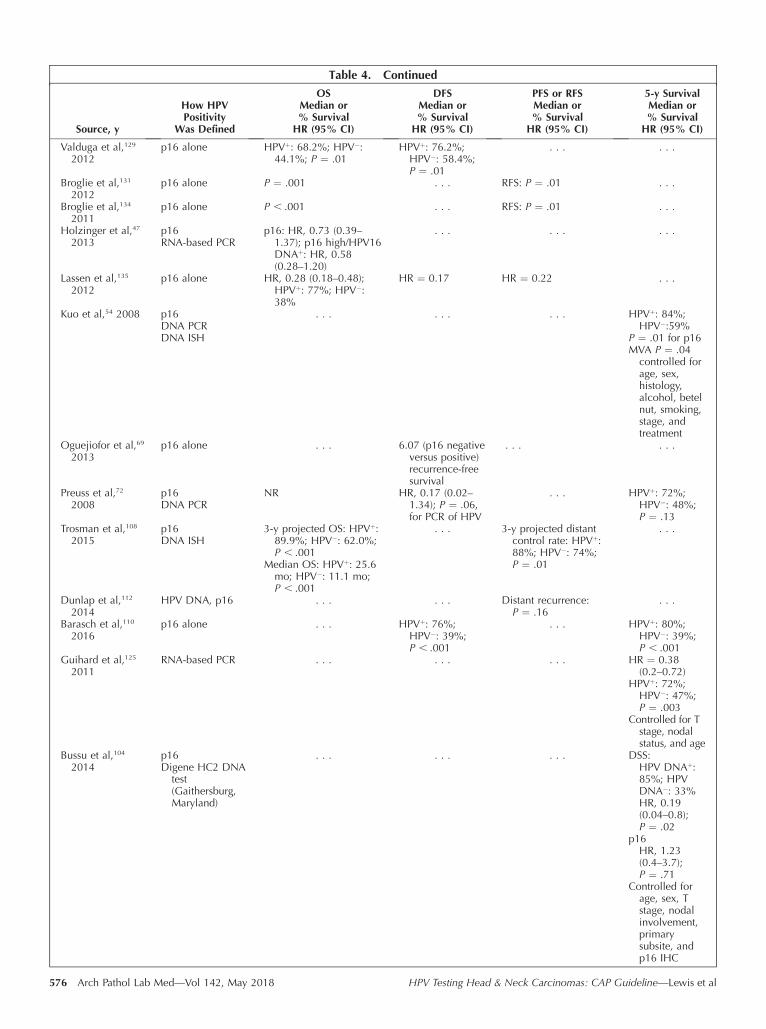
Table 4. Continued
Source, y
How HPVPositivity
Was Defined
OSMedian or% Survival
HR (95% CI)
DFSMedian or% Survival
HR (95% CI)
PFS or RFSMedian or% Survival
HR (95% CI)
5-y SurvivalMedian or% Survival
HR (95% CI)
Valduga et al,129
2012p16 alone HPVþ: 68.2%; HPV�:
44.1%; P ¼ .01HPVþ: 76.2%;
HPV�: 58.4%;P ¼ .01
. . . . . .
Broglie et al,131
2012p16 alone P ¼ .001 . . . RFS: P ¼ .01 . . .
Broglie et al,134
2011p16 alone P , .001 . . . RFS: P ¼ .01 . . .
Holzinger et al,47
2013p16RNA-based PCR
p16: HR, 0.73 (0.39–1.37); p16 high/HPV16DNAþ: HR, 0.58(0.28–1.20)
. . . . . . . . .
Lassen et al,135
2012p16 alone HR, 0.28 (0.18–0.48);
HPVþ: 77%; HPV�:38%
HR ¼ 0.17 HR ¼ 0.22 . . .
Kuo et al,54 2008 p16DNA PCRDNA ISH
. . . . . . . . . HPVþ: 84%;HPV�:59%
P ¼ .01 for p16MVA P ¼ .04
controlled forage, sex,histology,alcohol, betelnut, smoking,stage, andtreatment
Oguejiofor et al,69
2013p16 alone . . . 6.07 (p16 negative
versus positive)recurrence-freesurvival
. . . . . .
Preuss et al,72
2008p16DNA PCR
NR HR, 0.17 (0.02–1.34); P ¼ .06,for PCR of HPV
. . . HPVþ: 72%;HPV�: 48%;P ¼ .13
Trosman et al,108
2015p16DNA ISH
3-y projected OS: HPVþ:89.9%; HPV�: 62.0%;P , .001
Median OS: HPVþ: 25.6mo; HPV�: 11.1 mo;P , .001
. . . 3-y projected distantcontrol rate: HPVþ:88%; HPV�: 74%;P ¼ .01
. . .
Dunlap et al,112
2014HPV DNA, p16 . . . . . . Distant recurrence:
P ¼ .16. . .
Barasch et al,110
2016p16 alone . . . HPVþ: 76%;
HPV�: 39%;P , .001
. . . HPVþ: 80%;HPV�: 39%;P , .001
Guihard et al,125
2011RNA-based PCR . . . . . . . . . HR ¼ 0.38
(0.2–0.72)HPVþ: 72%;
HPV�: 47%;P ¼ .003
Controlled for Tstage, nodalstatus, and age
Bussu et al,104
2014p16Digene HC2 DNA
test(Gaithersburg,Maryland)
. . . . . . . . . DSS:HPV DNAþ:85%; HPVDNA�: 33%HR, 0.19(0.04–0.8);P ¼ .02
p16HR, 1.23(0.4–3.7);P ¼ .71
Controlled forage, sex, Tstage, nodalinvolvement,primarysubsite, andp16 IHC
576 Arch Pathol Lab Med—Vol 142, May 2018 HPV Testing Head & Neck Carcinomas: CAP Guideline—Lewis et al

Statement 4.—Recommendation.—Pathologists should notroutinely perform HR-HPV testing on patients with non-oropharyngeal primary tumors of the head and neck.
The strength of evidence is adequate.This recommendation is supported by 1 subgroup analysis
of 3 RCTs and 28 observational studies that met theinclusion criteria for our systematic review.66,158,162–187 Twoof these studies were available only in abstract form and, assuch, did not undergo formal quality assessment.186 Theevaluable 27 studies were deemed to have varying risk ofbias: 5 were deemed low risk of bias,162,164,169,176,183 8 low tomoderate,¶ 10 moderate,# and 4 high.66,158,175,179 The highrisk of bias for the 4 studies was due to funding fromindustry and missing information on the other assessmentcriteria. Refer to Supplemental Table 4 for the qualityassessment results for statement 4 studies.
There is confusion among many pathologists and treatingphysicians regarding the appropriateness of HPV testing innonoropharyngeal head and neck carcinomas. Because ofthe considerable attention that HPV-positive OPSCC hasreceived, it is understandable that pathologists and treatingphysicians may want to generalize that experience tocarcinomas arising outside the oropharynx, but the EP didnot find evidence to support this practice. Routine HPVtesting for nonoropharyngeal head and neck carcinomas isnot indicated because there is no proven prognostic ortherapeutic difference based on its presence or absence(either by any of the various HPV-specific tests or by thesurrogate marker p16). If HPV testing were to yield apositive result in a nonoropharyngeal carcinoma, it mightmislead treating physicians and patients as to the origin andlikely biologic behavior of the carcinoma. This does notmean that there is no potential biological and clinicalsignificance for transcriptionally active HR-HPV in non-oropharyngeal head and neck carcinomas (particularly inspecific anatomic subsites such as the nasopharynx orsinonasal tract); it simply means that the clinical significanceand ramifications are not established at this time.
Although the EP does not recommend routinely testingnonoropharyngeal carcinomas for HPV, it does recognizethat there are occasional situations where it may beindicated. For example, if the anatomic site of tumor originis not provided, is ambiguous, and/or includes both anoropharyngeal and a nonoropharyngeal site (eg, for largetumors), then HPV testing may be appropriate. As anotherexample, for a patient who had a prior HPV-positive
OPSCC, HPV testing in a new non-OPSCC may beappropriate to understand the relationship between the 2carcinomas (ie, recurrence versus new primary). In thesesettings, when HPV testing is performed, p16 IHC alone isinsufficient because of its suboptimal positive predictivevalue in nonoropharyngeal sites. p16 IHC can be used toscreen a tumor using the same criteria as in the oropharynx.If it is negative, then one can conclude that the tumor is notrelated to transcriptionally active HR-HPV. If it is positive,however, HPV-specific testing must be performed by one ofthe available platforms (see algorithm).
The systematic review uncovered 16 studies that investi-gated HPV testing in nonoropharyngeal head and neckcarcinomas (Table 5). These studies were heterogeneous inanatomic subsites evaluated (eg, larynx, oral cavity, sino-nasal tract, and other) and in the HPV testing methods used.The studies found that the prevalence of HPV-positivecarcinomas, when considering all tests for HR-HPV, isgenerally low in these nonoropharyngeal sites, ranging from5.9% to 58.3%.** When more specific, RNA-based methodsfor HPV detection were used, or p16 was combined withHPV-specific testing in order to establish the presence oftranscriptionally active HR-HPV, the rates were 2.7% to5.9%.175,189 Importantly, when the interpretation criteriawere reported and appropriate, the positive predictive valueof p16 IHC in nonoropharyngeal carcinomas was low,ranging from 22% to 50%, because of the very low overallrates of transcriptionally active HR-HPV in these tu-mors.166,173,175,183,189
The systematic review identified 28 studies†† that inves-tigated the clinical outcomes of nonoropharyngeal HPV-positive carcinomas (Table 6). Once again, these studieswere highly variable in anatomic subsites examined andHPV testing methods used and were also heterogeneous intheir reported outcomes. Only 7 studies reported astatistically significant difference in overall, disease-free,and/or progression-free survival between HPV-positive andHPV-negative groups, including 5 that found that thepatients with HPV-positive carcinomas had better surviv-al162,172,184,185 and 2 that actually found that the HPV-positivegroup had worse survival.168,187,190 One additional studyfound that the HPV-positive group had significantly lowerrates of recurrence.169
In addition to the aforementioned studies, systematicreviews, and meta-analyses, Li et al192 and Syrjanen and
Table 4. Continued
Source, y
How HPVPositivity
Was Defined
OSMedian or% Survival
HR (95% CI)
DFSMedian or% Survival
HR (95% CI)
PFS or RFSMedian or% Survival
HR (95% CI)
5-y SurvivalMedian or% Survival
HR (95% CI)
Driessen et al,111
2016p16DNA PCR
HPVþ: 85%; HPV�: 63%;P ¼ .18
HPVþ: 85%; HPV�:51%; P ¼ .09
. . .
Isayeva et al,52
2014p16RNA-based PCR
. . . HR, 0.31 (95% CI0.09–1.06)
HR, 0.27 (diseaseprogression); P ¼ .01
. . .
Abbreviations: ACE-27, adult comorbidity evaluation-27; DFS, disease-free survival; DSS, disease-specific survival; ECOG, Eastern CooperativeOncology Group; EGFR, epidermal growth factor receptor; HR, hazard ratio; IHC, immunohistochemistry; ISH, in situ hybridization; MVA,multivariate analysis; NR, not reported; OS, overall survival; PCR, polymerase chain reaction; PFS, progression-free survival; qRT-PCR, quantitativeRT-PCR; RFS, regression-free survival; RT-PCR, reverse transcriptase PCR; SEER, Surveillance, Epidemiology, and End Results Program; TNM, tumor-node-metastasis; UVA, univariate analysis.
¶ References 163, 165–167, 170, 177, 180, 185.# References 168, 171–174, 178, 181, 182, 184, 187.
** References 162, 164, 166, 168, 169, 173, 174, 176, 183, 185,188.
†† References 66, 162, 165, 167–173, 177–188, 190–195.
Arch Pathol Lab Med—Vol 142, May 2018 HPV Testing Head & Neck Carcinomas: CAP Guideline—Lewis et al 577

Syrjanen196 pooled data from numerous heterogeneousstudies and found that the overall rates of HPV DNA inlaryngeal and sinonasal carcinomas were 28% and 27%,respectively. Other studies using RNA-based HPV detectionmethods on oral cavity and laryngeal carcinomas reportedHPV prevalences of 1.3% and 2.3%, respectively.197,198
In the open comment period, there were 154 respondents,of whom 79.7% (n¼ 123) agreed with the recommendationand 12.3% (n ¼ 19) disagreed. There were 28 writtencomments, including some that expressed disagreementbecause they believed this statement included anogenital
sites. The recommendation was revised to specify that it was
applicable only to the head and neck. Some respondents
disagreed with the recommendation because of their
experience or reports of occasional HPV-positive tumors
arising in other nonoropharyngeal head and neck sites
(nasopharynx, oral cavity, hypopharynx, sinonasal tract, and
conjunctiva/lacrimal sac were all mentioned). Finally, a few
respondents felt the language should be changed to allow
testing in cases for which the precise anatomic location was
not given.
Table 5. Summary of Laboratory Data for Human Papillomavirus (HPV) Testing in Nonoropharyngeal Carcinomas
Source, yNo. of Cases
and Tumor Type
Method toDetermineHPV Status p16þ Criteria
Oral cavity
Lingen et al,176
2013409Oral cavity
qRT-PCR for E6/E7mRNA alone
H-score (combination of intensity anddistribution) .60
Elango et al,169
201160Oral tongue SCC
PCR alone . . .
Duncan etal,166 2013
81Oral cavity SCC
P16DNA PCR
.50% ¼ p16þ
Kaminagakuraet al,173 2012
114Oral SCC
p16DNA PCR
. . .
Ramshankar etal,188 2014
156Oral tongue
p16DNA PCR
.50% of cells ¼ positive
Duray et al,168
2012162Oral cavity SCC
PCR alone Any þ ¼ p16þ
Chaudhary etal,164 2010
430 (222 specimens)Oral submucous fibrosis
and oral SCC
PCR alone . . .
Laryngeal and/or hypopharyngeal
Laco et al,174
200888Laryngeal lesions
ISH alone Scaled, not clear what is positive
Wendt et al,183
2014142 (109 specimens)Hypopharyngeal
PCR alone .75% of cells ¼ positive
Sinonasal
Alos et al,162
200960Sinonasal SCC
PCR alone Only positive if ‘‘diffuse’’ in basal and parabasallayers excluding results in center
Larque et al,185
201470Sinonasal SCC
PCR alone Strong and diffuse cytoplasmic and nuclear inbasal and suprabasal cells in all tumor nests
Bishop et al,163
2013161Sinonasal
p16 and HPVDNA ISH
�70%
Other sites
Lewis et al,175
201287Oral cavity, laryngeal,
hypopharyngeal SCC
RNA ISH alone .70% ¼ p16þ
Skalova et al,158
201355Salivary gland
PCR alone They reported as 1%–25%, 26%–50%, morethan 51%
Chung et al,187
2014322Non-OPSCC
p16 alone .70% of cells ¼ positive
Abbreviations: ISH, in situ hybridization; mRNA, messenger RNA; NPV, negative predictive value; OPSCC, oropharyngeal squamous cell carcinoma;PCR, polymerase chain reaction; PPV, positive predictive value; qRT-PCR, quantitative reverse transcription PCR; RTOG, Radiation TherapyOncology Group; SCC, squamous cell carcinoma.a Prevalence calculated by true positiveþ false negative/total.
578 Arch Pathol Lab Med—Vol 142, May 2018 HPV Testing Head & Neck Carcinomas: CAP Guideline—Lewis et al

Statement 5.—Recommendation.—Pathologists shouldroutinely perform HR-HPV testing on patients withmetastatic SCC of unknown primary in a cervical upper–or mid–jugular chain lymph node. An explanatory note onthe significance of a positive HPV result is recommended.
The strength of evidence is adequate.This statement is supported by 4 observational studies
that met the inclusion criteria for the systematic re-view.199–202 Risk of bias assessments was deemed low in 2studies200,202 and low to moderate in 2.199,201 None of thesestudies were found to have methodologic flaws that wouldraise concerns about the findings. Refer to SupplementalTable 5 in the supplemental digital content for the qualityassessment results of included studies for statement 5.
Unknown primary is defined as any metastasis for whichthe primary site has not been clinically identified at the timethe biopsy of the metastasis is performed. In this setting,HR-HPV testing may aid in determining the most likelyprimary site. Hence, HR-HPV status is important for patientmanagement as it informs the clinical team where to searchfor the primary, or limits the likely area of primary if adefinitive lesion is not identified. There are inadequate dataat this time to support HR-HPV as a prognostic marker inthis setting.
In patients who have an unknown primary SCC in theneck, it is common clinical practice to search for the primarysite by performing a thorough endoscopic examination anddirected biopsies of likely sources of disease, such as thebase of tongue, tonsils, and nasopharynx, as well as
Table 5. Extended
Source, y ISHþ Criteria Control Method Intervention
No. TestPositive,DiseasePositive
No. TestPositive,Disease
Negative
Oral cavity
Lingen et al,176
2013Punctate or diffuse
nuclear signalsHPV qRT-PCR for
E6/E7 mRNAp16 19 27
Elango et al,169
2011Punctate signals PCR for HPV DNA p16 10 8
Duncan etal,166 2013
. . . PCR for HPV DNA p16 7 7
Kaminagakuraet al,173 2012
. . . PCR for HPV DNA p16 10 12
Ramshankar etal,188 2014
. . . PCR for HPV DNA p16 10 14
Duray et al,168
2012. . . PCR for HPV DNA ISH for HPV DNA 13 0
Chaudhary etal,164 2010
. . . PCR for HPV DNA Digene Hybrid Capture II(Gaithersburg, Maryland)
61 9
Laryngeal and/or hypopharyngeal
Laco et al,174
2008Brown staining
of nucleiISH for HPV DNA p16 14 0
Wendt et al,183
2014. . . PCR for HPV DNA p16 4 14
Sinonasal
Alos et al,162
2009. . . PCR for HPV DNA p16 12 0
Larque et al,185
2014Punctate signals PCR for HPV DNA p16 14 0
Bishop et al,163
2013Punctate signals
localized totumor nuclei
PCR for HPV DNA p16 8 0
Other sites
Lewis et al,175
2012RNA ISH, granular
cytoplasmic orpunctate nuclear
ISH for E6/E7 RNA p16 2 2
Skalova et al,158
2013. . . PCR for HPV DNA p16 0 45
Chung et al,187
2014Nuclear signals ISH for HPV p16 20 7
Arch Pathol Lab Med—Vol 142, May 2018 HPV Testing Head & Neck Carcinomas: CAP Guideline—Lewis et al 579
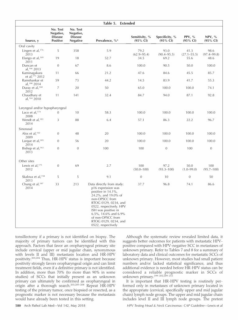
tonsillectomy if a primary is not identified on biopsy. Themajority of primary tumors can be identified with thisapproach. Factors that favor an oropharyngeal primary siteinclude cervical (upper or mid jugular chain, synonymouswith levels II and III) metastasis location and HR-HPVpositivity.203,204 Thus, HR-HPV status is important becausepositivity strongly favors oropharyngeal origin and can limittreatment fields, even if a definitive primary is not identified.In addition, more than 70% (to more than 90% in somestudies) of SCCs that initially present as an unknownprimary can ultimately be confirmed as oropharyngeal inorigin after a thorough search.203,205–208 Repeat HR-HPVtesting of the primary tumor, once biopsied or resected, as aprognostic marker is not necessary because the metastasiswould have already been tested in this setting.
Although the systematic review revealed limited data, itsuggests better outcomes for patients with metastatic HPV-positive compared with HPV-negative SCC in metastases ofunknown primary. Refer to Tables 7 and 8 for a summary oflaboratory data and clinical outcomes for metastatic SCCs ofunknown primary. However, most studies had small patientnumbers and/or lacked statistical significance, and thusadditional evidence is needed before HR-HPV status can beconsidered a reliable prognostic marker in SCCs ofunknown primary.199–202,209–211
It is important that HR-HPV testing is routinely per-formed only in metastases of unknown primary located inthe appropriate (cervical, specifically upper and mid jugularchain) lymph node groups. The upper and mid jugular chainincludes level II and III lymph node groups. The pretest
Table 5. Extended
Source, y
No. TestNegative,DiseasePositive
No. TestNegative,DiseaseNegative Prevalence, %a
Sensitivity, %(95% CI)
Specificity, %(95% CI)
PPV, %(95% CI)
NPV, %(95% CI)
Oral cavity
Lingen et al,176
20135 358 5.9 79.2
(62.9–95.4)93.0
(90.4–95.5)41.3
(27.1–55.5)98.6
(97.4–99.8)Elango et al,169
201119 18 52.7 34.5 69.2 55.6 48.6
Duncan etal,166 2013
0 67 8.6 100.0 90.5 50.0 100.0
Kaminagakuraet al,173 2012
11 66 21.2 47.6 84.6 45.5 85.7
Ramshankar etal,188 2014
59 73 44.2 14.5 83.9 41.7 55.3
Duray et al,168
20127 20 50 65.0 100.0 100.0 74.1
Chaudhary etal,164 2010
11 141 32.4 84.7 94.0 87.1 92.8
Laryngeal and/or hypopharyngeal
Laco et al,174
20080 10 58.3 100.0 100.0 100.0 100.0
Wendt et al,183
20143 88 6.4 57.1 86.3 22.2 96.7
Sinonasal
Alos et al,162
20090 48 20 100.0 100.0 100.0 100.0
Larque et al,185
20140 56 20 100.0 100.0 100.0 100.0
Bishop et al,163
20130 0 100 100 0 100 0
Other sites
Lewis et al,175
20120 69 2.7 100
(50.0–100)97.2
(93.3–100)50.0
(1.0–99.0)100
(95.7–100)
Skalova et al,158
20135 5 9.1 0 10 0 50
Chung et al,187
201433 213 Data directly from study:
p16 expression waspositive in 14.1%,24.2%, and 19.0% ofnon-OPSCC fromRTOG 0129, 0234, and0522, respectively. HPVISH was positive in6.5%, 14.6% and 6.9%of non-OPSCC fromRTOG 0129, 0234, and0522, respectively
37.7 96.8 74.1 86.6
580 Arch Pathol Lab Med—Vol 142, May 2018 HPV Testing Head & Neck Carcinomas: CAP Guideline—Lewis et al

probability of a positive HR-HPV result is high in thislocation because HPV-positive oropharyngeal SCCs almostalways metastasize here, and the primary tumor is oftenclinically occult.212,213 Up to one-third of HPV-positiveOPSCCs present as an unknown primary (compared with5%–10% of all head and neck cancers).212,214 Thus, HR-HPVtesting should be performed in all unknown primary cervicalupper– or mid–jugular chain metastases.
In contrast, HR-HPV testing should not be routinelyperformed on metastases of unknown primary outside ofthe cervical upper– and mid–jugular chain region. Non–upper- and mid-jugular chain lymph node groups includelevel I (submandibular/submental), level IV (lower jugular),level V (posterior triangle), level VI (pretracheal/prelaryng-eal/Delphian) and level VII (superior mediastinal), as well asparotid and supraclavicular nodes. The probability of anHPV-positive metastasis of unknown primary in the abovelocations is extremely low in the absence of concurrentupper/mid–jugular involvement, and therefore routine HR-HPV testing is not indicated. However, HR-HPV testingmay be performed on a non–upper/mid-jugular chainmetastasis when clinical suspicion for an HPV-positiveSCC is high (‘‘nonroutine’’ HR-HPV testing).
It is recognized that the specific lymph node group maynot always be known to the pathologist examining themetastasis. Some may simply be labeled ‘‘right neck’’ or‘‘left neck’’ by the ordering physician. Review of the medicalrecord (clinical notes and/or radiology reports) or directlycontacting the ordering physician will likely providesufficient information to identify the involved lymph nodegroup in most cases. There may be rare cases that cannot belocalized even after attempts to obtain clinical information.In such cases, HR-HPV testing should be performed.
An explanatory note in the pathology report describingthe clinical significance of a positive HR-HPV result isrecommended, specifying that HPV-positive SCC metasta-ses most likely originate from the oropharyngeal tonsilsand/or base of tongue but rarely may be from other sites (ie,nasopharynx).
In the open comment period, there were 149 respondents;85.23% (n ¼ 127) agreed, and 11.41% (n ¼ 17) disagreed.There were 22 written comments, including several com-ments questioning the method of HR-HPV testing, which isthe subject of statement 6. Several comments were either insupport of or against an explanatory note.
Statement 6.—Expert Consensus Opinion.—For tissuespecimens (ie, noncytology) from patients presenting withmetastatic SCC of unknown primary in a cervical upper– ormid–jugular chain lymph node, pathologists should performp16 IHC.
Note: Additional HPV-specific testing on p16-positivecases should be performed for tumors located outside oflevel II or III (nonroutine testing) in the neck and/or fortumors with keratinizing morphology.
Strength of evidence is insufficient.p16 IHC is a very sensitive surrogate marker for HR-HPV
that also maintains high positive predictive value when thepretest probability of an HPV-positive SCC is high.44,146,147
However, the positive predictive value decreases when thepretest probability is low because p16 overexpression canoccur by mechanisms other than HR-HPV signaling. Forexample, as many as 20% to 30% of aggressive head andneck cutaneous SCCs overexpress p16 unrelated to HR-HPV.215,216
An algorithmic approach for HR-HPV testing, shown inFigure 1, efficiently triages SCC metastases of unknownprimary and reduces unnecessary testing. Following thealgorithm, p16 IHC alone is sufficient to determine HR-HPVtumor status when the metastasis is located in one of theupper– or mid–jugular chain (level II and III) lymph nodegroups and the tumor morphology is nonkeratinizing.9,213,217
The pretest probability of an HR-HPV–positive SCC is veryhigh in this setting (and therefore p16 IHC is an excellenttest). High-risk HPV–specific testing is required to confirm apositive p16 IHC test result only when the tumormorphology is keratinizing and/or the metastasis is locatedoutside of the upper or mid jugular chain (the latter wouldapply to the setting of nonroutine HPV testing). Confirma-tory HR-HPV–specific testing should also be performed ifthe specific involved lymph node group cannot be deter-mined, regardless of tumor morphology. If the p16 IHC isnegative, no further HR-HPV testing is indicated, and thetumor is considered HPV negative because a p16-negativeresult essentially excludes a transcriptionally active HR-HPV–positive SCC.44,146,147
The ability to recognize keratinizing versus nonkeratiniz-ing tumor morphology is important in order to apply theabove HR-HPV testing algorithm. Nonkeratinizing SCCresembles transitional epithelium.9,217 The tumor cells haveoval to spindled nuclei, high nuclear to cytoplasmic ratios,and indistinct cell borders, often forming broad, pushing,ribbonlike nests. Limited keratinization may be present anddoes not mean that the tumor is keratinizing type (as long asthe above described histologic features are present). Mitoticactivity is typically brisk, with apoptotic debris and/ornecrosis in the background. When in doubt as to whethera particular SCC is keratinizing or nonkeratinizing, as maybe the case in small biopsy material, HR-HPV–specifictesting is recommended. However, one should attempt tohistologically classify most SCC metastases. In addition, onemust consider that the morphology of the tumor maychange in posttreatment specimens so that more keratini-zation may be present.
In the open comment period, there were 145 respondents;84.14% (n ¼ 122) agreed, and 11.72% (n ¼ 17) disagreed.There were 21 written comments, with most weighing in onthe method of HR-HPV testing: several comments favoredp16 IHC testing alone in all settings, some were in supportof HR-HPV–specific testing in all cases, and others felt themethod should be at the discretion of the pathologist.Several pointed out that the location of the lymph nodemetastasis (needed to apply the HR-HPV testing algorithm)is not always known to the pathologist.
Statement 7.—Expert Consensus Opinion.—Pathologistsshould perform HR-HPV testing on head and neck FNASCC samples from all patients with known oropharyngealSCC not previously tested for HR-HPV, with suspectedoropharyngeal SCC, or with metastatic SCC of unknownprimary.
Note: No recommendation is made for or against anyspecific testing methodology for HR-HPV testing in FNAsamples. If the result of HR-HPV testing on the FNA sampleis negative, testing should be performed on tissue if itbecomes available. If pathologists use cytology samples forp16 IHC testing, they should validate the criteria (ie, cutoff)for a positive result.
The strength of evidence is adequate.This statement is supported by 16 studies26,201,210,218–230
that met the inclusion criteria for our systematic review. Five
Arch Pathol Lab Med—Vol 142, May 2018 HPV Testing Head & Neck Carcinomas: CAP Guideline—Lewis et al 581

Table 6. Summary of Clinical Outcomes for Human Papillomavirus (HPV) Testing in Nonoropharyngeal Carcinomas
Source, yMethod toHPV Status
OSMedian or% Survival
HR (95% CI)
DFS, PFS, or RFSMedian or% Survival
HR (95% CI)
5-y SurvivalMedian or% Survival
HR (95% CI)
OtherClinical
OutcomeReported
Zhao et al,184
2009PCR alone HR, 0.13 (0.02–0.98);
P ¼ .048HPV, grade, stage,
tobacco
. . . . . . . . .
Alos et al,162
2009PCR alone HR, 0.17 (0.04–0.81);
P ¼ .07Controlling for age,
sex, p53, previouspapilloma, tumorgrade, smoking,tumor location assinus versus nasal,tumor stage at I–IIversus III–IV
DFS: HPVþ: 36%; HPV�:14%
PFS: HR, 0.21 (0.06–0.71);P ¼ .01
Controlling for age, sex,p53, previous papilloma,tumor grade, smoking,tumor location as sinusversus nasal, tumor stageat I–II versus III–IV
. . . . . .
Elango et al,169
2011PCR alone . . . . . . . . . Recurrence: HPVþ:
7%; HPV�: 32%;P ¼ .01
Sugiyama et al,182
2007PCR alone . . . . . . OR, 0.30
(0.08–1.2);P ¼ .08
DSS
Duray et al,167
2011PCR alone . . . DFS: HPVþ: 67%; HPV�:
77%; P ¼ NS. . . . . .
Robinson et al,180
2013p16DNA PCRDNA ISHAll 3 (p16, DNA
PCR, DNAISH) had tobe positive toconsider acase positivefor HPV
The data were notreported purely basedon HPV, but rather incombination withEBV
Mean overall survival:EBV�/HPV� ¼ 47.6mo (19.9–75.3)
EBVþ/HPV�: 67.9 mo(52.1–83.7)
EBV�/HPVþ: 53.6 mo(18.3–88.8)P ¼ .57
. . . . . . . . .
Larque et al,185
2014PCR alone HPVþ: 156.8 mo
(mean); HPV�: 72 mo(mean); P ¼ .03
DFS: HPVþ: mean 65.8mo; HPV�: mean 30.5mo; P ¼ .01
. . . . . .
Jiang et al,172
2013ISH alone HPV overexpression:
66.2%; HPV normal:86.6%; P ¼ .06
HPV overexpression:60.0%; HPV normal:83.9%; P ¼ .03
. . . . . .
Ernoux-Neufcoeuret al,170
2011
PCR alone . . . 5-y DFS: HPVþ: 58%;HPV�: 88%; P ¼ NS
RFS: HPVþ: 32% recurred;HPV�: 8% recurred
. . . 5-y DFS in p16;p16þ: 100%;p16�: 58%;P ¼ NS
Nemes et al,178
2006PCR alone . . . . . . . . . 2-y survival: HPVþ:
45.1%; HPV�:52.2%; P ¼ .73
Stephen et al,181
2012DNA PCR HPVþ: 79.7 mo; HPV�:
75 mo; P ¼ .35. . . . . . . . .
Morshed et al,177
2008PCR alone OS: 0.66 (HPVþ versus
HPV�). . . . . . DSS: 0.49 (HPVþ
versus HPV�)Huang et al,171
2012PCR alone P ¼ .44 P ¼ .43 . . . . . .
Kirby et al,186
2014p16DNA ISHRNA-based ISH
P ¼ .17 . . . P ¼ NS . . .
Kaminagakura etal,173 2012
p16DNA PCR
HPVþ: 66.8%; HPV�:38.4%; P ¼ .12
DFS: HPVþ: 52.7%; HPV�:40.4%; P ¼ .36
. . . . . .
Wendt et al,183
2014PCR alone OS: .16 (any HPV DNA
positive) (0.03–0.70);P ¼ .15
Controlling for age,stage, sex, p16
DFS for patients withhypopharyngeal cancerstratified HPV16 status:P ¼ .06
DFS for patients withhypopharyngeal cancerstratified p16 status:P ¼ .86
. . . . . .
Xu et al,193 2014 p16RNA-based PCR
P ¼ NS DFS: P ¼ NS . . . DSS: P ¼ NS
582 Arch Pathol Lab Med—Vol 142, May 2018 HPV Testing Head & Neck Carcinomas: CAP Guideline—Lewis et al

Table 6. Continued
Source, y
Methodto HPVStatus
OSMedian or% Survival
HR (95% CI)
DFS, PFS, or RFSMedian or% Survival
HR (95% CI)
5-y SurvivalMedian or% Survival
HR (95% CI)
OtherClinical
OutcomeReported
Duray et al,168
2012PCR alone . . . 5-y DFS
HPVþ: 40%; HPV�:76%; P ¼ .01HR, 2.81; P ¼ .01
Controlling for TNMstaging and node status
. . . . . .
Lee et al,191 2012 PCR alone P ¼ .11 DFS: HPVþ: 55%; HPV�:61%; P ¼ .21
HPVþ: 37%;HPV�: 50%;P ¼ .11
DSS: HPVþ:58%; HPV�:68%; P ¼ .21
Chuang et al,190
2012PCR alone Only a graph provided:
HPVþ did worse;P ¼ .13
Only a graph provided forRFS: HPVþ did worse;P ¼ .03
HR, 1.77(0.79–03.95)
HPVþ: 46.3%;HPV�: 72.6%;P ¼ .17
Controlling forsex, smoking,drinking, betelquid use
. . .
Leidy et al,194
2012p16 alone At 10 y: HPVþ: 31%;
HPV�: 34%; P ¼ .28. . . . . . . . .
Reuschenbach etal,179 2013
Not specified HPV EIA (PCR): P ¼ .88HPV-EIA (PCR and diffuse
p16 IHC): P ¼ .80No staining versus focal
p16 staining: P ¼ .09No staining versus diffuse
p16 staining: P ¼ .25
DFS: HPV EIA (PCR):P ¼ .86
HPV-EIA (PCR and diffusep16 IHC): P ¼ .35
. . . . . .
Li et al,192 2013 . . . . . . . . . . . . Risk of laryngealSCC; OR, 5.39;(3.25–8.94)
Nichols et al,66
2013PCR alone HR, 0.19 (0.06–0.60);
P ¼ .004Controlling for age, sex,
site, TNM stage,smoking, alcohol, p16,HPV 16 only
DFS: HR, 0.24 (0.1–0.56);P ¼ .001
Controlling for age, sex,site, TNM stage,smoking, alcohol, p16,HPV 16 only
. . . . . .
Chernock et al,165
2013HPV DNA ISHHPV PCR
P ¼ .06 DFS ¼ NS . . . . . .
Chung et al,187
2014p16 alone For p16: HR, 0.56
(0.35–0.89); P ¼ .01Controlling for age, sex,
TNM stageFor ISH: HR, 0.64
(0.34–1.21), P ¼ .17
For p16 PFS: HR, 0.63(0.42–0.95); P ¼ .03
Controlled for age, sex,TNM stage
For ISH: HR, 0.77(0.44–1.33); P ¼ .35
. . . . . .
Ramshankar etal,188 2014
p16DNA PCR
For p16: HR, 2.4(1.3–4.4); P ¼ .01
Controlling for age, sex,stage
For HPV16: HR, 0.6(0.38–0.10); P ¼ .049
Controlling for age, sex,stage
DFS for p16: HR, 2.6(1.4–4.6); P ¼ .01
Controlling for age, sex,stage
. . . . . .
Stenmark et al,195
2014p16DNA PCR
HR, 1.83 (0.71–4.72)Controlling for age,
tobacco exposure,WHO grade, and viralstatus
Median OS: HPVþ ¼71%; HPV� ¼ 47.2%;P ¼ .39
Controlling for age,tobacco exposure,WHO grade, and viralstatus
PFS: HR, 1.86 (0.77–4.47);P ¼ .36
Controlling for age,tobacco exposure, WHOgrade, and viral status
. . . Locoregionalcontrol: HR,3.0 (0.78–11.5); P ¼ .24
Controlling forage, tobaccoexposure,WHO grade,and viral status
Abbreviations: DFS, disease-free survival; DSS, disease-specific survival; EBV, Epstein-Barr virus; EIA, enzyme immunoassay; HR, hazard ratio; ISH,in situ hybridization; NS, not significant (no exact P value reported); OR, odds ratio; OS, overall survival; PCR, polymerase chain reaction; PFS,progression-free survival; RFS, recurrence-free survival; SCC, squamous cell carcinoma; TNM, tumor-node-metastasis; WHO, World HealthOrganization.
Arch Pathol Lab Med—Vol 142, May 2018 HPV Testing Head & Neck Carcinomas: CAP Guideline—Lewis et al 583

of these studies210,224–227 were reported only in abstract formand did not undergo quality assurance. Of the 11 studiesassessed, all were retrospective cohort designs, with theexception of 1 cross-sectional study.222 All included studieshad prospective data collection, 2 reported blinding ofoutcome assessors,222,230 and only 1 reported funding fromindustry.230 Ultimately, 4 studies219,222,229,230 were deemed tohave a low to moderate risk of bias, 6 moderate,‡‡ and 1high,228 although this study did not have methodologicflaws that would raise concerns about its findings. SeeSupplemental Table 6 in the supplemental digital contentfor the quality assessment results for all studies included inthe statement 7 evidentiary base.
Among patients with HPV-positive OPSCC, approxi-mately 85% develop lymph node metastases, and approx-imately 50% of these patients are first diagnosed based uponan enlarged cervical lymph node.11,53,55,231–233 Fine-needleaspiration is frequently used to sample and diagnosemetastatic head and neck carcinomas in cervical lymphnodes. Because of the marked tendency for HPV-positiveHNSCC to metastasize to cervical lymph nodes, FNA playsa very important diagnostic role in the initial detection ofthese cancers.234–237 In some cases, cytologic material may bethe only tumor specimen available for diagnostic workup,and in a subset of cases, a primary site of origin will not beidentified even after exhaustive clinical and radiologicevaluation. The aspirated material obtained by FNA canbe tested by any of a variety of methods for HR-HPV andused to classify the metastatic HNSCC as HPV positive orHPV negative, thus indicating an oropharynx origin.
The systematic review identified a limited number ofstudies that used samples obtained by FNA for analysis ofHR-HPV status of metastatic HNSCC. The range ofcytologic studies includes HPV determination using forma-lin-fixed, paraffin-embedded FNA material in a cell block;liquid-based specimens (Surepath, Becton, Dickinson andCompany, Franklin Lakes, New Jersey, and ThinPrep,Hologic, Marlborough, Massachusetts); and scrapes fromair-dried or alcohol-fixed smears. Testing methodologiesevaluated included p16 IHC,222,229,230,238 DNA ISH,239 cobasHPV test (Roche Molecular Systems, Inc, Pleasanton,California),26,228 Cervista HPV HR and Cervista HPV16/18
assays (Hologic),220,240,241 and Hybrid-Capture 2 assay(Qiagen, Germantown, Maryland).219,224,242 The literaturesupports the use of FNA as a valid method for obtainingmaterial for HR-HPV testing.218,221,223 Sensitivities andspecificities of HPV assays for detecting HR-HPV in FNAsamples are reported to be greater than 90%; however, thereare limited data about the accuracy of any one particularHPV testing method.§§
A particular difficulty in the assessment of HR-HPV inFNA samples pertains to the interpretation of p16 immu-noreactivity in cell block specimens. Although use of thestandard criterion of more than 70% positive cells isaccepted when applied to tissue biopsy, 3 recent studiessuggest that thresholds as low as 10% to 15% for thepercentage of positive cells may be valid for cellblocks.23,229,230 Whichever method is selected to assess theHR-HPV status of an FNA sample, individual laboratoryvalidation is required. Because of the limited data availablepertaining to HR-HPV testing in FNA specimens at thecurrent time, HR-HPV testing is recommended on anysubsequent tissue specimens that may become available ifHR-HPV testing was negative in the FNA specimen.
In the open comment period there were 142 respondents;84.51% (n ¼ 120) agreed, and 13.38% (n ¼ 19) disagreed.There were 16 written comments, including a number thatsuggested that either p16 IHC or liquid-based testingmethodologies be used. Others commented that confirma-tory testing should be performed for FNA samples testingpositive by p16 IHC, particularly in cases where the patientis known to have a nonoropharyngeal HNSCC.
Refer to Tables 9 and 10 for the summaries of laboratorydata and clinical outcomes for studies where FNAs wereused.
Statement 8.—Expert Consensus Opinion.—Pathologistsshould report p16 IHC positivity as a surrogate for HR-HPVin tissue specimens (ie, noncytology) when there is at least70% nuclear and cytoplasmic expression with at leastmoderate to strong intensity.
The strength of evidence is insufficient.p16 is a tumor suppressor protein that inhibits CDK4 and
CDK6–dependent/cyclin D–mediated phosphorylation ofRB required for cell proliferation.243 Overexpression of p16
Table 7. Summary of Laboratory Data for Metastatic Squamous Cell Carcinomas of Unknown Primary
Source, y
No. ofPatients orSpecimens
SpecimenType
HowHPV WasDefined p16þ Criteria ISH Criteria
PCRAssay
Compton et al,199
201125 Resection and
biopsyp16DNA ISH
. . . Nuclear signals . . .
Tribius et al,200
201263 Specimen type
not reportedp16DNA PCR
,1% is negative, isolatedcells ,5% is sporadic,focal is small clusters,25%, and diffuse is.25% nuclear andcytoplasmic staining
. . . QualitativePCR assay
Vent et al,201
201347 Specimen type
not reportedp16 versus
HPV alone.60% . . . Qualitative
PCR assaySivars et al,202
201450 Resection and
biopsyPCR alone .70% . . . Quantitative
PCR assay—MFI 100
Abbreviations: HPV, human papillomavirus; ISH, in situ hybridization; MFI, median fluorescence intensity; NPV, negative predictive value; PCR,polymerase chain reaction; PPV, positive predictive value.
‡‡ References 26, 201, 218, 220, 221, 223. §§ References 26, 219, 220, 222, 224, 228, 229.
584 Arch Pathol Lab Med—Vol 142, May 2018 HPV Testing Head & Neck Carcinomas: CAP Guideline—Lewis et al

Table 7. Extended
Source, yControlMethod Intervention
Sensitivity, %(95% CI)
Specificity, %(95% CI)
PPV, %(95% CI)
NPV, %(95% CI)
Compton et al,199
2011ISH for HPV DNA p16 100 (57.1–100) 77.8 (58.6–97.0) 63.6 (35.2–92.1) 100 (78.6–100)
Tribius et al,200
2012PCR for HPV DNA p16 72.7 (57.5–87.9) 60.0 (42.5–77.5) 66.7 (51.3–82.1) 66.7 (48.9–84.4)
Vent et al,201
2013ISH for HPV DNA p16 100 (66.7–100) 96.4 (89.6–100) 90.0 (71.4–100) 100 (88.9–100)
Sivars et al,202
2014PCR for HPV DNA p16 100.0 90.6 85.7 100.0
Table 8. Clinical Outcomes of Human Papillomavirus Testing in Metastatic Squamous Cell Carcinomasof Unknown Primary
Source, y
Method toDetermineHPV Status
OSMedian or% Survival
HR (95% CI)
DFSMedian or% Survival
HR (95%CI)
PFS or RFSMedian or% Survival
HR (95%CI)
3-y or 5-y SurvivalMedian or %
SurvivalHR (95% CI)
OtherClinical
OutcomeReported
Compton et al,199
2011p16DNA ISH
. . . HPVþ: 66.7%;HPV�: 48.5%;P ¼ .54
. . . 5-y: HPVþ: 66.7%;HPV�: 48.5%;P ¼ .35
. . .
Tribius et al,200
2012p16DNA PCR
2-y OS: HPVþ:75.7%; HPV�:75.3%; P ¼ .53
. . . PFS: HPVþ: 79.5%;HPV�: 67.8%;P ¼ .30
. . . . . .
Vent et al,201
2013p16 versus
HPV alone5-y: p16þ: 83%;
p16�: 32%;P ¼ NS
p16þ: 83%;P16�: 32.4%;P ¼ .18
. . . . . . . . .
Sivars et al,202
2014PCR alone . . . HPVþ: 85.0%;
HPV�: 63.3%;P ¼ .05
. . . 5-y: HPVþ: 80%;HPV�: 36.7%;P ¼ .01
HR ¼ 0.29 (0.09–0.91); P ¼ .03
Controlled for p53expression,gender, age, andsmoking habits
5-y survival HPVDNA and p16þ:77%; HPV DNAand p16�: 40.6%;P ¼ .02
. . .
Straetmans et al,209
2014p16DNA PCRHPV 16 FISH
. . . . . . RFS: HPVþ: 100%;HPV�: 77.8%;P ¼ NS
5-y: HPVþ: 75%;HPV�: 66.7%;P ¼ NS
DistantmetastasisHPVþ: 0%;HPV�: 5.6%
Fowler et al,210
2012 [abstract]p16DNA ISH
P ¼ .001 P ¼ .07 . . . 3-y: HPVþ: 83%;HPV�: 40%
5-y: HPVþ: 71%;HPV�: 40%
1-y survival:HPVþ: 97%;HPV�: 64%
Straetmans et al,211
2011 [abstract]p16DNA PCRDNA ISH
. . . . . . . . . 5-y: p16þ:69%; p16�:33%; P¼ .05
HPVþ: 65%; HPV�:37%; P¼ .09
42% ofprimariesfound duringfollow-up.SignificantcorrelationbetweenHPVþ andlater detectionof oropharyn-geal primary(P ¼ .04)
Abbreviations: DFS, disease-free survival; FISH, fluorescence in situ hybridization; HPV, human papillomavirus; HR, hazard ratio; ISH, in situhybridization; NS, not significant (no exact P value reported); OS, overall survival; PCR, polymerase chain reaction; PFS, progression-free survival;RFS, regression-free survival.
Arch Pathol Lab Med—Vol 142, May 2018 HPV Testing Head & Neck Carcinomas: CAP Guideline—Lewis et al 585

is an established surrogate marker for transcriptionallyactive HR-HPV infection.139,140,243,244
The criteria for p16 IHC have been established in multipleretrospective and prospective studies that validated theassociation of p16 immunopositivity with a more favorableprognosis in OPSCCs compared with p16-negative carci-nomas.11,35,81,245 It is important to note that staining must beboth nuclear and cytoplasmic to be considered positive.Definitions for what percentage of positive cells is necessaryhave varied substantially; however, some of the largest andprospective studies, such as Ang et al,11 have supported astringent cutoff of 70% to 75%. However, in high-incidencecountries such as the United States, lesser staining cutoffsmay function similarly. With these criteria, the sensitivity of
p16 IHC for transcriptionally active HR-HPV approaches100%. The specificity of p16 IHC in the oropharynx is lower(~85%–95%) for transcriptionally active HR-HPV, in partbecause of p16 expression unrelated to HPV.53,81,246 Theinterrater agreement among pathologists for p16 IHCinterpretation is excellent (j ¼ 0.95–0.98).53
Rare tissue specimens may exhibit an equivocal pattern ofp16 staining that fails to meet the recommended thresholdfor positivity. For instance, cases can exhibit more than 50%and less than 70% moderate to strong nuclear andcytoplasmic staining or diffuse low-intensity nuclear andcytoplasmic staining. Limited evidence suggests a subset ofthese cases may have transcriptionally active HR-HPV.53,247
Table 9. Summary of Laboratory Data for Studies Using Fine-Needle Aspirations
Source, yNo. of Patientsor Specimens Specimen Type Cancer Type
Method toDetermineHPV Status
Vent et al,201
201347 patients . . . CUP p16 versus HPV alone
Begum et al,218
200777 specimens FNA . . . . . .
Bishop et al,219
201224 patients Cytologic preparations
(FNAs and brushes) wereobtained from surgicalresections
OPSCC, non-OPSCC, cervicalnodal metastatic carcinomaof known primary, CUP
p16DNA PCRDNA ISH
Guo et al,220
201464 patients FNA OPSCC, non-OPSCC, cervical
nodal metastatic carcinomaof known primary, CUP
p16DNA PCRDNA ISH
Lau et al,263 2011 67 patients FNA Cervical nodal metastaticcarcinoma of known primary,CUP
Cervista on FNA fluidfor HR-HPV DNA
Jakscha et al,222
2013OPSCC Resection, biopsy, FNA . . . p16 alone
Jannapureddy etal,223 2010
40 patients FNA OPSCC, non-OPSCC p16DNA ISH
Smith et al,224
201425 patients Resection, FNA OPSCC p16
DNA ISH
Kerr et al,26 2014 33 patients Resection, biopsy, FNA OPSCC, non-OPSCC Compared Roche cobas(Roche, Pleasanton,California), ISH, and p16
Davis et al,225
201474 patients Surgical specimens, FNA OPSCC, cervical nodal metastatic
carcinoma of known primaryp16DNA ISHHPV L1 IHC
Fatima et al,226
2012 [abstract]. . . FNA Cervical nodal metastatic
carcinoma of known primaryp16DNA ISH
Baldassarri etal,228 2015
37 patients FNA with correlatingbiopsy or resectionspecimens
OPSCC, non-OPSCC, cervicalnodal metastatic carcinomaof known primary, CUP
Roche cobas oncytology fluid
Holmes et al,229
201585 patients Metastatic specimens
sampled by FNA andprimary tumors byresection or biopsy
CUP p16DNA ISH
Jalaly et al,230
201548 patients Resection, biopsy, FNA OPSCC, cervical nodal metastatic
carcinoma of known primaryp16RNA-based ISH
Abbreviations: CUP, cancer of unknown primary; FNA, fine-needle aspiration; HPV, human papillomavirus; HR-HPV, high-risk humanpapillomavirus; IHC, immunohistochemistry; ISH, in situ hybridization; L1, level 1; NA, not applicable; non-OPSCC, nonoropharyngeal squamouscell carcinoma; NPV, negative predictive value; OPSCC, oropharyngeal squamous cell carcinoma; PCR, polymerase chain reaction; PPV, positivepredictive value.
586 Arch Pathol Lab Med—Vol 142, May 2018 HPV Testing Head & Neck Carcinomas: CAP Guideline—Lewis et al
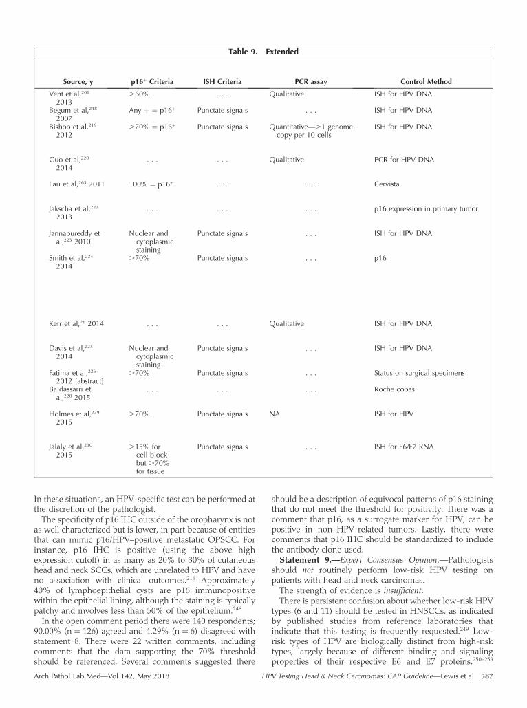
In these situations, an HPV-specific test can be performed atthe discretion of the pathologist.
The specificity of p16 IHC outside of the oropharynx is notas well characterized but is lower, in part because of entitiesthat can mimic p16/HPV–positive metastatic OPSCC. Forinstance, p16 IHC is positive (using the above highexpression cutoff) in as many as 20% to 30% of cutaneoushead and neck SCCs, which are unrelated to HPV and haveno association with clinical outcomes.216 Approximately40% of lymphoepithelial cysts are p16 immunopositivewithin the epithelial lining, although the staining is typicallypatchy and involves less than 50% of the epithelium.248
In the open comment period there were 140 respondents;90.00% (n ¼ 126) agreed and 4.29% (n ¼ 6) disagreed withstatement 8. There were 22 written comments, includingcomments that the data supporting the 70% thresholdshould be referenced. Several comments suggested there
should be a description of equivocal patterns of p16 stainingthat do not meet the threshold for positivity. There was acomment that p16, as a surrogate marker for HPV, can bepositive in non–HPV-related tumors. Lastly, there werecomments that p16 IHC should be standardized to includethe antibody clone used.
Statement 9.—Expert Consensus Opinion.—Pathologistsshould not routinely perform low-risk HPV testing onpatients with head and neck carcinomas.
The strength of evidence is insufficient.There is persistent confusion about whether low-risk HPV
types (6 and 11) should be tested in HNSCCs, as indicatedby published studies from reference laboratories thatindicate that this testing is frequently requested.249 Low-risk types of HPV are biologically distinct from high-risktypes, largely because of different binding and signalingproperties of their respective E6 and E7 proteins.250–253
Table 9. Extended
Source, y p16þ Criteria ISH Criteria PCR assay Control Method
Vent et al,201
2013.60% . . . Qualitative ISH for HPV DNA
Begum et al,218
2007Any þ ¼ p16þ Punctate signals . . . ISH for HPV DNA
Bishop et al,219
2012.70% ¼ p16þ Punctate signals Quantitative—.1 genome
copy per 10 cellsISH for HPV DNA
Guo et al,220
2014. . . . . . Qualitative PCR for HPV DNA
Lau et al,263 2011 100% ¼ p16þ . . . . . . Cervista
Jakscha et al,222
2013. . . . . . . . . p16 expression in primary tumor
Jannapureddy etal,223 2010
Nuclear andcytoplasmicstaining
Punctate signals . . . ISH for HPV DNA
Smith et al,224
2014.70% Punctate signals . . . p16
Kerr et al,26 2014 . . . . . . Qualitative ISH for HPV DNA
Davis et al,225
2014Nuclear and
cytoplasmicstaining
Punctate signals . . . ISH for HPV DNA
Fatima et al,226
2012 [abstract].70% Punctate signals . . . Status on surgical specimens
Baldassarri etal,228 2015
. . . . . . . . . Roche cobas
Holmes et al,229
2015.70% Punctate signals NA ISH for HPV
Jalaly et al,230
2015.15% for
cell blockbut .70%for tissue
Punctate signals . . . ISH for E6/E7 RNA
Arch Pathol Lab Med—Vol 142, May 2018 HPV Testing Head & Neck Carcinomas: CAP Guideline—Lewis et al 587

Although low-risk HPV types are an established etiologicagent in benign squamous papillomas and warts of varioussites, they do not play a significant role in the developmentof HPV-positive OPSCC.253
Because there is little (if any) benefit of identifying low-risk HPV types in the head and neck, the EP determinedthat there is no role for routine low-risk HPV in this context.Although the systematic review did not specifically addresslow-risk HPV types, the expert consensus opinion is thatlow-risk testing should not be routinely performed.
In the open comment period, there were 140 respondents;95.71% (n¼134) agreed, and 4.29% (n¼6) disagreed. Therewere 7 written comments, 2 of which focused on a beliefthat low-risk HPV can lead to dysplasia. The publishedliterature does not support this association. One commentsuggested that low-risk HPV testing should be done inpatients with HIV; however, there is no evidence that low-risk HPV results in carcinoma in HIV-positive patients.
Statement 10.—Expert Consensus Opinion.—Pathologistsshould not repeat HPV testing on patients with locallyrecurrent, regionally recurrent, or persistent tumor ifprimary tumor HR-HPV status has already been established.If initial HR-HPV status was never assessed or results areunknown, testing is recommended. Testing for HPV may beperformed on a case-by-case basis for diagnostic purposes ifthere is uncertainty regarding whether the tumor inquestion is a recurrence or a new primary SCC.
The strength of evidence is insufficient.High-risk HPV status established on the primary tumor is
the basis for its prognostic value. Recurrences are readilytestable and have been demonstrated to show the same HR-HPV status.254 As such, there is no documented value ofrepeating testing for HR-HPV on locoregionally recurrent orpersistent HNSCC. However, because of this consistency ofphenotype, HR-HPV testing on a recurrence when thestatus of the primary OPSCC is unknown would accurately
Table 9. Extended
Source, y InterventionSensitivity, %
(95% CI)Specificity, %
(95% CI)PPV, %
(95% CI)NPV, %(95% CI)
Vent et al,201
2013p16 100 (66.7–100) 96.4 (89.6–100) 90.0 (71.4–100) 100 (88.9–100)
Begum et al,218
2007p16 90.0 (71.4–100) 55.6 (23.1–88.0) 69.2 (44.1–94.3) 83.3 (53.5–100)
Bishop et al,219
2012Hybrid Capture II
(Digene,Gaithersburg,Maryland)
100 (76.9–100) 90.9 (73.9–100) 92.9 (79.4–100) 100 (70–100)
Guo et al,220
2014Cervista (Hologic,
Marlborough,Massachusetts)
97.4 (92.3–100) 90.9 (73.9–100) 97.4 (92.3–100) 100 (73.9–100)
Lau et al,263 2011 p16 72.4 (56.1–88.7) 83.3 (53.5–100) 95.5 (86.8–100) 38.5 (12.0–64.9)
Jakscha et al,222
2013FNA—p16 expression
in lymph nodemetastasis
92.3 (77.8–100) 95.1 (88.5–100) 85.7 (67.4–100) 97.5 (92.7–100)
Jannapureddy etal,223 2010
p16 100 (66.7–100) 77.4 (62.7–92.1) 56.3 (31.9–80.6) 100 (87.5–100)
Smith et al,224
2014Hybrid Capture II 100 (62.5–100)
cytologictumor cellsrequired
100 (62.5–100) 100 (62.5–100) 100 (62.5–100)
66.7 (40–93.3)withoutrequiringcytologic cells
100 (62.5–100) 100 (62.5–100) 66.7 (40–93.3)
Kerr et al,26 2014 Roche cobas 100 (75–100) 86.4 (72–100) 80 (59.8–100) 100 (84.2–100)
Davis et al,225
2014p16 75.0 (63.2–86.8) 63.6 (43.5–83.7) 83.0 (72.2–93.7) 51.9 (33.0–70.7)
HPV L1 76.6 (64.5–88.7) 31.6 (10.7–52.5) 73.5 (61.1–85.8) 35.3 (12.6–58.0)Fatima et al,226
2012 [abstract]p16 and HPV ISH 64
43100100
100100
3425
Baldassarri etal,228 2015
p16 100 (72.7–100) 100 (66.7–100) 100 (72.7–100) 100 (66.7–100)
Holmes et al,229
2015p16 100 (94.5–100) 92 (81.4–100) 96.5 (91.7–100) 100 (87–100)
Jalaly et al,230
2015p16 . . . . . . . . . . . .
588 Arch Pathol Lab Med—Vol 142, May 2018 HPV Testing Head & Neck Carcinomas: CAP Guideline—Lewis et al

reflect the HPV status and is thus recommended. Particu-larly with delayed recurrences, a logical clinical question thatmay arise is the possibility of a new primary tumor. Suchscenarios require correlation with clinical and morphologicfeatures, and HPV status may be informative in separating arecurrence from a new primary SCC.
Our systematic review yielded limited data addressing thelevel of concordance between the HPV status of the primarytumor and the corresponding recurrence. In one study,254 16locoregional recurrences and 21 metastases were testedalong with their untreated primaries. Thirty-six of 37 cases(97%) demonstrated a concordant HPV status. For the 1discordant case, a second primary was not entirely excluded.However, the study also suggests that technical variability intesting may affect findings in a recurrent site.
In the open comment period there were 94 respondents;92.47% (n¼ 86) agreed, and 7.53% (n¼ 7) disagreed. Therewere 8 comments, most notably the suggestion to indicatethat HPV testing may help separate true recurrence from aseparate primary tumor. Concern about technical differenc-es among laboratories affecting repeat testing was raised, aswell as a concern about treatment effect.
Statement 11.—Expert Consensus Opinion.—Pathologistsshould not routinely perform HR-HPV testing on patientswith distant metastases if primary tumor HR-HPV statushas been established. Testing for HPV may be performed ona case-by-case basis for diagnostic purposes if there isuncertainty regarding whether the tumor in question is ametastasis or a new primary SCC.
The strength of evidence is insufficient.High-risk HPV status established on the primary tumor is
the basis for its prognostic value. Distant metastases arereadily testable and limited data show that they retain thesame HR-HPV status, including p16 overexpression.137,254,255
As such, there is no documented value of repeating testingon a metastatic tumor. However, because of this consistencyof phenotype, HR-HPV testing on a metastasis when thestatus of the primary is unknown would accurately reflectthe HPV status of the primary HNSCC and is thusrecommended. In some cases, however, there is thepossibility that the metastasis represents a new primarySCC. Such scenarios require correlation with clinical and
morphologic features, and HPV status may be informative inseparating a distant metastasis from a new primary SCC.
Our systematic review yielded limited data addressing thelevel of concordance between the HPV status of primarytumor and corresponding distant metastasis. Collectively,concordance is noted in about 44 of 45 (97.7%) tested pairedprimary tumors and metastases in the literature.137,254,255 Inone study,137 20 of 20 tested metastases demonstratedconcordant HPV status as compared with their primaries. Inanother study,254 16 locoregional recurrences and 21 distantmetastases were tested along with their untreated primaries.Thirty-six of 37 cases (97%) demonstrated concordant HPVstatus. For the 1 discordant case, a second primary was notentirely excluded. One small series of 4 patients outside ofour systematic review confirmed concordance in HPV statusbetween 4 primary and metastatic SCCs.255
However, as with recurrences, technical variability intesting may affect findings in a metastatic site,254 and thefindings are insufficient to recommend a specific testingalgorithm at recurrent sites. Particularly with possible lungmetastases, an approach that includes HPV-specific testingfor p16-positive tumors should be considered, because ofp16 expression in a subset of lung SCCs not associated withHR-HPV.
In the open comment period there were 90 respondents;95.56% (n¼ 86) agreed, and 4.44% (n¼ 4) disagreed. Therewere 7 comments, most notably echoing the statement thatHPV testing may help separate true recurrence from aseparate primary tumor. Concern about p16 IHC testing as astand-alone test in this context was raised, as well asconcern about the effect of tumor heterogeneity.
Statement 12.—Expert Consensus Opinion.—Pathologistsshould report primary OPSCCs that test positive for HR-HPV or its surrogate marker p16 as HPV positive and/or p16positive.
The strength of evidence is insufficient.Oropharyngeal SCC with transcriptionally active HR-HPV
is a distinct subtype of head and neck cancer. Because HPVdefines this subtype, the HR-HPV status (by HPV-specificand/or surrogate marker p16 testing) should be included inthe pathologic diagnosis. In the literature, a number ofdifferent terms have been used to describe the HPV status of
Table 10. Summary of Clinical Outcomes for Studies Using Fine-Needle Aspiration
Source, y
Method toDefine HPV
Status
OS, Medianor %
HR (95% CI)
DFS, Medianor %
HR (95% CI)
3-y Survival,Median or %HR (95% CI)
5-y Survival,Median or %HR (95% CI)
Other ClinicalOutcomes
Vent et al,201
2013p16 versus
HPV alonep16þ: 83%;
p16�: 32%;P ¼ NS
p16þ: 83%; p16�:32.4%; P ¼ .18
HPVþ: 67%; HPV�:48%; P ¼ .94
. . . . . . . . .
Fowler et al,210
2012p16DNA ISH
P ¼ .001 5-y: HPV/p16þ:75%; HPV/p16�:56%; P ¼ .07
HPVþ: 83%;HPV�: 40%
HPVþ: 71%;HPV�:40%
1-y survival: HPVþ:97%; HPV�: 64%
Davis et al,225
2014p16DNA ISHHPV L1 IHC
P ¼ .10 . . . . . . . . . . . .
Inohara et al,227
2012PCR alone . . . . . . HPVþ: 100%;
HPV�: 89%;P ¼ .67
. . . Complete response ofadvanced nodalmetastases (N2c) toconcomitantchemoradiotherapy
HPVþ: 100%; HPV�:40%
Abbreviations: DFS, disease-free survival; HPV, human papillomavirus; HR, hazard ratio; IHC, immunohistochemistry; ISH, in situ hybridization; L1,level 1; NS, not significant (no exact P value reported); OS, overall survival; PCR, polymerase chain reaction.
Arch Pathol Lab Med—Vol 142, May 2018 HPV Testing Head & Neck Carcinomas: CAP Guideline—Lewis et al 589

these tumors, including HPV positive, HPV related, HPVdriven, HPV mediated, and p16 positive.12,55,148,256 The aboveexpert consensus opinion is consistent with the terminologyused in contemporary classifications of OPSCCs.
The term HPV positive refers to OPSCCs with detectablevirus by HPV-specific methods in a tumor that is alreadyestablished to be p16 positive, referring thus to transcrip-tionally active HPV. It can also be used to describe tumorsthat are just positive for p16 by IHC. Although p16 IHC is asurrogate marker for HR-HPV, a positive result in theappropriate clinical and pathologic context is sufficient toclassify a tumor as HPV positive for risk stratification.55 If theterm p16 positive is used in clinical reporting on its own, acomment should be added that describes the strongrelationship between p16 immunopositivity and HPV inthe respective setting. For tumors that are positive for p16 byIHC with or without accompanying HPV-specific testing,both HPV positive and p16 positive can be used forreporting (ie, HPV-positive or p16-positive OPSCC).
The World Health Organization (WHO) also recommendsthe term SCC, HPV positive for patients who are either p16positive (when it is the only test performed) or p16 plusHPV-specific test positive. The new Union for InternationalCancer Control257 and AJCC staging systems14 prefer ahybrid term, HPV-mediated (p16þ) oropharyngeal cancer. Thepanel considers these synonymous.
The above terminology should be used at the time ofdiagnosis, if possible. Delays in the reporting of HPV statusshould be avoided, as the HPV status of OPSCCs defines thetumor subtype, predicts prognosis, and may affect thera-peutic decisions. Given the widespread availability and rapidturnaround time of p16 IHC, the HPV status of OPSCCshould be rapidly available in the majority of cases.243
In the open comment period there were 136 respondents;92.65% (n¼126) agreed, and 5.15% (n¼7) disagreed. Therewere 22 written comments. There were several commentsthat HPV positive and p16 positive are not equivalent, inpart because of a small percentage of p16-positive tumorsthat are unrelated to HPV. Others commented thatreporting terminology needs to explicitly describe the HPVstatus, given that some clinicians may not understand therelationship between p16 IHC and HPV status. There was asuggestion to avoid the term HPV positive because of thestigma of sexually transmitted diseases. Lastly, there was asuggestion to use the terminology approved by the WHOClassification of Head and Neck Tumours,258 which thisguideline agrees with, in accordance with the new 4thedition.
Statement 13.—Expert Consensus Opinion.—Pathologistsshould not provide a tumor grade or differentiation status forHPV-positive/p16-positive OPSCC.
The strength of evidence is insufficient.Tumor grade is a measurement of differentiation, that is,
how closely a tumor resembles the normal tissue fromwhich it presumably arises. Tumor grade generally corre-lates with biologic behavior, so that well-differentiatedtumors typically behave less aggressively than poorlydifferentiated tumors. For SCCs, highly keratinizing tumorswith keratin pearl formation and small nuclei are consideredwell differentiated. Most HPV-positive OPSCCs have acharacteristic morphologic appearance. As they are usuallynonkeratinizing, with high nuclear to cytoplasmic ratio sandhyperchromatic nuclei, and are arranged in lobules andsheets, they have often been classified as poorly differen-tiated or high-grade carcinomas. However, these classifiers
were developed in head and neck SCC in general and notspecifically for HPV-positive OPSCC. In these tumors, thismorphology does not predict poor outcomes, but rather isparadoxically associated with a better prognosis in amajority of cases because it predicts HPV positivity.2,55,90
Human papillomavirus–positive/p16-positive OPSCCsarise from the tonsillar crypts rather than the surfaceepithelium. The tonsillar crypts are lined by a specializedreticulated epithelium with an associated lymphocyticinfiltrate. Deep in the normal crypts, this epithelium hasbasaloid cytologic features, absent keratinization, highnuclear to cytoplasmic ratio, and permeating lymphocytes.Westra161 has proposed that HPV-positive oropharyngealSCCs might best be considered as well-differentiatedcarcinomas given their resemblance to the nonneoplasticreticulated crypt lining epithelium.
In the open comment period there were 136 respondents;57.35% (n ¼ 78) agreed, and 36.03% (n ¼ 49) disagreed,making this the recommendation with the lowest agreementrate. There were 26 written comments. Eight comments wereacknowledgment of the respondents’ lack of expertise toanswer the question. Thirteen respondents stated thatgrading should be performed. Some of these 13 felt thattumor grade and differentiation might assist in staging andfuture therapy. Some saw ‘‘no harm’’ in providing tumorgrade even if it did not correlate with clinical behavior. Someof the respondents confused tumor grade and staging.Several respondents pointed out that the CAP protocol forthe examination of specimens from patients with carcinomasof the pharynx259 and the 7th edition of the AJCC stagingmanual260 require histologic grading on all head and neckcancers. The concerns regarding AJCC 7th edition will be lessrelevant as the 8th edition of the AJCC staging manual14
comes into clinical use by early 2018, and familiarity with the4th edition of the WHO Classification of Head and NeckTumours258 grows. The latter states that grading is notapplicable for HPV-positive OPSCC. In addition, somepointed out the practical complication that one might notknow what the p16 status was before signing out a case, andthat one would have to provide tumor grade in an addendumif the tumor turned out to be p16 negative.
Statement 14.—Expert Consensus Opinion.—Pathologistsshould not alter HR-HPV testing strategy based on patientsmoking history.
The strength of evidence is insufficient.Patients with HPV-positive OPSCC often have improved
disease-free and overall survival when compared with thosewith HPV-negative OPSCC. The strong historical associa-tion of OPSCC with tobacco and alcohol use has led to theexamination of these and other potential prognostic factorsto further risk stratify HPV-positive OPSCC to allow theidentification of patients who might benefit from treatmentdeintensification and subsequent reduction in short- andlong-term side effects without compromising overall sur-vival. The initial observation that tobacco use significantlydecreased the improved survival seen in HPV-positiveOPSCC resulted from a retrospective analysis of RadiationTherapy Oncology Group 0129 data using recursivepartition analysis. Although HPV status was the mainpredictor of survival, tobacco use as measured by pack-yearsincreased the risk of death by 1% per year, independent ofOPSCC HPV status.11 The authors concluded that tumorHPV status and tobacco use (.10 pack-years) were robustand independent predictors of survival after chemoradiationtherapy. A subsequent retrospective analysis based on the
590 Arch Pathol Lab Med—Vol 142, May 2018 HPV Testing Head & Neck Carcinomas: CAP Guideline—Lewis et al

subset of OPSCC patients from Radiation Therapy Oncol-ogy Groups 9003 and 0129 confirmed these findings.29
Careful and exhaustive quantitation of tobacco exposure inthis analysis demonstrated that the risk of death increasedlinearly with tobacco smoking as measured by pack-years inHPV-related OPSCC. The increased rate of locoregionalfailure seen in this cohort suggests a likely direct effect oftobacco use on treatment effectiveness, rather than fromcompeting causes of mortality commonly seen in smokers.Since these initial studies, a number of additional analysesbased on recursive partitioning analysis have validatedtobacco use as an important variable in treatment responsein HPV-positive OPSCC.70,261 There is also no publishedevidence that smoking changes the results of any of theHPV-specific tests or p16 IHC. Consequently, the EP doesnot recommend altering the HR-HPV testing strategy basedon smoking history. Rather, tobacco use, as measured bypack-years, is one of several variables, along with HR-HPVstatus, that the treating physician will use when counselingpatients regarding likely treatment outcomes and potentiallywhen selecting therapy in the context of a clinical trial.
In the open comment period there were 134 respondents;97.76% (n¼ 131) agreed, and 1.49% (n¼ 2) disagreed. Of allthe recommendations, this one had the highest agreementrate. There were only 8 written comments. Two respondentscommented that the smoking history was irrelevant, onerespondent expressed concern regarding pathology accessto the smoking history, and another expressed concernabout the lack of standards in reporting a smoking history.One respondent agreed that HPV and smoking may coexist,and another stressed smoking does not rule out HPVinfection. One respondent suggested performing HPVtesting in non-OPSCC in the absence of a smoking historyand limited keratinization.
CONCLUSIONS
The emergence of HPV-positive OPSCC, which isbiologically and clinically a unique type of HNSCC, hasmade it critical that such patients be identified by routinetesting for HR-HPV in clinical practice. Knowledge of theHPV tumor status is important for patient prognosis and forthe establishment of specific treatments better matched tosuch tumors. This EP, through a rigorous systematic review,has provided 14 formal recommendations or expert con-sensus opinions on the nature of HPV testing in varioushead and neck specimens, scenarios, and settings, with thegoal of standardizing what is performed across diversepathology practice settings. These recommendations will beexpected to evolve with future research, literature updates,and reviews in the coming years.
We thank advisory panel members Maura Gillison, MD, PhD;Amy Lynn, MD; Dina Mody, MD; Evan R. Myers, MD, MPH;Cherie Paquette, MD; Michael B. Prystowsky, MD, PhD; HarryQuon, MD; Brian Hill of the Oral Cancer Foundation; and BertNoojin, JD. We also thank the members of the ASCO Head andNeck Guideline Advisory Group and the ASCO Clinical PracticeGuideline Committee, and in particular E. Rosenthal, MD, and A.Loren, MD, for their thoughtful review.
References
1. Chaturvedi AK, Engels EA, Pfeiffer RM, et al. Human papillomavirus andrising oropharyngeal cancer incidence in the United States. J Clin Oncol. 2011;29(32):4294–4301.
2. Gillison ML, Koch WM, Capone RB, et al. Evidence for a causal associationbetween human papillomavirus and a subset of head and neck cancers. J NatlCancer Inst. 2000;92(9):709–720.
3. Adelstein DJ, Ridge JA, Gillison ML, et al. Head and neck squamous cellcancer and the human papillomavirus: summary of a National Cancer InstituteState of the Science Meeting, November 9–10, 2008, Washington, D.C. HeadNeck. 2009;31(11):1393–1422.
4. D’Souza G, Kreimer AR, Viscidi R, et al. Case-control study of humanpapillomavirus and oropharyngeal cancer. N Engl J Med. 2007;356(19):1944–1956.
5. Marur S, D’Souza G, Westra WH, Forastiere AA. HPV-associated head andneck cancer: a virus-related cancer epidemic. Lancet Oncol. 2010;11(8):781–789.
6. Viens LJ, Henley SJ, Watson M, et al. Human papillomavirus-associatedcancers—United States, 2008–2012. MMWR Morb Mortal Wkly Rep. 2016;65(26):661–666.
7. Bhosale PG, Pandey M, Desai RS, et al. Low prevalence of transcriptionallyactive human papilloma virus in Indian patients with HNSCC and leukoplakia.Oral Surg Oral Med Oral Pathol Oral Radiol. 2016;122(5):609–618.e7.
8. Castellsague X, Alemany L, Quer M, et al. HPV involvement in head andneck cancers: comprehensive assessment of biomarkers in 3680 patients. J NatlCancer Inst. 2016;108(6):djv403. doi:10.1093/jnci/djv403.
9. Gondim DD, Haynes W, Wang X, Chernock RD, El-Mofty SK, Lewis JS Jr.Histologic typing in oropharyngeal squamous cell carcinoma: a 4-yearprospective practice study with p16 and high-risk HPV mRNA testing correlationAm J Surg Pathol. 2016;40(8):1117–1124.
10. O’Sullivan B, Huang SH, Perez-Ordonez B, et al. Outcomes of HPV-related oropharyngeal cancer patients treated by radiotherapy alone using alteredfractionation. Radiother Oncol. 2012;103(1):49–56.
11. Ang KK, Harris J, Wheeler R, et al. Human papillomavirus and survival ofpatients with oropharyngeal cancer. N Engl J Med. 2010;363(1):24–35.
12. Fakhry C, Westra WH, Li S, et al. Improved survival of patients withhuman papillomavirus–positive head and neck squamous cell carcinoma in aprospective clinical trial. J Natl Cancer Inst. 2008;100(4):261–269.
13. Ragin CCR, Taioli E. Survival of squamous cell carcinoma of the head andneck in relation to human papillomavirus infection: review and meta-analysis. IntJ Cancer. 2007;121(8):1813–1820.
14. Amin MB, Edge SB, Greene F, et al. AJCC Cancer Staging Manual. 8th ed.New York, NY: Springer; 2017.
15. Bonilla-Velez J, Mroz EA, Hammon RJ, Rocco JW. Impact of humanpapillomavirus on oropharyngeal cancer biology and response to therapy:implications for treatment. Otolaryngol Clin North Am. 2013;46(4):521–543.
16. Mirghani H, Amen F, Blanchard P, et al. Treatment de-escalation in HPV-positive oropharyngeal carcinoma: ongoing trials, critical issues and perspectives.Int J Cancer. 2015;136(7):1494–1503.
17. Westra W. Detection of human papillomavirus (HPV) in clinical samples:evolving methods and strategies for the accurate determination of HPV status ofhead and neck carcinomas. Oral Oncol. 2014;50(9):771–779.
18. Begum S, Gillison ML, Ansari-Lari MA, Shah K, Westra WH. Detection ofhuman papillomavirus in cervical lymph nodes: a highly effective strategy forlocalizing site of tumor origin. Clin Cancer Res. 2003;9(17):6469–6475.
19. Bishop JA, Ogawa T, Chang X, et al. HPV analysis in distinguishingsecond primary tumors from lung metastases in patients with head and necksquamous cell carcinoma. Am J Surg Pathol. 2012;36(1):142–148.
20. Min KW, Houck JR, Jr; Cancer Committee, College of AmericanPathologists. Protocol for the examination of specimens removed from patientswith carcinomas of the upper aerodigestive tract: carcinomas of the oral cavityincluding lip and tongue, nasal and paranasal sinuses, pharynx, larynx, salivaryglands, hypopharynx, oropharynx, and nasopharynx. Arch Pathol Lab Med. 1998;122(3):222–230.
21. Hou Y, Chaudhary S, Shen R, Li Z. Fine-needle aspiration of cervicallymph nodes yields adequate materials for accurate HPV testing in metastatichead and neck squamous cell carcinomas. Diagn Cytopathol. 2016;44(10):792–798.
22. Channir HI, Gronhoj Larsen C, Ahlborn LB, et al. Validation study of HPVDNA detection from stained FNA smears by polymerase chain reaction:improving the diagnostic workup of patients with a tumor on the neck. Cancer.2016;124(11):820–827.
23. Xu B, Ghossein R, Lane J, Lin O, Katabi N. The utility of p16immunostaining in fine needle aspiration in p16-positive head and necksquamous cell carcinoma. Hum Pathol. 2016;54:193–200.
24. Sivars L, Landin D, Haeggblom L, et al. Human papillomavirus DNAdetection in fine-needle aspirates as indicator of human papillomavirus–positiveoropharyngeal squamous cell carcinoma: a prospective study. Head Neck. 2016;39(3):419–426.
25. Takes RP, Kaanders JH, van Herpen CM, Merkx MA, Slootweg PJ,Melchers WJ. Human papillomavirus detection in fine needle aspiration cytologyof lymph node metastasis of head and neck squamous cell cancer. J Clin Virol.2016;85:22–26.
26. Kerr DA, Pitman MB, Sweeney B, Arpin RN III, Wilbur DC, Faquin WC.Performance of the Roche cobas 4800 high-risk human papillomavirus test incytologic preparations of squamous cell carcinoma of the head and neck. CancerCytopathol. 2014;122(3):167–174.
27. Institute of Medicine (IOM). Clinical Practice Guidelines We Can Trust.Washington, DC: National Academies Press; 2011.
28. O’Rorke MA, Ellison MV, Murray LJ, Moran M, James J, Anderson LA.Human papillomavirus related head and neck cancer survival: a systematicreview and meta-analysis. Oral Oncol. 2012;48(12):1191–1201.
Arch Pathol Lab Med—Vol 142, May 2018 HPV Testing Head & Neck Carcinomas: CAP Guideline—Lewis et al 591

29. Gillison ML, Zhang Q, Jordan R, et al. Tobacco smoking and increasedrisk of death and progression for patients with p16-positive and p16-negativeoropharyngeal cancer. J Clin Oncol. 2012;30(17):2102–2111.
30. Kumar B, Cordell KG, Lee JS, et al. EGFR, p16, HPV titer, Bcl-xL and p53,sex, and smoking as indicators of response to therapy and survival inoropharyngeal cancer. J Clin Oncol. 2008;26(19):3128–3137.
31. Posner MR, Lorch JH, Goloubeva O, et al. Survival and humanpapillomavirus in oropharynx cancer in TAX 324: a subset analysis from aninternational phase III trial. Ann Oncol. 2011;22(5):1071–1077.
32. Rischin D, Young RJ, Fisher R, et al. Prognostic significance of p16INK4Aand human papillomavirus in patients with oropharyngeal cancer treated onTROG 02.02 phase III trial. J Clin Oncol. 2010;28(27):4142–4148.
33. Wu Y, Posner MR, Schumaker LM, et al. Novel biomarker panel predictsprognosis in human papillomavirus-negative oropharyngeal cancer: an analysisof the TAX 324 trial. Cancer. 2012;118(7):1811–1817.
34. Lassen P, Overgaard J, Eriksen JG. Expression of EGFR and HPV-associated p16 in oropharyngeal carcinoma: correlation and influence onprognosis after radiotherapy in the randomized DAHANCA 5 and 7 trials.Radiother Oncol. 2013;108(3):489–494.
35. Fakhry C, Zhang Q, Nguyen-Tan PF, et al. Human papillomavirus andoverall survival after progression of oropharyngeal squamous cell carcinoma. JClin Oncol. 2014;32(30):3365–3373.
36. Al-Swiahb JN, Huang C-C, Fang F-M, et al. Prognostic impact of p16,p53, epidermal growth factor receptor, and human papillomavirus in oropha-ryngeal cancer in a betel nut-chewing area. Arch Otolaryngol Head Neck Surg.2010;136(5):502–508.
37. Attner P, Du J, Nasman A, et al. Human papillomavirus and survival inpatients with base of tongue cancer [published correction appears in Int J Cancer.2012;131(9):E1182]. Int J Cancer. 2011;128(12):2892–2897.
38. Cerezo L, de la Torre A, Hervas A, et al. Oropharyngeal cancer related tohuman papilloma virus: incidence and prognosis in Madrid, Spain. Clin TranslOncol. 2014;16(3):301–306.
39. Cheng NM, Chang JTC, Huang CG, et al. Prognostic value of pretreatment8F-FDG PET/CT and human papillomavirus type 16 testing in locally advancedoropharyngeal squamous cell carcinoma. Eur J Nucl Med Mol Imaging. 2012;39(11):1673–1684.
40. Chien CY, Su CY, Fang FM, et al. Lower prevalence but favorable survivalfor human papillomavirus-related squamous cell carcinoma of tonsil in Taiwan.Oral Oncol. 2008;44(2):174–179.
41. Cohen MA, Weinstein GS, O’Malley BW Jr, Feldman M, Quon H.Transoral robotic surgery and human papillomavirus status: oncologic results.Head Neck. 2011;33(4):573–580.
42. Cooper T, Biron V, Adam B, Klimowicz AC, Puttagunta L, Seikaly H.Prognostic utility of basaloid differentiation in oropharyngeal cancer. JOtolaryngol Head Neck Surg. 2013;42:57. doi:10.1186/1916-0216-42-57.
43. El-Mofty SK, Patil S. Human papillomavirus (HPV)-related oropharyngealnonkeratinizing squamous cell carcinoma: characterization of a distinctphenotype. Oral Surg Oral Med Oral Pathol Oral Radiol Endod. 2006;101(3):339–345.
44. Gao G, Chernock RD, Gay HA, et al. A novel RT-PCR method forquantification of human papillomavirus transcripts in archived tissues and itsapplication in oropharyngeal cancer prognosis. Int J Cancer. 2013;132(4):882–890.
45. Granata R, Miceli R, Orlandi E, et al. Tumor stage, human papillomavirusand smoking status affect the survival of patients with oropharyngeal cancer: anItalian validation study. Ann Oncol. 2012;23(7):1832–1837.
46. Hannisdal K, Schjolberg A, De Angelis PM, Boysen M, Clausen OPF.Human papillomavirus (HPV)-positive tonsillar carcinomas are frequent and havea favourable prognosis in males in Norway. Acta Otolaryngol. 2010;130(2):293–299.
47. Holzinger D, Flechtenmacher C, Henfling N, et al. Identification oforopharyngeal squamous cell carcinomas with active HPV16 involvement byimmunohistochemical analysis of the retinoblastoma protein pathway. Int JCancer. 2013;133(6):1389–1399.
48. Holzinger D, Schmitt M, Dyckhoff G, Benner A, Pawlita M, Bosch FX.Viral RNA patterns and high viral load reliably define oropharynx carcinomaswith active HPV16 involvement. Cancer Res. 2012;72(19):4993–5003.
49. Hong A, Jones D, Chatfield M, et al. HPV status of oropharyngeal cancerby combination HPV DNA/p16 testing: biological relevance of discordant results.Ann Surg Oncol. 2013;20(suppl 3):S450–S458.
50. Hong AM, Martin A, Armstrong BK, et al. Human papillomavirus modifiesthe prognostic significance of T stage and possibly N stage in tonsillar cancer.Ann Oncol. 2013;24(1):215–219.
51. Hong AM, Martin A, Chatfield M, et al. Human papillomavirus, smokingstatus and outcomes in tonsillar squamous cell carcinoma. Int J Cancer. 2013;132(12):2748–2754.
52. Isayeva T, Xu J, Dai Q, et al. African Americans with oropharyngealcarcinoma have significantly poorer outcomes despite similar rates of humanpapillomavirus-mediated carcinogenesis. Hum Pathol. 2014;45(2):310–319.
53. Jordan RC, Lingen MW, Perez-Ordonez B, et al. Validation of methods fororopharyngeal cancer HPV status determination in US cooperative group trials.Am J Surg Pathol. 2012;36(7):945–954.
54. Kuo KT, Hsiao CH, Lin CH, Kuo LT, Huang SH, Lin MC. The biomarkers ofhuman papillomavirus infection in tonsillar squamous cell carcinoma—molecular basis and predicting favorable outcome. Mod Pathol. 2008;21(4):376–386.
55. Lewis JS Jr, Thorstad WL, Chernock RD, et al. p16 positive oropharyngealsquamous cell carcinoma: an entity with a favorable prognosis regardless oftumor HPV status. Am J Surg Pathol. 2010;34(8):1088–1096.
56. Licitra L, Perrone F, Bossi P, et al. High-risk human papillomavirus affectsprognosis in patients with surgically treated oropharyngeal squamous cellcarcinoma. J Clin Oncol. 2006;24(36):5630–5636.
57. Lin RJ, Lubpairee T, Liu KY, Anderson DW, Durham S, Poh CF. Cyclin D1overexpression is associated with poor prognosis in oropharyngeal cancer. JOtolaryngol Head Neck Surg. 2013;42:23. doi:10.1186/1916-0216-42-23.
58. Lindel K, Beer KT, Laissue J, Greiner RH, Aebersold DM. Humanpapillomavirus positive squamous cell carcinoma of the oropharynx: aradiosensitive subgroup of head and neck carcinoma. Cancer. 2001;92(4):805–813.
59. Marklund L, Nasman A, Ramqvist T, Dalianis T, Munck-Wikland E,Hammarstedt L. Prevalence of human papillomavirus and survival in oropha-ryngeal cancer other than tonsil or base of tongue cancer. Cancer Med. 2012;1(1):82–88.
60. Maxwell JH, Kumar B, Feng FY, et al. Tobacco use in humanpapillomavirus–positive advanced oropharynx cancer patients related toincreased risk of distant metastases and tumor recurrence. Clin Cancer Res.2010;16(4):1226–1235.
61. Mills AM, Beck AH, Pourmand N, Le QT, Kong CS. Evaluation of ProExCas a prognostic marker in oropharyngeal squamous cell carcinomas. Am J SurgPathol. 2012;36(8):1158–1164.
62. Mizumachi T, Kano S, Sakashita T, et al. Improved survival of Japanesepatients with human papillomavirus-positive oropharyngeal squamous cellcarcinoma. Int J Clin Oncol. 2013;18(5):824–828.
63. Mooren JJ, Kremer B, Claessen SMH, et al. Chromosome stability intonsillar squamous cell carcinoma is associated with HPV16 integration andindicates a favorable prognosis. Int J Cancer. 2013;132(8):1781–1789.
64. Nasman A, Andersson E, Marklund L, et al. HLA class I and II expressionin oropharyngeal squamous cell carcinoma in relation to tumor HPV status andclinical outcome. PLoS One. 2013;8(10):e77025. doi:10.1371/journal.pone.0077025.
65. Nasman A, Nordfors C, Grun N, et al. Absent/weak CD44 intensity andpositive human papillomavirus (HPV) status in oropharyngeal squamous cellcarcinoma indicates a very high survival. Cancer Med. 2013;2(4):507–518.
66. Nichols AC, Dhaliwal SS, Palma DA, et al. Does HPV type affect outcomein oropharyngeal cancer? J Otolaryngol Head Neck Surg. 2013;42:9. doi:10.1186/1916-0216-42-9.
67. Nichols AC, Finkelstein DM, Faquin WC, et al. Bcl2 and humanpapilloma virus 16 as predictors of outcome following concurrent chemo-radiation for advanced oropharyngeal cancer. Clin Cancer Res. 2010;16(7):2138–2146.
68. Nichols AC, Palma DA, Dhaliwal SS, et al. The epidemic of humanpapillomavirus and oropharyngeal cancer in a Canadian population. Curr Oncol.2013;20(4):212–219.
69. Oguejiofor KK, Hall JS, Mani N, et al. The prognostic significance of thebiomarker p16 in oropharyngeal squamous cell carcinoma. Clin Oncol (R CollRadiol). 2013;25(11):630–638.
70. O’Sullivan B, Huang SH, Siu LL, et al. Deintensification candidatesubgroups in human papillomavirus–related oropharyngeal cancer according tominimal risk of distant metastasis. J Clin Oncol. 2013;31(5):543–550.
71. Park K, Cho KJ, Lee M, Yoon DH, Kim S-B. Importance of FOXP3 inprognosis and its relationship with p16 in tonsillar squamous cell carcinoma.Anticancer Res. 2013;33(12):5667–5673.
72. Preuss SF, Weinell A, Molitor M, et al. Nuclear survivin expression isassociated with HPV-independent carcinogenesis and is an indicator of poorprognosis in oropharyngeal cancer. Br J Cancer. 2008;98(3):627–632.
73. Psychogios G, Mantsopoulos K, Agaimy A, et al. Prognostic factors inlimited (T1-2, N0-1) oropharyngeal carcinoma treated with surgery 6 adjuvanttherapy. Head Neck. 2013;35(12):1752–1758.
74. Reimers N, Kasper HU, Weissenborn SJ, et al. Combined analysis of HPV-DNA, p16 and EGFR expression to predict prognosis in oropharyngeal cancer. IntJ Cancer. 2007;120(8):1731–1738.
75. Rietbergen MM, Brakenhoff RH, Bloemena E, et al. Human papilloma-virus detection and comorbidity: critical issues in selection of patients withoropharyngeal cancer for treatment de-escalation trials. Ann Oncol. 2013;24(11):2740–2745.
76. Rietbergen MM, Leemans CR, Bloemena E, et al. Increasing prevalencerates of HPV attributable oropharyngeal squamous cell carcinomas in theNetherlands as assessed by a validated test algorithm. Int J Cancer. 2013;132(7):1565–1571.
77. Rietbergen MM, Snijders PJ, Beekzada D, et al. Molecular characteriza-tion of p16-immunopositive but HPV DNA-negative oropharyngeal carcinomas.Int J Cancer. 2014;134(10):2366–2372.
78. Rodrigo JP, Heideman DAM, Garcia-Pedrero JM, et al. Time trends in theprevalence of HPV in oropharyngeal squamous cell carcinomas in northern Spain(1990–2009). Int J Cancer. 2014;134(2):487–492.
79. Rotnaglova E, Tachezy R, Salakova M, et al. HPV involvement in tonsillarcancer: prognostic significance and clinically relevant markers. Int J Cancer.2011;129(1):101–110.
80. Scantlebury JB, Luo J, Thorstad WL, El-Mofty SK, Lewis JS Jr. Cyclin D1—aprognostic marker in oropharyngeal squamous cell carcinoma that is tightlyassociated with high-risk human papillomavirus status. Hum Pathol. 2013;44(8):1672–1680.
592 Arch Pathol Lab Med—Vol 142, May 2018 HPV Testing Head & Neck Carcinomas: CAP Guideline—Lewis et al

81. Schache AG, Liloglou T, Risk JM, et al. Validation of a novel diagnosticstandard in HPV-positive oropharyngeal squamous cell carcinoma. Br J Cancer.2013;108(6):1332–1339.
82. Sedaghat AR, Zhang Z, Begum S, et al. Prognostic significance of humanpapillomavirus in oropharyngeal squamous cell carcinomas. Laryngoscope.2009;119(8):1542–1549.
83. Semrau R, Duerbaum H, Temming S, et al. Prognostic impact of humanpapillomavirus status, survivin, and epidermal growth factor receptor expressionon survival in patients treated with radiochemotherapy for very advancednonresectable oropharyngeal cancer. Head Neck. 2013;35(9):1339–1344.
84. Shi W, Kato H, Perez-Ordonez B, et al. Comparative prognostic value ofHPV16 E6 mRNA compared with in situ hybridization for human oropharyngealsquamous carcinoma. J Clin Oncol. 2009;27(36):6213–6221.
85. Straetmans JM, Olthof N, Mooren JJ, de Jong J, Speel EJM, Kremer B.Human papillomavirus reduces the prognostic value of nodal involvement intonsillar squamous cell carcinomas. Laryngoscope. 2009;119(10):1951–1957.
86. Tahtali A, Hey C, Geissler C, et al. HPV status and overall survival ofpatients with oropharyngeal squamous cell carcinoma—a retrospective study of aGerman head and neck cancer center. Anticancer Res. 2013;33(8):3481–3485.
87. Thavaraj S, Stokes A, Guerra E, et al. Evaluation of human papillomavirustesting for squamous cell carcinoma of the tonsil in clinical practice. J Clin Pathol.2011;64(4):308–312.
88. Tural D, Elicin O, Batur S, et al. Human papillomavirus is independentprognostic factor on outcome of oropharyngeal squamous cell carcinoma.Tumour Biol. 2013;34(6):3363–3369.
89. Ukpo OC, Flanagan JJ, Ma XJ, Luo Y, Thorstad WL, Lewis JS Jr. High-riskhuman papillomavirus E6/E7 mRNA detection by a novel in situ hybridizationassay strongly correlates with p16 expression and patient outcomes inoropharyngeal squamous cell carcinoma. Am J Surg Pathol. 2011;35(9):1343–1350.
90. Ukpo OC, Pritchett CV, Lewis JE, Weaver AL, Smith DI, Moore EJ. Humanpapillomavirus–associated oropharyngeal squamous cell carcinomas: primarytumor burden and survival in surgical patients. Ann Otol Rhinol Laryngol. 2009;118(5):368–373.
91. Weinberger PM, Yu Z, Haffty BG, et al. Molecular classification identifiesa subset of human papillomavirus–associated oropharyngeal cancers withfavorable prognosis. J Clin Oncol. 2006;24(5):736–747.
92. Weinberger PM, Yu Z, Kountourakis P, et al. Defining molecularphenotypes of human papillomavirus–associated oropharyngeal squamous cellcarcinoma: validation of three-class hypothesis. Otolaryngol Head Neck Surg.2009;141(3):382–389.
93. Worden FP, Kumar B, Lee JS, et al. Chemoselection as a strategy for organpreservation in advanced oropharynx cancer: response and survival positivelyassociated with HPV16 copy number. J Clin Oncol. 2008;26(19):3138–3146.
94. Worsham MJ, Stephen JK, Chen KM, et al. Improved survival with HPVamong African Americans with oropharyngeal cancer. Clin Cancer Res. 2013;19(9):2486–2492.
95. Ali SMA, Awan MS, Ghaffar S, et al. Human papillomavirus infection inoral squamous cell carcinomas: correlation with histologic variables and survivaloutcome in a high risk population. Oral Surg. 2008;1(2):111–113.
96. Bledsoe TJ, Noble AR, Hunter GK, et al. Oropharyngeal squamous cellcarcinoma with known human papillomavirus status treated with definitivechemoradiotherapy: patterns of failure and toxicity outcomes. Radiat Oncol.2013;8:174. doi:10.1186/1748-717X-8-174.
97. Cerezo L, Lopez C, de la Torre A, et al. Incidence of humanpapillomavirus-related oropharyngeal cancer and outcomes after chemoradiationin a population of heavy smokers. Head Neck. 2014;36(6):782–786.
98. Fujimaki M, Fukumura Y, Mitani K, et al. Histological subtypes andcharacteristic structures of HPV-associated oropharyngeal carcinoma; study withJapanese cases. Diagn Pathol. 2013;8(1):211. doi:10.1186/1746-1596-8-211.
99. Habbous S, Harland LT, La Delfa A, et al. Comorbidity and prognosis inhead and neck cancers: differences by subsite, stage, and human papillomavirusstatus. Head Neck. 2014;36(6):802–810.
100. Hess CB, Rash DL, Daly ME, et al. Competing causes of death andmedical comorbidities among patients with human papillomavirus–positive vshuman papillomavirus–negative oropharyngeal carcinoma and impact onadherence to radiotherapy. JAMA Otolaryngol Head Neck Surg. 2014;140(4):312–316.
101. Song JS, Kim MS, Park JW, Lee YS, Kang CS. Expression of humanpapillomavirus-related proteins and its clinical implication in tonsillar squamouscell carcinoma. Korean J Pathol. 2012;46(2):177–186.
102. Spector ME, Gallagher KK, Bellile E, et al. Patterns of nodal metastasis andprognosis in human papillomavirus–positive oropharyngeal squamous cellcarcinoma. Head Neck. 2013;36(9):1233–1240.
103. Vainshtein JM, Spector ME, McHugh JB, et al. Refining risk stratificationfor locoregional failure after chemoradiotherapy in human papillomavirus–associated oropharyngeal cancer. Oral Oncol. 2014;50(5):513–519.
104. Bussu F, Sali M, Gallus R, et al. Human papillomavirus (HPV) infection insquamous cell carcinomas arising from the oropharynx: detection of HPV DNAand p16 immunohistochemistry as diagnostic and prognostic indicators—a pilotstudy. Int J Radiat Oncol Biol Phys. 2014;89(5):1115–1120.
105. Cai C, Chernock RD, Pittman ME, El-Mofty SK, Thorstad WL, Lewis JS Jr.Keratinizing-type squamous cell carcinoma of the oropharynx: p16 overexpres-sion is associated with positive high-risk HPV status and improved survival. Am JSurg Pathol. 2014;38(6):809–815.
106. Liu SZ, Zandberg DP, Schumaker LM, Papadimitriou JC, Cullen KJ.Correlation of p16 expression and HPV type with survival in oropharyngealsquamous cell cancer. Oral Oncol. 2015;51(9):862–869.
107. Rios Velazquez E, Hoebers F, Aerts HJ, et al. Externally validated HPV-based prognostic nomogram for oropharyngeal carcinoma patients yields moreaccurate predictions than TNM staging. Radiother Oncol. 2014;113(3):324–330.
108. Trosman SJ, Koyfam SA, Ward MC, et al. Effect of human papillomaviruson patterns of distant metastatic failure in oropharyngeal squamous cellcarcinoma treated with chemoradiotherapy. JAMA Otolaryngol Head Neck Surg.2015;141(5):457–462.
109. Hatekeyama H, Mizumachi T, Sakashita T, Kano S, Homma A, Fukuda S.Epithelial-mesenchymal transition in human papillomavirus-positive and -negative oropharyngeal squamous cell carcinoma. Oncol Rep. 2014;32(6):2673–2679.
110. Barasch S, Mohindra P, Hennrick K, Hartig GK, Harari PM, Yang DT.Assessing p16 status of oropharyngeal squamous cell carcinoma by combinedassessment of the number of cells stained and the confluence of p16 staining: avalidation by clinical outcomes. Am J Surg Pathol. 2016;40(9):1261–1269.
111. Driessen CM, Janssens GO, van der Graaf WT, et al. Toxicity and efficacyof accelerated radiotherapy with concurrent weekly cisplatin for locallyadvanced head and neck carcinoma. Head Neck. 2016;38(suppl 1):E559–E565.
112. Dunlap NE, Narayan R, Shaughnessy J, et al. HPV/p16 status and patternsof failure for squamous cell carcinoma of the oropharynx after definitivechemoradiation: establishing a relationship to elective nodal irradiation:definitive management of head-and-neck squamous cell carcinoma [ASTROabstract 216]. Int J Radiat Oncol Biol Phys. 2014;88(suppl 2):483–484.
113. Zandberg DP, Liu SZ, Goloubeva O, Schumaker LM, Cullen KJ. HPV-positive oropharyngeal cancer increased for both black and white patients overtime, 1992–2007: epidemiology and prevention [ASTRO abstract 107]. Int JRadiat Oncol Biol Phys. 2014;88(suppl 2):494–495.
114. Sweeney BJ, Ring L, Rego M, Smith HL, Faquin W, Wilbur DC.Automated extraction of FFPE (formalin fixed paraffin embedded) tissue for headand neck HR-HPV (high risk human papillomavirus) testing on the Roche cobast4800 System [ASC abstract 120]. J Am Soc Cytopathol. 2013;2(1):S52–S53.
115. Bahl A, Dar L, Mohanti B, et al. Prevalence, trends, and survival impact ofhuman papillomavirus on oropharyngeal cancer in Indian population [ASCOabstract 5540]. J Clin Oncol. 2012;30(suppl 15):5540.
116. Ang M-K, Ang SH, Krishna SS, et al. Association of smoking status withp16 and cyclin D1 (CCND1) expression with clinical characteristics and overallsurvival (OS) in oropharyngeal squamous cell carcinoma (OSC) [ASCO abstract5551]. J Clin Oncol. 2012;30(suppl 15):5551.
117. Lorch JH, Hanna G, Dai W, et al. HPV and survival in patients withoropharyngeal squamous cell cancer of the head and neck (OPC) treated withinduction chemotherapy followed by chemoradiotherapy (ST) versus chemo-radiotherapy alone (CRT): the Dana-Farber experience [ASCO abstract 5582]. JClin Oncol. 2012;30(suppl 15):5582.
118. Austin M, Schmidt R, Parvathaneni U, et al. Expression of p16, ERCC1,and EGFR amplification as predictors of responsiveness of locally advancedsquamous cell carcinomas of head and neck (SCCHN) to cisplatin, radiotherapy,and erlotinib: a phase II randomized trial [ASCO abstract 5515]. J Clin Oncol.2012;30(suppl 15):5515.
119. Knoedler M, Zakarneh A, Zimmermann U, Woelke K, Kaschke O,Keilholz U. Effects of human papillomavirus (HPV) and other potential risk factorson survival in patients with oropharyngeal cancer [ASCO abstract 5577]. J ClinOncol. 2011;29(suppl 15):5577.
120. Xu J, Isayeva T, Brandwein-Gensler M. p16 overexpression: a betterprognostic discriminator than transcriptionally active human papillomavirus 16[CAP abstract 4]. Arch Pathol Lab Med. 2013;137(10):1481.
121. Shaw R, Schache A, Jones T, Risk J, Robinson M, Liloglou L. Smoking maynot influence prognosis in HPV-16 mediated oropharyngeal squamous cellcarcinoma [AAO-AHNS abstract 1948]. Presented at the 8th InternationalConference on Head & Neck Cancer; July 21–25, 2012; Toronto, ON, Canada.
122. Smith G, Muller S, Moore C, et al. Racial disparity in p16 positiveoropharyngeal squamous cell carcinoma [USCAP abstract 1354]. Mod Pathol.2014;27(suppl 2):328A.
123. Xu J, Dai Q, Isayeva T, Hebert-Magee S, Brandwein-Gensler M. AfricanAmericans with oropharyngeal carcinoma: decreased transcriptionally activehigh-risk human papillomavirus contributes to poorer survival [USCAP abstract1340]. Mod Pathol. 2012;92(suppl 1):318A.
124. Hasegawa M, Meda H, Agena S, Suzuki M. HPV E6/E7 mRNA expressionin oropharyngeal carcinoma [AAO-HNS abstract]. Otolaryngol Head Neck Surg.2011;145(suppl 2):P168.
125. Guihard S, Jung AC, Abecassis J, et al. Prognostic value of HPV E6/E7mRNA expression in a retrospective series of 144 French patients [ASTROabstract 2589]. Int J Radiat Oncol Biol Phys. 2011;81(2):S491–S492.
126. Rakusic Z, Seiwerth S, Jakovcevic A, Prgomet D, Juretic A. Impact ofhuman papillomavirus on clinicopathological characteristics of oropharyngealcarcinomas [ASTRO abstract 2677]. Int J Radiat Oncol Biol Phys. 2012;84(3):S473–S474.
127. Brookes L, Allibone R, Christian JA. The impact of human papillomaviruson oropharyngeal cancer in Nottingham, UK [ESTRO abstract EP-1142].Radiother Oncol. 2014;111(suppl 1):S34–S35.
128. Lorch J, Thotakura V, Posner M, et al. HPV and survival in patients withoropharyngeal squamous cell cancer of the head and neck (OPC) treated withinduction chemotherapy followed by chemoradiotherapy (ST) versus chemo-radiotherapy alone (CRT): a retrospective analysis [ASTRO abstract 145].
Arch Pathol Lab Med—Vol 142, May 2018 HPV Testing Head & Neck Carcinomas: CAP Guideline—Lewis et al 593

Multidisciplinary Head and Neck Cancer Symposium; January 26–28, 2012;Phoenix, AZ. http://www.abstractsonline.com/Plan/ViewAbstract.aspx?sKey¼b1c590bb-c362-40ea-bf96-b8f21b8936d8&cKey¼2c0d661a-453a-4dbd-8d42-32fa94f6c890&mKey¼82897973-8a41-4f26-bb8e-7e358fc53b94. Ac-cessed June 6, 2017.
129. Valduga F, Caldara A, Vanoni V, et al. Clinical outcome according to p16status and treatment modalities in oropharyngeal cancer (OC) patients (PTS)[ASTRO abstract 220]. Multidisciplinary Head and Neck Cancer Symposium;January 26–28, 2012; Phoenix, AZ. http://www.abstractsonline.com/Plan/ViewAbstract.aspx?sKey=b1c590bb-c362-40ea-bf96-b8f21b8936d8&cKey=da2c4be7-f301-46c7-9ff2-93d0285d561e&mKey=%7b82897973-8A41-4F26-BB8E-7E358FC53B94%7d. Accessed June 6, 2017.
130. Maxwell J, Ferris R. Effect of HPV and smoking on survival and recurrencein oropharyngeal cancer patients [AHNS abstract S035]. Presented at the annualmeeting of the American Head and Neck Society; April 27–28, 2011; Chicago,IL.
131. Broglie M, Soltermann A, Pawlita M, Huber G, Studer G, Stoeckli S.Impact of p16, p53, smoking, alcohol and staging on survival in oropharyngealsquamous cell carcinoma [AHNS abstract S220]. Presented at the 8thInternational Conference on Head & Neck Cancer; July 21–25, 2012; Toronto,ON, Canada.
132. Upile N, Lancaster J, Kinshuck A, et al. Is human papillomavirus (HPV)status really important? Transoral laser surgery in the management oforopharyngeal squamous cell carcinoma (OPSCC) [AHNS abstract P467].Presented at the 8th International Conference on Head & Neck Cancer; July21–25, 2012; Toronto, ON, Canada.
133. Saraiya M, Thompson T, Lynch C, et al. HPV genotype-specific survival oforopharyngeal cancers—United States, 1994–2005 [IPS abstract]. Presented atthe 28th International Papillomavirus Conference; November 30–December 6,2012; San Juan, PR.
134. Broglie M, Soltermann A, Pawlita M, Probst R, Stoeckli S. Riskstratification based on p16-expression and other factors in OPSCC [IPS abstractP-16.23]. Presented at the 27th Annual International Papillomavirus Conference& Clinical Workshops; September 17–22, 2011; Berlin, Germany.
135. Lassen P, Primdahl H, Johansen J, et al. HPV, smoking and RT-outcome inadvanced OPC treated without chemotherapy—analysis of DAHANCA patients[ESTRO abstract OC-0149]. Radiother Oncol. 2012;103(suppl 1):S58.
136. Chernock RD, Lewis JS Jr, Zhang Q, El-Mofty SK. Human papillomavirus–positive basaloid squamous cell carcinomas of the upper aerodigestive tract: adistinct clinicopathologic and molecular subtype of basaloid squamous cellcarcinoma. Hum Pathol. 2010;41(7):1016–1023.
137. Mehrad M, Carpenter DH, Chernock RD, et al. Papillary squamous cellcarcinoma of the head and neck: clinicopathologic and molecular features withspecial reference to human papillomavirus. Am J Surg Pathol. 2013;37(9):1349–1356.
138. Begum S, Westra WH. Basaloid squamous cell carcinoma of the head andneck is a mixed variant that can be further resolved by HPV status. Am J SurgPathol. 2008;32(7):1044–1050.
139. Moody CA, Laimins LA. Human papillomavirus oncoproteins: pathwaysto transformation. Nat Rev Cancer. 2010;10(8):550–560.
140. Munger K, Baldwin A, Edwards KM, et al. Mechanisms of humanpapillomavirus–induced oncogenesis. J Virol. 2004;78(21):11451–11460.
141. Sedghizadeh PP, Billington WD, Paxton D, et al. Is p16-positiveoropharyngeal squamous cell carcinoma associated with favorable prognosis?:a systematic review and meta-analysis. Oral Oncol. 2016;54:15–27.
142. Ma C, Lewis J Jr. Small biopsy specimens reliably indicate p16 expressionstatus of oropharyngeal squamous cell carcinoma. Head Neck Pathol. 2012;6(2):208–215.
143. Bishop JA, Ma X-J, Wang H, et al. Detection of transcriptionally activehigh-risk HPV in patients with head and neck squamous cell carcinoma asvisualized by a novel E6/E7 mRNA in situ hybridization method. Am J SurgPathol. 2012;36(12):1874–1882.
144. Kerr DA, Arora KS, Mahadevan KK, et al. Performance of a branch chainRNA in situ hybridization assay for the detection of high-risk humanpapillomavirus in head and neck squamous cell carcinoma. Am J Surg Pathol.2016;39(12):1643–1652.
145. Rosenthal DI, Harari PM, Giralt J, et al. Association of humanpapillomavirus and p16 status with outcomes in the IMCL-9815 phase IIIregistration trial for patients with locoregionally advanced oropharyngealsquamous cell carcinoma of the head and neck treated with radiotherapy withor without cetuximab. J Clin Oncol. 2016;34(12):1300–1308.
146. Mirghani H, Casiraghi O, Amen F, et al. Diagnosis of HPV-driven headand neck cancer with a single test in routine clinical practice. Mod Pathol. 2015;28(12):1518–1527.
147. Rooper LM, Gandhi M, Bishop JA, Westra WH. RNA in-situ hybridizationis a practical and effective method for determining HPV status of oropharyngealsquamous cell carcinoma including discordant cases that are p16 positive byimmunohistochemistry but HPV negative by DNA in-situ hybridization. OralOncol. 2016;5511–5516.
148. Schache AG, Liloglou T, Risk JM, et al. Evaluation of human papillomavirus diagnostic testing in oropharyngeal squamous cell carcinoma: sensitivity,specificity, and prognostic discrimination. Clin Cancer Res. 2011;17(19):6262–6271.
149. Schlecht NF, Brandwein-Gensler M, Nuovo GJ, et al. A comparison ofclinically utilized human papillomavirus detection methods in head and neckcancer. Mod Pathol. 2011;24(10):1295–1305.
150. Fitzgibbons PL, Bradley LA, Fatheree LA, et al. Principles of analyticvalidation of immunohistochemical assays: guideline from the College ofAmerican Pathologists Pathology and Laboratory Quality Center. Arch PatholLab Med. 2014;138(11):1432–1443.
151. Bishop JA, Westra WH. Human papillomavirus-related small cellcarcinoma of the oropharynx. Am J Surg Pathol. 2011;35(11):1679–1684.
152. Kraft S, Faquin WC, Krane JF. HPV-associated neuroendocrine carcinomaof the oropharynx: a rare new entity with potentially aggressive clinical behavior.Am J Surg Pathol. 2012;36(3):321–330.
153. Bates T, McQueen A, Iqbal MS, Kelly C, Robinson M. Small cellneuroendocrine carcinoma of the oropharynx harbouring oncogenic HPV-infection. Head Neck Pathol. 2014;8(1):127–131.
154. Isayeva T, Said-Al-Naief N, Ren Z, Li R, Gnepp D, Brandwein-Gensler M.Salivary mucoepidermoid carcinoma: demonstration of transcriptionally activehuman papillomavirus 16/18. Head Neck Pathol. 2013;7(2):135–148.
155. Boland JM, McPhail ED, Garcia JJ, Lewis JE, Schembri-Wismayer DJ.Detection of human papilloma virus and p16 expression in high-grade adenoidcystic carcinoma of the head and neck. Mod Pathol. 2012;25(4):529–536.
156. Bishop JA, Yonescu R, Batista D, Yemelyanova A, Ha PK, Westra WH.Mucoepidermoid carcinoma does not harbor transcriptionally active high riskhuman papillomavirus even in the absence of the MAML2 translocation. HeadNeck Pathol. 2014;8(3):298–302.
157. Brunner M, Koperek O, Wrba F, et al. HPV infection and p16 expressionin carcinomas of the minor salivary glands. Eur Arch Otorhinolaryngol. 2012;269(10):2265–2269.
158. Skalova A, Kaspirkova J, Andrle P, Hosticka L, Vanecek T. Humanpapillomaviruses are not involved in the etiopathogenesis of salivary glandtumors. Cesk Patol. 2013;49(2):72–75.
159. Perez-Ordonez B, Irish JC, Yu ES, Gillison ML. Human papillomavirus-16associated adenocarcinoma NOS of base of tongue. Head Neck Pathol. 2012;7(3):268–273.
160. Chang AM, Nikiforova MN, Johnson JT, et al. Human papillomavirus-associated adenocarcinoma of the base of tongue: potentially actionable geneticchanges. Head Neck Pathol. 2013;8(2):151–156.
161. Westra WH. The pathology of HPV-related head and neck cancer:implications for the diagnostic pathologist. Semin Diagn Pathol. 2015;32(1):42–53.
162. Alos L, Moyano S, Nadal A, et al. Human papillomaviruses are identifiedin a subgroup of sinonasal squamous cell carcinomas with favorable outcome.Cancer. 2009;115(12):2701–2709.
163. Bishop JA, Guo TW, Smith DF, et al. Human papillomavirus-relatedcarcinomas of the sinonasal tract. Am J Surg Pathol. 2013;37(2):185–192.
164. Chaudhary AK, Pandya S, Mehrotra R, Bharti AC, Singh M, Singh M.Comparative study between the Hybrid Capture II test and PCR based assay forthe detection of human papillomavirus DNA in oral submucous fibrosis andoral squamous cell carcinoma. Virol J. 2010;7:253. doi:10.1186/1743-422X-7-253.
165. Chernock RD, Wang X, Gao G, et al. Detection and significance ofhuman papillomavirus, CDKN2A(p16) and CDKN1A(p21) expression in squa-mous cell carcinoma of the larynx. Mod Pathol. 2013;26(2):223–231.
166. Duncan LD, Winkler M, Carlson ER, Heidel RE, Kang E, Webb D. p16immunohistochemistry can be used to detect human papillomavirus in oral cavitysquamous cell carcinoma. J Oral Maxillofac Surg. 2013;71(8):1367–1375.
167. Duray A, Descamps G, Arafa M, et al. High incidence of high-risk HPV inbenign and malignant lesions of the larynx. Int J Oncol 2011;39(1):51–59.
168. Duray A, Descamps G, Decaestecker C, et al. Human papillomavirusDNA strongly correlates with a poorer prognosis in oral cavity carcinoma.Laryngoscope. 2012;122(7):1558–1565.
169. Elango KJ, Suresh A, Erode EM, et al. Role of human papilloma virus inoral tongue squamous cell carcinoma. Asian Pac J Cancer Prev. 2011;12(4):889–896.
170. Ernoux-Neufcoeur P, Arafa M, Decaestecker C, et al. Combined analysisof HPV DNA, p16, p21 and p53 to predict prognosis in patients with stage IVhypopharyngeal carcinoma. J Cancer Res Clin Oncol. 2011;137(1):173–181.
171. Huang SF, Li HF, Liao CT, et al. Association of HPV infections with secondprimary tumors in early-staged oral cavity cancer. Oral Dis. 2012;18(8):809–815.
172. Jiang H, Lin P-F. Human papillomavirus infection a favorable prognosticfactor in laryngeal squamous cell carcinoma is associated with the expression ofproliferating cell nuclear antigen. Pak J Med Sci. 2013;29(5):1173–1177.
173. Kaminagakura E, Villa LL, Andreoli MA, et al. High-risk humanpapillomavirus in oral squamous cell carcinoma of young patients. Int J Cancer.2012;130(8):1726–1732.
174. Laco J, Slaninka I, Jirasek M, Celakovsky P, Vosmikova H, Ryska A. High-risk human papillomavirus infection and p16INK4a protein expression inlaryngeal lesions. Pathol Res Pract. 2008;204(8):545–552.
175. Lewis JS Jr, Ukpo OC, Ma XJ, et al. Transcriptionally-active high-riskhuman papillomavirus is rare in oral cavity and laryngeal/hypopharyngealsquamous cell carcinomas—a tissue microarray study utilizing E6/E7 mRNA insitu hybridization. Histopathology. 2012;60(6):982–991.
176. Lingen MW, Xiao W, Schmitt A, et al. Low etiologic fraction for high-riskhuman papillomavirus in oral cavity squamous cell carcinomas. Oral Oncol.2013;49(1):1–8.
177. Morshed K, Polz-Dacewicz M, Szymanski M, Polz D. Short-fragment PCRassay for highly sensitive broad-spectrum detection of human papillomaviruses inlaryngeal squamous cell carcinoma and normal mucosa: clinico-pathologicalevaluation. Eur Arch Otorhinolaryngol. 2008;265(suppl 1):S89–S96.
594 Arch Pathol Lab Med—Vol 142, May 2018 HPV Testing Head & Neck Carcinomas: CAP Guideline—Lewis et al

178. Nemes JA, Deli L, Nemes Z, Marton IJ. Expression of p16(INK4A), p53,and Rb proteins are independent from the presence of human papillomavirusgenes in oral squamous cell carcinoma. Oral Surg Oral Med Oral Pathol OralRadiol Endod. 2006;102(3):344–352.
179. Reuschenbach M, Kansy K, Garbe K, et al. Lack of evidence of humanpapillomavirus-induced squamous cell carcinomas of the oral cavity in southernGermany. Oral Oncol. 2013;49(9):937–942.
180. Robinson M, Suh YE, Paleri V, et al. Oncogenic human papillomavirus-associated nasopharyngeal carcinoma: an observational study of correlation withethnicity, histological subtype and outcome in a UK population. Infect AgentsCancer. 2013;8(1):30. doi:10.1186/1750-9378-8-30.
181. Stephen JK, Chen KM, Shah V, et al. Human papillomavirus outcomes inan access-to-care laryngeal cancer cohort. Otolaryngol Head Neck Surg. 2012;146(5):730–738.
182. Sugiyama M, Bhawal UK, Kawamura M, et al. Human papillomavirus-16in oral squamous cell carcinoma: clinical correlates and 5-year survival. Br J OralMaxillofac Surg. 2007;45(2):116–122.
183. Wendt M, Romanitan M, Nasman A, et al. Presence of humanpapillomaviruses and p16 expression in hypopharyngeal cancer. Head Neck.2014;36(1):107–112.
184. Zhao D, Xu QG, Chen XM, Fan MW. Human papillomavirus as anindependent predictor in oral squamous cell cancer. Int J Oral Sci. 2009;1(3):119–125.
185. Larque AB, Hakim S, Ordi J, et al. High-risk human papillomavirus istranscriptionally active in a subset of sinonasal squamous cell carcinomas. ModPathol. 2014;27(3):343–351.
186. Kirby S, Marcinow A, Teknos T, Iwenofu O. Does p16 status matter in thelarynx? A study of overall survival in p16 positive squamous cell carcinomas ofthe larynx regardless of HPV status [USCAP abstract 1326]. Mod Pathol. 2014;27(suppl 2):321A.
187. Chung CH, Zhang Q, Kong CS, et al. p16 protein expression and humanpapillomavirus status as prognostic biomarkers of nonoropharyngeal head andneck squamous cell carcinoma. J Clin Oncol. 2014;32(35):3930–3938.
188. Ramshankar V, Soundara VT, Shyamsundar V, Ramani P, Krishnamurthy A.Risk stratification of early stage oral tongue cancers based on HPV status and p16immunoexpression. Asian Pac J Cancer Prev. 2014;15(19):8351–8359.
189. Furrer VE, Benitez MB, Furnes M, Lanfranchi HE, Modesti NM. Biopsy vs.superficial scraping: detection of human papillomavirus 6, 11, 16, and 18 inpotentially malignant and malignant oral lesions. J Oral Pathol Med. 2006;35(6):338–344.
190. Chuang CY, Sung WW, Wang L, et al. Differential impact of IL-10expression on survival and relapse between HPV16-positive and -negative oralsquamous cell carcinomas. PLoS One. 2012;7(10):e47541. doi:10.1371/journal.pone.0047541.
191. Lee LA, Huang CG, Liao CT, et al. Human papillomavirus-16 infection inadvanced oral cavity cancer patients is related to an increased risk of distantmetastases and poor survival. PLoS One. 2012;7(7):e40767. doi:10.1371/journal.pone.0040767.
192. Li X, Gao L, Li H, et al. Human papillomavirus infection and laryngealcancer risk: a systematic review and meta-analysis. J Infect Dis. 2013;207(3):479–488.
193. Xu J, Isayeva T, BrandweinGensler M. P16Ink4A: no correlation withtranscriptionally active HPV16/18 or outcomes in oral cavity carcinoma [USCAPabstract 1367]. Mod Pathol. 2014;27(suppl 2):332A.
194. Leidy J, Chen K, Stockl T, et al. p16 Expression and prognostic value inlaryngeal squamous cell carcinomas—a large cohort study from Chinese patients[USCAP abstract 1311]. Mod Pathol. 2012;25(suppl 2):311A.
195. Stenmark MH, McHugh JB, Schipper M, et al. Nonendemic HPV-positivenasopharyngeal carcinoma: association with poor prognosis. Int J Radiat OncolBiol Phys. 2014;88(3):580–588.
196. Syrjanen K, Syrjanen S. Detection of human papillomavirus in sinonasalcarcinoma: systematic review and meta-analysis. Hum Pathol. 2013;44(6):983–991.
197. Poling JS, Ma XJ, Bui S, et al. Human papillomavirus (HPV) status of non-tobacco related squamous cell carcinomas of the lateral tongue. Oral Oncol.2014;50(4):306–310.
198. Young RJ, Urban D, Angel C, et al. Frequency and prognostic significanceof p16(INK4A) protein overexpression and transcriptionally active humanpapillomavirus infection in laryngeal squamous cell carcinoma. Br J Cancer.2015;112(6):1098–1104.
199. Compton AM, Moore-Medlin T, Herman-Ferdinandez L, et al. Humanpapillomavirus in metastatic lymph nodes from unknown primary head andneck squamous cell carcinoma. Otolaryngol Head Neck Surg. 2011;145(1):51–57.
200. Tribius S, Hoffmann AS, Bastrop S, et al. HPV status in patients with headand neck of carcinoma of unknown primary site: HPV, tobacco smoking, andoutcome. Oral Oncol. 2012;48(11):1178–1184.
201. Vent J, Haidle B, Wedemeyer I, et al. p16 expression in carcinoma ofunknown primary: diagnostic indicator and prognostic marker. Head Neck.2013;35(11):1521–1526.
202. Sivars L, Nasman A, Tertipis N, et al. Human papillomavirus and p53expression in cancer of unknown primary in the head and neck region in relationto clinical outcome. Cancer Med. 2014;3(2):376–384.
203. Cianchetti M, Mancuso AA, Amdur RJ, et al. Diagnostic evaluation ofsquamous cell carcinoma metastatic to cervical lymph nodes from an unknownhead and neck primary site. Laryngoscope. 2009;119(12):2348–2354.
204. Zengel P, Assmann G, Mollenhauer M, et al. Cancer of unknown primaryoriginating from oropharyngeal carcinomas are strongly correlated to HPVpositivity. Virchows Arch. 2012;461(3):283–290.
205. Byrd JK, Smith KJ, de Almeida JR, et al. Transoral robotic surgery and theunknown primary: a cost-effectiveness analysis. Otolaryngol Head Neck Surg.2014;150(6):976–982.
206. Durmus K, Rangarajan SV, Old MO, Agrawal A, Teknos TN, Ozer E.Transoral robotic approach to carcinoma of unknown primary. Head Neck. 2014;36(6):848–852.
207. Karni RJ, Rich JT, Sinha P, Haughey BH. Transoral laser microsurgery: anew approach for unknown primaries of the head and neck. Laryngoscope. 2011;121(6):1194–1201.
208. Patel SA, Magnuson JS, Holsinger FC, et al. Robotic surgery for primaryhead and neck squamous cell carcinoma of unknown site. JAMA OtolaryngolHead Neck Surg. 2013;139(11):1203–1211.
209. Straetmans J, Vent J, Lacko M, et al. Management of neck metastases ofunknown primary origin united in two European centers. Eur Arch Otorhinolar-yngol. 2014;272(1):1–11.
210. Fowler N, Manzoor N, Rajasekaran K, et al. Prevalence of humanpapilloma virus & p16 and predictors of survival in patients with cervicalunknown primary squamous cell carcinoma [AAO-AHNS abstract S168]. 8thInternational Conference on Head and Neck Cancer; July 21–25, 2012; Toronto,ON, Canada. http://ahns.jnabstracts.com/2012/Detail?ID¼0168. Accessed June6, 2017.
211. Straetmans J, Vent J, Henfling M, et al. Value of HPV in neck metastasis ofunknown primary tumours [IPS abstract O-16.08]. Presented at the 27thInternational Papillomavirus Conference and Clinical Workshop; September17–22, 2011; Berlin, Germany.
212. Weiss D, Koopmann M, Rudack C. Prevalence and impact onclinicopathological characteristics of human papillomavirus-16 DNA in cervicallymph node metastases of head and neck squamous cell carcinoma. Head Neck.2011;33(6):856–862.
213. Sanguineti G, Pai S, Agbahiwe H, et al. HPV-related oropharyngealcarcinoma with overt level II and/or III metastases at presentation: the risk ofsubclinical disease in ipsilateral levels IB, IV and V. Acta Oncol. 2014;53(5):662–668.
214. Pavlidis N, Briasoulis E, Hainsworth J, Greco FA. Diagnostic andtherapeutic management of cancer of an unknown primary. Eur J Cancer. 2003;39(14):1990–2005.
215. Beadle BM, William WN Jr, McLemore MS, Sturgis EM, Williams MD.p16 expression in cutaneous squamous carcinomas with neck metastases: apotential pitfall in identifying unknown primaries of the head and neck. HeadNeck. 2013;35(11):1527–1533.
216. McDowell LJ, Young RJ, Johnston ML, et al. p16-positive lymph nodemetastases from cutaneous head and neck squamous cell carcinoma: noassociation with high-risk human papillomavirus or prognosis and implicationsfor the workup of the unknown primary. Cancer. 2016;122(8):1201–1208.
217. Chernock RD, El-Mofty SK, Thorstad WL, Parvin CA, Lewis JS Jr. HPV-related nonkeratinizing squamous cell carcinoma of the oropharynx: utility ofmicroscopic features in predicting patient outcome. Head Neck Pathol. 2009;3(3):186–194.
218. Begum S, Gillison ML, Nicol TL, Westra WH. Detection of humanpapillomavirus-16 in fine-needle aspirates to determine tumor origin in patientswith metastatic squamous cell carcinoma of the head and neck. Clin Cancer Res.2007;13(4):1186–1191.
219. Bishop JA, Maleki Z, Valsamakis A, et al. Application of the hybridcapture 2 assay to squamous cell carcinomas of the head and neck: a convenientliquid-phase approach for the reliable determination of human papillomavirusstatus. Cancer Cytopathol. 2012;120(1):18–25.
220. Guo M, Khanna A, Dhillon J, et al. Cervista HPV assays for fine-needleaspiration specimens are a valid option for human papillomavirus testing inpatients with oropharyngeal carcinoma. Cancer Cytopathol. 2014;122(2):96–103.
221. Lastra RR, Pramick MR, Nakashima MO, et al. Adequacy of fine-needleaspiration specimens for human papillomavirus infection molecular testing inhead and neck squamous cell carcinoma. Cytojournal. 2013;10:21. doi:10.1186/1750-9378-8-30.
222. Jakscha J, Zlobec I, Storck C, et al. The clinical impact of p16 status infine-needle aspirates of cervical lymph node metastasis of head and necksquamous cell carcinomas. Eur Arch Otorhinolaryngol. 2013;270(2):661–667.
223. Jannapureddy S, Cohen C, Lau S, Beitler JJ, Siddiqui MT. Assessing forprimary oropharyngeal or nasopharyngeal squamous cell carcinoma from fineneedle aspiration of cervical lymph node metastases. Diagn Cytopathol. 2010;38(11):795–800.
224. Smith DF, Maleki Z, Coughlan D, et al. Human papillomavirus status ofhead and neck cancer as determined in cytologic specimens using the hybrid-capture 2 assay. Oral Oncol. 2014;50(6):600–604.
225. Davis D, Braxton D, Fatima N, Momin S, Cynthia C. HPV L1 inoropharyngeal squamous cell carcinomas: comparison and correlation with p16,HPV ISH, and outcome [USCAP abstract 1309]. Mod Pathol. 2014;27(suppl 2):317A.
226. Fatima N, Cohen C, Siddiqui M. Comparing HPV ISH and p16 inassessing metastatic oropharyngeal carcinoma [USCAP abstract 361]. ModPathol. 2012;25(suppl 2):89A.
227. Inohara H, Yasui T, Maruyama H. Human papilloma virus in neckmetastasis from head and neck carcinoma and unknown primary carcinoma
Arch Pathol Lab Med—Vol 142, May 2018 HPV Testing Head & Neck Carcinomas: CAP Guideline—Lewis et al 595

[AHNS abstract P346]. Presented at the 8th International Conference on Head &Neck Cancer; July 21–25, 2012; Toronto, ON, Canada.
228. Baldassarri R, Aronberg R, Levi AW, Yarbrough WG, Kowalski D, ChhiengD. Detection and genotype of high-risk human papillomavirus in fine-needleaspirates of patients with metastatic squamous cell carcinoma is helpful indetermining tumor origin. Am J Clin Pathol. 2015;143(5):694–700.
229. Holmes BJ, Maleki Z, Westra WH. The fidelity of p16 staining as asurrogate marker of human papillomavirus status in fine-needle aspirates andcore biopsies of neck node metastases: implications for HPV testing protocols.Acta Cytol. 2015;59(1):97–103.
230. Jalaly JB, Lewis JS Jr, Collins BT, et al. Correlation of p16 immunohis-tochemistry in FNA biopsies with corresponding tissue specimens in HPV-relatedsquamous cell carcinomas of the oropharynx. Cancer Cytopathol. 2015;123(12):723–731.
231. Goldenberg D, Begum S, Westra WH, et al. Cystic lymph node metastasisin patients with head and neck cancer: an HPV-associated phenomenon. HeadNeck. 2008;30(7):898–903.
232. McIlwain WR, Sood AJ, Nguyen SA, Day TA. Initial symptoms in patientswith HPV-positive and HPV-negative oropharyngeal cancer. JAMA OtolaryngolHead Neck Surg. 2014;140(5):441–447.
233. O’Sullivan B, Huang SH, Su J, et al. Development and validation of astaging system for HPV-related oropharyngeal cancer by the InternationalCollaboration on Oropharyngeal cancer Network for Staging (ICON-S): amulticentre cohort study. Lancet Oncol. 2016;17(4):440–451.
234. Holmes BJ, Westra WH. The expanding role of cytopathology in thediagnosis of HPV-related squamous cell carcinoma of the head and neck. DiagnCytopathol. 2014;42(1):85–93.
235. Krane JF. Role of cytology in the diagnosis and management of HPV-associated head and neck carcinoma. Acta Cytol. 2013;57(2):117–126.
236. Pusztaszeri MP, Faquin WC. Cytologic evaluation of cervical lymph nodemetastases from cancers of unknown primary origin. Semin Diagn Pathol. 2015;32(1):32–41.
237. Allison DB, Miller JA, Coquia SF, Maleki Z. Ultrasonography-guided fine-needle aspiration with concurrent small core biopsy of neck masses and lymphnodes yields adequate material for HPV testing in head and neck squamous cellcarcinomas. J Am Soc Cytopathol. 2016;5(1):22–30.
238. Grimes R, Garcia-Buitrago MT, Jorda M, Ganjei-Azar P, Ferrell A, Gomez-Fernandez C. P16INKa immunocytochemistry in fine-needle aspiration cytologysmears of metastatic head and neck squamous cell carcinoma. Acta Cytol. 2013;57(1):33–37.
239. Zhang MQ, El-Mofty SK, Davila RM. Detection of human papillomavirus-related squamous cell carcinoma cytologically and by in situ hybridization infine-needle aspiration biopsies of cervical metastasis: a tool for identifying the siteof an occult head and neck primary. Cancer. 2008;114(2):118–123.
240. Anderson CE, McLaren KM, Rae F, Sanderson RJ, Cuschieri KS. Humanpapilloma virus in squamous carcinoma of the head and neck: a study of cases insouth east Scotland. J Clin Pathol. 2007;60(4):439–441.
241. Han M, Bernadt CT, Murray B, et al. Aptima HR-HPV testing from Diff-Quik-stained fine-needle aspiration smears of oropharyngeal squamous cellcarcinoma. J Am Soc Cytopathol. 2016;5(4):221–226.
242. Chute DJ, Aramouni GT, Brainard JA, Hoschar AP, Kroeger A, Yen-Lieberman B. Hybrid Capture 2 human papilloma virus testing for head and neckcytology specimens. J Am Soc Cytopathol. 2014;3(4):173–182.
243. Bishop JA, Lewis JS Jr, Rocco JW, Faquin WC. HPV-related squamous cellcarcinoma of the head and neck: an update on testing in routine pathologypractice. Semin Diagn Pathol. 2015;32(5):344–351.
244. Venuti A, Paolini F. HPV detection methods in head and neck cancer.Head Neck Pathol. 2012;6(suppl 1):S63–S74.
245. Miah AB, Schick U, Bhide SA, et al. A phase II trial of inductionchemotherapy and chemo-IMRT for head and neck squamous cell cancers at risk
of bilateral nodal spread: the application of a bilateral superficial lobe parotid-sparing IMRT technique and treatment outcomes. Br J Cancer. 2015;112(1):32–38.
246. Gronhoj Larsen C, Gyldenlove M, Jensen DH, et al. Correlation betweenhuman papillomavirus and p16 overexpression in oropharyngeal tumours: asystematic review. Br J Cancer. 2014;110(6):1587–1594.
247. Lewis JS Jr, Chernock RD, Ma XJ, et al. Partial p16 staining inoropharyngeal squamous cell carcinoma: extent and pattern correlate withhuman papillomavirus RNA status. Mod Pathol. 2012;25(9):1212–1220.
248. Cao D, Begum S, Ali SZ, Westra WH. Expression of p16 in benign andmalignant cystic squamous lesions of the neck. Hum Pathol. 2010;41(4):535–539.
249. Witt BL, Albertson DJ, Coppin MG, Horrocks CF, Post M, Gulbahce HE.Use of in situ hybridization for HPV in head and neck tumors: experience from anational reference laboratory. Head Neck Pathol. 2015;9(1):60–64.
250. Munger K, Phelps WC, Bubb V, Howley PM, Schlegel R. The E6 and E7genes of the human papillomavirus type 16 together are necessary and sufficientfor transformation of primary human keratinocytes. J Virol. 1989;63(10):4417–4421.
251. Hawley-Nelson P, Vousden KH, Hubbert NL, Lowy DR, Schiller JT.HPV16 E6 and E7 proteins cooperate to immortalize human foreskinkeratinocytes. EMBO J. 1989;8(12):3905–3910.
252. zur Hausen H. Papillomaviruses and cancer: from basic studies to clinicalapplication. Nat Rev Cancer. 2002;2(5):342–350.
253. Rautava J, Syrjanen S. Biology of human papillomavirus infections inhead and neck carcinogenesis. Head Neck Pathol. 2012;6(suppl 1):S3–S15.
254. Vainshtein J, McHugh JB, Spector ME, et al. Human papillomavirus–related oropharyngeal cancer: HPV and p16 status in the recurrent versus parenttumor. Head Neck. 2015;37(1):8–11.
255. Muller S, Khuri FR, Kono SA, Beitler JJ, Shin DM, Saba NF. HPV positivesquamous cell carcinoma of the oropharynx. Are we observing an unusualpattern of metastases? Head Neck Pathol. 2012;6(3):336–344.
256. El-Naggar AK, Westra WH. p16 expression as a surrogate marker for HPV-related oropharyngeal carcinoma: a guide for interpretative relevance andconsistency. Head Neck. 2012;34(4):459–461.
257. Brierley JD, Gospodarowicz MK, Wittekind E. TNM Classification ofMalignant Tumours. 8th ed. Chichester, UK: John Wiley & Sons; 2017.
258. El-Naggar AK, Chan JKC, Grandis JR, Takata T, Slootweg PJ, eds. WHOClassification of Head and Neck Tumours. 4th ed. Geneva, Switzerland: IARC/WHO Press; 2017.
259. Seethala RR, Weinreb I, Carlson DL, et al; for Cancer Committee,College of American Pathologists. Protocol for the examination of specimensfrom patients with carcinomas of the pharynx. http://www.cap.org/ShowProperty?nodePath¼/UCMCon/Contribution%20Folders/WebContent/pdf/pharynx-13protocol-3300.pdf. Published October 2013. Accessed June 6,2017.
260. Edge SB, Byrd DR, Compton CC, Fritz AG, Greene FL, Trotti A. AJCCCancer Staging Manual. 7th ed. New York, NY: Springer; 2011.
261. Huang SH, Xu W, Waldron J, et al. Refining American Joint Committee onCancer/Union for International Cancer Control TNM stage and prognostic groupsfor human papillomavirus–related oropharyngeal carcinomas. J Clin Oncol.2015;33(8):836–845.
262. Andrews J, Guyatt G, Oxman AD, et al. GRADE guidelines, 14: goingfrom evidence to recommendations: the significance and presentation ofrecommendations. J Clin Epidemiol. 2013;66(7):719–725.
263. Lau HY, Brar S, Klimowicz AC, et al. Prognostic significance of p16 inlocally advanced squamous cell carcinoma of the head and neck treated withconcurrent cisplatin and radiotherapy. Head Neck. 2011;33(2):251–256.
APPENDIX. Disclosed Interests and Activities From April 2013 to December 2016
Name Interest/Activity Type Entity
Justin A. Bishop, MD Grants/contracted research/collaborative agreements NCI SPORERoyalties Springer
Rebecca D. Chernock, MD Grants/contracted research/collaborative agreements AffymetrixBarnes Jewish Hospital–Cancer Frontier Fund
Lecture fees/honoraria USCAPRemuneration from relevant commercial entities ElsevierOther International Collaboration on Cancer Reporting
William C. Faquin, MD, PhD Boards, advisory boards Acta CytologicaAdvances in Anatomic PathologyArchives of Pathology & Laboratory MedicineCancer CytopathologyDiagnostic CytopathologyHead and Neck PathologyJournal of the American Society of Cytopathology
Consultancies Navigant ConsultingGuidepoint Global Consulting
596 Arch Pathol Lab Med—Vol 142, May 2018 HPV Testing Head & Neck Carcinomas: CAP Guideline—Lewis et al

APPENDIX. Continued
Name Interest/Activity Type Entity
Expert witness Schochor, Federico and Staton, PARoxanne Ward, PCMartin, Magnuson, McCarthy & KenneyJudith A. Berman, PLLCKatherine E. Poindexter
Grants/contracted research/collaborative agreements Adenoid Cystic Carcinoma Research FoundationNCI
Lecture fees/honoraria ASCASCPCAPHarvard Medical SchoolNew England Thyroid ClubPacific Northwest Society of PathologyUSCAP
Intellectual properties/patents USPTORoyalties Springer-Verlag
James S. Lewis Jr, MD Boards, advisory boards The American Journal of Surgical PathologyHead and Neck PathologyAnnals of Otology, Rhinology, and Laryngology
Lecture fees/honoraria ASCPUSCAPAAOMPThe Ohio State University Medical Center
Grants/contracted research/collaborative agreements NCCNBarnes Jewish Hospital–Cancer Frontier FundAffymetrixNIH
Expert witness Fox GalvinStamos & Trucco, LLPHare, Wynn, Newell, and Newton
Other International Collaboration on Cancer ReportingJoel Todd Moncur, MD, PhD Board, advisory boards Murtha Cancer CenterJames W. Rocco, MD, PhD Board, advisory boards Johns Hopkins Head and Neck SPORE
MD Anderson Head and Neck SPOREOral Oncology
Grants/contracted research/collaborative agreements NIDCRIntellectual properties/patents USPTORoyalties UpToDate
Mary R. Schwartz, MD Board, advisory boards ASC FoundationArchives of Pathology & Laboratory MedicineCancer CytopathologyCenter for Medicine After the HolocaustDiagnostic CytopathologyJournal of American Society of Cytopathology
Grants/contracted research/collaborative agreements Houston Methodist Department of Pathologyand Genomic Medicine microgrants
United States Department of DefenseRaja R. Seethala, MD Lecture fees/honoraria ASCP
Grants/contracted research/collaborative agreements NIHNIDCR
Royalties Demos Medical PublishingWilliam H. Westra, MD Consultancies Merck
Board, advisory boards AstraZenecaExpert witness Robert E. Schack PALecture fees/honoraria AAOMP
ASCMassachusetts General HospitalMemorial Sloan Kettering Cancer CenterYale University School of Medicine
Abbreviations: AAOMP, American Academy of Oral and Maxillofacial Pathology; ASC, American Society of Cytopathology; ASCP, American Societyof Clinical Pathology; CAP, College of American Pathologists; NCCN, National Comprehensive Cancer Network; NCI, National Cancer Institute;NIDCR, National Institute of Dental and Craniofacial Research; NIH, National Institutes of Health; SPORE, Specialized Programs of ResearchExcellence; USCAP, United States and Canadian Academy of Pathology; USPTO, United States Patent and Trademark Office.
Arch Pathol Lab Med—Vol 142, May 2018 HPV Testing Head & Neck Carcinomas: CAP Guideline—Lewis et al 597