Human non-decalcified histology of three dental implants ...
6
CASE REPORT Open Access Human non-decalcified histology of three dental implants 45 months under function—a case report Rafael Silveira Faeda 1 , Suzana Clesia Silverio do Nascimento 2 , Pâmela Leticia Santos 1* , Rodolfo Jorge Boeck Neto 1 , Rafael Sartori 3 , Rogério Margonar 1 and Elcio Marcantonio Jr. 3 Abstract Background: Fracture of an implant is a quite rare event but represents an important opportunity to evaluate the peri-implant bone tissue response to implant overload in human beings. This study aimed to evaluate bone tissue around three fractured titanium implants retrieved from a human maxilla, by histomorphometric and birefringence analyses. Case report: For this, the implants and the surrounding bone were removed after having been united to a tooth in function for 45 months, by a 4-mm internal diameter trephine bur, following an undecalcified section was obtained. The results showed a rate of 77.3% of bone-to-implant contact (BIC) and 80.3% of bone area filling within the limits of the implant threads. Under circularly polarized light microscopy investigation, the amount of the transverse collagen fibers was of 48.11%, and the amount of the longitudinal collagen fibers was of 51.89%. Conclusion: Within the limitation of this study, the possible cause of the implant fracture could be the association of overload, inadequate implant diameter, and fragile internal hexagon connection. Keywords: Dental implants, Circularly polarized light, Collagen fiber orientation, Tooth-implant connection Background The osseointegration process is defined as the direct contact of living bone and a loaded implant at the microscopic level [1]. This event has been shown by dif- ferent animal models [2–4] and in a few human histo- logical reports [5], being influenced by many variable like systemic conditions (diabetes mellitus [3] and cigarette smoking [4]), implant surface macro- and microtexture (roughness and treatment methods) [6, 7], and prosthetic restoration stability [8]. The Branemark protocol recommends the isolation of the implants from the natural teeth abutments for par- tially edentulous situations, due to the potential differ- ence in the way natural teeth and implants would react to static and dynamic loading [9]. While a natural tooth with a healthy periodontal ligament has a mobility of 50 to 200 μm, an osseointegrated implant may move only 10 μm, which is primarily a result of bone flexibility [10]. Eventually, there are different stress and strain patterns in the bone surrounding an implant compared with a natural tooth under masticatory forces [10]. The clinical outcomes associated with this problem include bone re- sorption around the implant neck, bone or implant frac- ture, fracture of attachment screws, loosening of attachment screws, cement failure, prosthetic cantilever comportment, and intrusion of a natural tooth [11]. Implant fracture is the most catastrophic failure of im- plant components because it usually causes the loss of the implant and prosthesis. However, an osseointegrated fractured implant represents a very useful opportunity to study, in human beings, the effects of overloading of the peri-implant bone microstructure [8]. The aim of this case report was to describe three sandblasted and acid- etched implants fractured after 45 months of load, and analyze the peri-implant bone microstructure. © The Author(s). 2019 Open Access This article is distributed under the terms of the Creative Commons Attribution 4.0 International License (http://creativecommons.org/licenses/by/4.0/), which permits unrestricted use, distribution, and reproduction in any medium, provided you give appropriate credit to the original author(s) and the source, provide a link to the Creative Commons license, and indicate if changes were made. * Correspondence: [email protected] 1 Department of Health Sciences, Post-graduation Program in Implantology, University of Araraquara - UNIARA Dental School, Rua Carlos Gomes, 1338, Centro, Araraquara, SP 14801-340, Brazil Full list of author information is available at the end of the article International Journal of Implant Dentistry Faeda et al. International Journal of Implant Dentistry (2019) 5:33 https://doi.org/10.1186/s40729-019-0184-4
Transcript of Human non-decalcified histology of three dental implants ...

CASE REPORT Open Access
Human non-decalcified histology of threedental implants 45 months underfunction—a case reportRafael Silveira Faeda1, Suzana Clesia Silverio do Nascimento2, Pâmela Leticia Santos1* ,Rodolfo Jorge Boeck Neto1, Rafael Sartori3, Rogério Margonar1 and Elcio Marcantonio Jr.3
Abstract
Background: Fracture of an implant is a quite rare event but represents an important opportunity to evaluate theperi-implant bone tissue response to implant overload in human beings. This study aimed to evaluate bone tissuearound three fractured titanium implants retrieved from a human maxilla, by histomorphometric and birefringenceanalyses.
Case report: For this, the implants and the surrounding bone were removed after having been united to a toothin function for 45 months, by a 4-mm internal diameter trephine bur, following an undecalcified section wasobtained. The results showed a rate of 77.3% of bone-to-implant contact (BIC) and 80.3% of bone area filling withinthe limits of the implant threads. Under circularly polarized light microscopy investigation, the amount of thetransverse collagen fibers was of 48.11%, and the amount of the longitudinal collagen fibers was of 51.89%.
Conclusion: Within the limitation of this study, the possible cause of the implant fracture could be the associationof overload, inadequate implant diameter, and fragile internal hexagon connection.
Keywords: Dental implants, Circularly polarized light, Collagen fiber orientation, Tooth-implant connection
BackgroundThe osseointegration process is defined as the directcontact of living bone and a loaded implant at themicroscopic level [1]. This event has been shown by dif-ferent animal models [2–4] and in a few human histo-logical reports [5], being influenced by many variablelike systemic conditions (diabetes mellitus [3] andcigarette smoking [4]), implant surface macro- andmicrotexture (roughness and treatment methods) [6, 7],and prosthetic restoration stability [8].The Branemark protocol recommends the isolation of
the implants from the natural teeth abutments for par-tially edentulous situations, due to the potential differ-ence in the way natural teeth and implants would reactto static and dynamic loading [9]. While a natural toothwith a healthy periodontal ligament has a mobility of 50
to 200 μm, an osseointegrated implant may move only10 μm, which is primarily a result of bone flexibility [10].Eventually, there are different stress and strain patternsin the bone surrounding an implant compared with anatural tooth under masticatory forces [10]. The clinicaloutcomes associated with this problem include bone re-sorption around the implant neck, bone or implant frac-ture, fracture of attachment screws, loosening ofattachment screws, cement failure, prosthetic cantilevercomportment, and intrusion of a natural tooth [11].Implant fracture is the most catastrophic failure of im-
plant components because it usually causes the loss ofthe implant and prosthesis. However, an osseointegratedfractured implant represents a very useful opportunity tostudy, in human beings, the effects of overloading of theperi-implant bone microstructure [8]. The aim of thiscase report was to describe three sandblasted and acid-etched implants fractured after 45 months of load, andanalyze the peri-implant bone microstructure.
© The Author(s). 2019 Open Access This article is distributed under the terms of the Creative Commons Attribution 4.0International License (http://creativecommons.org/licenses/by/4.0/), which permits unrestricted use, distribution, andreproduction in any medium, provided you give appropriate credit to the original author(s) and the source, provide a link tothe Creative Commons license, and indicate if changes were made.
* Correspondence: [email protected] of Health Sciences, Post-graduation Program in Implantology,University of Araraquara - UNIARA Dental School, Rua Carlos Gomes, 1338,Centro, Araraquara, SP 14801-340, BrazilFull list of author information is available at the end of the article
International Journal ofImplant Dentistry
Faeda et al. International Journal of Implant Dentistry (2019) 5:33 https://doi.org/10.1186/s40729-019-0184-4
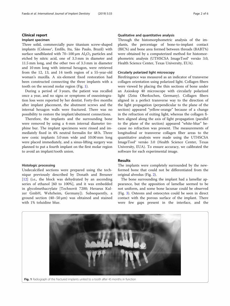
Clinical reportImplant specimenThree solid, commercially pure titanium screw-shapedimplants (Colosso®, Emfils, Itu, São Paulo, Brazil) withsurface sandblasted with 70–100 μm Al2O3 particles andetched by nitric acid, one of 3.3 mm in diameter and11.5 mm long, and the other two of 3.3 mm in diameterand 10 mm long with internal hexagon, were retrievedfrom the 12, 13, and 14 teeth region of a 55-year-oldwoman’s maxilla. A six-element fixed restoration hadbeen constructed connecting the three implants with atooth on the second molar region (Fig. 1).During a period of 3 years, the patient was recalled
once a year, and no signs or symptoms of osseointegra-tion loss were reported by her dentist. Forty-five monthsafter implant placement, the abutment screws and theinternal hexagon walls were fractured. There was nopossibility to restore the implant/abutment connections.Therefore, the implants and the surrounding bone
were removed by using a 4-mm internal diameter tre-phine bur. The implant specimens were rinsed and im-mediately fixed in 4% neutral formalin for 48 h. Threenew conic implants 5.0 mm wide and 10.00 mm longwere placed immediately, and a sinus-lifting surgery wasplanned to put a fourth implant on the first molar regionto avoid an implant/tooth union.
Histologic processingUndecalcified sections were prepared using the tech-nique previously described by Donath and Breuner[12] (i.e., the block was dehydrated by an ascendingseries of ethanol [60 to 100%], and it was embeddedin glycolmethacrylate [Technovit 7200; Heraeus Kul-zer GmbH, Wehrheim, Germany]). Subsequently, aground section (40–50 μm) was obtained and stainedwith 1% toluidine blue.
Qualitative and quantitative analysisThrough the histomorphometric analysis of the im-plants, the percentage of bone-to-implant contact(BIC%) and bone area formed between threads (BABT%)were obtained by a computerized method for histomor-phometric analysis (UTHSCSA ImageTool® versão 3.0,Health Science Center, Texas University, EUA).
Circularly polarized light microscopyBirefringence was measured as an indicator of transversecollagen orientation using polarized light. Collagen fiberswere viewed by placing the thin sections of bone underan Axioskop 40 microscope with circularly polarizedlight (Zeiss Oberkochen, Germany). Collagen fibersaligned in a perfect transverse way to the direction ofthe light propagation (perpendicular to the plane of thesection) appeared “yellow-orange” because of a changein the refraction of exiting light, whereas the collagen fi-bers aligned along the axis of light propagation (parallelto the plane of the section) appeared “white-blue” be-cause no refraction was present. The measurements oflongitudinal or transverse collagen fiber areas to thequantitative analysis were made using the UTHSCSAImageTool® versão 3.0 (Health Science Center, TexasUniversity, EUA). To ensure accuracy, we calibrated thesoftware for each experimental image.
ResultsThe implants were completely surrounded by the new-formed bone that could not be differentiated from theoriginal alveolus (Fig. 2).The bone surrounding the implant had a lamellar ap-
pearance, but the apposition of lamellae seemed to benot uniform, and some bone lacunae could be observed(Fig. 3). Osteons and osteocytes could be seen in directcontact with the porous surface of the implant. Therewere few gaps present in the interface, and the
Fig. 1 Radiograph of the fractured implants united to a tooth after 45 months in function
Faeda et al. International Journal of Implant Dentistry (2019) 5:33 Page 2 of 6

histomorphometric analysis revealed a mean bone-to-implant contact of 80.3% ± 4.1% (mean ± standard devi-ation) and a mean bone area of 77.3% ± 9.6% formedwithin the limits of the implant threads Orientation ofthe collagen fibers in the peri-implant bone was 63.14%,40.91%, and 40.29% for transverse collagen fibers and36.86%, 59.09%, and 59.71% for the longitudinal collagenfibers, respectively, for implants 12, 13, and 14 (Fig. 4).The comparison of the proportions between transverse
and longitudinal collagen fibers revealed a high percentageof transverse fibers in contact with the implants posi-tioned on the posterior region (Fig. 5). The histologicalobservations on bone microstructure revealed an intensebone remodeling activity near the dental implant surface.
DiscussionHistomorphometric analysis of human retrieved im-plants is the method available to analyze the bone-to-im-plant interface behavior over time [13]. Thereproduction of a human’s mouth environment in ani-mals is tremendously difficult. Therefore, this study cancontribute to the knowledge of human bone response toa dental implant under loading conditions.Many efforts have been made by researchers and man-
ufacturers to produce implant surfaces attractive to liv-ing cells and, consequently, to improve the quantity andquality of osseointegration. It has been reported that mi-cro-rough topography observed in a porous implantcould favorably affect angiogenesis, as well as cellularmigratory events, activity, and function [14], resulting ina faster and higher bone-implant contact and mechanicalinterlocking [6, 7].
Fig. 2 General overview of sandblasted and acid-etched implant histologic section. Note the presence of abutment screws and the internalhexagon walls which are fractured, and high bone density within threads of implant (toluidine blue, original magnification × 1.5)
Fig. 3 Histologic appearance of sandblasted and acid-etchedimplant. Direct bone contact with well-organized bone and lamellarapposition is observed
Faeda et al. International Journal of Implant Dentistry (2019) 5:33 Page 3 of 6

In this study, three commercially pure, titanium screw-type implants were used. They formerly received a sand-blasting treatment with aluminum oxide to promotemacroporosities, and they were acid-etched to achieve mi-croporosities. Rates of 80.3% of bone-to-implant contactand 77.3% of bone area within the limits of the implantthreads were found. These findings are similar to the re-sult reported by Hayakawa et al. [15] (76.60%) when a
sandblasted and acid-etched implant was placed into thepalatal bone as anchorage for orthodontic treatment.Other investigators reported similar results with differ-
ent surfaces and follow-up. Piattelli et al. [5] found 60 to70% of bone-to-implant contact to titanium plasmaspray implant. Brunel et al [16] reported 74% with hy-droxyapatite coating in maxilla after 14 months of fol-low-up, and Degidi et al. [17] found 60% after 9 months
Fig. 4 Under circularly polarized light microscopy (a) (original magnification × 50), the transverse collagen fibers appear in yellow-orange, whilethe longitudinal ones appear in white-blue. The transverse collagen fibers are mainly associated to the lower flank of the threads. b Originalmagnification × 200. Bone appears mainly constituted by transverse collagen fibers. The presence of an osteon shows bone remodeling activity
Fig. 5 Percentage of the collagen fiber orientation of each implant
Faeda et al. International Journal of Implant Dentistry (2019) 5:33 Page 4 of 6

of follow-up in porous anodized implant submitted toimmediate loading.Based on the trustworthiness of the macro- and micro-
implant systems, the implant-tooth splinting has beenconsidered as an alternative in some clinical situations.Although some studies show satisfactory success in shortand near future [18, 19], the previsibility of the implant/tooth system is still unclear.The amount of tooth movement with healthy peri-
odontal ligaments against that of an osseointegrateddental implant can be 5–20 times greater [20]. This dis-parity causes the implant side to receive a higher bend-ing moment as a result of the bridge function as acantilever construction and is only supported by the im-plant when the occlusal load acts on the tooth [21]. Aseries of potential problems such as tooth intrusion,osseointegration loss, screw loosening, and implant orprosthesis fracture can arise, with resulting complicatedphysiological and engineering aspects [18, 19, 22].In the present case, three implants with 3.3mm in
diameter and with internal hexagon abutment connectionwere positioned at the 12, 13, and 14 teeth region andunited to the second molar with reduced periodontal sup-port, having the first molar suspended between them. Asdescribed by the literature, the occlusal load over the firstand second molars resulted in a cantilever force that con-centrated on the implant neck [23]. The association ofoverload, inadequate implant diameter to the case, and in-ternal hexagon connection resulted in the abutment screwand the internal hexagon wall fracture.The studies already showed the relation between the
load and the collagen fiber orientation in bone nearthreaded dental [24, 25]. The spatial orientation of colla-gen fibers has a direct bearing on its mechanical proper-ties [24]. Based on a number of studies, several authorsalso correlate strongly the collagen fiber orientation tothe loading regimen [26, 27].In 1958, Evans [28] described the relation between the
bone stiffness and predominant direction of the collagenfibers in the bone matrix. When collagen fibers ran par-allel to the loading vector, the bone was more resistant.McElhaney [29] found that the ultimate compressivestrength and modulus of elasticity of the cortical boneincreased with increasing strain rate. The energy absorp-tion capacity had a maximum at an intermediate strainrate. It was suggested that low strain rate shear failuresresult from a distortion of the lamellar substructure andfracture along several weaker planes. High strain ratefailures appeared to follow the cement lines, constitutingthe boundaries of the haversian and lamellar systems.Traini et al. observed that the load can influence the col-
lagen fiber orientation in bone near threaded dental im-plants in immediately loaded implants [24, 30]. Theyfound that loading has a relevant influence in the
distribution of the collagen fibers in the peri-implantbone. Transverse collagen fibers, related to compressiveloads, were found in a higher and statistically significantquantity in loaded than in unloaded implants or inthe alveolar bone. The bone tissue responded to anoverloading (until the threshold of the implant frac-ture was reached) by modeling and remodeling itsmicrostructure. The predominance of transverse colla-gen fiber orientation should be related to a high com-pression state [24, 30].Regarding BIC and high load occlusion, Chang et
al. [31] performed a systematic literature review andconcluded that the greatest peri-implant bone remod-eling activity is found around implants subjected tohigh loading forces, when the applied force exceedsthe biological adaptable limit. The authors reportedthat there was a limitation in the research due to theabsence of experimental studies in humans. But theauthors suggest that a possible correlation betweenocclusal overload and implant failures is related tothe degree tolerance of the alveolar bone which variesaccording to the individual, the location, and otheranatomic and physiological parameters.
ConclusionWithin the limitation of this study, the possible cause ofthe implant fracture could be the association of overload,inadequate implant diameter, and fragile internal hexa-gon connection.
AbbreviationsBABT: Bone area formed between threads; BIC: Bone-to-implant contact
AcknowledgementsNot applicable
Authors’ contributionsRSF designed the study, wrote the protocol and the first draft of themanuscript, and contributed to its critical revision. SCSdN managed theliterature searches and wrote the protocol and the first draft of themanuscript. PLS performed the surgical procedures, followed up the patient,managed the study analyses, and contributed to the manuscript writing.RJBN performed the surgical procedures, followed up the patient, andmanaged the study analyses. RS contributed to the acquisition, analysis, andinterpretation of data. RM performed the prosthetic procedures, followed upthe patient, managed the study analyses, and contributed to the manuscriptwriting. EMJ contributed to the conception and design of the study, and thecritical revision of the manuscript. All authors read and approved the finalmanuscript.
FundingThe authors declare that they have no funding.
Availability of data and materialsNot applicable
Ethics approval and consent to participateOk
Consent for publicationOk
Faeda et al. International Journal of Implant Dentistry (2019) 5:33 Page 5 of 6

Competing interestsRafael Silveira Faeda, Suzana Clesia Silverio do Nascimento, Pâmela LeticiaSantos, Rodolfo Jorge Boeck Neto, Rafael Sartori, Rogerio Margonar, and ElcioMarcantonio Jr. declare that they have no competing interests.
Author details1Department of Health Sciences, Post-graduation Program in Implantology,University of Araraquara - UNIARA Dental School, Rua Carlos Gomes, 1338,Centro, Araraquara, SP 14801-340, Brazil. 2Department of Health Sciences,University of Araraquara (UNIARA), Araraquara, SP, Brazil. 3Department ofDiagnosis and Surgery, School of Dentistry at Araraquara, São Paulo StateUniversity (UNESP), Araraquara, SP, Brazil.
Received: 4 June 2019 Accepted: 23 August 2019
References1. Brånemark PI, Adell R, Breine U, et al. Intra-osseous anchorage of dental
prosthesis I. Experimental studies. Scand J Plast Reconsrt Surg. 1969;3:81–100.
2. Santos PL, Molon RS, Queiroz TP, Okamoto R, Faloni APS, Gulinelli JL,Luvizuto ER, Garcia-Junior IR. Evaluation of bone substitutes for treatment ofperi-implant bone defects: biomechanical, histological, andimmunohistochemical analyses in the rabbit tibia. J Periodontal Implant Sci.2016;46(3):176–96.
3. Margonar R, Sakakura CE, Holzhausen M, et al. The influence of diabetesmellitus and insulin therapy on biomechanical retention around dentalimplants: a biomechanical study in rabbits. Implant Dent. 2003;12:333–9.
4. Linden MSS, Paranhos LR, De Carli JP, Trentin MS, de Bittencourt ME, Santos PL,Groppo FC, Ramacciato JC. Influence of nicotine on machined- and anodized-surface implants. Histometric analysis. J Clin Exp Dent. 2017;9(10):e1207–11.
5. Piattelli A, Degidi M, Marchetti C, et al. Histologic analysis of the interface oftitanium implant retrieved from a nonvascularized mandibular block graftafter a 10- month loading period. Int J Maxillofac Implants. 1997;12:840–3.
6. Cordioli G, Majzoub Z, Piattelli A, et al. Removal torque andhistomorphometric investigation of 4 different surfaces: an experimentalstudy in the rabbit tibia. Int J Oral Maxillofac Implants. 2000;15:668–74.
7. Linden MSS, Bittencourt ME, Carli JP, Miyagaki DC, Santos PLD, Paranhos LR,Groppo FC, Ramacciato JC. The effects of subcutaneous injection ofnicotine on osseointegration of machined and anodized implants in rabbits.Acta Cir Bras. 2018;33(1):31–9.
8. Traini T, De Paoli S, Caputi S, Iezzi G, Piattelli A. Collagen fiber orientationnear a fractured dental implant after a 5-year loading period: case report.Implant Dent. 2006;15(1):70–6.
9. Langer B, Sullivan DY. Osseointegration: its impact on the interrelationshipof periodontics and restorative dentistry: part II. Int J PeriodonticsRestorative Dent. 1989;9:165–83.
10. Lundgren D, Laurell L. Biomechanical aspect of fixed bridgework supportedby natural teeth and endosseous implants. Periodontol. 1994;4:23–40.
11. Ozçelik T, Ersoy AE. An investigation of tooth/implant-supported fixedprosthesis designs with two different stress analysis methods: an in vitrostudy. J Prosthodont. 2007;16:107–16.
12. Donath K, Breuner G. A method for study of undecalcified bones and teethwith attached soft tissue. The sage-Scliff (sawing and grinding) technique. JOral Pathol. 1982;11:318–26.
13. Spin-Neto R, Bedran TB, De Paula WN, et al. Incisive canal deflation forcorrect implant placement: case report. Implant Dent. 2009;18:473–9.
14. Schneider GB, Perinpanayagam H, Clegg M, Zaharias R, Seabold D, Keller J,Stanford C. Implant surface roughness affect osteoblast gene expression. JDent Res. 2003;82:372.
15. Hayakawa T, Kiba H, Yasuda S, et al. A histologic and histomorphometricevaluation of two types of retrieved human titanium implants. Int JPeriodontics Restorative Dent. 2002;22:164–71.
16. Brunel G, Armand S, Miller N, et al. Histologic analysis of fractured implant: acase report. Int J Periodontics Restorative Dent. 2000;20:521–6.
17. Degidi M, Petrone G, Iezzi G, et al. Histologic evaluation of a humanimmediately loaded titanium implant with a porous anodized surface. ClinImplant Dent Relat Res. 2002;4:110–4.
18. Gross M, Laufer BZ. Splinting osseointegrated implants and natural teeth inrehabitation of partially edentulous patients. Part I: laboratory and clinicalstudies. J Oral Rehabil. 1997;24:863–70.
19. Laufer BZ, Gross M. Splinting osseointegrated implants and natural teeth inrehabilitation of partially edentulous patients. Part ¼ 2 II: principles andapplications. J Oral Rehabil. 1998;25:69–80.
20. Nyman S, Lang NP. Tooth mobility and the biological rationale for splintingteeth. Periodontol. 1994;4:15–22.
21. Lin CL, Wang JC, Kuo YC. Numerical simulation on the biomechanicalinteractions of tooth/implant-supported system under various occlusalforces with rigid/non-rigid connections. J Biomech. 2006;39(3):453.
22. Becker CM, Kaiser DA, Jones JD. Guidelines for splinting implants. J ProsthetDent. 2000;84:210–4.
23. Menicucci G, Mossolov A, Mozzati M, Lorenzetti M, Preti G. Tooth– implantconnection: some biomechanical aspects based on finite element analyses.Clin Oral Implants Res. 2002;13:334–41.
24. Traini T, Degidi M, Strocchi R, et al. Collagen fibers orientation near dentalimplants in human bone: do their organization reflect differences inloading? J Biomed Mater Res B Appl Biomater. 2005;74:538–46.
25. Delgado-Ruiz RA, Abboud M, Romanos G, Aguilar-Salvatierra A, Gomez-Moreno G, Calvo-Guirado JL. Peri-implant bone organization surroundingzirconia-microgrooved surfaces circularly polarized light and confocal laserscanning microscopy study. Clin Oral Implants Res. 2015;26(11):1328–37.
26. Kalmey JK, Lovejoy CO. Collagen fiber orientation in the femoral necks ofapes and humans: do their histological structures reflect differences inlocomotor loading? Bone. 2002;31:327–32.
27. Tami AE, Nasser P, Verborgt O, Schaffler MB, Knothe Tate ML. The role ofinterstitial fluid flow in the remodeling response to fatigue loading. J BoneMiner Res. 2002;17:2030–7.
28. Evans FG. Relations between the microscopic structure and tensile strengthof human bone. Acta Anat (Basel). 1958;35:285–301.
29. McElhaney JH. Dynamic response of bone and muscle tissue. J Appl Physiol.1966;21:1231–6.
30. Traini T, Degidi M, Caputi S, et al. Collagen fiber orientation in humanperiimplant bone of immediately loaded and unloaded titanium dentalimplants. J Periodontol. 2005;76:83–9.
31. Chang M, Chronopoulos V, Mattheos N. Impact of excessive occlusal loadon successfully osseointegrated dental implants: a literature review. JInvestig Clin Dent. 2013;4:142–50.
Publisher’s NoteSpringer Nature remains neutral with regard to jurisdictional claims inpublished maps and institutional affiliations.
Faeda et al. International Journal of Implant Dentistry (2019) 5:33 Page 6 of 6


![Histology Slides - mediconotes.commediconotes.com/freenotes/basic/histology_laboratory_slides.pdf[Histology] Histology Slides MedicoNotes provides real laboratory Histological slides](https://static.fdocuments.net/doc/165x107/5ae110e87f8b9a5a668e6aa3/histology-slides-histology-histology-slides-mediconotes-provides-real-laboratory.jpg)