Human Coronaviruses: A Review of Virus–Host Interactions Human coronaviruses A... · HCoV-OC43,...
28
diseases Review Human Coronaviruses: A Review of Virus–Host Interactions Yvonne Xinyi Lim, Yan Ling Ng, James P. Tam and Ding Xiang Liu * School of Biological Sciences, Nanyang Technological University, 60 Nanyang Drive, Singapore 637551, Singapore; [email protected] (Y.X.L.); [email protected] (Y.L.N.); [email protected] (J.P.T.) * Correspondence: [email protected]; Tel.: +65-6316-2861 Academic Editor: Maurizio Battino Received: 8 June 2016; Accepted: 18 July 2016; Published: 25 July 2016 Abstract: Human coronaviruses (HCoVs) are known respiratory pathogens associated with a range of respiratory outcomes. In the past 14 years, the onset of severe acute respiratory syndrome coronavirus (SARS-CoV) and Middle East respiratory syndrome coronavirus (MERS-CoV) have thrust HCoVs into spotlight of the research community due to their high pathogenicity in humans. The study of HCoV-host interactions has contributed extensively to our understanding of HCoV pathogenesis. In this review, we discuss some of the recent findings of host cell factors that might be exploited by HCoVs to facilitate their own replication cycle. We also discuss various cellular processes, such as apoptosis, innate immunity, ER stress response, mitogen-activated protein kinase (MAPK) pathway and nuclear factor kappa B (NF-κB) pathway that may be modulated by HCoVs. Keywords: human coronavirus; virus–host interactions; apoptosis; innate immunity; ER stress; MAPK; NF-κB 1. Introduction Human coronaviruses (HCoVs) represent a major group of coronaviruses (CoVs) associated with multiple respiratory diseases of varying severity, including common cold, pneumonia and bronchilitis [1]. Today, HCoVs are recognised as one of the most rapidly evolving viruses owing to its high genomic nucleotide substitution rates and recombination [2]. In recent years, evolution of HCoVs has also been expedited by factors such as urbanization and poultry farming. These have permitted the frequent mixing of species and facilitated the crossing of species barrier and genomic recombination of these viruses [3]. To date, six known HCoVs have been identified, namely HCoV-229E, HCoV-NL63, HCoV-OC43, HCoV-HKU1, severe acute respiratory syndrome coronavirus (SARS-CoV) and Middle East respiratory syndrome coronavirus (MERS-CoV); of which, four HCoVs (HCoV-229E, HCoV-NL63, HCoV-OC43 and HCoV-HKU1) are globally circulated in the human population and contribute to approximately one-third of common cold infections in humans [4]. In severe cases, these four HCoVs can cause life-threatening pneumonia and bronchiolitis especially in elderly, children and immunocompromised patients [1,5,6]. Besides respiratory illnesses, they may also cause enteric and neurological diseases [7–11]. SARS-CoV first emerged in 2002–2003 in Guangdong, China as an atypical pneumonia marked by fever, headache and subsequent onset of respiratory symptoms such as cough and pneumonia, which may later develop into life-threatening respiratory failure and acute respiratory distress syndrome [12]. Being highly transmissible among humans, it quickly spread across 29 countries, infecting more than 8000 individuals with a mortality rate of about 10% [13,14]. Originally, palm civets were thought to be the natural reservoir for the virus [15]. However, subsequent phylogenetic studies pointed to the bat origin of SARS-CoV based on sequences of SARS-like virus found in bats [16]. The MERS-CoV epidemic surfaced in Saudi Arabia in 2012 with similar clinical symptoms as SARS-CoV Diseases 2016, 4, 26; doi:10.3390/diseases4030026 www.mdpi.com/journal/diseases
Transcript of Human Coronaviruses: A Review of Virus–Host Interactions Human coronaviruses A... · HCoV-OC43,...

diseases
Review
Human Coronaviruses: A Review ofVirus–Host InteractionsYvonne Xinyi Lim, Yan Ling Ng, James P. Tam and Ding Xiang Liu *
School of Biological Sciences, Nanyang Technological University, 60 Nanyang Drive, Singapore 637551,Singapore; [email protected] (Y.X.L.); [email protected] (Y.L.N.); [email protected] (J.P.T.)* Correspondence: [email protected]; Tel.: +65-6316-2861
Academic Editor: Maurizio BattinoReceived: 8 June 2016; Accepted: 18 July 2016; Published: 25 July 2016
Abstract: Human coronaviruses (HCoVs) are known respiratory pathogens associated with a range ofrespiratory outcomes. In the past 14 years, the onset of severe acute respiratory syndrome coronavirus(SARS-CoV) and Middle East respiratory syndrome coronavirus (MERS-CoV) have thrust HCoVsinto spotlight of the research community due to their high pathogenicity in humans. The study ofHCoV-host interactions has contributed extensively to our understanding of HCoV pathogenesis.In this review, we discuss some of the recent findings of host cell factors that might be exploited byHCoVs to facilitate their own replication cycle. We also discuss various cellular processes, such asapoptosis, innate immunity, ER stress response, mitogen-activated protein kinase (MAPK) pathwayand nuclear factor kappa B (NF-κB) pathway that may be modulated by HCoVs.
Keywords: human coronavirus; virus–host interactions; apoptosis; innate immunity; ER stress;MAPK; NF-κB
1. Introduction
Human coronaviruses (HCoVs) represent a major group of coronaviruses (CoVs) associatedwith multiple respiratory diseases of varying severity, including common cold, pneumonia andbronchilitis [1]. Today, HCoVs are recognised as one of the most rapidly evolving viruses owing to itshigh genomic nucleotide substitution rates and recombination [2]. In recent years, evolution of HCoVshas also been expedited by factors such as urbanization and poultry farming. These have permitted thefrequent mixing of species and facilitated the crossing of species barrier and genomic recombination ofthese viruses [3]. To date, six known HCoVs have been identified, namely HCoV-229E, HCoV-NL63,HCoV-OC43, HCoV-HKU1, severe acute respiratory syndrome coronavirus (SARS-CoV) and MiddleEast respiratory syndrome coronavirus (MERS-CoV); of which, four HCoVs (HCoV-229E, HCoV-NL63,HCoV-OC43 and HCoV-HKU1) are globally circulated in the human population and contributeto approximately one-third of common cold infections in humans [4]. In severe cases, these fourHCoVs can cause life-threatening pneumonia and bronchiolitis especially in elderly, children andimmunocompromised patients [1,5,6]. Besides respiratory illnesses, they may also cause enteric andneurological diseases [7–11].
SARS-CoV first emerged in 2002–2003 in Guangdong, China as an atypical pneumonia markedby fever, headache and subsequent onset of respiratory symptoms such as cough and pneumonia,which may later develop into life-threatening respiratory failure and acute respiratory distresssyndrome [12]. Being highly transmissible among humans, it quickly spread across 29 countries,infecting more than 8000 individuals with a mortality rate of about 10% [13,14]. Originally, palmcivets were thought to be the natural reservoir for the virus [15]. However, subsequent phylogeneticstudies pointed to the bat origin of SARS-CoV based on sequences of SARS-like virus found in bats [16].The MERS-CoV epidemic surfaced in Saudi Arabia in 2012 with similar clinical symptoms as SARS-CoV
Diseases 2016, 4, 26; doi:10.3390/diseases4030026 www.mdpi.com/journal/diseases
Diseases 2016, 4, 26 2 of 28
but with a much higher mortality rate of about 35% [17]. Unlike SARS-CoV, which exhibits super-spreaderevents, transmission of MERS-CoV is geographically limited [12]. In fact, reported cases of MERS-CoVoften stem from outbreaks within the Middle Eastern countries or recent travel to the region [18,19].
Taxonomy, Genomic Structure and Morphology
CoVs are a group of large enveloped RNA viruses under the Coronaviridae family. Togetherwith Artierivirdae and Roniviridae, Coronaviridae is classified under the Nidovirale order [20].As proposed by the International Committee for Taxonomy of Viruses, CoVs are further categorizedinto four main genera, Alpha-, Beta-, Gamma- and Deltacoronaviruses based on sequence comparisons ofentire viral genomes [21,22]. These CoVs can infect a wide variety of hosts, including avian, swineand humans. HCoVs are identified to be either in the Alpha- or Betacoronavirus genera, includingAlphacoronaviruses, HCoV-229E and HCoV-NL63, and Betacoronaviruses, HCoV-HKU1, SARS-CoV,MERS-CoV and HCoV-OC43 (Table 1).
Under the electron microscope, the CoV virions appear to be roughly spherical or moderatelypleomorphic, with distinct “club-like” projections formed by the spike (S) protein [23,24]. Within thevirion interior lies a helically symmetrical nucleocapsid that encloses a single-stranded and positivesense RNA viral genome of an extraordinarily large size of about 26 to 32 kilobases [20]. The positivesense viral genomic RNA acts as a messenger RNA (mRNA), comprising a 51 terminal cap structureand a 31 poly A tail. This genomic RNA acts in three capacities during the viral life cycle: (1) as an initialRNA of the infectious cycle; (2) as a template for replication and transcription; and (3) as a substrate forpackaging into the progeny virus. The replicase-transcriptase is the only protein translated from thegenome, while the viral products of all downstream open reading frames are derived from subgenomicmRNAs. In all CoVs, the replicase gene makes up approximately 51 two-thirds of the genome and iscomprised of two overlapping open reading frames (ORFs), ORF1a and ORF1b, which encodes 16non-structural proteins. The final one-third of the CoV genomic RNA encodes CoV canonical set offour structural protein genes, in the order of spike (S), envelope (E), membrane (M) and nucleocapsid(N). In addition, several accessory ORFs are also interspersed along the structural protein genes andthe number and location varies among CoV species [25] (Figure 1).
Diseases 2016, 4, 26 2 of 27
SARS-CoV but with a much higher mortality rate of about 35% [17]. Unlike SARS-CoV, which exhibits super-spreader events, transmission of MERS-CoV is geographically limited [12]. In fact, reported cases of MERS-CoV often stem from outbreaks within the Middle Eastern countries or recent travel to the region [18,19].
Taxonomy, Genomic Structure and Morphology
CoVs are a group of large enveloped RNA viruses under the Coronaviridae family. Together with Artierivirdae and Roniviridae, Coronaviridae is classified under the Nidovirale order [20]. As proposed by the International Committee for Taxonomy of Viruses, CoVs are further categorized into four main genera, Alpha-, Beta-, Gamma- and Deltacoronaviruses based on sequence comparisons of entire viral genomes [21,22]. These CoVs can infect a wide variety of hosts, including avian, swine and humans. HCoVs are identified to be either in the Alpha- or Betacoronavirus genera, including Alphacoronaviruses, HCoV-229E and HCoV-NL63, and Betacoronaviruses, HCoV-HKU1, SARS-CoV, MERS-CoV and HCoV-OC43 (Table 1).
Under the electron microscope, the CoV virions appear to be roughly spherical or moderately pleomorphic, with distinct “club-like” projections formed by the spike (S) protein [23,24]. Within the virion interior lies a helically symmetrical nucleocapsid that encloses a single-stranded and positive sense RNA viral genome of an extraordinarily large size of about 26 to 32 kilobases [20]. The positive sense viral genomic RNA acts as a messenger RNA (mRNA), comprising a 5′ terminal cap structure and a 3′ poly A tail. This genomic RNA acts in three capacities during the viral life cycle: (1) as an initial RNA of the infectious cycle; (2) as a template for replication and transcription; and (3) as a substrate for packaging into the progeny virus. The replicase-transcriptase is the only protein translated from the genome, while the viral products of all downstream open reading frames are derived from subgenomic mRNAs. In all CoVs, the replicase gene makes up approximately 5′ two-thirds of the genome and is comprised of two overlapping open reading frames (ORFs), ORF1a and ORF1b, which encodes 16 non-structural proteins. The final one-third of the CoV genomic RNA encodes CoV canonical set of four structural protein genes, in the order of spike (S), envelope (E), membrane (M) and nucleocapsid (N). In addition, several accessory ORFs are also interspersed along the structural protein genes and the number and location varies among CoV species [25] (Figure 1).
Figure 1. Genome organisation of human coronaviruses (HCoVs). HCoV genomes range from about 26 to 32 kilobases (kb) in size, as indicated by the black lines above the scale. Coronavirus (CoV) genome is typically arranged in the order of 5′-ORF1a-ORF1b-S-E-M-N-3′. The overlapping open reading frames (ORF) ORF1a and ORF1b comprise two-thirds of the coronavirus genome, which encodes for all the viral components required for viral RNA synthesis. The other one-third of the genome at the 3′ end encodes for a set of structural (orange) and non-structural proteins (green).
Figure 1. Genome organisation of human coronaviruses (HCoVs). HCoV genomes range from about 26to 32 kilobases (kb) in size, as indicated by the black lines above the scale. Coronavirus (CoV) genome istypically arranged in the order of 51-ORF1a-ORF1b-S-E-M-N-31. The overlapping open reading frames(ORF) ORF1a and ORF1b comprise two-thirds of the coronavirus genome, which encodes for all theviral components required for viral RNA synthesis. The other one-third of the genome at the 31 endencodes for a set of structural (orange) and non-structural proteins (green).

Diseases 2016, 4, 26 3 of 28
Table 1. Classification of human coronavirus.
CoronaviriniaeGenera Strains Discovery Cellular Receptor Host References
Alpha-coronavirus HCoV-229E 1966 Human Aminopeptidase N (CD13) Bats [1,2,21]HCoV-NL63 2004 ACE2 Palm Civets, Bats [3,21]
Beta-coronavirus
HCoV-OC43 1967 9-O-Acetylated sialic acid Cattle [4,5]HcoV-HKU1 2005 9-O-Acetylated sialic acid Mice [6,7]SARS-CoV 2003 ACE2 Palm Civets, Bats [8,19,21]MERS-CoV 2012 DPP4 Bats, Camels [9]
2. Involvement of Host Factors in Viral Replication and Pathogenesis
As intracellular obligate parasites, HCoVs exploit the host cell machinery for their own replicationand spread. Since virus–host interactions form the basis of diseases, knowledge about their interplayis of great research interest. Here, we describe what is currently known of the cell’s contribution inCoV infection cycle: attachment; entry into the host cell; translation of the replicase-transcriptase;replication of genome and transcription of mRNAs; and assembly and budding of newly packagedvirions (Figure 2).
Diseases 2016, 4, 26 3 of 27
Table 1. Classification of human coronavirus.
Coronaviriniae Genera
Strains Discovery Cellular Receptor Host References
Alpha-coronavirus HCoV-229E 1966 Human Aminopeptidase N (CD13) Bats [1,2,21] HCoV-NL63 2004 ACE2 Palm Civets, Bats [3,21]
Beta-coronavirus
HCoV-OC43 1967 9-O-Acetylated sialic acid Cattle [4,5] HcoV-HKU1 2005 9-O-Acetylated sialic acid Mice [6,7] SARS-CoV 2003 ACE2 Palm Civets, Bats [8,19,21] MERS-CoV 2012 DPP4 Bats, Camels [9]
2. Involvement of Host Factors in Viral Replication and Pathogenesis
As intracellular obligate parasites, HCoVs exploit the host cell machinery for their own replication and spread. Since virus–host interactions form the basis of diseases, knowledge about their interplay is of great research interest. Here, we describe what is currently known of the cell’s contribution in CoV infection cycle: attachment; entry into the host cell; translation of the replicase-transcriptase; replication of genome and transcription of mRNAs; and assembly and budding of newly packaged virions (Figure 2).
Figure 2. Coronavirus replication cycle. Coronavirus infection begins with the attachment of the S1 domain of the spike protein (S) with its cognate receptor. This drives the conformational change in the S2 subunit in S, promoting the fusion of the viral and cell plasma membrane. Following the release of the nucleocapsid to the cytoplasm, the viral gRNA is translated through ribosomal frameshifting to produce polyproteins pp1a and pp1ab. pp1a and pp1ab are autoproteolytically processed by host and viral proteases to generate 16 non-structural proteins (NSPs), which will then be assembled to form the replicase-polymerase. The replicase-polymerase is involved in the coronaviral replication, a process in which the genomic RNA are replicated and the subgenomic RNA will be transcribed and translated to form the structural proteins. The viral products produced will be assembled in the ERGIC, and bud out as a smooth-wall vesicle to the plasma membrane to egress via exocytosis. Host factors that promote infection and inhibit infection are highlighted in green and red, respectively. APN, aminopeptidase N; ACE2, Angiotensin converting enzyme 2; DPP4, dipeptidyl peptidase 4; 9-O-Ac Sialic Acid, 9-O-Acetylated Sialic Acid; IFITM, Interferon induced transmembrane protein; ATP1A1, ATPase, Na+/K+ Transporting, Alpha 1 Polypeptide; HnRNP A1, Heterogeneous nuclear ribonucleoprotein A1; MADP1, Zinc Finger CCHC-Type and RNA Binding Motif 1; DDX1, ATP-dependent RNA Helicase; PCBP1/2, Poly r(C) binding protein 1/2; PABP, Poly A binding protein; COPB2, Coatomer protein complex, subunit beta 2 (beta prime); GAPDH, Glyceraldehyde 3-phosphate dehydrogenase; ERGIC, Endoplasmic reticulum Golgi intermediate compartment; ER, endoplasmic reticulum; VCP, Valosin-Containing Protein.
Figure 2. Coronavirus replication cycle. Coronavirus infection begins with the attachment of theS1 domain of the spike protein (S) with its cognate receptor. This drives the conformational changein the S2 subunit in S, promoting the fusion of the viral and cell plasma membrane. Followingthe release of the nucleocapsid to the cytoplasm, the viral gRNA is translated through ribosomalframeshifting to produce polyproteins pp1a and pp1ab. pp1a and pp1ab are autoproteolyticallyprocessed by host and viral proteases to generate 16 non-structural proteins (NSPs), which will then beassembled to form the replicase-polymerase. The replicase-polymerase is involved in the coronaviralreplication, a process in which the genomic RNA are replicated and the subgenomic RNA will betranscribed and translated to form the structural proteins. The viral products produced will beassembled in the ERGIC, and bud out as a smooth-wall vesicle to the plasma membrane to egress viaexocytosis. Host factors that promote infection and inhibit infection are highlighted in green and red,respectively. APN, aminopeptidase N; ACE2, Angiotensin converting enzyme 2; DPP4, dipeptidylpeptidase 4; 9-O-Ac Sialic Acid, 9-O-Acetylated Sialic Acid; IFITM, Interferon induced transmembraneprotein; ATP1A1, ATPase, Na+/K+ Transporting, Alpha 1 Polypeptide; HnRNP A1, Heterogeneousnuclear ribonucleoprotein A1; MADP1, Zinc Finger CCHC-Type and RNA Binding Motif 1; DDX1,ATP-dependent RNA Helicase; PCBP1/2, Poly r(C) binding protein 1/2; PABP, Poly A binding protein;COPB2, Coatomer protein complex, subunit beta 2 (beta prime); GAPDH, Glyceraldehyde 3-phosphatedehydrogenase; ERGIC, Endoplasmic reticulum Golgi intermediate compartment; ER, endoplasmicreticulum; VCP, Valosin-Containing Protein.

Diseases 2016, 4, 26 4 of 28
2.1. Coronavirus Attachment and Entry
CoV infection is initiated by the attachment to specific host cellular receptors via the spike (S)protein. The host receptor is a major determinant of pathogenicity, tissue tropism and host range of thevirus. The S protein comprises of two domains: S1 and S2. The interaction between the S1 domain andits cognate receptor triggers a conformational change in the S protein, which then promotes membranefusion between the viral and cell membrane through the S2 domain. Today, the main host cell receptorsutilised by all HCoVs are known: aminopeptidase N by HCoV-229E [26], angiotensin-convertingenzyme 2 (ACE2) by SARS-CoV [27] and HCoV-NL63 [28,29], dipeptidyl peptidase 4 (DPP4) byMERS-CoV [30] and 9-O-acetylated sialic acid by HCoV-OC43 and HCoV-HKU1 [31,32].
Apart from the conventional endosomal route of entry, some CoVs may also gain entry intothe cell via the non-endosomal pathway, or a combination of both. The low pH in the cellularenvironment and endosomal cysteine protease cathepsins may help to facilitate membrane fusion andendosomal CoV cell entry [33]. Recent evidence has supported the role of cathepsin L in SARS-CoV andMERS-CoV entry [34–36]. Other host proteases, such as transmembrane protease serine 2 (TMPRSS2)and airway trypsin-like protease TMPRSS11D, could also perform S1/S2 cleavage to activate the Sprotein for non-endosomal virus entry at the cell plasma membrane during HCoV-229E and SARS-CoVinfection [37,38]. In addition, MERS-CoV is also activated by furin, a serine endopeptidase that hasbeen implicated in the cell entry of other RNA viruses and S1/S2 cleavage during viral egress [39].
Many host cells also utilise its own factors to restrict viral entry. Using cell culture system andpseudotype virus, many groups have identified a family of interferon inducible transmembraneproteins (IFITM), which could inhibit global circulating HCoV-229E and HCoV-NL63 S proteinmediated entry, and also the highly pathogenic SARS-CoV and MERS-CoV [12,40]. While the IFITMmode of action remains elusive, cell-to-cell fusion assays performed by some research groups suggestthat IFITM3 blocks the enveloped virus entry by preventing fusion of the viral envelope with theplasma membrane or endosomal membranes through modulating the host membrane fluidity [41].
2.2. Coronavirus Replication
Following the release and uncoating of viral nucleocapsid to the cytoplasm, CoV replication beginswith the translation of ORF 1a and 1b into polyproteins pp1a (4382 amino acids) and pp1ab (7073 aminoacids). Here, the downstream ORF1b is translated through ribosomal frameshifting mechanism, inwhich a translating ribosome shifts one nucleotide in the ´1 direction, from the ORF1a reading frameinto ORF1b reading frame. This repositioning is enabled by two RNA elements—a 51-UUUAAAC-31
heptanucleotide slippery sequence and RNA pseudoknot structure. Subsequently, polyproteins pp1aand pp1ab are cleaved into at least 15 nsp, which assemble and form the replication-transcriptioncomplex. With the assembly of the replicase-polymerase, the full-length positive strand of genomicRNA is transcribed to form a full-length negative-strand template for the synthesis of new genomicRNAs and overlapping subgenomic negative-strand templates. These subgenomic mRNAs are thentranscribed and translated to produce the structural and accessory proteins. Several heterologousnuclear ribonucleoprotein (hnRNA) family members (hnRNPA1, PTB, SYN-CRYP) have been found tobe essential for efficient RNA replication [42]. Other RNA-binding proteins have also been suggestedto play a role in CoV replication, such as m-aconitase and poly-A-binding protein (PABP), DDX1,PCBP1/2 and zinc finger CCHC-type and RNA-binding motif 1 (MADP1) [43–45].
2.3. Coronavirus Assembly and Egress
The assembly of virions is quickly ensued with the accumulation of new genomic RNA andstructural components. In this phase of the infection cycle, the helical nucleocapsid containing thegenomic RNA interacts with other viral structural proteins (S, E and M proteins) to form the assembledvirion. The assembly of CoV particles is completed through budding of the helical nucleocapsidthrough membranes early in the secretory pathway from the endoplasmic reticulum to the Golgi

Diseases 2016, 4, 26 5 of 28
intermediate compartment (ERGIC). The contributions of the host in this phase of the infection cyclehave rarely been explored. Currently, it is known that the M protein orchestrates the entire assemblyprocess by selecting and organizing the viral envelope components at the assembly sites and bymediating the interactions with the nucleocapsid to allow the budding of virions [46]. The M proteininteracts with different viral structural proteins, such as the E protein, to assemble into a maturevirus. This interaction generates the scaffold of the virion envelope and induces the budding andrelease of the M protein-modified membrane and with the S protein to assemble the spikes into theviral envelope [46,47]. Following assembly and budding, the virions are transported in vesicles andeventually released by exocytosis. In a recent study, an inhibition of a Valosin-containing protein(VCP/p97) resulted in virus accumulation in early endosome in infectious bronchitis virus (IBV),suggesting a role for VCP in the maturation of virus-loaded endosomes [48].
3. Human Coronavirus Infection and Apoptosis
Apoptosis is a process of programmed cell death that is tightly regulated and anti-inflammatory.When cells undergo apoptosis, they demonstrate specific hallmarks such as cell shrinkage, extensiveplasma membrane blebbing, nuclear pykosis, DNA fragmentation and asymmetrical distribution ofplasma membrane [49–51]. To date, two main mechanisms of apoptosis have been established—theextrinsic and intrinsic pathways. The extrinsic pathway is initiated by the binding of extracellulardeath ligands (such as Fas ligand (FasL) and TNF-receptor-related apoptosis-inducing ligands (TRAIL))to death receptors from the tumour necrosis factor (TNF) super-family [52]. These death receptorsthen recruit various death adapter proteins, such as Fas-associated death domain protein (FADD) [53],and initiator procaspases 8 and 10 to form the death-inducing signalling complex (DISC) [54,55].Consequently, the two initiator procapases are cleaved to their active forms and induce a signallingcascade to eventually activate effector caspases 3 and 7. On the other hand, the intrinsic pathwayoccurs internally in the cell and involves changes in the mitochondrial outer membrane permeability(MOMP) based on the ratio of pro-apoptotic and anti-apoptotic B-cell lymphoma 2 (Bcl2) family ofproteins (Figure 3). Enhanced MOMP causes the release of pro-apoptotic factors such as cytochrome cto activate initiator caspase 9. Like the extrinsic pathway, activation of initiator caspase 9 in the intrinsicpathway results in the proteolytic cleavage effector caspases 3 and 7, which in turn process manykey cellular proteins essential for apoptosis [56]. Convergence between the two pathways may alsooccur even before effector caspase activation, when Bid, a pro-apoptotic Bcl2 family protein, is directlycleaved by caspase 8 [57].
During viral infections, apoptosis is induced as one of the host antiviral responses to limit virusreplication and production. Many viruses have evolved distinct strategies to subvert apoptosis [58].For example, some viruses encode for viral proteins that act as Bcl2 family protein homologues [59].Alternatively, viruses might develop mechanisms to regulate Bcl2 family proteins or caspase activationeither directly or indirectly through other molecular pathways such as mitogen-activated protein(MAPK) and nuclear factor kappa B (NF-κB) pathways [60–65]. Interestingly, some viruses may engagethe apoptotic machineries for efficient viral infection. For instance, alphaviruses and flavivirusescontain phosphatidylserine-rich viral membranes to imitate apoptotic cells to promote viral entry [66].
3.1. Cell Tropism and Apoptosis
As HCoVs are respiratory pathogens known to infect tissue cultures and cell lines derived fromthe respiratory tract, these viruses may also infect other tissue cultures and cell lines. Infection of thesetissues and cells may induce apoptosis [67,68]. However, although HCoVs mainly target the respiratorytract during infection, they have also been associated to apoptosis induction in a wide spectrum ofcell types, including intestinal mucosal cells, kidney tubular cells and neuronal cells [69–74]. Autopsystudies of SARS-CoV-infected tissues revealed apoptosis induced in lung, spleen and thyroid [75].HCoVs have also been shown to infect the immune system and induce apoptosis in immune cells suchas macrophages, monocytes, T lymphocytes and dendritic cells [69,76–79]. Because these immune cells
Diseases 2016, 4, 26 6 of 28
are associated with the activation of the innate and acquired immunity, it is reasonable to speculatethat the massive elimination of these cells could be a viral strategy to suppress the host innate andadaptive immune responses. In a recent study, it was reported that HCoV-229E infection resulted inmassive CPE and cell death in dendritic cells [80]. Since dendritic cells are prevalent throughout ourbodies, it is possible that they are used as a vehicle to facilitate viral spread and impair our immunesystems [80,81].
Diseases 2016, 4, 26 6 of 27
suppress the host innate and adaptive immune responses. In a recent study, it was reported that HCoV-229E infection resulted in massive CPE and cell death in dendritic cells [80]. Since dendritic cells are prevalent throughout our bodies, it is possible that they are used as a vehicle to facilitate viral spread and impair our immune systems [80,81].
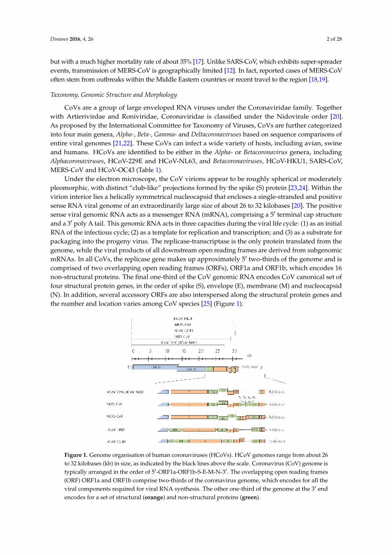
Figure 3. Regulation of MOMP by Bcl2 family of proteins. (a) The Bcl2 family of proteins is categorized into three main classes according to their functions and number of Bcl2 homology (BH) domains. The pro-survival Bcl2-like family members (Bcl2, B-cell lymphoma-extra-large (Bcl-XL), myeloid cell leukemia (Mcl1)) contain all four BH domains and are anti-apoptotic. A second class, known as Bcl2-associated X (BAX)-like proteins, which includes BAX and Bcl2 homologous antagonist killer (BAK), is pro-apoptotic and lacks the BH4 domain. Finally, the third class, known as BH3-only proteins (Bid, Bcl2-associated death promoter (Bad), and p53-upregulated modulator of apoptosis (PUMA)), contain only BH3 domain and is pro-apoptotic. (b) Two models have been proposed to account for the role of the Bcl2 family proteins in MOMP—the indirect activator model and direct activator–depressor model [11]. In the indirect activator model, the anti-apoptotic Bcl2-like proteins suppress the insertion of Bax-Bak pore complex into the mitochondria to promote MOMP and release of cytochrome c. However, when BH3-only proteins are activated beyond a certain threshold, the inhibitory effects of Bcl2-like proteins can be subverted. In the direct activator–depressor model, BH3-only protein acts as direct activators to induce Bak-Bak insertion into the outer mitochondrial membrane. These BH3-only proteins can be suppressed by Bcl2-like protein, which can in turn be inhibited by another subset of BH3-only proteins. This figure is modified from [12].
3.2. Molecular Mechanisms in Apoptosis
On a molecular level, HCoV infections have been reported to trigger apoptosis through multiple mechanisms. SARS-CoV-induced apoptosis was shown to be caspase-dependent and could be inhibited by caspase inhibitor Z-VAD-FMK or overexpression of Bcl2 [82,83]. Although viral replication was required for apoptosis induction [83], apoptosis did not affect the viral replication kinetics of SARS-CoV [82]. On the other hand, infection of primary T lymphocytes by MERS-CoV was shown to induce DNA fragmentation and caspase 8 and 9 activation, suggesting that both extrinsic and intrinsic pathways were activated. Unlike SARS-CoV infection, MERS-CoV replication was not necessary to induce apoptosis in infected T lymphocytes [79]. Apoptosis can also be induced by the less pathogenic strains of HCoVs, as substantiated by microarray data showing significant changes in pro-apoptotic and anti-apoptotic gene expression of Bcl2 family members during HCoV-
Figure 3. Regulation of MOMP by Bcl2 family of proteins. (a) The Bcl2 family of proteins is categorizedinto three main classes according to their functions and number of Bcl2 homology (BH) domains.The pro-survival Bcl2-like family members (Bcl2, B-cell lymphoma-extra-large (Bcl-XL), myeloid cellleukemia (Mcl1)) contain all four BH domains and are anti-apoptotic. A second class, known asBcl2-associated X (BAX)-like proteins, which includes BAX and Bcl2 homologous antagonist killer(BAK), is pro-apoptotic and lacks the BH4 domain. Finally, the third class, known as BH3-only proteins(Bid, Bcl2-associated death promoter (Bad), and p53-upregulated modulator of apoptosis (PUMA)),contain only BH3 domain and is pro-apoptotic. (b) Two models have been proposed to account for therole of the Bcl2 family proteins in MOMP—the indirect activator model and direct activator–depressormodel [11]. In the indirect activator model, the anti-apoptotic Bcl2-like proteins suppress the insertionof Bax-Bak pore complex into the mitochondria to promote MOMP and release of cytochrome c.However, when BH3-only proteins are activated beyond a certain threshold, the inhibitory effects ofBcl2-like proteins can be subverted. In the direct activator–depressor model, BH3-only protein acts asdirect activators to induce Bak-Bak insertion into the outer mitochondrial membrane. These BH3-onlyproteins can be suppressed by Bcl2-like protein, which can in turn be inhibited by another subset ofBH3-only proteins. This figure is modified from [12].
3.2. Molecular Mechanisms in Apoptosis
On a molecular level, HCoV infections have been reported to trigger apoptosis through multiplemechanisms. SARS-CoV-induced apoptosis was shown to be caspase-dependent and could beinhibited by caspase inhibitor Z-VAD-FMK or overexpression of Bcl2 [82,83]. Although viral replicationwas required for apoptosis induction [83], apoptosis did not affect the viral replication kinetics ofSARS-CoV [82]. On the other hand, infection of primary T lymphocytes by MERS-CoV was shown toinduce DNA fragmentation and caspase 8 and 9 activation, suggesting that both extrinsic and intrinsicpathways were activated. Unlike SARS-CoV infection, MERS-CoV replication was not necessary toinduce apoptosis in infected T lymphocytes [79]. Apoptosis can also be induced by the less pathogenic
Diseases 2016, 4, 26 7 of 28
strains of HCoVs, as substantiated by microarray data showing significant changes in pro-apoptoticand anti-apoptotic gene expression of Bcl2 family members during HCoV-229E infection [84]. Infectionof HCoV-OC43 was shown to promote BAX translocation to the mitochondria in human neuronalcells [74]. Although caspases 3 and 9 were activated in HCoV-OC43-infected murine and humanneuronal cells [9,74], addition of pan-caspase inhibitor Z-VAD-FMK and the caspase-9 inhibitorZ-LEHD-FMK did not affect the viability of these infected neuronal cells, indicating that programmedcell death induced by HCoV-OC43 could be caspase-independent [74]. This highlights the possibilityof a non-classical programmed cell death mechanism induced in HCoV infection.
Apoptotic mechanisms during HCoV infection are likely to be manipulated by viral proteins(Figure 4), although this has only mostly been studied in SARS-CoV. Specifically, SARS-CoV S, N,E, M, ORF-6, 7a and 9b proteins have been shown to serve pro-apoptotic functions in their hostcells [77,85–91]. Expression of SARS-CoV E protein and 7a protein promoted mitochondrial-mediatedapoptosis by sequestering the anti-apoptotic Bcl-XL protein to the endoplasmic reticulum (ER)membranes [77,92]. SARS-CoV M protein is also highly pro-apoptotic and mediates activation ofboth caspases 8 and 9 [90]. Additionally, HCoV-OC43 wild type S protein has been shown to induceunfolded protein response (UPR) in human neuronal NT2-N and LA-N-5 cell lines, which maylead to apoptosis [93]. A recombinant HCoV-OC43 harbouring point mutations at its S proteininduced stronger caspase 3 activation and nuclear fragmentation than the wild-type virus [93]. It isinteresting to note that the localisation of SARS-CoV N and 9b protein is associated with induction ofcaspase-dependent apoptosis [89,94]. This finding opens up to novel perspectives of the link betweensubcellular localisation of viral proteins and caspase activation as a mode of apoptosis regulation by HCoVs.
Diseases 2016, 4, 26 7 of 27
229E infection [84]. Infection of HCoV-OC43 was shown to promote BAX translocation to the mitochondria in human neuronal cells [74]. Although caspases 3 and 9 were activated in HCoV-OC43-infected murine and human neuronal cells [9,74], addition of pan-caspase inhibitor Z-VAD-FMK and the caspase-9 inhibitor Z-LEHD-FMK did not affect the viability of these infected neuronal cells, indicating that programmed cell death induced by HCoV-OC43 could be caspase-independent [74]. This highlights the possibility of a non-classical programmed cell death mechanism induced in HCoV infection.
Apoptotic mechanisms during HCoV infection are likely to be manipulated by viral proteins (Figure 4), although this has only mostly been studied in SARS-CoV. Specifically, SARS-CoV S, N, E, M, ORF-6, 7a and 9b proteins have been shown to serve pro-apoptotic functions in their host cells [77,85–91]. Expression of SARS-CoV E protein and 7a protein promoted mitochondrial-mediated apoptosis by sequestering the anti-apoptotic Bcl-XL protein to the endoplasmic reticulum (ER) membranes [77,92]. SARS-CoV M protein is also highly pro-apoptotic and mediates activation of both caspases 8 and 9 [90]. Additionally, HCoV-OC43 wild type S protein has been shown to induce unfolded protein response (UPR) in human neuronal NT2-N and LA-N-5 cell lines, which may lead to apoptosis [93]. A recombinant HCoV-OC43 harbouring point mutations at its S protein induced stronger caspase 3 activation and nuclear fragmentation than the wild-type virus [93]. It is interesting to note that the localisation of SARS-CoV N and 9b protein is associated with induction of caspase-dependent apoptosis [89,94]. This finding opens up to novel perspectives of the link between subcellular localisation of viral proteins and caspase activation as a mode of apoptosis regulation by HCoVs.
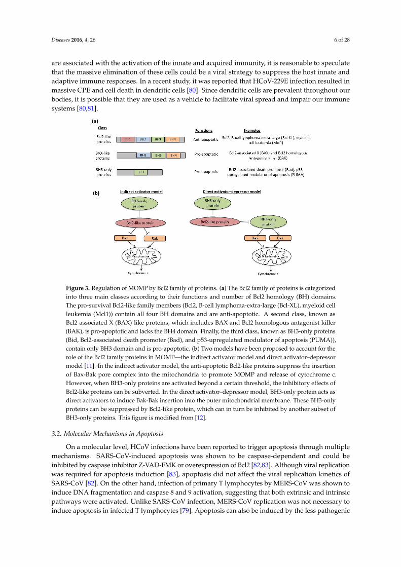
Figure 4. Activation of apoptosis by HCoVs. Binding of death ligands to death receptor induces caspase 8 activation, which in turn activates effector caspases 3 and 7 to stimulate apoptosis. On the other hand, intrinsic pathway is regulated by pro-apoptotic and anti-apoptotic Bcl2 family proteins, such as Bcl-XL, Bcl2, Bax and Bak to induce MOMP. Subsequent caspase 9 activation caused by enhanced MOMP stimulates caspases 3 and 7 activation. During HCoV infection, the virus or specific viral proteins (yellow-orange boxes) target at multiple stages of both the extrinsic and intrinsic apoptosis signalling pathways.
4. Human Coronavirus Infection and Innate Immunity
When the cells are exposed to pathogens such as viruses, immune responses are induced as a form of host defence. The immune response is modulated during pathogen exposure in a cell-type dependent fashion. Innate immunity is the first line of defence mounted against the virus before the adaptive immune system is generated. Both the host and virus can manipulate innate immune mechanisms as a form of defence or evasion strategy [95,96].
Figure 4. Activation of apoptosis by HCoVs. Binding of death ligands to death receptor induces caspase8 activation, which in turn activates effector caspases 3 and 7 to stimulate apoptosis. On the otherhand, intrinsic pathway is regulated by pro-apoptotic and anti-apoptotic Bcl2 family proteins, such asBcl-XL, Bcl2, Bax and Bak to induce MOMP. Subsequent caspase 9 activation caused by enhancedMOMP stimulates caspases 3 and 7 activation. During HCoV infection, the virus or specific viralproteins (yellow-orange boxes) target at multiple stages of both the extrinsic and intrinsic apoptosissignalling pathways.
4. Human Coronavirus Infection and Innate Immunity
When the cells are exposed to pathogens such as viruses, immune responses are induced as aform of host defence. The immune response is modulated during pathogen exposure in a cell-typedependent fashion. Innate immunity is the first line of defence mounted against the virus beforethe adaptive immune system is generated. Both the host and virus can manipulate innate immunemechanisms as a form of defence or evasion strategy [95,96].
Diseases 2016, 4, 26 8 of 28
4.1. Pattern Recognition Receptors
Cells in the immune system detect the viral pathogens via several recognition strategies. Of which,the most well characterized is the pattern recognition receptors (PRR), which engage various microbialpathogens via evolutionarily conserved structures known as pathogen-associated molecular patterns(PAMPs). PRRs are mainly categorized into three classes, namely Toll-like receptors (TLRs), retinoicacid-inducible gene I (RIG-I)-like receptors (RLRs) and nucleotide oligomerisation domain (NOD)-likereceptors (NLRs).
TLR is a type I transmembrane protein localized to either the cell surface or endosomal vesicles.Their leucine-rich repeats (LRR) domain mediates the recognition of PAMPs and damage-associatedmolecular patterns (DAMPs) from various sources including bacteria, fungi and viruses [97]. Activationof TLRs occurs mainly in antigen-presenting cells such as dendritic cells (DCs), macrophages,monocytes and B cells. Of the 10 known TLRs in human, TLR2, 3, 4, 7 and 9 are found to be involvedin viral detection [98,99]. TLR3 recognizes double-stranded RNA (dsRNA), a replicative intermediateproduced during viral RNA replication [100]. TLR7 and 8 detect single-stranded RNA (ssRNA) andTLR9 recognizes unmethylated CpG DNA present in DNA viruses [101–103]. Besides nucleic acids,other TLRs, such as TLR2 and 4, sense viral proteins as exemplified in respiratory syncytial virus (RSV),hepatitis virus, measles virus and human immunodeficiency virus [104–107]. Upon recognition of viralcomponents, TLRs recruit Toll/interleukin-1 receptor (TIR)-containing signalling adaptor molecules,such as MyD88 (myeloid differentiation primary response protein 88) and TIR-domain-containingadapter-inducing interferon-β (TRIF) [108–110]. MyD88 and TRIF then stimulate the MAPK andNF-κB pathways to boost IFN and pro-inflammatory cytokine production [111].
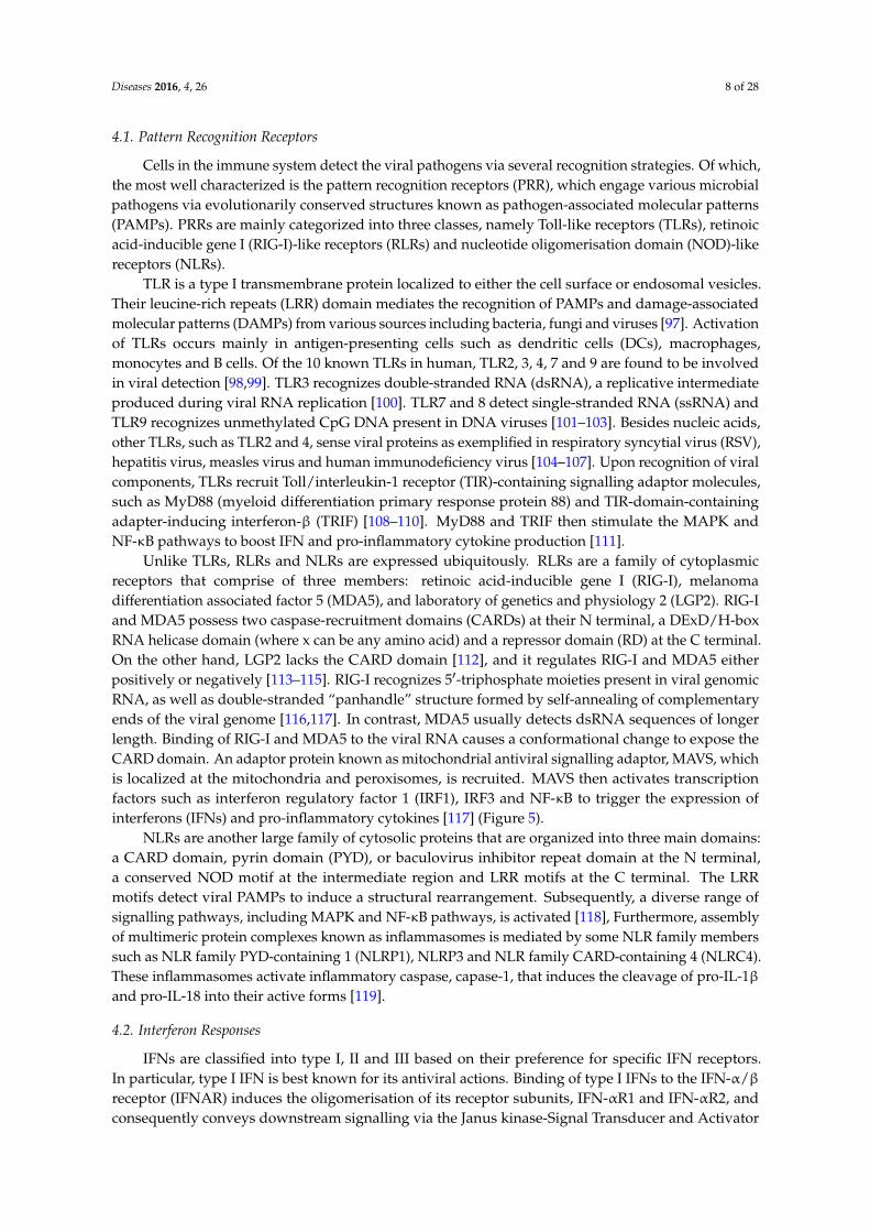
Unlike TLRs, RLRs and NLRs are expressed ubiquitously. RLRs are a family of cytoplasmicreceptors that comprise of three members: retinoic acid-inducible gene I (RIG-I), melanomadifferentiation associated factor 5 (MDA5), and laboratory of genetics and physiology 2 (LGP2). RIG-Iand MDA5 possess two caspase-recruitment domains (CARDs) at their N terminal, a DExD/H-boxRNA helicase domain (where x can be any amino acid) and a repressor domain (RD) at the C terminal.On the other hand, LGP2 lacks the CARD domain [112], and it regulates RIG-I and MDA5 eitherpositively or negatively [113–115]. RIG-I recognizes 51-triphosphate moieties present in viral genomicRNA, as well as double-stranded “panhandle” structure formed by self-annealing of complementaryends of the viral genome [116,117]. In contrast, MDA5 usually detects dsRNA sequences of longerlength. Binding of RIG-I and MDA5 to the viral RNA causes a conformational change to expose theCARD domain. An adaptor protein known as mitochondrial antiviral signalling adaptor, MAVS, whichis localized at the mitochondria and peroxisomes, is recruited. MAVS then activates transcriptionfactors such as interferon regulatory factor 1 (IRF1), IRF3 and NF-κB to trigger the expression ofinterferons (IFNs) and pro-inflammatory cytokines [117] (Figure 5).
NLRs are another large family of cytosolic proteins that are organized into three main domains:a CARD domain, pyrin domain (PYD), or baculovirus inhibitor repeat domain at the N terminal,a conserved NOD motif at the intermediate region and LRR motifs at the C terminal. The LRRmotifs detect viral PAMPs to induce a structural rearrangement. Subsequently, a diverse range ofsignalling pathways, including MAPK and NF-κB pathways, is activated [118], Furthermore, assemblyof multimeric protein complexes known as inflammasomes is mediated by some NLR family memberssuch as NLR family PYD-containing 1 (NLRP1), NLRP3 and NLR family CARD-containing 4 (NLRC4).These inflammasomes activate inflammatory caspase, capase-1, that induces the cleavage of pro-IL-1βand pro-IL-18 into their active forms [119].
4.2. Interferon Responses
IFNs are classified into type I, II and III based on their preference for specific IFN receptors.In particular, type I IFN is best known for its antiviral actions. Binding of type I IFNs to the IFN-α/βreceptor (IFNAR) induces the oligomerisation of its receptor subunits, IFN-αR1 and IFN-αR2, andconsequently conveys downstream signalling via the Janus kinase-Signal Transducer and Activator

Diseases 2016, 4, 26 9 of 28
of Transcription (JAK-STAT) pathway. Autophosphorylation of JAK domains in the IFNAR resultsin subsequent phosphorylation of STAT1 and STAT2 proteins at their tyrosine residues. This isfollowed by dimerisation and nuclear translocation of the activated STAT proteins, which recruit IFNregulatory factor 9 (IRF9) to form the IFN-stimulated gene factor 3 (ISGF3). ISFG3 is a transcriptionfactor that binds to its cognate DNA sequence known as IFN-stimulated response elements (ISREs) toactivate transcription of IFN-stimulated genes (ISGs) [120] (Figure 5). Many of these ISGs such as 21–51
oligoadenylatesynthetase and protein kinase R (PKR) confer resistance against virus invasions [121].Additionally, type I IFNs facilitates the maturation of dendritic cells (DCs), cytotoxicity of natural killer(NK) cells, and differentiation of T lymphocytes [98].
Diseases 2016, 4, 26 9 of 27
in subsequent phosphorylation of STAT1 and STAT2 proteins at their tyrosine residues. This is followed by dimerisation and nuclear translocation of the activated STAT proteins, which recruit IFN regulatory factor 9 (IRF9) to form the IFN-stimulated gene factor 3 (ISGF3). ISFG3 is a transcription factor that binds to its cognate DNA sequence known as IFN-stimulated response elements (ISREs) to activate transcription of IFN-stimulated genes (ISGs) [120] (Figure 5). Many of these ISGs such as 2′–5′ oligoadenylatesynthetase and protein kinase R (PKR) confer resistance against virus invasions [121]. Additionally, type I IFNs facilitates the maturation of dendritic cells (DCs), cytotoxicity of natural killer (NK) cells, and differentiation of T lymphocytes [98].
Figure 5. HCoV viral proteins on innate immunity. During HCoV infection, PRRs such as TLRs, RIG-I and MDA5 are activated to trigger a series of signalling pathway, including MAPK and NF-κB, for IFN production. These IFNs then act on IFNAR and activate the JAK-STAT signalling pathway to induce ISGs. The yellow-orange boxes show the viral proteins that have been reported to modulate host innate immunity at multiple stages.
4.3. Modulation of Innate Immunity
Infection by HCoVs, especially the highly pathogenic SARS-CoV and MERS-CoV, is associated with suppression of IFN synthesis [122–126]. The capability of the virus to regulate type I IFN signalling is an important hallmark for virulence [127]. As compared to SARS-CoV and MERS-CoV, a huge rise in type I IFNs were detected in cells infected with HCoV strain 229E [80,124,128].
Based on studies from SARS-CoV and Mouse Hepatitis Virus (MHV)-infected cells, two mechanisms have been proposed to explain the HCoV-mediated inhibition of type I IFN production [13,126]. Firstly, CoV genomic and subgenomic RNA replication takes place in double membrane vesicles to prevent detection by PRRs [13,129]. Secondly, proteins encoded by the virus could interfere with innate immune pathways [13,130]. The structural proteins, nonstructural proteins and accessory proteins of HCoVs have been shown to modify innate immune responses (Figure 5).
4.3.1. Viral Proteins Involved in Innate Immunity
Structural Proteins of HCoVs
Expression of SARS-CoV M protein could suppress type I IFN production mediated by RIG-I, but not MDA5, in infected HEK293 cells [131], likely through its first transmembrane domain. However, this inhibition was not observed when expressing the M protein of HCoV-HKU1, suggesting that this activity is not conserved among all HCoV strains [132]. In another study, it was shown that the MERS-CoV M protein could also suppress type I IFN by inhibiting the translocation
Figure 5. HCoV viral proteins on innate immunity. During HCoV infection, PRRs such as TLRs, RIG-Iand MDA5 are activated to trigger a series of signalling pathway, including MAPK and NF-κB, for IFNproduction. These IFNs then act on IFNAR and activate the JAK-STAT signalling pathway to induceISGs. The yellow-orange boxes show the viral proteins that have been reported to modulate host innateimmunity at multiple stages.
4.3. Modulation of Innate Immunity
Infection by HCoVs, especially the highly pathogenic SARS-CoV and MERS-CoV, is associatedwith suppression of IFN synthesis [122–126]. The capability of the virus to regulate type I IFN signallingis an important hallmark for virulence [127]. As compared to SARS-CoV and MERS-CoV, a huge risein type I IFNs were detected in cells infected with HCoV strain 229E [80,124,128].
Based on studies from SARS-CoV and Mouse Hepatitis Virus (MHV)-infected cells, twomechanisms have been proposed to explain the HCoV-mediated inhibition of type I IFNproduction [13,126]. Firstly, CoV genomic and subgenomic RNA replication takes place in doublemembrane vesicles to prevent detection by PRRs [13,129]. Secondly, proteins encoded by the viruscould interfere with innate immune pathways [13,130]. The structural proteins, nonstructural proteinsand accessory proteins of HCoVs have been shown to modify innate immune responses (Figure 5).
4.3.1. Viral Proteins Involved in Innate Immunity
Structural Proteins of HCoVs
Expression of SARS-CoV M protein could suppress type I IFN production mediated by RIG-I,but not MDA5, in infected HEK293 cells [131], likely through its first transmembrane domain. However,
Diseases 2016, 4, 26 10 of 28
this inhibition was not observed when expressing the M protein of HCoV-HKU1, suggesting thatthis activity is not conserved among all HCoV strains [132]. In another study, it was shown that theMERS-CoV M protein could also suppress type I IFN by inhibiting the translocation of IRF3 into thenucleus, although the exact mechanism has not yet been elucidated [133]. Additionally, SARS-CoV Nprotein was also shown to interfere with the function of IRF3 [134]. The N protein of SARS-CoV likelyact at the initial recognition stage of viral RNA via its RNA binding activity, although it neither formsa complex with RIG-I nor MDA5 [135]. This implies that the N protein possibly acts on other viralRNA recognition strategies of the host.
Non-Structural and Accessory Proteins of HCoVs
Besides the structural proteins, other nonstructural proteins (nsp) and accessory proteins ofHCoVs have also been implicated in the modulation of innate immunity. For instance, nsp1 of bothSARS-CoV and MERS-CoV has been demonstrated to modify capped non-viral RNAs to facilitateendonucleolytic cleavage of host messenger RNA (mRNA) [136,137]. Additionally, SARS-CoV nsp1interacted with the 40S subunit of ribosome to prevent host mRNA translation [136]. This inducesthe host shutoff mechanism, as transcription and translation of viral RNA are more favoured overthat of host mRNA. In a recent study, several residues of SARS-CoV nsp1 were identified to affectIFN-dependent signalling [138]. In addition to nsp1, SARS-CoV and MERS-CoV nsp3 proteins, whichpossess papain-like protease (PLpro) domain and a PLP2 domain, also antagonize IFN production.Both SARS-CoV and MERS-CoV PLpro domains are deISGylating enzymes and they downregulatedmRNA levels of pro-inflammatory cytokines including CCL5, IFNβ, and CXCL10 [139]. Suppressionof IFN responses by SARS-CoV PLpro is not mediated by its protease activity. Rather, SARS-CoVPLpro inhibited the phosphorylation of interferon-regulatory factor 3 (IRF3) and its translocation to thenucleus to enhance IFN gene transcription [140]. Expression of MERS-CoV PLpro also antagonizes IFNproduction and is required for suppression on RIG-I and MDA5 [139,141]. Furthermore, it has beenidentified that the ADP-ribose-1-monophosphatase macrodomain encoded within nsp3 in HCoV-229Eand SARS-CoV is responsible for suppressing IFN induction [142].
Despite being dispensable in viral replication, HCoV accessory proteins are essential in diversecellular signalling, such as cell proliferation, apoptosis and interferon signalling [25]. In SARS-CoV,ORF3b and -6 are shown to interfere with IFNβ synthesis by inhibiting the phosphorylation andnuclear translocation of IRF3. Furthermore, these accessory proteins also disrupt IFN signalling bypreventing IFNβ-induced activation of interferon-stimulated response element (ISRE) found in thepromoter region of ISG [134]. The accessory proteins of MERS-CoV, ORF4a, -4b and -5, could similarlysuppress IRF3 nuclear translocation, hence significantly reducing IFN-β promoter-driven luciferaseactivity in cells transfected with these accessory proteins [133].
5. Human Coronavirus and ER Stress Response
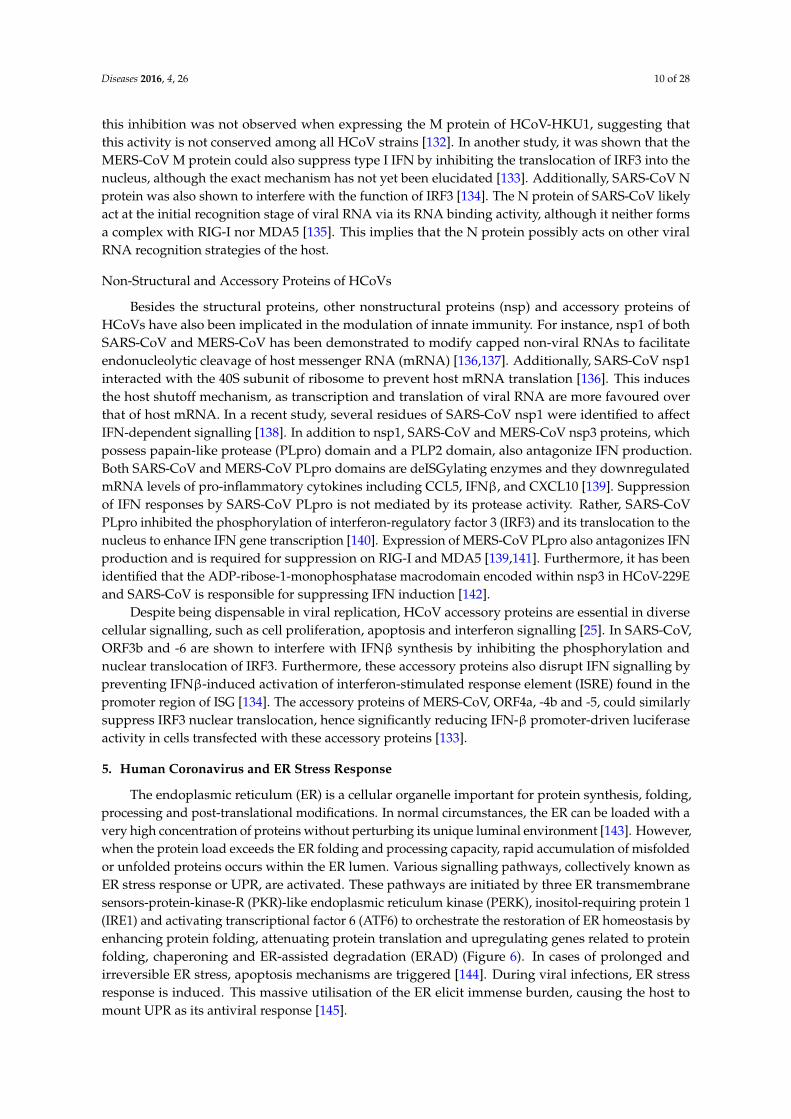
The endoplasmic reticulum (ER) is a cellular organelle important for protein synthesis, folding,processing and post-translational modifications. In normal circumstances, the ER can be loaded with avery high concentration of proteins without perturbing its unique luminal environment [143]. However,when the protein load exceeds the ER folding and processing capacity, rapid accumulation of misfoldedor unfolded proteins occurs within the ER lumen. Various signalling pathways, collectively known asER stress response or UPR, are activated. These pathways are initiated by three ER transmembranesensors-protein-kinase-R (PKR)-like endoplasmic reticulum kinase (PERK), inositol-requiring protein 1(IRE1) and activating transcriptional factor 6 (ATF6) to orchestrate the restoration of ER homeostasis byenhancing protein folding, attenuating protein translation and upregulating genes related to proteinfolding, chaperoning and ER-assisted degradation (ERAD) (Figure 6). In cases of prolonged andirreversible ER stress, apoptosis mechanisms are triggered [144]. During viral infections, ER stressresponse is induced. This massive utilisation of the ER elicit immense burden, causing the host tomount UPR as its antiviral response [145].

Diseases 2016, 4, 26 11 of 28
Diseases 2016, 4, 26 11 of 27
Figure 6. HCoVs on ER stress response. During HCoV infections, the ER stress response, which comprises of three signalling pathways, PERK, ATF6 and IRE1, is activated. HCoVs encode many viral proteins (yellow-orange boxes) that target the various signalling pathway of ER stress during viral infections.
5.1. PERK Signalling Pathway
Activation of PERK is initiated by its dissociation of the luminal domain from the ER chaperone, binding immunoglobulin protein (BiP). This is followed by the oligomerisation and autophosphorylation of PERK. In its active form, PERK phosphorylates Ser51 at the α-subunit of eukaryotic initiation factor 2 (eIF2α) to attenuate protein translation [146]. Activation of PERK plays a pro-survival role in cells, as clearly demonstrated by PERK−/− mouse embryonic fibroblasts, which exhibited higher cell death when treated with cycloheximide, an ER stress-inducing agent [147]. Phosphorylated eIF2α not only triggers a shutdown of global protein synthesis, but also enhances the translation of activating transcription factor ATF4 [148]. ATF4 stimulates target gene expression such as GADD153 (also known as CHOP or C/EBP-homologous protein), to enhance transcription of pro-apoptotic genes [149]. Additionally, eIF2α can be phosphorylated by other kinases such as PKR, heme-regulated inhibitor kinase (HRI), and general control non-derepressible-2 (GCN2) [144]. These kinases activate various downstream signalling pathways, which together form the integrated stress response [146,150].
PKR and eIF2α phosphorylation was detected in SARS-CoV-infected cells and inhibition of PKR using antisense peptide-conjugated phosphorodiamidate morpholino oligomers did not affect eIF2α phosphorylation but significantly reduced SARS-CoV-induced apoptosis. SARS-CoV protein replication and virus production were not affected by PKR knockdown. Therefore, it is likely that SARS-CoV adopts a strategy to counteract against the antiviral effects of PKR, thus enabling viral mRNA translation to proceed regardless of eIF2α phosphorylation. PERK was also found to be activated during SARS-CoV infection [75], possibly through its S and 3a proteins [151,152]. In another study, it was demonstrated that expression of a dominant-negative PERK mutant, that inhibited PERK kinase activity, suppressed the transcriptional activation of Grp78 and Grp94 promoters mediated by S proteins of SARS-CoV and HCoV-HKU1 [153]. However, PERK activation is unlikely to occur in all HCoV strains. In neuronal cells lines infected with HCoV-OC43, it was shown that eIF2α was only transiently phosphorylated at the early stage of infection, but was subsequently suppressed and returned back to its basal level of phosphorylation, similar to the mock-infected cells [93]. On the other hand, previous studies from this group showed that PKR, PERK and eIF2α were moderately induced at the early stages of IBV infection, but were subsequently suppressed at late
Figure 6. HCoVs on ER stress response. During HCoV infections, the ER stress response, whichcomprises of three signalling pathways, PERK, ATF6 and IRE1, is activated. HCoVs encode manyviral proteins (yellow-orange boxes) that target the various signalling pathway of ER stress duringviral infections.
5.1. PERK Signalling Pathway
Activation of PERK is initiated by its dissociation of the luminal domain from the ERchaperone, binding immunoglobulin protein (BiP). This is followed by the oligomerisation andautophosphorylation of PERK. In its active form, PERK phosphorylates Ser51 at the α-subunit ofeukaryotic initiation factor 2 (eIF2α) to attenuate protein translation [146]. Activation of PERK playsa pro-survival role in cells, as clearly demonstrated by PERK´/´ mouse embryonic fibroblasts,which exhibited higher cell death when treated with cycloheximide, an ER stress-inducing agent [147].Phosphorylated eIF2α not only triggers a shutdown of global protein synthesis, but also enhancesthe translation of activating transcription factor ATF4 [148]. ATF4 stimulates target gene expressionsuch as GADD153 (also known as CHOP or C/EBP-homologous protein), to enhance transcriptionof pro-apoptotic genes [149]. Additionally, eIF2α can be phosphorylated by other kinases such asPKR, heme-regulated inhibitor kinase (HRI), and general control non-derepressible-2 (GCN2) [144].These kinases activate various downstream signalling pathways, which together form the integratedstress response [146,150].
PKR and eIF2α phosphorylation was detected in SARS-CoV-infected cells and inhibition ofPKR using antisense peptide-conjugated phosphorodiamidate morpholino oligomers did not affecteIF2α phosphorylation but significantly reduced SARS-CoV-induced apoptosis. SARS-CoV proteinreplication and virus production were not affected by PKR knockdown. Therefore, it is likely thatSARS-CoV adopts a strategy to counteract against the antiviral effects of PKR, thus enabling viralmRNA translation to proceed regardless of eIF2α phosphorylation. PERK was also found to beactivated during SARS-CoV infection [75], possibly through its S and 3a proteins [151,152]. In anotherstudy, it was demonstrated that expression of a dominant-negative PERK mutant, that inhibited PERKkinase activity, suppressed the transcriptional activation of Grp78 and Grp94 promoters mediatedby S proteins of SARS-CoV and HCoV-HKU1 [153]. However, PERK activation is unlikely to occurin all HCoV strains. In neuronal cells lines infected with HCoV-OC43, it was shown that eIF2αwasonly transiently phosphorylated at the early stage of infection, but was subsequently suppressed andreturned back to its basal level of phosphorylation, similar to the mock-infected cells [93]. On theother hand, previous studies from this group showed that PKR, PERK and eIF2α were moderatelyinduced at the early stages of IBV infection, but were subsequently suppressed at late infection
Diseases 2016, 4, 26 12 of 28
stages [154,155]. Nevertheless, the moderate and transient increase in eIF2α phosphorylation wassufficient to activate ATF4 protein translation and upregulate the downstream targets of ATF4, ATF3and GADD153. Knockdown of PKR and PERK in IBV-infected cells attenuated IBV-induced GADD153upregulation and IBV-induced apoptosis, although the viral protein replication was unaffected [155].Upregulation of GADD153 is postulated to induce pro-apoptotic protein TRIB3 and inhibit pro-survivalERK protein [154], as well as provide a negative feedback to rapidly dephosphorylate eIF2α at latestages of IBV infection [155]. Based on these findings, we speculate the HCoVs might use similarmechanism to modulate the PKR/PERK/eIF2α pathway in infected cells. More studies could be done onHCoV infection to analyze the activation of the PKR/PERK/eIF2α pathway at various stages of infection.
5.2. ATF6 Signalling Pathway
Like PERK, activation of ATF6 is initiated by dissociation from the ER chaperone, BiP,although alternative mechanisms such as deglycosylation and reduction of disulphide bonds couldoccur [156,157]. ATF6 then translocates into the Golgi apparatus, where it is proteolyzed by site-1and site-2 proteases (S1P and S2P). The processed ATF6 then migrates to the nucleus where it turnson expression of genes containing an ER stress response element (ERSE) in their promoters [158].Like ATF4, ATF6 also induces expression of ER chaperone proteins such as GRP78, GRP94 and transcriptionfactors CHOP and X box-binding protein 1 (XBP1) [150]. XBP1 is essential for IRE signalling [159].
As compared to the two other UPR branches, PERK and IRE1, the ATF6 branch is less wellstudied. As GRP94/78 are also target genes of ATF6, and their promoter activities were enhancedby SARS-CoV S protein, one could hypothesize that ATF6 pathway could also be induced bySARS-CoV S. Surprisingly, overexpression of SARS-CoV S protein did not affect ATF6 promoterluciferase activity [152]. Deletion of E protein in recombinant SARS-CoV also did not significantlyactivate ATF6 [91]. Intriguingly, 8ab protein, an accessory protein of SARS-CoV, was shown to reside inthe luminal surface of the ER surface and activate ATF6 via facilitating its proteolysis and translocationof the processed ATF6 into the nucleus [160]. 8ab protein from SARS-CoV was found in civet catsand early human isolates, but was subsequently split into two accessory proteins, 8a and 8b, with acharacteristic 29-nucleotide deletion [161].
5.3. IRE1 Signalling Pathway
IRE1 is believed to be the last UPR branch to be activated in cells undergoing ER stress [162].It is also the most conserved among all UPR arms [163]. Although IRE1 was initially proposedto be activated in the same mechanism as PERK [162], later studies suggested that the N-terminalluminal domain (NLD) of IRE1 can directly bind unfolded proteins [164,165]. Activation of its RNasedomain results in unconventional splicing of a 252-nucleotide intron from homologous to Atf/Creb1(HAC1) mRNA in yeasts and a 26-nucleotide intron from X-box binding protein 1 (XBP1) mRNAin humans [166]. Splicing of XBP1 generates a potent transcription factor, XBP1s, that inducesexpression of genes related to protein entry into the ER, folding and ERAD [159]. In a negativefeedback mechanism, XBP1s also promotes the transcription of E3 ubiquitin ligase synoviolin toenhance IRE1 ubiquitination [167]. The unspliced variant XBP1u contained a nuclear exclusionsignal to sequester XBP1s from the nucleus, thus making XBP1u another negative feedback regulatorof XBP1s. [168]. In a separate mechanism, IRE1 can cleave ER-associated mRNA species throughregulated IRE1-dependent decay (RIDD) during late stages of ER stress [145]. It is believed that initialXBP1/HAC1 splicing by IRE1 promotes survival but subsequent activation of RIDD upon prolongedER stress leads to cell death, thus allowing IRE1 to play dual role in apoptosis [169,170]. Anotherimportant enzymatic activation of IRE1 is its kinase activity. The kinase domain of phosphorylated IRE1recruits the TNF receptor-associated factor 2 (TRAF2), which then activates other kinases to eventuallyactivate the c-Jun N-terminal kinase (JNK) and regulates ER stress-dependent apoptosis [171].
Previous studies have investigated the role of IRE1-XBP1 pathway during SARS-CoV infection.Although no increase in XBP1 splicing was observed in SARS-CoV-infected cells [172], deletion

Diseases 2016, 4, 26 13 of 28
of E protein in recombinant SARS-CoV resulted in significant XBP1 splicing and higher rate ofapoptosis [91]. On the other hand, infection of HCoV-OC43 caused an induction in XBP1 splicing andenhanced expression of genes regulated by XBP1s, namely Edem, Herp, Grp94 and P58-ipk. However,introduction of two point mutations (H183R and Y241H) in the S protein of HCoV-OC43 led to a higherdegree of XBP1 cleavage, followed by a strong activation of caspase-3 and nuclear fragmentation [93].Since IRE1 pathway is closely associated to JNK activation, it is possible that the JNK pathway is alsoimplicated during HCoV-OC43 infection.
Similar to HCoV infections, it has been shown that the IRE1-XBP1 pathway is activatedduring IBV infection. Knockdown of IRE1 using specific siRNA in IBV-infected cells augmentedIBV-induced apoptosis; however, an opposite effect was observed by XBP1 knockdown in IBV-infectedcells. Consistent with the knockdown experiments, transient overexpression of the full-lengthIRE1α attenuated IBV-induced apoptosis. When both spliced and unspliced forms of XBP1 wereoverexpressed in IBV-infected cells, the spliced form of XBP1 was shown to be anti-apoptotic andthe unspliced form was pro-apoptotic. Overexpression of a dominant-negative XBP1 enhancedIBV-induced apoptosis. Therefore, our findings showed that the anti-apoptotic function of IRE1 duringIBV infection could be mediated by its splicing of XBP1, hence converting XBP1 from a pro-apoptoticto anti-apoptotic form. Finally, IRE1 induction during IBV infection was shown to mediate JNKhyperphosphorylation and Akt hypophosphorylation to potentiate the IBV-infected cells to apoptosis [173].
6. Human Coronavirus and MAPK Pathways
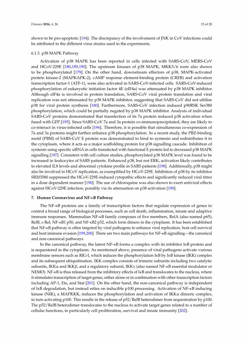
The MAPKs are a group of evolutionally conserved serine/theronine kinases that are activatedin response to environmental stresses including oxidative stress, DNA damage, cancer developmentand viral infections [174–176]. To date, multiple MAPK pathways have been identified in mammals,and they can be broadly classified into three major categories—the extracellular signal-regulated kinase(ERK), p38 MAPK and stress-activated protein kinase/c-Jun N-terminal kinase (SAPK/JNK) [177].In all MAPK pathways, the signals are transduced downstream via a three-tier protein kinase cascade.In each tier, the kinases are activated by upstream kinases by dual phosphorylation at the Thr-X-Tyrmotif (X represents any amino acid). Presence of extracellular stimuli triggers the activation ofMAP kinase kinase kinases (MAPKKKs), which then activate the MAP kinase kinases (MAPKKs).These sequential phosphorylation events eventually activate MAPKs which in turn regulate a variety offundamental cellular processes such as cell proliferation, survival, motility, differentiation, autophagy,apoptosis and regulation of cytokine production (Figure 7) [178].
Diseases 2016, 4, 26 13 of 27
protein in recombinant SARS-CoV resulted in significant XBP1 splicing and higher rate of apoptosis [91]. On the other hand, infection of HCoV-OC43 caused an induction in XBP1 splicing and enhanced expression of genes regulated by XBP1s, namely Edem, Herp, Grp94 and P58-ipk. However, introduction of two point mutations (H183R and Y241H) in the S protein of HCoV-OC43 led to a higher degree of XBP1 cleavage, followed by a strong activation of caspase-3 and nuclear fragmentation [93]. Since IRE1 pathway is closely associated to JNK activation, it is possible that the JNK pathway is also implicated during HCoV-OC43 infection.
Similar to HCoV infections, it has been shown that the IRE1-XBP1 pathway is activated during IBV infection. Knockdown of IRE1 using specific siRNA in IBV-infected cells augmented IBV-induced apoptosis; however, an opposite effect was observed by XBP1 knockdown in IBV-infected cells. Consistent with the knockdown experiments, transient overexpression of the full-length IRE1α attenuated IBV-induced apoptosis. When both spliced and unspliced forms of XBP1 were overexpressed in IBV-infected cells, the spliced form of XBP1 was shown to be anti-apoptotic and the unspliced form was pro-apoptotic. Overexpression of a dominant-negative XBP1 enhanced IBV-induced apoptosis. Therefore, our findings showed that the anti-apoptotic function of IRE1 during IBV infection could be mediated by its splicing of XBP1, hence converting XBP1 from a pro-apoptotic to anti-apoptotic form. Finally, IRE1 induction during IBV infection was shown to mediate JNK hyperphosphorylation and Akt hypophosphorylation to potentiate the IBV-infected cells to apoptosis [173].
6. Human Coronavirus and MAPK Pathways
The MAPKs are a group of evolutionally conserved serine/theronine kinases that are activated in response to environmental stresses including oxidative stress, DNA damage, cancer development and viral infections [174–176]. To date, multiple MAPK pathways have been identified in mammals, and they can be broadly classified into three major categories—the extracellular signal-regulated kinase (ERK), p38 MAPK and stress-activated protein kinase/c-Jun N-terminal kinase (SAPK/JNK) [177]. In all MAPK pathways, the signals are transduced downstream via a three-tier protein kinase cascade. In each tier, the kinases are activated by upstream kinases by dual phosphorylation at the Thr-X-Tyr motif (X represents any amino acid). Presence of extracellular stimuli triggers the activation of MAP kinase kinase kinases (MAPKKKs), which then activate the MAP kinase kinases (MAPKKs). These sequential phosphorylation events eventually activate MAPKs which in turn regulate a variety of fundamental cellular processes such as cell proliferation, survival, motility, differentiation, autophagy, apoptosis and regulation of cytokine production (Figure 7) [178].
Figure 7. HCoVs on MAPK signalling pathways. MAPK pathways comprises of the ERK, JNK and p38 MAPK pathways. During HCoV infections, the signals are transduced by the MAPK pathways by a three-tier protein kinase cascade, with the kinase of each tier being phosphorylated by upstream kinases at the Thr and Tyr residues. HCoVs and their viral proteins (yellow-orange boxes) have been shown to induce these MAPK pathways as shown in the figure.
Figure 7. HCoVs on MAPK signalling pathways. MAPK pathways comprises of the ERK, JNK andp38 MAPK pathways. During HCoV infections, the signals are transduced by the MAPK pathwaysby a three-tier protein kinase cascade, with the kinase of each tier being phosphorylated by upstreamkinases at the Thr and Tyr residues. HCoVs and their viral proteins (yellow-orange boxes) have beenshown to induce these MAPK pathways as shown in the figure.

Diseases 2016, 4, 26 14 of 28
6.1. Modulation of MAPK Pathways
Phosphorylation of all three MAPK members has been detected in cells infected withSARS-CoV [179,180]. Additionally, the MAPK pathways are also activated during infection by otherHCoVs, as discussed below (Figure 7).
6.1.1. ERK Pathway
Activation of ERK and its upstream kinases, MEK1/2, was detected in cells infected withSARS-CoV or overexpressed with SARS-CoV S protein [179,181]. However, ERK activation didnot contribute to phosphorylation of its downstream target, p90 ribosomal S6 kinase (p90RSK) inSARS-CoV-infected Vero E6 cells [182]. This could be attributed to the higher phosphorylation levels ofERK1 compared to ERK2 in the SARS-CoV-infected cells, as ERK1 has been shown to suppress p90RSKphosphorylation and functional activity [183]. In another recent study, vimentin, a type III intermediatefilament protein, was shown to be critical for entry of SARS-CoV, via direct interaction with the viral Sprotein [184]. Since vimentin could associate with β-adrenergic receptor to regulate ERK activation,one could speculate that induction of ERK pathway by SARS-CoV S protein might be through theinteraction between vimentin and the ACE2 receptor, which is required for entry of SARS-CoV [185].Additionally, binding of SARS-CoV S protein to ACE2 receptor stimulates upregulation of chemokine(C–C motif) ligand 2 (CCL2) mediated by ERK/AP-1 activation. CCL2 is believed to be responsiblefor respiratory inflammatory symptoms in SARS patients [181]. SARS-CoV S protein-induced ERKphosphorylation was shown to enhance IL8 release [186]. Other viral proteins of SARS-CoV have alsobeen shown to induce ERK activation. Expression of SARS-CoV PLpro was shown to increase ERK1ubiquitin-mediated degradation to suppress IFN-induced responses [187]. SARS-CoV 3b protein wasalso involved in ERK phosphorylation to potentiate AP-1 dependent activity of pro-inflammatorycytokine monocyte chemoattractant protein-1 (MCP-1) [188]. Besides SARS-CoV, MERS-CoV infectionis also associated with enhanced ERK phosphorylation profile. The use of ER pathway inhibitorinhibited MERS-CoV infection by approximately 50% [189]. Although ERK phosphorylation was notsignificantly affected by HCoV-229E infection, the level of phosphorylated ERK can be enhanced bythe use of chloroquine, a known antiviral agent against viruses [190]. Therefore, targeting the ERKpathway might have significant antiviral potential during HCoV infection.
6.1.2. JNK Pathway
Phosphorylation of JNK and its upstream kinases, MKK4/7, was detected in SARS-CoV-infectedcells [191]. Overexpression of SARS-CoV 3a and 7a protein increased JNK activation and augmentedIL8 promoter activity [192]. SARS-CoV 3b protein was shown to induce JNK/c-Jun/AP-1 activationto mediate transcription of MCP-1 [188]. Concurrently, the S protein of SARS-CoV was also shownto induce the activation of protein kinase epsilon via JNK activation [193]. Expression of SARS-CoVN protein is associated with the downregulation of prosurvival factors and apoptosis induction inCOS-1 cells, possibly mediated by JNK activation [86]. Apoptosis induced by SARS-CoV 6 and 7aprotein in Vero E6 and COS-7 cells were blocked by a JNK inhibitor [88]. Therefore, these findingssuggest that JNK could act as a pro-apoptotic protein during SARS-CoV infection. However, in anotherstudy, phosphorylation of JNK was required for the maintenance of Vero E6 cells persistently infectedwith SARS-CoV. As persistent infection is only established after apoptotic events, it was proposedthat JNK might act as a pro-apoptotic during acute phase of infection, but subsequently switched tobecome anti-apoptotic during prolonged infection [191]. It is uncertain if these observations notedwere dependent on cell-type specificity. In our recent study, our group has found out that JNK was alsoactivated during HCoV-229E infection and serves an anti-apoptotic role via modulation of Bcl2 familyproteins. Furthermore, JNK contributes to the production of IFNβ and IL8 in HCoV-229E-infectedcells (unpublished). The anti-apoptotic role of JNK during HCoV-229E infection contradicts ourobservations in H1299 cells infected with animal coronaviruses such as IBV, in which JNK has been
Diseases 2016, 4, 26 15 of 28
shown to be pro-apoptotic [194]. The discrepancy of the involvement of JNK in CoV infections couldbe attributed to the different virus strains used in the experiments.
6.1.3. p38 MAPK Pathway
Activation of p38 MAPK has been reported in cells infected with SARS-CoV, MERS-CoVand HCoV-229E [180,189,190]. The upstream kinases of p38 MAPK, MKK3/6 were also shownto be phosphorylated [179]. On the other hand, downstream effectors of p38, MAPK-activatedprotein kinase-2 (MAPKAPK-2), cAMP response element-binding protein (CREB) and activationtranscription factor-1 (ATF-1), were also activated in SARS-CoV-infected cells. SARS-CoV-inducedphosphorylation of eukaryotic initiation factor 4E (eIF4a) was attenuated by p38 MAPK inhibitor.Although eIF4a is involved in protein translation, SARS-CoV viral protein translation and viralreplication was not attenuated by p38 MAPK inhibitor, suggesting that SARS-CoV did not ultilisep38 for viral protein synthesis [180]. Furthermore, SARS-CoV infection induced p90RSK Ser380phosphorylation, which could be partially negated by p38 MAPK inhibitor. Analysis of individualSARS-CoV proteins demonstrated that transfection of its 7a protein induced p38 activation whenfused with GFP [195]. Since SARS-CoV 7a and 3a protein co-immunoprecipitated, they are likely toco-interact in virus-infected cells [196]. Therefore, it is possible that simultaneous co-expression of7a and 3a proteins might further enhance p38 phosphorylation. In a recent study, the PBZ-bindingmotif (PBM) of SARS-CoV E protein was demonstrated to bind to syntenin and redistributes it tothe cytoplasm, where it acts as a major scaffolding protein for p38 signalling cascade. Inhibition ofsyntenin using specific siRNA in cells transfected with functional E protein led to decreased p38 MAPKsignalling [197]. Consistent with cell culture studies, phosphorylated p38 MAPK level was found to beincreased in leukocytes of SARS patients. Enhanced p38, but not ERK, activation likely contributesto elevated IL8 levels and abnormal cytokine profile in SARS patients [198]. Additionally, p38 mightalso be involved in HCoV replication, as exemplified by HCoV-229E. Inhibition of p38 by its inhibitorSB203580 suppressed the HCoV-229E-induced cytopathic effects and significantly reduced viral titresin a dose dependent manner [190]. The use of chloroquine was also shown to exert antiviral effectsagainst HCoV-229E infection, possibly via its attenuation on p38 activation [190].
7. Human Coronavirus and NF-κB Pathway
The NF-κB proteins are a family of transcription factors that regulate expression of genes tocontrol a broad range of biological processes, such as cell death, inflammation, innate and adaptiveimmune responses. Mammalian NF-κB family composes of five members, RelA (also named p65),RelB, c-Rel, NF-κB1 p50, and NF-κB2 p52, which form dimers in the cytoplasm. It has been establishedthat NF-κB pathway is often targeted by viral pathogens to enhance viral replication, host cell survivaland host immune evasion [199,200]. There are two main pathways for NF-κB signalling—the canonicaland non-canonical pathways.
In the canonical pathway, the latent NF-κB forms a complex with its inhibitor IκB protein andis sequestered in the cytoplasm. As mentioned above, presence of viral pathogens activate variousmembrane sensors such as RIG-I, which induces the phosphorylation IκB by IκB kinase (IKK) complexand its subsequent ubiquitination. IKK complex consists of trimeric subunits including two catalyticsubunits, IKKα and IKKβ, and a regulatory subunit, IKKγ (also named NF-κB essential modulator orNEMO). NF-κB is thus released from the inhibitory effects of IκB and translocates to the nucleus, whereit stimulates transcription of target genes, either alone or in combination with other transcription factorsincluding AP-1, Ets, and Stat [201]. On the other hand, the non-canonical pathway is independentof IκB degradation, but instead relies on inducible p100 processing. Activation of NF-κB inducingkinase (NIK), a MAPKKK, induces the phosphorylation and activation of IKKα dimeric complex,in turn activating p100. This results in the release of p52/RelB heterodimer from sequestration by p100.The p52/RelB heterodimer translocates to the nucleus to activate target genes related to a number ofcellular functions, in particularly cell proliferation, survival and innate immunity [202].
Diseases 2016, 4, 26 16 of 28
7.1. Modulation of NF-κB Pathway
NF-κB pathway has been shown to play an important role in HCoV infections. NF-κB wasactivated in lungs of mice infected with recombinant SARS-CoV [203]. In the same study, however,subsequent treatment of these infected lung cells with NF-κB inhibitors did not affect virus titresbut reduce expression of TNF, CCL2 and CXCL2, hence suggesting that NF-κB is essential forSARS-CoV-mediated induction of pro-inflammatory cytokines [203]. HCoV-229E was also shown tomediate IL8 induction in peripheral blood mononuclear cells (PBMC), which could be attenuated byNF-κB inhibitor [204]. Modulation of NF-κB is mediated via several viral proteins of HCoVs (Figure 5).
7.1.1. Structural Proteins
In SARS-CoV, the S, M, E and N structural proteins have been demonstrated to interfere withNF-κB signalling. A recent study reported an enhanced nuclear NF-κB activity in PBMCs treatedwith a purified and recombinant SARS-CoV S protein. Synthesis and secretion of IL8 in these Sprotein-treated cells could be suppressed by NF-κB inhibitor, hence suggesting that NF-κB regulatespro-inflammatory cytokine levels in these cells [204]. SARS-CoV S protein likely stimulates NF-κB viathe upregulation of an upstream protein kinase C (PKC) isozyme PKCα, since S activated ERK and JNKwere PKC dependent [192]. Although IL8 synthesis and secretion was not promoted by SARS-CoVE protein [204], deletion of E protein in recombinant SARS-CoV reduced NF-κB activation [203].Additionally, overexpression of SARS-CoV N protein significantly increased NF-κB luciferase activityin a dose-dependent manner in Vero E6 cells, but not Vero, HeLa and Huh-7 cells, suggesting thatthis induction of NF-κB might be cell-specific [205,206]. On the other hand, co-immunoprecipitationexperiments showed that SARS-CoV M protein physically bind to IKKb to sequester it in the cytoplasm,hence inhibiting the activation of NF-κB [207]. However, the MERS-CoV M protein did not affect theluciferase activity controlled by a promoter with NF-κB binding sites, although we cannot rule out thepossibility that some other structural or non-structural MERS-CoV protein might be involved [133].For HCoV-OC43, expression of its N protein alone was unable to activate NF-κB, unless under thestimulation of TNFα. This potentiation of NF-κB activation was through the interaction betweenHCoV-OC43 N and microRNA 9, which inhibits NF-κB [208].
7.1.2. Nonstructural and Accessory Proteins
Previously, it was demonstrated that overexpression of SARS-CoV nsp1, but not HCoV-229E nsp1,could induce NF-κB activation [138]. The use of NF-κB inhibitor suppressed SARS-CoV nsp1-inducedchemokine expression in a dose-dependent manner [209]. The PLpro domain found within nsp3 ofSARS-CoV and MERS-CoV was shown to antagonize IFN and NF-κB activities [139,210]. However,in another study, SARS-CoV PLpro domain did not significantly negate the induction of expression ofNFκB-dependent genes by Sendai virus infection [140]. Nevertheless, it was shown that SARS-CoVPLpro repressed NF-κB activation by removing K48-linked ubiquitination from IκBα [211].
Other accessory proteins of HCoVs have also been shown to interfere with NF-κB signalling.Expression of SARS-CoV 3a and 7a proteins was shown to significantly induce NF-κB-dependentluciferase activity. Enhancement of IL8 promoter activity by SARS-CoV 3a and 7a protein was negatedby mutating the NF-κB binding site on the promoter [192]. Previously, expression of MERS-CoVORF4a, but not ORF4b and ORF5, was shown to inhibit Sendai virus-induced firefly luciferase activityunder the control of a NFκB-responsive promoter [133]. However, in another study, MERS-CoV ORF4bwas demonstrated to moderately attenuate NF-κB-dependent luciferase activity induced upon TNFαtreatment [212].
8. Conclusions
The relationship between a virus and its host is a complicated affair: a myriad of factors fromthe virus and the host are involved in viral infection and consequential pathogenesis. During viral
Diseases 2016, 4, 26 17 of 28
infections, the host must respond to the virus by putting multiple lines of defence mechanisms inplace. As intracellular obligate parasites, viruses have also evolved various strategies to hijack thehost machineries. In this review, we first showed how viral factors could manipulate the host cell toexpedite its own replication cycle and pathogenesis. We also highlighted how multiple cellular andviral factors come into play in their long-standing battle against one another.
For years, HCoVs have been identified as mild respiratory pathogens that affect the humanpopulation. However, it was the emergence of SARS-CoV that thrust these human viruses into thespotlight of the research field. Therefore, most of the HCoV research today is pertained towardsSARS-CoV. While the recent MERS-CoV outbreak has been mostly limited to the Middle East region,it is likely that more emerging or re-emerging HCoVs might surface to threaten the global public health,as seen from the high mortality rates in the past two outbreaks: SARS-CoV (10%) and MERS-CoV (35%).Therefore, study of the pathogenesis of all HCoVs would gain more insights for the development ofantiviral therapeutics and vaccines.
Acknowledgments: This work was partially supported by a Competitive Research Programme (CRP) grant(NRF-CRP8-2011-05), the National Research Foundation, Singapore, an Academic Research Fund (AcRF) Tier 1grant (RGT17/13), Nanyang Technological University and Ministry of Education, Singapore, and an AcRF Tier 2grant (ACR47/14), Ministry of Education, Singapore.
Author Contributions: Yvonne Xinyi Lim and Yan Ling Ng wrote the paper; and James P. Tam and Ding Xiang Liurevised the manuscript.
Conflicts of Interest: The authors declare no conflict of interest.
References
1. Pene, F.; Merlat, A.; Vabret, A.; Rozenberg, F.; Buzyn, A.; Dreyfus, F.; Cariou, A.; Freymuth, F.; Lebon, P.Coronavirus 229E-Related Pneumonia in Immunocompromised Patients. Clin. Infect. Dis. 2003, 37, 929–932.[CrossRef] [PubMed]
2. Vijgen, L.; Keyaerts, E.; Moës, E.; Maes, P.; Duson, G.; van Ranst, M. Development of One-Step, Real-Time,Quantitative Reverse Transcriptase PCR Assays for Absolute Quantitation of Human Coronaviruses OC43and 229E. J. Clin. Microbiol. 2005, 43, 5452–5456. [CrossRef] [PubMed]
3. Jones, B.A.; Grace, D.; Kock, R.; Alonso, S.; Rushton, J.; Said, M.Y.; McKeever, D.; Mutua, F.; Young, J.;McDermott, J.; et al. Zoonosis emergence linked to agricultural intensification and environmental change.Proc. Natl. Acad. Sci. USA 2013, 21, 8399–8340. [CrossRef] [PubMed]
4. Van der Hoek, L. Human coronaviruses: What do they cause? Antivir. Ther. 2007, 12, 651–658. [PubMed]5. Walsh 2007, E.E.; Shin, J.H.; Falsey, A.R. Clinical Impact of Human Coronaviruses 229E and OC43 Infection
in Diverse Adult Populations. J. Infect. Dis. 2013, 208, 1634–1642. [CrossRef] [PubMed]6. Gorse, G.J.; O’Connor, T.Z.; Hall, S.L.; Vitale, J.N.; Nichol, K.L. Human Coronavirus and Acute Respiratory
Illness in Older Adults with Chronic Obstructive Pulmonary Disease. J. Infect. Dis. 2009, 199, 847–857.[CrossRef] [PubMed]
7. Arbour, N.; Day, R.; Newcombe, J.; Talbot, P.J. Neuroinvasion by Human Respiratory Coronaviruses. J. Virol.2000, 74, 8913–8921. [CrossRef] [PubMed]
8. Arbour, N.; Ekandé, S.; Côté, G.; Lachance, C.; Chagnon, F.; Tardieu, M.; Cashman, N.R.; Talbot, P.J. PersistentInfection of Human Oligodendrocytic and Neuroglial Cell Lines by Human Coronavirus 229E. J. Virol. 1999,73, 3326–3337. [PubMed]
9. Jacomy, H.; Fragoso, G.; Almazan, G.; Mushynski, W.E.; Talbot, P.J. Human coronavirus OC43 infectioninduces chronic encephalitis leading to disabilities in BALB/C mice. Virology 2006, 349, 335–346. [CrossRef][PubMed]
10. Vabret, A.; Mourez, T.; Gouarin, S.; Petitjean, J.; Freymuth, F. An Outbreak of Coronavirus OC43 RespiratoryInfection in Normandy, France. Clin. Infect. Dis. 2003, 36, 985–989. [CrossRef] [PubMed]
11. Smuts, H. Human coronavirus NL63 infections in infants hospitalised with acute respiratory tract infectionsin South Africa. Influenza Other Respir. Viruses 2008, 2, 135–138. [CrossRef] [PubMed]
12. Graham, R.L.; Donaldson, E.F.; Baric, R.S. A decade after SARS: Strategies for controlling emergingcoronaviruses. Nat. Rev. Microbiol. 2013, 11, 836–848. [CrossRef] [PubMed]

Diseases 2016, 4, 26 18 of 28
13. Frieman, M.; Baric, R. Mechanisms of Severe Acute Respiratory Syndrome Pathogenesis and InnateImmunomodulation. Microbiol. Mol. Biol. Rev. MMBR 2008, 72, 672–685. [CrossRef] [PubMed]
14. Peiris, J.S.M.; Guan, Y.; Yuen, K.Y. Severe acute respiratory syndrome. Nat. Med. 2004, 10, S88–S97. [CrossRef][PubMed]
15. Wang, M.; Yan, M.; Xu, H.; Liang, W.; Kan, B.; Zheng, B.; Chen, H.; Zheng, H.; Xu, Y.; Zhang, E.; et al.SARS-CoV Infection in a Restaurant from Palm Civet. Emerg. Infect. Dis. 2005, 11, 1860–1865. [CrossRef][PubMed]
16. Hu, B.; Ge, X.; Wang, L.-F.; Shi, Z. Bat origin of human coronaviruses. Virol. J. 2015, 12, 221. [CrossRef][PubMed]
17. Kim, Y.; Cheon, S.; Min, C.-K.; Sohn, K.M.; Kang, Y.J.; Cha, Y.-J.; Kang, J.I.; Han, S.K.; Ha, N.Y.; Kim, G.; et al.Spread of Mutant Middle East Respiratory Syndrome Coronavirus with Reduced Affinity to Human CD26during the South Korean Outbreak. mBio 2016, 7. [CrossRef] [PubMed]
18. Oboho, I.K.; Tomczyk, S.M.; Al-Asmari, A.M.; Banjar, A.A.; Al-Mugti, H.; Aloraini, M.S.; Alkhaldi, K.Z.;Almohammadi, E.L.; Alraddadi, B.M.; Gerber, S.I.; et al. 2014 MERS-CoV Outbreak in Jeddah—A Link toHealth Care Facilities. N. Engl. J. Med. 2015, 372, 846–854. [CrossRef] [PubMed]
19. The Korean Society of Infectious Diseases; Korean Society for Healthcare-associated Infection Control andPrevention. An Unexpected Outbreak of Middle East Respiratory Syndrome Coronavirus Infection in theRepublic of Korea, 2015. Infect. Chemother. 2015, 47, 120–122.
20. Masters, P.S. The Molecular Biology of Coronaviruses Advances in Virus Research; Academic Press: Massachusetts,MA, USA, 2006; Volume 66, pp. 193–292.
21. McBride, R.; Fielding, B.C. The Role of Severe Acute Respiratory Syndrome (SARS)-Coronavirus AccessoryProteins in Virus Pathogenesis. Viruses 2012, 4, 2902–2923. [CrossRef] [PubMed]
22. Gorbalenya, A.E.; Snijder, E.J.; Spaan, W.J.M. Severe Acute Respiratory Syndrome Coronavirus Phylogeny:Toward Consensus. J. Virol. 2004, 78, 7863–7866. [CrossRef] [PubMed]
23. Kolesnikova, L.; Slenczka, W.; Brodt, H.; Klenk, H.; Becker, S. Electron microscopy in diagnostics of SARScase. Microsc. Microanal. 2003, 9, 438–439.
24. Marsolais, G.; Berthiaume, L.; DiFranco, E.; Marois, P. Rapid Diagnosis by Electron Microscopy of AvianCoronavirus Infection. Can. J. Comp. Med. 1971, 35, 285–288. [PubMed]
25. Liu, D.X.; Fung, T.S.; Chong, K.K.-L.; Shukla, A.; Hilgenfeld, R. Accessory proteins of SARS-CoV and othercoronaviruses. Antivir. Res. 2014, 109, 97–109. [CrossRef] [PubMed]
26. Yeager, C.L.; Ashmun, R.A.; Williams, R.K.; Cardellichio, C.B.; Shapiro, L.H.; Look, A.T.; Holmes, K.V.Human aminopeptidase N is a receptor for human coronavirus 229E. Nature 1992, 357, 420–422. [CrossRef][PubMed]
27. Li, W.; Moore, M.J.; Vasilieva, N.; Sui, J.; Wong, S.K.; Berne, M.A.; Somasundaran, M.; Sullivan, J.L.;Luzuriaga, K.; Greenough, T.C.; et al. Angiotensin-converting enzyme 2 is a functional receptor for the SARScoronavirus. Nature 2003, 426, 450–454. [CrossRef] [PubMed]
28. Li, W.; Sui, J.; Huang, I.C.; Kuhn, J.H.; Radoshitzky, S.R.; Marasco, W.A.; Choe, H.; Farzan, M. The S proteinsof human coronavirus NL63 and severe acute respiratory syndrome coronavirus bind overlapping regionsof ACE2. Virology 2007, 367, 367–374. [CrossRef] [PubMed]
29. Wu, K.; Li, W.; Peng, G.; Li, F. Crystal structure of NL63 respiratory coronavirus receptor-binding domaincomplexed with its human receptor. Proc. Natl. Acad. Sci. USA 2009, 106, 19970–19974. [CrossRef] [PubMed]
30. Van Doremalen, N.; Miazgowicz, K.L.; Milne-Price, S.; Bushmaker, T.; Robertson, S.; Scott, D.; Kinne, J.;McLellan, J.S.; Zhu, J.; Munster, V.J. Host Species Restriction of Middle East Respiratory SyndromeCoronavirus through Its Receptor, Dipeptidyl Peptidase 4. J. Virol. 2014, 88, 9220–9232. [CrossRef] [PubMed]
31. Huang, X.; Dong, W.; Milewska, A.; Golda, A.; Qi, Y.; Zhu, Q.K.; Marasco, W.A.; Baric, R.S.; Sims, A.C.;Pyrc, K.; et al. Human Coronavirus HKU1 Spike Protein Uses O-Acetylated Sialic Acid as an AttachmentReceptor Determinant and Employs Hemagglutinin-Esterase Protein as a Receptor-Destroying Enzyme.J. Virol. 2015, 89, 7202–7213. [CrossRef] [PubMed]
32. Butler, N.; Pewe, L.; Trandem, K.; Perlman, S. Murine encephalitis caused by HCoV-OC43, a humancoronavirus with broad species specificity, is partly immune-mediated. Virology 2006, 347, 410–421. [CrossRef][PubMed]
33. Zumla, A.; Chan, J.W.; Azhar, E.I.; Hui, D.C.; Yuen, K. Coronaviruses—Drug discovery and therapeuticoptions. Nat. Rev. Drug Discov. 2016, 15, 327–347. [CrossRef] [PubMed]
Diseases 2016, 4, 26 19 of 28
34. Bosch, B.J.; Bartelink, W.; Rottier, P.J.M. Cathepsin L Functionally Cleaves the Severe Acute RespiratorySyndrome Coronavirus Class I Fusion Protein Upstream of Rather than Adjacent to the Fusion Peptide.J. Virol. 2008, 82, 8887–8890. [CrossRef] [PubMed]
35. Qian, Z.; Dominguez, S.R.; Holmes, K.V. Role of the Spike Glycoprotein of Human Middle East RespiratorySyndrome Coronavirus (MERS-CoV) in Virus Entry and Syncytia Formation. PLoS ONE 2013, 8, e76469.[CrossRef] [PubMed]
36. Simmons, G.; Gosalia, D.N.; Rennekamp, A.J.; Reeves, J.D.; Diamond, S.L.; Bates, P. Inhibitors of cathepsinL prevent severe acute respiratory syndrome coronavirus entry. Proc. Natl. Acad. Sci. USA 2005, 102,11876–11881. [CrossRef] [PubMed]
37. Bertram, S.; Dijkman, R.; Habjan, M.; Heurich, A.; Gierer, S.; Glowacka, I.; Welsch, K.; Winkler, M.;Schneider, H.; Hofmann-Winkler, H.; et al. TMPRSS2 Activates the Human Coronavirus 229E forCathepsin-Independent Host Cell Entry and Is Expressed in Viral Target Cells in the Respiratory Epithelium.J. Virol. 2013, 87, 6150–6160. [CrossRef] [PubMed]
38. Bertram, S.; Glowacka, I.; Müller, M.A.; Lavender, H.; Gnirss, K.; Nehlmeier, I.; Niemeyer, D.; He, Y.;Simmons, G.; Drosten, C.; et al. Cleavage and Activation of the Severe Acute Respiratory SyndromeCoronavirus Spike Protein by Human Airway Trypsin-Like Protease. J. Virol. 2011, 85, 13363–13372.[CrossRef] [PubMed]
39. Millet, J.K.; Whittaker, G.R. Host cell entry of Middle East respiratory syndrome coronavirus after two-step,furin-mediated activation of the spike protein. Proc. Natl. Acad. Sci. USA 2014, 111, 15214–15219. [CrossRef][PubMed]
40. Huang, I.C.; Bailey, C.C.; Weyer, J.L.; Radoshitzky, S.R.; Becker, M.M.; Chiang, J.J.; Brass, A.L.; Ahmed, A.A.;Chi, X.; Dong, L.; et al. Distinct Patterns of IFITM-Mediated Restriction of Filoviruses, SARS Coronavirus,and Influenza A Virus. PLoS Pathog. 2011, 7, e1001258. [CrossRef] [PubMed]
41. Li, K.; Markosyan, R.M.; Zheng, Y.-M.; Golfetto, O.; Bungart, B.; Li, M.; Ding, S.; He, Y.; Liang, C.;Lee, J.C.; et al. IFITM Proteins Restrict Viral Membrane Hemifusion. PLoS Pathog. 2013, 9, e1003124.[CrossRef] [PubMed]
42. Luo, H.; Chen, Q.; Chen, J.; Chen, K.; Shen, X.; Jiang, H. The nucleocapsid protein of SARS coronavirus has ahigh binding affinity to the human cellular heterogeneous nuclear ribonucleoprotein A1. FEBS Lett. 2005,579, 2623–2628. [CrossRef] [PubMed]
43. Nanda, S.K.; Leibowitz, J.L. Mitochondrial Aconitase Binds to the 31 Untranslated Region of the MouseHepatitis Virus Genome. J. Virol. 2001, 75, 3352–3362. [CrossRef] [PubMed]
44. Wu, C.-H.; Chen, P.-J.; Yeh, S.-H. Nucleocapsid Phosphorylation and RNA Helicase DDX1 RecruitmentEnables Coronavirus Transition from Discontinuous to Continuous Transcription. Cell Host Microb. 2014, 16,462–472. [CrossRef] [PubMed]
45. Tan, Y.W.; Hong, W.; Liu, D.X. Binding of the 51-untranslated region of coronavirus RNA to zinc fingerCCHC-type and RNA-binding motif 1 enhances viral replication and transcription. Nucleic Acids Res. 2012,40, 5065–5077. [CrossRef] [PubMed]
46. Neuman, B.W.; Kiss, G.; Kunding, A.H.; Bhella, D.; Baksh, M.F.; Connelly, S.; Droese, B.; Klaus, J.P.;Makino, S.; Sawicki, S.G.; et al. A structural analysis of M protein in coronavirus assembly and morphology.J. Struct. Biol. 2011, 174, 11–22. [CrossRef] [PubMed]
47. Luo, H.; Wu, D.; Shen, C.; Chen, K.; Shen, X.; Jiang, H. Severe acute respiratory syndrome coronavirusmembrane protein interacts with nucleocapsid protein mostly through their carboxyl termini by electrostaticattraction. Int. J. Biochem. Cell Biol. 2006, 38, 589–599. [CrossRef] [PubMed]
48. Wong, H.H.; Kumar, P.; Tay, F.P.L.; Moreau, D.; Liu, D.X.; Bard, F. Genome-Wide Screen RevealsValosin-Containing Protein Requirement for Coronavirus Exit from Endosomes. J. Virol. 2015, 89, 11116–11128.[CrossRef] [PubMed]
49. Kerr, J.F.R.; Wyllie, A.H.; Currie, A.R. Apoptosis: A Basic Biological Phenomenon with Wide-rangingImplications in Tissue Kinetics. Br. J. Cancer 1972, 26, 239–257. [CrossRef] [PubMed]
50. Boulares, A.H.; Yakovlev, A.G.; Ivanova, V.; Stoica, B.A.; Wang, G.; Iyer, S.; Smulson, M. Role ofPoly(ADP-ribose) Polymerase (PARP) Cleavage in Apoptosis: Caspase 3-resistant parp mutant increasesrates of apoptosis in transfected cells. J. Biol. Chem. 1999, 274, 22932–22940. [CrossRef] [PubMed]
Diseases 2016, 4, 26 20 of 28
51. Segawa, K.; Kurata, S.; Yanagihashi, Y.; Brummelkamp, T.R.; Matsuda, F.; Nagata, S. Caspase-mediatedcleavage of phospholipid flippase for apoptotic phosphatidylserine exposure. Science 2014, 344, 1164–1168.[CrossRef] [PubMed]
52. Walczak, H.; Krammer, P.H. The CD95 (APO-1/Fas) and the TRAIL (APO-2L) Apoptosis Systems.Exp. Cell Res. 2000, 256, 58–66. [CrossRef] [PubMed]
53. Bender, L.M.; Morgan, M.J.; Thomas, L.R.; Liu, Z.G.; Thorburn, A. The adaptor protein TRADD activatesdistinct mechanisms of apoptosis from the nucleus and the cytoplasm. Cell Death Differ. 2005, 12, 473–481.[CrossRef] [PubMed]
54. Stennicke, H.R.; Jürgensmeier, J.M.; Shin, H.; Deveraux, Q.; Wolf, B.B.; Yang, X.; Zhou, Q.; Ellerby, H.M.;Ellerby, L.M.; Bredesen, D.; et al. Pro-caspase-3 Is a Major Physiologic Target of Caspase-8. J. Biol. Chem.1998, 273, 27084–27090. [CrossRef] [PubMed]
55. Taylor, R.C.; Cullen, S.P.; Martin, S.J. Apoptosis: Controlled demolition at the cellular level. Nat. Rev. Mol.Cell Biol. 2008, 9, 231–241. [CrossRef] [PubMed]
56. Sebbagh, M.; Renvoize, C.; Hamelin, J.; Riche, N.; Bertoglio, J.; Breard, J. Caspase-3-mediated cleavage ofROCK I induces MLC phosphorylation and apoptotic membrane blebbing. Nat. Cell Biol. 2001, 3, 346–352.[CrossRef] [PubMed]
57. Li, H.; Zhu, H.; Xu, C.-J.; Yuan, J. Cleavage of BID by Caspase 8 Mediates the Mitochondrial Damage in theFas Pathway of Apoptosis. Cell 1998, 94, 491–501. [CrossRef]
58. Benedict, C.A.; Norris, P.S.; Ware, C.F. To kill or be killed: Viral evasion of apoptosis. Nat. Immunol. 2002, 3,1013–1018. [CrossRef] [PubMed]
59. Kvansakul, M.; Hinds, M.G. Structural biology of the Bcl-2 family and its mimicry by viral proteins.Cell Death Dis. 2013, 4, e909. [CrossRef] [PubMed]
60. Pearce, A.F.; Lyles, D.S. Vesicular Stomatitis Virus Induces Apoptosis Primarily through Bak Rather than Baxby Inactivating Mcl-1 and Bcl-XL. J. Virol. 2009, 83, 9102–9112. [CrossRef] [PubMed]
61. Aillet, F.; Masutani, H.; Elbim, C.; Raoul, H.; Chêne, L.; Nugeyre, M.T.; Paya, C.; Barré-Sinoussi, F.;Gougerot-Pocidalo, M.A.; Israël, N. Human immunodeficiency virus induces a dual regulation of Bcl-2,resulting in persistent infection of CD4(+) T- or monocytic cell lines. J. Virol. 1998, 72, 9698–9705. [PubMed]
62. Tamura, R.; Kanda, T.; Imazeki, F.; Wu, S.; Nakamoto, S.; Tanaka, T.; Arai, M.; Fujiwara, K.; Saito, K.;Roger, T.; et al. Hepatitis C Virus Nonstructural 5A Protein Inhibits Lipopolysaccharide-Mediated Apoptosisof Hepatocytes by Decreasing Expression of Toll-Like Receptor 4. J. Infect. Dis. 2011, 204, 793–801. [CrossRef][PubMed]
63. Aweya, J.J.; Sze, C.W.; Bayega, A.; Mohd-Ismail, N.K.; Deng, L.; Hotta, H.; Tan, Y.J. NS5B inducesup-regulation of the BH3-only protein, BIK, essential for the hepatitis C virus RNA replication and viralrelease. Virology 2015, 474, 41–51. [CrossRef] [PubMed]
64. Nakamura-Lopez, Y.; Villegas-Sepúlveda, N.; Gómez, B. RSV P-protein impairs extrinsic apoptosis pathwayin a macrophage-like cell line persistently infected with respiratory syncytial virus. Virus Res. 2015, 204,82–87. [CrossRef] [PubMed]
65. Mocarski, E.S.; Upton, J.W.; Kaiser, W.J. Viral infection and the evolution of caspase 8-regulated apoptoticand necrotic death pathways. Nat. Rev. Immunol. 2012, 12, 79–88. [CrossRef] [PubMed]
66. Amara, A.; Mercer, J. Viral apoptotic mimicry. Nat. Rev. Microbiol. 2015, 13, 461–469. [CrossRef] [PubMed]67. Collins, A.R. Induction of Apoptosis in MRC-5, Diploid Human Fetal Lung Cells after Infection with Human
Coronavirus OC43. In The Nidoviruses: Coronaviruses and Arteriviruses; Lavi, E., Weiss, S.R., Hingley, S.T., Eds.;Springer US: Boston, MA, USA, 2001; pp. 677–682.
68. Pyrc, K.; Berkhout, B.; van der Hoek, L. The Novel Human Coronaviruses NL63 and HKU1. J. Virol. 2007, 81,3051–3057. [CrossRef] [PubMed]
69. Gu, J.; Korteweg, C. Pathology and Pathogenesis of Severe Acute Respiratory Syndrome. Am. J. Pathol. 2007,170, 1136–1147. [CrossRef] [PubMed]
70. Tao, X.; Hill, T.E.; Morimoto, C.; Peters, C.J.; Ksiazek, T.G.; Tseng, C.-T.K. Bilateral Entry and Release ofMiddle East Respiratory Syndrome Coronavirus Induces Profound Apoptosis of Human Bronchial EpithelialCells. J. Virol. 2013, 87, 9953–9958. [CrossRef] [PubMed]
71. Yeung, M.-L.; Yao, Y.; Jia, L.; Chan, J.F.W.; Chan, K.-H.; Cheung, K.-F.; Chen, H.; Poon, V.K.M.; Tsang, A.K.L.;To, K.K.W.; et al. MERS coronavirus induces apoptosis in kidney and lung by upregulating Smad7 andFGF2. Nat. Microbiol. 2016, 1, 16004. [CrossRef]
Diseases 2016, 4, 26 21 of 28
72. Desforges, M.; Coupanec, A.; Brison, É.; Meessen-Pinard, M.; Talbot, P.J. Neuroinvasive and NeurotropicHuman Respiratory Coronaviruses: Potential Neurovirulent Agents in Humans. In Proceedings of theInfectious Diseases and Nanomedicine I: First International Conference (ICIDN-2012), Kathmandu, Nepal,15–18 December 2012; Adhikari, R., Thapa, S., Eds.; Springer: New Delhi, India, 2014; pp. 75–96.
73. Desforges, M.; Le Coupanec, A.; Stodola, J.K.; Meessen-Pinard, M.; Talbot, P.J. Human coronaviruses: Viraland cellular factors involved in neuroinvasiveness and neuropathogenesis. Virus Res. 2014, 194, 145–158.[CrossRef] [PubMed]
74. Favreau, D.J.; Meessen-Pinard, M.; Desforges, M.; Talbot, P.J. Human Coronavirus-Induced NeuronalProgrammed Cell Death Is Cyclophilin D Dependent and Potentially Caspase Dispensable. J. Virol. 2012, 86,81–93. [CrossRef] [PubMed]
75. Krähling, V.; Stein, D.A.; Spiegel, M.; Weber, F.; Mühlberger, E. Severe Acute Respiratory SyndromeCoronavirus Triggers Apoptosis via Protein Kinase R but Is Resistant to Its Antiviral Activity. J. Virol.2009, 83, 2298–2309. [CrossRef] [PubMed]
76. Collins, A.R. In Vitro Detection of Apoptosis in Monocytes/Macrophages Infected with Human Coronavirus.Clin. Diagn. Lab. Immunol. 2002, 9, 1392–1395. [CrossRef] [PubMed]
77. Yang, Y.; Xiong, Z.; Zhang, S.; Yan, Y.; Nguyen, J.; Ng, B.; Lu, H.; Brendese, J.; Yang, F.; Wang, H.; et al. Bcl-xLinhibits T-cell apoptosis induced by expression of SARS coronavirus E protein in the absence of growthfactors. Biochem. J. 2005, 392, 135–143. [CrossRef] [PubMed]
78. Ying, T.; Li, W.; Dimitrov, D.S. Discovery of T-Cell Infection and Apoptosis by Middle East RespiratorySyndrome Coronavirus. J. Infect. Dis. 2015. [CrossRef] [PubMed]
79. Chu, H.; Zhou, J.; Wong, B.H.-Y.; Li, C.; Chan, J.F.-W.; Cheng, Z.-S.; Yang, D.; Wang, D.; Lee, A.C.; Li, C.; et al.Middle East Respiratory Syndrome Coronavirus Efficiently Infects Human Primary T Lymphocytes andActivates the Extrinsic and Intrinsic Apoptosis Pathways. J. Infect. Dis. 2016, 213, 904–914. [CrossRef][PubMed]
80. Mesel-Lemoine, M.; Millet, J.; Vidalain, P.-O.; Law, H.; Vabret, A.; Lorin, V.; Escriou, N.; Albert, M.L.; Nal, B.;Tangy, F. A Human Coronavirus Responsible for the Common Cold Massively Kills Dendritic Cells but NotMonocytes. J. Virol. 2012, 86, 7577–7587. [CrossRef] [PubMed]
81. Spiegel, M.; Schneider, K.; Weber, F.; Weidmann, M.; Hufert, F. Interaction of severe acute respiratorysyndrome-associated coronavirus with dendritic cells. J. Gen. Virol. 2006, 87, 1953–1960. [CrossRef][PubMed]
82. Bordi, L.; Castilletti, C.; Falasca, L.; Ciccosanti, F.; Calcaterra, S.; Rozera, G.; Di Caro, A.; Zaniratti, S.;Rinaldi, A.; Ippolito, G.; et al. Bcl-2 inhibits the caspase-dependent apoptosis induced by SARS-CoV withoutaffecting virus replication kinetics. Arch. Virol. 2005, 151, 369–377. [CrossRef] [PubMed]
83. Ren, L.; Yang, R.; Guo, L.; Qu, J.; Wang, J.; Hung, T. Apoptosis Induced by the SARS-Associated Coronavirusin Vero Cells Is Replication-Dependent and Involves Caspase. DNA Cell Biol. 2005, 24, 496–502. [CrossRef][PubMed]
84. Tang, B.S.F.; Chan, K.-h.; Cheng, V.C.C.; Woo, P.C.Y.; Lau, S.K.P.; Lam, C.C.K.; Chan, T.L.; Wu, A.K.; Hung, I.F.;Leung, S.Y.; et al. Comparative Host Gene Transcription by Microarray Analysis Early after Infection of theHuh7 Cell Line by Severe Acute Respiratory Syndrome Coronavirus and Human Coronavirus 229E. J. Virol.2005, 79, 6180–6193. [CrossRef] [PubMed]
85. Chow, K.Y.C.; Yeung, Y.S.; Hon, C.C.; Zeng, F.; Law, K.M.; Leung, F.C.C. Adenovirus-mediated expression ofthe C-terminal domain of SARS-CoV spike protein is sufficient to induce apoptosis in Vero E6 cells. FEBS Lett.2005, 579, 6699–6704. [CrossRef] [PubMed]
86. Surjit, M.; Liu, B.; Jameel, S.; Chow, V.T.K.; Lal, S.K. The SARS coronavirus nucleocapsid protein inducesactin reorganization and apoptosis in COS-1 cells in the absence of growth factors. Biochem. J. 2004, 383,13–18. [CrossRef] [PubMed]
87. Tan, Y.-J.; Fielding, B.C.; Goh, P.-Y.; Shen, S.; Tan, T.H.P.; Lim, S.G.; Hong, W. Overexpression of 7a, a ProteinSpecifically Encoded by the Severe Acute Respiratory Syndrome Coronavirus, Induces Apoptosis via aCaspase-Dependent Pathway. J. Virol. 2004, 78, 14043–14047. [CrossRef] [PubMed]
88. Ye, Z.; Wong, C.K.; Li, P.; Xie, Y. A SARS-CoV protein, ORF-6, induces caspase-3 mediated, ER stress andJNK-dependent apoptosis. Biochim. Biophys. Acta 2008, 1780, 1383–1387. [CrossRef] [PubMed]
Diseases 2016, 4, 26 22 of 28
89. Sharma, K.; Åkerström, S.; Sharma, A.K.; Chow, V.T.K.; Teow, S.; Abrenica, B.; Booth, S.A.; Booth, T.F.;Mirazimi, A.; Lal, S.K. SARS-CoV 9b Protein Diffuses into Nucleus, Undergoes Active Crm1 MediatedNucleocytoplasmic Export and Triggers Apoptosis When Retained in the Nucleus. PLoS ONE 2011, 6, e19436.[CrossRef] [PubMed]
90. Tsoi, H.; Li, L.; Chen, Zhefan S.; Lau, K.-F.; Tsui, S.K.W.; Chan, H.Y.E. The SARS-coronavirus membraneprotein induces apoptosis via interfering with PDK1-PKB/Akt signalling. Biochem. J. 2014, 464, 439–447.[CrossRef] [PubMed]
91. De Diego, M.L.; Nieto-Torres, J.L.; JimÈnez-GuardeÒo, J.M.; Regla-Nava, J.A.; Álvarez, E.; Oliveros, J.C.;Zhao, J.; Fett, C.; Perlman, S.; Enjuanes, L. Severe Acute Respiratory Syndrome Coronavirus EnvelopeProtein Regulates Cell Stress Response and Apoptosis. PLoS Pathog. 2011, 7, e1002315.
92. Tan, Y.-X.; Tan, T.H.P.; Lee, M.J.R.; Tham, P.-Y.; Gunalan, V.; Druce, J.; Birch, C.; Catton, M.; Fu, N.Y.;Yu, V.C.; et al. Induction of Apoptosis by the Severe Acute Respiratory Syndrome Coronavirus 7a Protein IsDependent on Its Interaction with the Bcl-X(L) Protein. J. Virol. 2007, 81, 6346–6355. [CrossRef] [PubMed]
93. Favreau, D.J.; Desforges, M.; St-Jean, J.R.; Talbot, P.J. A human coronavirus OC43 variant harboringpersistence-associated mutations in the S glycoprotein differentially induces the unfolded protein responsein human neurons as compared to wild-type virus. Virology 2009, 395, 255–267. [CrossRef] [PubMed]
94. Diemer, C.; Schneider, M.; Seebach, J.; Quaas, J.; Frösner, G.; Schätzl, H.M.; Gilch, S. Cell Type-SpecificCleavage of Nucleocapsid Protein by Effector Caspases during SARS Coronavirus Infection. J. Mol. Biol.2008, 376, 23–34. [CrossRef] [PubMed]
95. Alcami, A.; Koszinowski, U.H. Viral mechanisms of immune evasion. Immunol. Today 2000, 21, 447–455.[CrossRef]
96. Bowie, A.G.; Unterholzner, L. Viral evasion and subversion of pattern-recognition receptor signalling.Nat. Rev. Immunol. 2008, 8, 911–922. [CrossRef] [PubMed]
97. Kawai, T.; Akira, S. Toll-like Receptors and Their Crosstalk with Other Innate Receptors in Infection andImmunity. Immunity 2011, 34, 637–650. [CrossRef] [PubMed]
98. Jensen, S.; Thomsen, A.R. Sensing of RNA Viruses: A Review of Innate Immune Receptors Involved inRecognizing RNA virus Invasion. J. Virol. 2012, 86, 2900–2910. [CrossRef] [PubMed]
99. Lee, S.M.Y.; Kok, K.-H.; Jaume, M.; Cheung, T.K.W.; Yip, T.-F.; Lai, J.C.C.; Guan, Y.; Webster, R.G.; Jin, D.Y.;Peiris, J.S. Toll-like receptor 10 is involved in induction of innate immune responses to influenza virusinfection. Proc. Natl. Acad. Sci. USA 2014, 111, 3793–3798. [CrossRef] [PubMed]
100. Weber, F.; Wagner, V.; Rasmussen, S.B.; Hartmann, R.; Paludan, S.R. Double-Stranded RNA Is Produced byPositive-Strand RNA Viruses and DNA Viruses but Not in Detectable Amounts by Negative-Strand RNAViruses. J. Virol. 2006, 80, 5059–5064. [CrossRef] [PubMed]
101. Hemmi, H.; Takeuchi, O.; Kawai, T.; Kaisho, T.; Sato, S.; Sanjo, H.; Matsumoto, M.; Hoshino, K.; Wagner, H.;Takeda, K.; et al. A Toll-like receptor recognizes bacterial DNA. Nature 2000, 408, 740–745. [PubMed]
102. Krug, A.; Luker, G.D.; Barchet, W.; Leib, D.A.; Akira, S.; Colonna, M. Herpes simplex virus type 1 activatesmurine natural interferon-producing cells through toll-like receptor 9. Blood 2003, 103, 1433–1437. [CrossRef][PubMed]
103. Krug, A.; French, A.R.; Barchet, W.; Fischer, J.A.A.; Dzionek, A.; Pingel, J.T.; Orihuela, M.M.; Akira, S.;Yokoyama, W.M.; Colonna, M. TLR9-Dependent Recognition of MCMV by IPC and DC GeneratesCoordinated Cytokine Responses that Activate Antiviral NK Cell Function. Immunity 2004, 21, 107–119.[CrossRef] [PubMed]
104. Hoffmann, M.; Zeisel, M.B.; Jilg, N.; Paranhos-Baccalà, G.; Stoll-Keller, F.; Wakita, T.; Hafkemeyer, P.;Blum, H.E.; Barth, H.; Henneke, P.; et al. Toll-like receptor 2 senses hepatitis C virus core protein but notinfectious viral particles. J. Innate Immunity 2009, 1, 446–454. [CrossRef] [PubMed]
105. Bieback, K.; Lien, E.; Klagge, I.M.; Avota, E.; Schneider-Schaulies, J.; Duprex, W.P.; Wagner, H.;Kirschning, C.J.; Ter Meulen, V.; Schneider-Schaulies, S. Hemagglutinin Protein of Wild-Type MeaslesVirus Activates Toll-Like Receptor 2 Signaling. J. Virol. 2002, 76, 8729–8736. [CrossRef] [PubMed]
106. Rallabhandi, P.; Phillips, R.L.; Boukhvalova, M.S.; Pletneva, L.M.; Shirey, K.A.; Gioannini, T.L.; Weiss, J.P.;Chow, J.C.; Hawkins, L.D.; Vogel, S.N.; et al. Respiratory Syncytial Virus Fusion Protein-InducedToll-Like Receptor 4 (TLR4) Signaling Is Inhibited by the TLR4 Antagonists Rhodobacter sphaeroidesLipopolysaccharide and Eritoran (E5564) and Requires Direct Interaction with MD-2. mBio 2012, 3. [CrossRef][PubMed]

Diseases 2016, 4, 26 23 of 28
107. Ben Haij, N.; Leghmari, K.; Planès, R.; Thieblemont, N.; Bahraoui, E. HIV-1 Tat protein binds to TLR4-MD2and signals to induce TNF-α and IL-10. Retrovirology 2013, 10, 1–12. [CrossRef] [PubMed]
108. Kumar, H.; Kawai, T.; Akira, S. Toll-like receptors and innate immunity. Biochem. Biophys. Res. Commun.2009, 388, 621–625. [CrossRef] [PubMed]
109. Honda, K.; Taniguchi, T. IRFs: Master regulators of signalling by Toll-like receptors and cytosolicpattern-recognition receptors. Nat. Rev. Immunol. 2006, 6, 644–658. [CrossRef] [PubMed]
110. Brown, J.; Wang, H.; Hajishengallis, G.N.; Martin, M. TLR-signaling Networks: An Integration of AdaptorMolecules, Kinases, and Cross-talk. J. Dent. Res. 2011, 90, 417–427. [CrossRef] [PubMed]
111. O’Neill, L.A.J.; Golenbock, D.; Bowie, A.G. The history of Toll-like receptors [mdash] redefining innateimmunity. Nat. Rev. Immunol. 2013, 13, 453–460. [CrossRef] [PubMed]
112. Yoneyama, M.; Kikuchi, M.; Matsumoto, K.; Imaizumi, T.; Miyagishi, M.; Taira, K.; Foy, E.; Loo, Y.M.;Gale, M., Jr.; Akira, S.; et al. Shared and Unique Functions of the DExD/H-Box Helicases RIG-I, MDA5,and LGP2 in Antiviral Innate Immunity. J. Immunol. 2005, 175, 2851–2858. [CrossRef] [PubMed]
113. Rodriguez, K.R.; Bruns, A.M.; Horvath, C.M. MDA5 and LGP2: Accomplices and Antagonists of AntiviralSignal Transduction. J. Virol. 2014, 88, 8194–8200. [CrossRef] [PubMed]
114. Satoh, T.; Kato, H.; Kumagai, Y.; Yoneyama, M.; Sato, S.; Matsushita, K.; Tsujimura, T.; Fujita, T.; Akira, S.;Takeuchi, O. LGP2 is a positive regulator of RIG-I- and MDA5-mediated antiviral responses. Proc. Natl. Acad.Sci. USA 2010, 107, 1512–1517. [CrossRef] [PubMed]
115. Zhu, Z.; Zhang, X.; Wang, G.; Zheng, H. The Laboratory of Genetics and Physiology 2: Emerging Insightsinto the Controversial Functions of This RIG-I-Like Receptor. BioMed Res. Int. 2014, 2014, 7. [CrossRef][PubMed]
116. Liu, G.; Park, H.-S.; Pyo, H.-M.; Liu, Q.; Zhou, Y. Influenza A Virus Panhandle Structure Is Directly Involvedin RIG-I Activation and Interferon Induction. J. Virol. 2015, 89, 6067–6079. [CrossRef] [PubMed]
117. Gack, M.U. Mechanisms of RIG-I-like Receptor Activation and Manipulation by Viral Pathogens. J. Virol.2014. [CrossRef] [PubMed]
118. Kanneganti, T.-D. Central roles of NLRs and inflammasomes in viral infection. Nat. Rev. Immunol. 2010, 10,688–698. [CrossRef] [PubMed]
119. Guo, H.; Callaway, J.B.; Ting, J.P.Y. Inflammasomes: Mechanism of action, role in disease, and therapeutics.Nat. Med. 2015, 21, 677–687. [CrossRef] [PubMed]
120. Ivashkiv, L.B.; Donlin, L.T. Regulation of type I interferon responses. Nat. Rev. Immunol. 2014, 14, 36–49.[CrossRef] [PubMed]
121. Schoggins, J.W.; Rice, C.M. Interferon-stimulated genes and their antiviral effector functions.Curr. Opin. Virol. 2011, 1, 519–525. [CrossRef] [PubMed]
122. Kindler, E.; Jónsdóttir, H.R.; Muth, D.; Hamming, O.J.; Hartmann, R.; Rodriguez, R.; Geffers, R.;Fouchier, R.A.; Drosten, C.; Müller, M.A.; et al. Efficient Replication of the Novel Human BetacoronavirusEMC on Primary Human Epithelium Highlights Its Zoonotic Potential. mBio 2013, 4, e00611–e00612.[CrossRef] [PubMed]
123. Frieman, M.; Heise, M.; Baric, R. SARS coronavirus and innate immunity. Virus Res. 2008, 133, 101–112.[CrossRef] [PubMed]
124. Clementz, M.A.; Chen, Z.; Banach, B.S.; Wang, Y.; Sun, L.; Ratia, K.; Baez-Santos, Y.M.; Wang, J.; Takayama, J.;Ghosh, A.K.; et al. Deubiquitinating and Interferon Antagonism Activities of Coronavirus Papain-LikeProteases. J. Virol. 2010, 84, 4619–4629. [CrossRef] [PubMed]
125. Lau, S.K.P.; Lau, C.C.Y.; Chan, K.-H.; Li, C.P.Y.; Chen, H.; Jin, D.-Y.; Chan, J.F.; Woo, P.C.; Yuen, K.Y. Delayedinduction of proinflammatory cytokines and suppression of innate antiviral response by the novel MiddleEast respiratory syndrome coronavirus: Implications for pathogenesis and treatment. J. Gen. Virol. 2013, 94,2679–2690. [CrossRef] [PubMed]
126. Cheung, C.Y.; Poon, L.L.M.; Ng, I.H.Y.; Luk, W.; Sia, S.-F.; Wu, M.H.S.; Chan, K.H.; Yuen, K.Y.;Gordon, S.; Guan, Y.; et al. Cytokine Responses in Severe Acute Respiratory Syndrome Coronavirus-InfectedMacrophages In Vitro: Possible Relevance to Pathogenesis. J. Virol. 2005, 79, 7819–7826. [CrossRef] [PubMed]
127. Randall, R.E.; Goodbourn, S. Interferons and viruses: An interplay between induction, signalling, antiviralresponses and virus countermeasures. J. Gen. Virol. 2008, 89, 1–47. [CrossRef] [PubMed]
Diseases 2016, 4, 26 24 of 28
128. Funk, C.J.; Wang, J.; Ito, Y.; Travanty, E.A.; Voelker, D.R.; Holmes, K.V.; Mason, R.J. Infection of humanalveolar macrophages by human coronavirus strain 229E. J. Gen. Virol. 2012, 93, 494–503. [CrossRef][PubMed]
129. Gosert, R.; Kanjanahaluethai, A.; Egger, D.; Bienz, K.; Baker, S.C. RNA Replication of Mouse Hepatitis VirusTakes Place at Double-Membrane Vesicles. J. Virol. 2002, 76, 3697–3708. [CrossRef] [PubMed]
130. Perlman, S.; Netland, J. Coronaviruses post-SARS: Update on replication and pathogenesis. Nature reviews.Microbiology 2009, 7, 439–450. [PubMed]
131. Siu, K.L.; Kok, K.H.; Ng, M.H.; Poon, V.K.; Yuen, K.Y.; Zheng, B.J.; Jin, D.Y. Severe Acute RespiratorySyndrome Coronavirus M Protein Inhibits Type I Interferon Production by Impeding the Formation ofTRAF3¨ TANK¨ TBK1/IKKε Complex. J. Biol. Chem. 2009, 284, 16202–16209. [CrossRef] [PubMed]
132. Siu, K.-L.; Chan, C.-P.; Kok, K.-H.; Chiu-Yat Woo, P.; Jin, D.-Y. Suppression of innate antiviral response bysevere acute respiratory syndrome coronavirus M protein is mediated through the first transmembranedomain. Cell. Mol. Immunol. 2014, 11, 141–149. [CrossRef] [PubMed]
133. Yang, Y.; Zhang, L.; Geng, H.; Deng, Y.; Huang, B.; Guo, Y.; Zhao, Z.; Tan, W. The structural and accessoryproteins M, ORF 4a, ORF 4b, and ORF 5 of Middle East respiratory syndrome coronavirus (MERS-CoV) arepotent interferon antagonists. Protein Cell 2013, 4, 951–961. [CrossRef] [PubMed]
134. Kopecky-Bromberg, S.A.; Martínez-Sobrido, L.; Frieman, M.; Baric, R.A.; Palese, P. Severe Acute RespiratorySyndrome Coronavirus Open Reading Frame (ORF) 3b, ORF 6, and Nucleocapsid Proteins Function asInterferon Antagonists. J. Virol. 2007, 81, 548–557. [CrossRef] [PubMed]
135. Lu, X.; Pan, J.A.; Tao, J.; Guo, D. SARS-CoV nucleocapsid protein antagonizes IFN-β response by targetinginitial step of IFN-β induction pathway, and its C-terminal region is critical for the antagonism. Virus Genes2010, 42, 37–45. [CrossRef] [PubMed]
136. Huang, C.; Lokugamage, K.G.; Rozovics, J.M.; Narayanan, K.; Semler, B.L.; Makino, S. SARS Coronavirusnsp1 Protein Induces Template-Dependent Endonucleolytic Cleavage of mRNAs: Viral mRNAs Are Resistantto nsp1-Induced RNA Cleavage. PLoS Pathog. 2011, 7, e1002433. [CrossRef] [PubMed]
137. Lokugamage, K.G.; Narayanan, K.; Nakagawa, K.; Terasaki, K.; Ramirez, S.I.; Tseng, C.-T.K.; Ramirez, S.I.;Tseng, C.T.; Makino, S. Middle East respiratory syndrome coronavirus nsp1 inhibits host gene expression byselectively targeting nuclear-transcribed mRNAs but spares mRNAs of cytoplasmic origin. J. Virol. 2015.[CrossRef] [PubMed]
138. Jauregui, A.R.; Savalia, D.; Lowry, V.K.; Farrell, C.M.; Wathelet, M.G. Identification of Residues of SARS-CoVnsp1 That Differentially Affect Inhibition of Gene Expression and Antiviral Signaling. PLoS ONE 2013, 8,e62416. [CrossRef] [PubMed]
139. Mielech, A.M.; Kilianski, A.; Baez-Santos, Y.M.; Mesecar, A.D.; Baker, S.C. MERS-CoV papain-like proteasehas deISGylating and deubiquitinating activities. Virology 2014, 450–451, 64–70. [CrossRef] [PubMed]
140. Devaraj, S.G.; Wang, N.; Chen, Z.; Chen, Z.; Tseng, M.; Barretto, N.; Lin, R.; Peters, C.J.; Tseng, C.T.;Baker, S.C.; et al. Regulation of IRF-3 dependent innate immunity by the papain-like protease domain of thesars coronavirus. J. Biol. Chem. 2007, 282, 32208–32221. [CrossRef] [PubMed]
141. Yang, X.; Chen, X.; Bian, G.; Tu, J.; Xing, Y.; Wang, Y.; Chen, Z. Proteolytic processing, deubiquitinaseand interferon antagonist activities of Middle East respiratory syndrome coronavirus papain-like protease.J. Gen. Virol. 2014, 95, 614–626. [CrossRef] [PubMed]
142. Kuri, T.; Eriksson, K.K.; Putics, A.; Züst, R.; Snijder, E.J.; Davidson, A.D.; Siddell, S.G.; Thiel, V.; Ziebuhr, J.;Weber, F. The ADP-ribose-111-monophosphatase domains of severe acute respiratory syndrome coronavirusand human coronavirus 229E mediate resistance to antiviral interferon responses. J. Gen. Virol. 2011, 92,1899–1905. [CrossRef] [PubMed]
143. Stevens, F.J.; Argon, Y. Protein folding in the ER. Semin. Cell Dev. Biol. 1999, 10, 443–454. [CrossRef] [PubMed]144. Ron, D.; Walter, P. Signal integration in the endoplasmic reticulum unfolded protein response. Nat. Rev. Mol.
Cell Biol. 2007, 8, 519–529. [CrossRef] [PubMed]145. Fung, T.S.; Liu, D.X. Coronavirus infection, ER stress, apoptosis and innate immunity. Front. Microbiol. 2014,
5, 296. [CrossRef] [PubMed]146. Teske, B.F.; Wek, S.A.; Bunpo, P.; Cundiff, J.K.; McClintick, J.N.; Anthony, T.G.; Wek, R.C. The eIF2 kinase
PERK and the integrated stress response facilitate activation of ATF6 during endoplasmic reticulum stress.Mol. Biol. Cell 2011, 22, 4390–4405. [CrossRef] [PubMed]

Diseases 2016, 4, 26 25 of 28
147. Harding, H.P.; Zhang, Y.; Bertolotti, A.; Zeng, H.; Ron, D. Perk Is Essential for Translational Regulation andCell Survival during the Unfolded Protein Response. Mol. Cell 2000, 5, 897–904. [CrossRef]
148. Harding, H.P.; Novoa, I.; Zhang, Y.; Zeng, H.; Wek, R.; Schapira, M.; Ron, D. Regulated Translation InitiationControls Stress-Induced Gene Expression in Mammalian Cells. Mol. Cell 2000, 6, 1099–1108. [CrossRef]
149. Oyadomari, S.; Mori, M. Roles of CHOP//GADD153 in endoplasmic reticulum stress. Cell Death Differ. 2003,11, 381–389. [CrossRef] [PubMed]
150. Fung, T.S.; Huang, M.; Liu, D.X. Coronavirus-induced ER stress response and its involvement in regulationof coronavirus-host interactions. Virus Res. 2014, 194, 110–123. [CrossRef] [PubMed]
151. Minakshi, R.; Padhan, K.; Rani, M.; Khan, N.; Ahmad, F.; Jameel, S. The SARS Coronavirus 3a Protein CausesEndoplasmic Reticulum Stress and Induces Ligand-Independent Downregulation of the Type 1 InterferonReceptor. PLoS ONE 2009, 4, e8342. [CrossRef] [PubMed]
152. Chan, C.-P.; Siu, K.-L.; Chin, K.-T.; Yuen, K.-Y.; Zheng, B.; Jin, D.-Y. Modulation of the Unfolded ProteinResponse by the Severe Acute Respiratory Syndrome Coronavirus Spike Protein. J. Virol. 2006, 80, 9279–9287.[CrossRef] [PubMed]
153. Siu, K.-L.; Chan, C.-P.; Kok, K.-H.; C-Y Woo, P.; Jin, D.-Y. Comparative analysis of the activation of unfoldedprotein response by spike proteins of severe acute respiratory syndrome coronavirus and human coronavirusHKU1. Cell Biosci. 2014, 4, 1–9. [CrossRef] [PubMed]
154. Liao, Y.; Fung, T.S.; Huang, M.; Fang, S.G.; Zhong, Y.; Liu, D.X. Upregulation of CHOP/GADD153 duringCoronavirus Infectious Bronchitis Virus Infection Modulates Apoptosis by Restricting Activation of theExtracellular Signal-Regulated Kinase Pathway. J. Virol. 2013, 87, 8124–8134. [CrossRef] [PubMed]
155. Wang, X.; Liao, Y.; Yap, P.L.; Png, K.J.; Tam, J.P.; Liu, D.X. Inhibition of Protein Kinase R Activation andUpregulation of GADD34 Expression Play a Synergistic Role in Facilitating Coronavirus Replication byMaintaining De Novo Protein Synthesis in Virus-Infected Cells. J. Virol. 2009, 83, 12462–12472. [CrossRef][PubMed]
156. Hong, M.; Luo, S.; Baumeister, P.; Huang, J.-M.; Gogia, R.K.; Li, M.; Lee, A.S. Underglycosylation of ATF6as a Novel Sensing Mechanism for Activation of the Unfolded Protein Response. J. Biol. Chem. 2004, 279,11354–11363. [CrossRef] [PubMed]
157. Nadanaka, S.; Okada, T.; Yoshida, H.; Mori, K. Role of Disulfide Bridges Formed in the Luminal Domain ofATF6 in Sensing Endoplasmic Reticulum Stress. Mol. Cell. Biol. 2007, 27, 1027–1043. [CrossRef] [PubMed]
158. Schröder, M.; Kaufman, R.J. The mammalian unfolded protein response. Annu. Rev. Biochem. 2005, 74,739–789. [CrossRef] [PubMed]
159. Yoshida, H.; Matsui, T.; Yamamoto, A.; Okada, T.; Mori, K. XBP1 mRNA Is Induced by ATF6 and Splicedby IRE1 in Response to ER Stress to Produce a Highly Active Transcription Factor. Cell 2001, 107, 881–891.[CrossRef]
160. Sung, S.-C.; Chao, C.-Y.; Jeng, K.-S.; Yang, J.-Y.; Lai, M.M.C. The 8ab protein of SARS-CoV is a luminal ERmembrane-associated protein and induces the activation of ATF6. Virology 2009, 387, 402–413. [CrossRef][PubMed]
161. Oostra, M.; de Haan, C.A.M.; Rottier, P.J.M. The 29-Nucleotide Deletion Present in Human but Not in AnimalSevere Acute Respiratory Syndrome Coronaviruses Disrupts the Functional Expression of Open ReadingFrame 8. J. Virol. 2007, 81, 13876–13888. [CrossRef] [PubMed]
162. Szegezdi, E.; Logue, S.E.; Gorman, A.M.; Samali, A. Mediators of endoplasmic reticulum stress-inducedapoptosis. EMBO Rep. 2006, 7, 880–885. [CrossRef] [PubMed]
163. Mori, K. Signalling Pathways in the Unfolded Protein Response: Development from Yeast to Mammals.J. Biochem. 2009, 146, 743–750. [CrossRef] [PubMed]
164. Credle, J.J.; Finer-Moore, J.S.; Papa, F.R.; Stroud, R.M.; Walter, P. On the mechanism of sensing unfoldedprotein in the endoplasmic reticulum. Proc. Natl. Acad. Sci. USA 2005, 102, 18773–18784. [CrossRef][PubMed]
165. Gardner, B.M.; Walter, P. Unfolded Proteins Are Ire1-Activating Ligands That Directly Induce the UnfoldedProtein Response. Science 2011, 333, 1891–1894. [CrossRef] [PubMed]
166. Prischi, F.; Nowak, P.R.; Carrara, M.; Ali, M.M.U. Phosphoregulation of Ire1 RNase splicing activity.Nat. Commun. 2014, 5. [CrossRef] [PubMed]

Diseases 2016, 4, 26 26 of 28
167. Gao, B.; Lee, S.-M.; Chen, A.; Zhang, J.; Zhang, D.D.; Kannan, K.; Ortmann, R.A.; Fang, D. Synoviolinpromotes IRE1 ubiquitination and degradation in synovial fibroblasts from mice with collagen-inducedarthritis. EMBO Rep. 2008, 9, 480–485. [CrossRef] [PubMed]
168. Yoshida, H.; Oku, M.; Suzuki, M.; Mori, K. pXBP1(U) encoded in XBP1 pre-mRNA negatively regulatesunfolded protein response activator pXBP1(S) in mammalian ER stress response. J. Cell Biol. 2006, 172,565–575. [CrossRef] [PubMed]
169. Tam, A.B.; Koong, A.C.; Niwa, M. Ire1 Has Distinct Catalytic Mechanisms for XBP1/HAC1 Splicing andRIDD. Cell Rep. 2014, 9, 850–858.
170. Walter, F.; Schmid, J.; Dussmann, H.; Concannon, C.G.; Prehn, J.H.M. Imaging of single cell responses to ERstress indicates that the relative dynamics of IRE1/XBP1 and PERK/ATF4 signalling rather than a switchbetween signalling branches determine cell survival. Cell Death Differ. 2015, 22, 1502–1516. [CrossRef][PubMed]
171. Urano, F.; Wang, X.; Bertolotti, A.; Zhang, Y.; Chung, P.; Harding, H.P.; Ron, D. Coupling of Stress in the ERto Activation of JNK Protein Kinases by Transmembrane Protein Kinase IRE1. Science 2000, 287, 664–666.[CrossRef] [PubMed]
172. Versteeg, G.A.; van de Nes, P.S.; Bredenbeek, P.J.; Spaan, W.J.M. The Coronavirus Spike Protein InducesEndoplasmic Reticulum Stress and Upregulation of Intracellular Chemokine mRNA Concentrations. J. Virol.2007, 81, 10981–10990. [CrossRef] [PubMed]
173. Fung, T.S.; Liao, Y.; Liu, D.X. The Endoplasmic Reticulum Stress Sensor IRE1α Protects Cells from ApoptosisInduced by the Coronavirus Infectious Bronchitis Virus. J. Virol. 2014, 88, 12752–12764. [CrossRef] [PubMed]
174. Dhillon, A.S.; Hagan, S.; Rath, O.; Kolch, W. MAP kinase signalling pathways in cancer. Oncogene 0000, 26,3279–3290. [CrossRef] [PubMed]
175. Davis, R.J. Signal Transduction to the Nucleus by MAP Kinase. In Signaling Networks and Cell Cycle Control:The Molecular Basis of Cancer and Other Diseases; Gutkind, J.S., Ed.; Humana Press: Totowa, NJ, USA, 2000;pp. 153–164.
176. Panteva, M.; Korkaya, H.; Jameel, S. Hepatitis viruses and the MAPK pathway: Is this a survival strategy?Virus Res. 2003, 92, 131–140. [CrossRef]
177. Arthur, J.S.C.; Ley, S.C. Mitogen-activated protein kinases in innate immunity. Nat. Rev. Immunol. 2013, 13,679–692. [CrossRef] [PubMed]
178. Keshet, Y.; Seger, R. The MAP Kinase Signaling Cascades: A System of Hundreds of Components Regulatesa Diverse Array of Physiological Functions. In MAP Kinase Signaling Protocols, 2nd ed.; Peger, R., Ed.;Humana Press: Totowa, NJ, USA, 2010; pp. 3–38.
179. Mizutani, T.; Fukushi, S.; Murakami, M.; Hirano, T.; Saijo, M.; Kurane, I.; Morikawa, S. Tyrosinedephosphorylation of STAT3 in SARS coronavirus-infected Vero E6 cells. FEBS Lett. 2004, 577, 187–192.[CrossRef] [PubMed]
180. Mizutani, T.; Fukushi, S.; Saijo, M.; Kurane, I.; Morikawa, S. Phosphorylation of p38 MAPK and itsdownstream targets in SARS coronavirus-infected cells. Biochem. Biophys. Res. Commun. 2004, 319, 1228–1234.[CrossRef] [PubMed]
181. Chen, I.Y.; Chang, S.C.; Wu, H.-Y.; Yu, T.-C.; Wei, W.-C.; Lin, S.; Chien, C.L.; Chang, M.F. Upregulation ofthe Chemokine (C-C Motif) Ligand 2 via a Severe Acute Respiratory Syndrome Coronavirus Spike-ACE2Signaling Pathway. J. Virol. 2010, 84, 7703–7712. [CrossRef] [PubMed]
182. Mizutani, T.; Fukushi, S.; Saijo, M.; Kurane, I.; Morikawa, S. Regulation of p90RSK phosphorylation bySARS-CoV infection in Vero E6 cells. FEBS Lett. 2006, 580, 1417–1424. [CrossRef] [PubMed]
183. Yao, Y.; Li, W.; Wu, J.; Germann, U.A.; Su, M.S.; Kuida, K.; Boucher, D.M. Extracellular Signal-RegulatedKinase 2 Is Necessary for Mesoderm Differentiation. Proc. Natl. Acad. Sci. USA 2003, 100, 12759–12764.[CrossRef] [PubMed]
184. Yu, Y.T.-C.; Chien, S.-C.; Chen, I.Y.; Lai, C.-T.; Tsay, Y.-G.; Chang, S.C.; Chang, M.F. Surface vimentin is criticalfor the cell entry of SARS-CoV. J. Biomed.Sci. 2016, 23, 14. [CrossRef] [PubMed]
185. Kumar, N.; Robidoux, J.; Daniel, K.W.; Guzman, G.; Floering, L.M.; Collins, S. Requirement of VimentinFilament Assembly for β3-Adrenergic Receptor Activation of ERK MAP Kinase and Lipolysis. J. Biol. Chem.2007, 282, 9244–9250. [CrossRef] [PubMed]

Diseases 2016, 4, 26 27 of 28
186. Chang, Y.-J.; Liu, C.Y.-Y.; Chiang, B.-L.; Chao, Y.-C.; Chen, C.-C. Induction of IL-8 Release inLung Cells via Activator Protein-1 by Recombinant Baculovirus Displaying Severe Acute RespiratorySyndrome-Coronavirus Spike Proteins: Identification of Two Functional Regions. J. Immunol. 2004, 173,7602–7614. [CrossRef] [PubMed]
187. Li, S.-W.; Lai, C.-C.; Ping, J.-F.; Tsai, F.-J.; Wan, L.; Lin, Y.-J.; Kung, S.H.; Lin, C.W. Severe acute respiratorysyndrome coronavirus papain-like protease suppressed alpha interferon-induced responses throughdownregulation of extracellular signal-regulated kinase 1-mediated signalling pathways. J. Gen. Virol.2011, 92, 1127–1140. [CrossRef] [PubMed]
188. Varshney, B.; Lal, S. SARS-CoV Accessory Protein 3b Induces AP-1 Transcriptional Activity throughActivation of JNK and ERK Pathways. Biochemistry 2011, 50, 5419–5425. [CrossRef] [PubMed]
189. Kindrachuk, J.; Ork, B.; Hart, B.J.; Mazur, S.; Holbrook, M.R.; Frieman, M.B.; Traynor, D.; Johnson, R.F.;Dyall, J.; Kuhn, J.H.; et al. Antiviral Potential of ERK/MAPK and PI3K/AKT/mTOR Signaling Modulationfor Middle East Respiratory Syndrome Coronavirus Infection as Identified by Temporal Kinome Analysis.Antimicrob. Agents Chemother. 2015, 59, 1088–1099. [CrossRef] [PubMed]
190. Kono, M.; Tatsumi, K.; Imai, A.M.; Saito, K.; Kuriyama, T.; Shirasawa, H. Inhibition of human coronavirus229E infection in human epithelial lung cells (L132) by chloroquine: Involvement of p38 MAPK and ERK.Antivir. Res. 2008, 77, 150–152. [CrossRef] [PubMed]
191. Mizutani, T.; Fukushi, S.; Saijo, M.; Kurane, I.; Morikawa, S. JNK and PI3k/Akt signaling pathways arerequired for establishing persistent SARS-CoV infection in Vero E6 cells. Biochim. Biophys. Acta 2005, 1741,4–10. [CrossRef] [PubMed]
192. Kanzawa, N.; Nishigaki, K.; Hayashi, T.; Ishii, Y.; Furukawa, S.; Niiro, A.; Yasui, F.; Kohara, M.; Morita, K.;Matsushima, K.; et al. Augmentation of chemokine production by severe acute respiratory syndromecoronavirus 3a/X1 and 7a/X4 proteins through NF-κB activation. FEBS Lett. 2006, 580, 6807–6812. [CrossRef][PubMed]
193. Liu, M.; Yang, Y.; Gu, C.; Yue, Y.; Wu, K.K.; Wu, J.; Zhu, Y. Spike protein of SARS-CoV stimulatescyclooxygenase-2 expression via both calcium-dependent and calcium-independent protein kinase Cpathways. FASEB J. 2007, 21, 1586–1596. [CrossRef] [PubMed]
194. Fung, T.S. Molecular Characterization of Cellular Stress Responses during Coronavirus Infection.Ph.D. Thesis, Nanyang Technological University, Singapore, Singapore, 2015.
195. Kopecky-Bromberg, S.A.; Martinez-Sobrido, L.; Palese, P. 7a Protein of Severe Acute Respiratory SyndromeCoronavirus Inhibits Cellular Protein Synthesis and Activates p38 Mitogen-Activated Protein Kinase. J. Virol.2006, 80, 785–793. [CrossRef] [PubMed]
196. Tan, Y.-J.; Teng, E.; Shen, S.; Tan, T.H.P.; Goh, P.-Y.; Fielding, B.C.; Ooi, E.E.; Tan, H.C.; Lim, S.G.; Hong, W.A Novel Severe Acute Respiratory Syndrome Coronavirus Protein, U274, Is Transported to the Cell Surfaceand Undergoes Endocytosis. J. Virol. 2004, 78, 6723–6734. [CrossRef] [PubMed]
197. Jimenez-Guardeño, J.M.; Nieto-Torres, J.L.; DeDiego, M.L.; Regla-Nava, J.A.; Fernandez-Delgado, R.;Castaño-Rodriguez, C.; Enjuanes, L. The PDZ-Binding Motif of Severe Acute Respiratory SyndromeCoronavirus Envelope Protein Is a Determinant of Viral Pathogenesis. PLoS Pathog. 2014, 10, e1004320.[CrossRef] [PubMed]
198. Lee, C.-H.; Chen, R.-F.; Liu, J.-W.; Yeh, W.-T.; Chang, J.-C.; Liu, P.-M.; Eng, H.L.; Lin, M.C.; Yang, K.D.Altered p38 Mitogen-Activated Protein Kinase Expression in Different Leukocytes with Increment ofImmunosuppressive Mediators in Patients with Severe Acute Respiratory Syndrome. J. Immunol. 2004, 172,7841–7847. [CrossRef] [PubMed]
199. Santoro, M.; Rossi, A.; Amici, C. NEW EMBO MEMBER’S REVIEW: NF-κB and virus infection: Who controlswhom. EMBO J. 2003, 22, 2552–2560. [CrossRef] [PubMed]
200. Hoesel, B.; Schmid, J.A. The complexity of NF-κB signaling in inflammation and cancer. Mol. Cancer 2013,12, 1–15. [CrossRef] [PubMed]
201. Hiscott, J.; Kwon, H.; Génin, P. Hostile takeovers: Viral appropriation of the NF-κB pathway. J. Clin. Investig.2001, 107, 143–151. [CrossRef] [PubMed]
202. Sun, S.-C. Non-canonical NF-κB signaling pathway. Cell Res. 2011, 21, 71–85. [CrossRef] [PubMed]
Diseases 2016, 4, 26 28 of 28
203. De Diego, M.L.; Nieto-Torres, J.L.; Regla-Nava, J.A.; Jimenez-Guardeño, J.M.; Fernandez-Delgado, R.; Fett, C.;Castaño-Rodriguez, C.; Perlman, S.; Enjuanes, L. Inhibition of NF-κB-Mediated Inflammation in SevereAcute Respiratory Syndrome Coronavirus-Infected Mice Increases Survival. J. Virol. 2014, 88, 913–924.[CrossRef] [PubMed]
204. Dosch, S.F.; Mahajan, S.D.; Collins, A.R. SARS Coronavirus Spike Protein-Induced Innate Immune Responseoccurs via Activation of the NF-κB pathway in Human Monocyte Macrophages in vitro. Virus Res. 2009, 142,19–27. [CrossRef] [PubMed]
205. Liao, Q.-J.; Ye, L.-B.; Timani, K.A.; Zeng, Y.-C.; She, Y.-L.; Ye, L.; Wu, Z.H. Activation of NF-κB by theFull-length Nucleocapsid Protein of the SARS Coronavirus. Acta Biochim. Biophys. Sin. 2005, 37, 607–612.[CrossRef] [PubMed]
206. He, R.; Leeson, A.; Andonov, A.; Li, Y.; Bastien, N.; Cao, J.; Osiowy, C.; Dobie, F.; Cutts, T.; Ballantine, M.; et al.Activation of AP-1 signal transduction pathway by SARS coronavirus nucleocapsid protein. Biochem. Biophys.Res. Commun. 2003, 311, 870–876. [CrossRef] [PubMed]
207. Fang, X.; Gao, J.; Zheng, H.; Li, B.; Kong, L.; Zhang, Y.; Wang, W.; Zeng, Y.; Ye, L. The membrane protein ofSARS-CoV suppresses NF-κB activation. J. Med. Virol. 2007, 79, 1431–1439. [CrossRef] [PubMed]
208. Lai, F.W.; Stephenson, K.B.; Mahony, J.; Lichty, B.D. Human Coronavirus OC43 Nucleocapsid Protein BindsMicroRNA 9 and Potentiates NF-κB Activation. J. Virol. 2014, 88, 54–65. [CrossRef] [PubMed]
209. Law, A.H.Y.; Lee, D.C.W.; Cheung, B.K.W.; Yim, H.C.H.; Lau, A.S.Y. Role for Nonstructural Protein 1 ofSevere Acute Respiratory Syndrome Coronavirus in Chemokine Dysregulation. J. Virol. 2007, 81, 416–422.[CrossRef] [PubMed]
210. Frieman, M.; Ratia, K.; Johnston, R.E.; Mesecar, A.D.; Baric, R.S. Severe Acute Respiratory SyndromeCoronavirus Papain-Like Protease Ubiquitin-Like Domain and Catalytic Domain Regulate Antagonism ofIRF3 and NF-κB Signaling. J. Virol. 2009, 83, 6689–6705. [CrossRef] [PubMed]
211. Ratia, K.; Kilianski, A.; Baez-Santos, Y.M.; Baker, S.C.; Mesecar, A. Structural Basis for the Ubiquitin-LinkageSpecificity and deISGylating Activity of SARS-CoV Papain-Like Protease. PLoS Pathog. 2014, 10, e1004113.[CrossRef] [PubMed]
212. Matthews, K.L.; Coleman, C.M.; van der Meer, Y.; Snijder, E.J.; Frieman, M.B. The ORF4b-encoded accessoryproteins of Middle East respiratory syndrome coronavirus and two related bat coronaviruses localize to thenucleus and inhibit innate immune signalling. J. Gen. Virol. 2014, 95, 874–882. [CrossRef] [PubMed]
© 2016 by the authors; licensee MDPI, Basel, Switzerland. This article is an open accessarticle distributed under the terms and conditions of the Creative Commons Attribution(CC-BY) license (http://creativecommons.org/licenses/by/4.0/).


![Insights into the Recent 2019 Novel Coronavirus (SARS-CoV-2) in … · 2020. 3. 22. · 2004, HCoV-HKU1 in 2005, Middle East Respiratory Syndrome-CoV (MERS-CoV) in 2012 [10]. Starting](https://static.fdocuments.net/doc/165x107/60a6995d5b38b53b5a2bb999/insights-into-the-recent-2019-novel-coronavirus-sars-cov-2-in-2020-3-22-2004.jpg)