
Human Brain Tissue Bank and Laboratory, Semmelweis...
42
Human Brain Tissue Bank and Laboratory, Semmelweis University, Budapest 2015
Transcript of Human Brain Tissue Bank and Laboratory, Semmelweis...

Human Brain Tissue Bank and Laboratory,
Semmelweis University, Budapest
2015

The Human Brain Tissue Bank is unique for two reasons:
1) Only microdissected brain samples are stored. The microdissections of
260 different brain regions, areas and nuclei are performed on slides of
frozen brains and the samples are kept on -70°C. These samples have
been improved to be excellent for neurochemistry, neuroendocrinology, as
well as for molecular biological, proteomical and genomical studies.
2) Very short post mortem delay. Most of the brains were removed from the
skull and frozen within 2-6 hours after death.

Special collections:
- Alzheimer’s disease
- „general arteriosclerosis” (non-Alzheimer’s dementia)
- stroke
- depressive suicides
- extreme elderly brain samples (over age 85)
-„control” brain samples (without neurological disorders)

Within a country-wide program (Lenhossék Program), the HBTB has been
contracted with Hungarian pathological, neurological and neurosurgical
departments or laboratories to collect human brains.
Post mortem delay: 1h - 6h
permissions:
- family consent or medicolegal cases
obligatory information:
- family report
- medical report (died in public place) or
- clinical report (died in hospital)
- pathological report
- tests for: HIV, hepatitis, syphylis, Tb, drugs
- neuropathological report

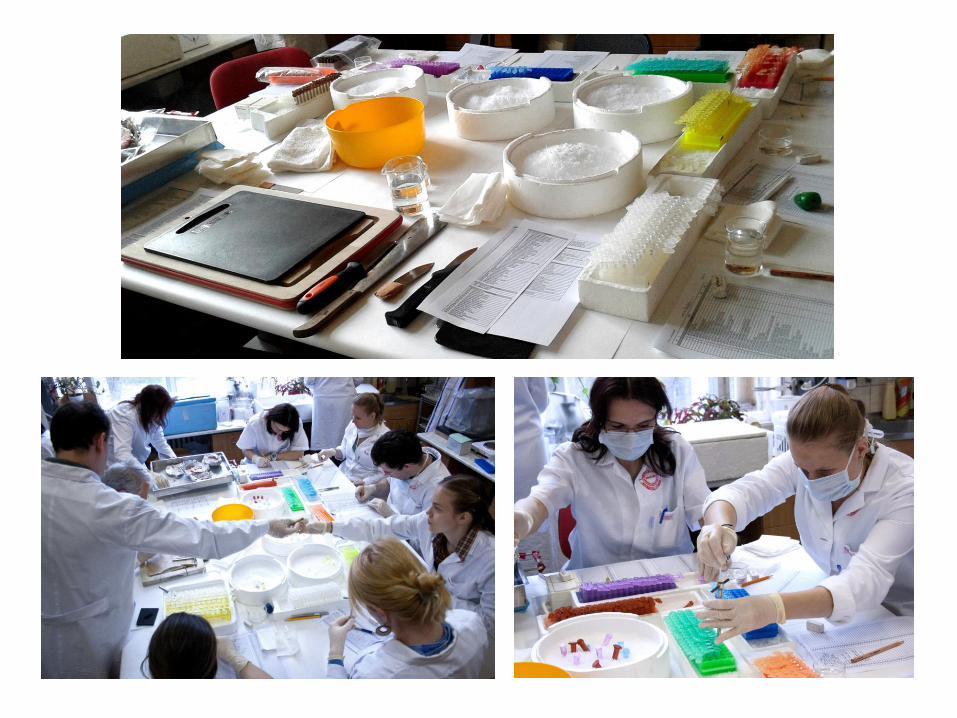
The microdissection of human brain nuclei is based on a simple procedure, called the “micropunch
technique”. This technique was introduced in 1973 to remove small, precisely localized areas from a
rat’s brain. Later, the technique was adapted for the microdissection of the human brain, and since
1992 the Human Brain Tissue Bank, Semmelweis University (HBTB) has been collecting and
storing thousands of microdissected human brain nuclei and areas. The bank is located in the
Department of Anatomy of the Semmelweis University, Budapest, and it operates under the
management of the Laboratory of Neuromorphology of the Hungarian Academy of Sciences.
The Human Brain Tissue Bank, Semmelweis University has been a member of the European Brain
Bank consortium, BrainNet II. Europe, since 2004.
The micropunch technique was published in the Brain Research in 1973. Punching out well
recognized and precisely localized areas from brain sections is a simple, quick, and reproducible
technique, as you will see here later. However, the correct use of technique requires a perfect
knowledge of the fine anatomy, or rather, the fine three-dimensional topography of the brain, whether
human or from experimental animals. By today, the slogan of the microdissection technique has been
realized: “anything that one can recognize and localize anatomically in the brain, can be removed
separately.”
References:
Palkovits M (1973) Isolated removal of hypothalamic or other brain nuclei of the rat, Brain Res
59:449-450
Palkovits M & Brownstein MJ, Maps and Guide to Microdissection of the Rat Brain, Elsevier, New
York-Amsterdam-London 1988


a) The hollow needles are constructed of hard stainless steel tubing mounted in a thicker handle.
Their inner diameters vary from 2.0 to 10.0 mm.
b) The needles are equipped with a spring-stylet that fits well into the lumen of the needle and reach
1-2.5 mm beyond the tip of the needle.
„Punch needles”
a b
For comfortable handling, needles should be 5.0-7.0 cm long with a thinner end, at least 5-7 mm
long, so that the tip of the needle is visible under the microscope.

„Needles” for macrodissection

General rule that the inner diameter of the needle should be as large as possible,
but smaller than the smallest diameter of the brain nuclei or areas

BRAIN SAMPLES
MACRODISSECTION 0.1 - 2.0 g tissue weight
57 different brain regions
MICRODISSECTION 1.0 -100 mg tissue weight
236 different brain nuclei or areas
MICRODISSECTION tissue blocks from 72 different brain areas
(size ~1.0 x 1.0 x 2.0 cm)
cryostat sections
300 mm thick 12-20 mm thick
in situ
hybridization
laser capture microscopy
genomics neurochemistry
proteomics
neurochemistry
proteomics

Brain nuclei can be removed from sections that have been placed on a black rubber plate. By using
upper illumination, the black plate provides a contrasting background.
Macrodissection from serial sections

Microdissection from serial section
by using small „punch needles”

The dissected tissue pellet can be pushed out with the stylet into a tube
or dish, or directly into the microhomogenizer.
If several samples are punched from the same brain nucleus, the pellets
can be collected in the lumen of the needle and pushed them all at
once.

BRAIN SAMPLES
MACRODISSECTION
FROM BRAIN SLICES 0.1-2.0 g tissue weight
57 brain regions
MICRODISSECTION
FROM BRAIN SLICES 1.0-100 mg tissue weight
236 brain regions
MICRODISSECTION
FROM TISSUE BLOCKS ~1.0 x 1.0 x 2.0 cm
dissected tissue blocks from 72 different brain
areas
cryostat sections
300 μm thick 12-20 μm thick
in situ
hybridization
laser capture microscopy
genomics
neurochemistry
proteomics
neurochemistry
proteomics

About 10 x 10 x 20 mm large, anatomically recognized and topographically
oriented blocks are cut out from the brain immediately after removal from the
skull (generally 20-40 brain areas), frozen on dry ice, ready for cryostat
sectioning without thawing of the tissue. Tissue blocks packed in aluminium
foil or in airtight bags are stored at -70°C.

1 2
3 4


Microdissection with
laser capture microscopic technique

Section (20 μm) from the hypothalamus stained with methylenblue
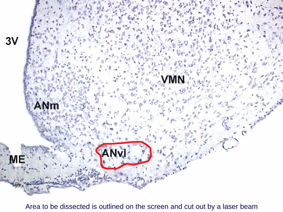
Area to be dissected is outlined on the screen and cut out by a laser beam

The dissected brain area has been sucked into a miniature Eppendorf-like tube
by the vacuum system

Testing for short ex-vivo („post mortem”)
alterations
Samples from same brain areas with
0 to 360 min ex-vivo changes

Step 1. Tissue samples from the same brain areas, immediately after removal
(0’). One drop saline on each samples.

Step 2. The first sample has been covered with dry ice powder within 1’ after
removal.

Step 3. The second sample has been covered with dry ice powder after 5’ after
removal.

Step 4. The third sample has been covered with dry ice powder after 15’ after
removal.

Step 5. The fourth sample has been covered with dry ice powder after 30’ after
removal.

Step 6. The fifth sample has been covered with dry ice powder after 60’ after
removal.
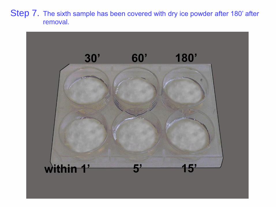
Step 7. The sixth sample has been covered with dry ice powder after 180’ after
removal.
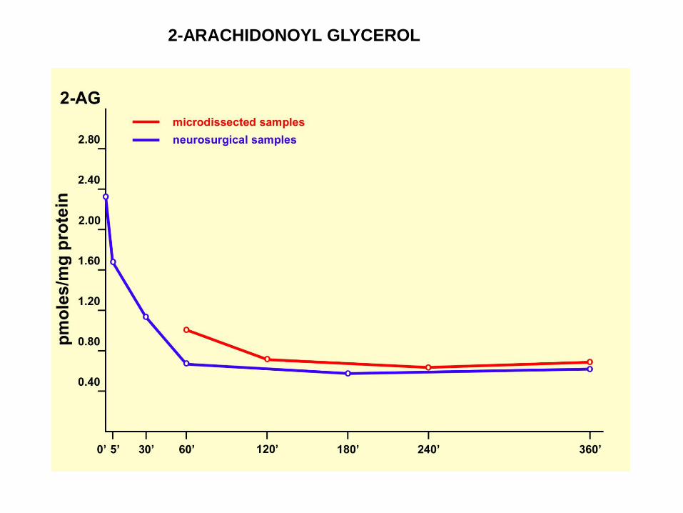
2-ARACHIDONOYL GLYCEROL

Database

PROTECTED
personal
data
large
brain
samples
neuro-
surgical
samples
„open”
personal
data
micro-
dissected
samples
macro-
dissected
samples
tissue
blocks
(color codes)
1. set
2. set
3. set
4. set
5. set
6. set
7. set
1. set
2. set
3. set
4. set
5. set
6. set
7. set
0’
5’
15’
30’
1 h
3 h
6 h
list and numbering of
name
code number
date of death gender
age
date of death
post mortem delay
cause of the death
pathological diagnosis
neuropathological record
date of dissection
storage time
code number
BNE-ID
The database contains several “compartments”. In order to fully respect personal rights, information about deceased persons
is divided into restricted and “open” compartments. The restricted compartment contains the personal data of the subjects.
The 7 parallel sets of the microdissected and macrodissected samples fill up two compartments.

RNA quality in different brain samples
RNA was extracted using the Qiagen lipid tissue kit
and the quality was checked by the Bioanalyser.
Dept. of Cellular and Molecular Neuroscience
Imperial College, Charing Cross Campus
London W6 8RF
„The samples have good quality RNA.”

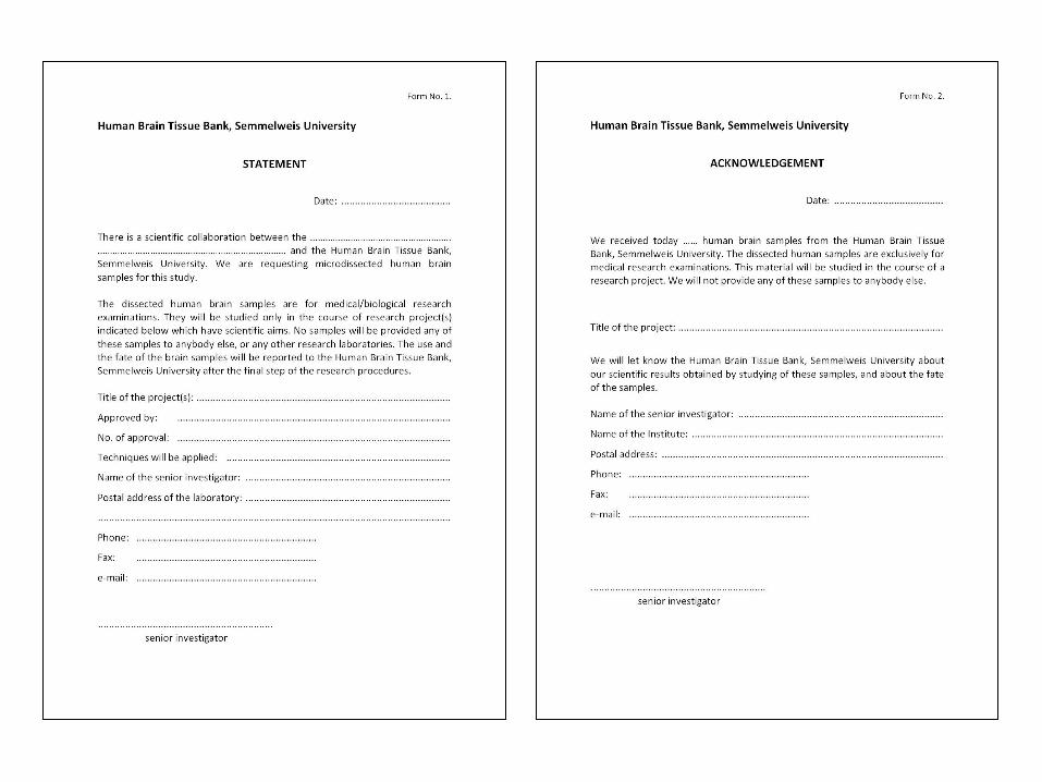
The activity of the Human Brain Tissue Bank, Semmelweis University,
Budapest has been authorized by the Committee of Science and Research
Ethic of the Ministry of Health of Hungary (ETT TUKEB) and the Semmelweis
University Regional Committee of Science and Research Ethic strictly for
research studies. The tissue samples are made available to qualified
researchers or research laboratories world-wide after signing a statement
about scientific collaborations with the HBTB (Form No. 1.) and a declaration
of the proper use and the fate of the microdissected human brain samples
provided by the HBTB (Form No. 2.).

2014
Du L, Merali Z, Poulter MO, Palkovits M, Faludi G & Anisman H (2014) Catechol-O-methyltransferase Val158Met polymorphism and altered COMT
gene expression in the prefrontal cortex of suicide brains, Prog Neuropsychopharmacol Biol Psychiatry 50: 178-183
IF: 4.025 (2013)
Fuxe K, Borroto-Escuela DO, Romero-Fernandez W, Palkovits M, Tarakanov AO, Ciruela F & Agnati LF (2014) Moonlighting proteins and protein-
protein interactions as neurotherapeutic targets in the G protein-coupled receptor field, Neuropsychopharmacology 39: 131-155
IF: 7.833 (2013)
2015
Durrenberger PF, Fernando FS, Kashefi SN, Bonnert TP, Seilhean D, Nait-Oumesmar B, Schmitt A, Gebicke-Haerter PJ, Falkai P, Grünblatt E,
Palkovits M, Arzberger T, Kretzschmar H, Dexter DT & Reynolds R (2015) Common mechanisms in neurodegeneration and neuroinflammation:
a BrainNet Europe gene expression microarray study, J Neural Transm 2014 Aug 13. [Epub ahead of print]
IF: 2.871 (2013)
Ádori C, Glück L, Barde S, Yoshitake T, Kovacs GG, Mulder J, Maglóczky Z, Havas L, Bölcskei K, Mitsios N, Uhlén M, Szolcsányi J, Kehr J, Rönnbäck
A, Schwartz T, Rehfeld JF, Harkany T, Palkovits M, Schulz S & Hökfelt T (2015) Critical role of somatostatin receptor 2 in the vulnerability of the
central noradrenergic system: new aspects on Alzheimer's disease, Acta Neuropathol 129: 541-563
IF: 9.777 (2013)
Dobolyi A, Bagó AG, Gál A, Molnár MJ, Palkovits M, Adam-Vizi V & Chinopoulos C (2015) Localization of SUCLA2 and SUCLG2 subunits of succinyl
CoA ligase within the cerebral cortex suggests the absence of matrix substrate-level phosphorylation in glial cells of the human brain, J Bioenerg
Biomembr 47: 33-41
IF: 2.708 (2013)
Dobolyi A, Ostergaard E, Bagó AG, Dóczi T, Palkovits M, Gál A, Molnár MJ, Adam-Vizi V & Chinopoulos C (2015) Exclusive neuronal expression of
SUCLA2 in the human brain, Brain Struct Funct 220: 135-151
IF: 4.567 (2013)
HBTB közlemények
darab impakt faktor idézettség
1991-2015 83 337.7 4955

Neuropeptides
Luteinizing hormone-releasing hormone Palkovits et al., Endocrinology, 1974
Thyrotropin-releasing hormone Brownstein et al., Science, 1974
Somatostatin Brownstein et al., Endocrinology, 1976
Growth hormone releasing factor Arimura et al., Peptides, 1984
Corticotropin-releasing hormone (CRF) Palkovits, Brownstein, Vale, Fed Proc, 1985
Vasopressin Zerbe and Palkovits, Neuroendocrinology, 1984
Substance P Brownstein et al., Brain Res, 1976
Cholecystokinin Beinfeld and Palkovits, Brain Res, 1982
Enkephalins Kobayashi et al., Life Sci, 1970
Zamir et al., Brain Res, 1985
Dynorphins Zamir et al., Brain Res, 1983, 1984
Pro-opiomelanocortin derived peptides,
ACTH, α-MSH, β-END
de Kloet, Palkovits, Mezey, Pharm Ther, 1981
Atrial natriuretic peptide (ANF) Bahner et al., Hypertension, 1988
Tuberoinfundibular peptide of 39 residues Dobolyi, Palkovits, Usdin, J Comp Neurol, 2003
Apelin / apelin receptors Reaux et al., J Neurochem, 2001 / Neuroscience, 2002
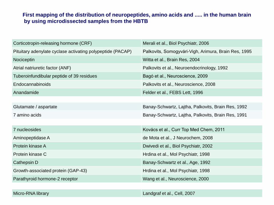
First mapping of the distribution of neuropeptides, amino acids and ..... in the human brain
by using microdissected samples from the HBTB
Corticotropin-releasing hormone (CRF) Merali et al., Biol Psychiatr, 2006
Pituitary adenylate cyclase activating polypeptide (PACAP) Palkovits, Somogyvári-Vigh, Arimura, Brain Res, 1995
Nociceptin Witta et al., Brain Res, 2004
Atrial natriuretic factor (ANF) Palkovits et al., Neuroendocrinology, 1992
Tuberoinfundibular peptide of 39 residues Bagó et al., Neuroscience, 2009
Endocannabinoids Palkovits et al., Neuroscience, 2008
Anandamide Felder et al., FEBS Lett, 1996
7 nucleosides Kovács et al., Curr Top Med Chem, 2011
Aminopeptidase A de Mota et al., J Neurochem, 2008
Protein kinase A Dwivedi et al., Biol Psychiatr, 2002
Protein kinase C Hrdina et al., Mol Psychiatr, 1998
Cathepsin D Banay-Schwartz et al., Age, 1992
Growth-associated protein (GAP-43) Hrdina et al., Mol Psychiatr, 1998
Parathyroid hormone-2 receptor Wang et al., Neuroscience, 2000
Glutamate / aspartate Banay-Schwartz, Lajtha, Palkovits, Brain Res, 1992
7 amino acids Banay-Schwartz, Lajtha, Palkovits, Brain Res, 1991
Micro-RNA library Landgraf et al., Cell, 2007

Adrenaline van der Gugten et al., Brain Res, 1976
PNMT Saavedra et al., Nature, 1974
Noradrenaline and dopamine Versteeg et al., Brain Res, 1976
Serotonin Palkovits, Brownstein, Saavedra, Brain Res, 1974
Histamine Brownstein et al., Brain Res, 1974
Tryptophan hydroxylase Brownstein et al., Brain Res, 1975
Histamine N-methyltransferase Saavedra, Brownstein, Palkovits, Brain Res, 1976
Choline acetyltransferase Kobayashi et al., J Neurochem, 1975
Brownstein et al., J Neurochem, 1975
Monoamine oxidase Saavedra, Brownstein, Palkovits, Brain Res, 1976
Catechol-0-methyltransferase Saavedra, Brownstein, Palkovits, Brain Res, 1976
Glutamate decarboxylase Tappaz, Brownstein, Palkovits, Brain Res, 1976
Acetylcholine Vizi and Palkovits, Brain Res Bull, 1978
Muscarinic cholinergic receptors Kobayashi et al., Brain Res, 1978

GABA and glycine Elekes et al., Neuropharmacology, 1986
Glutamate / aspartate Palkovits et al., Brain Res, 1986
Taurine Palkovits et al., J Neurochem, 1986
Calmodulin Zhou et al., J Neurochem, 1985
Prostaglandins E and F Cseh et al., Brain Res Bull, 1978
Trypsinogen-4 Tóth et al., Neurochem Res, 2007
Adenylyl cyclase IX Antoni et al., J Neurosci, 1998
Protein kinase, type 2 De Vente et al., Neuroscience, 2001
Biogenic amine transporters Hoffman et al., Front Neuroendocrin, 1998
Angiotensin receptors, type-2 Lenkei et al., J Comp Neurol, 1996
Angiotensin receptors, type-1 Lenkei et al., Front Neuroendocrin, 1997


