Management of a Giant Ovarian Cyst by Keyless Abdominal Rope ...
Clinical Pediatric Endocrinology
Received: May 21, 2020 Accepted: September 9, 2020Corresponding author: Mostafa Kotb, M.D., Department of Pediatric Surgery, Alexandria Faculty of Medicine, Alexandria 21615, EgyptE-mail: [email protected]
This is an open-access article distributed under the terms of the Creative Commons Attribution Non-Commercial No Derivatives (by-nc-nd) License <http://creativecommons.org/licenses/by-nc-nd/4.0/>.
pp 57–60January 2021Vol.30 / No.1
Case Report
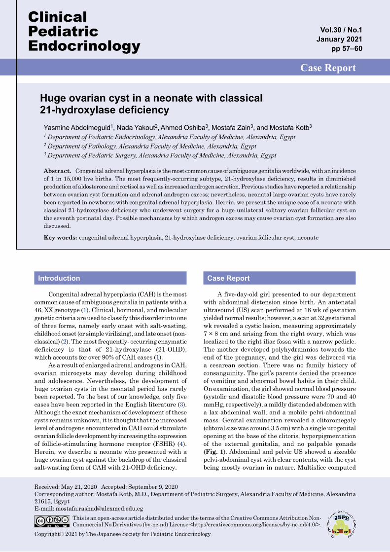
Huge ovarian cyst in a neonate with classical 21-hydroxylase deficiencyYasmine Abdelmeguid1, Nada Yakout2, Ahmed Oshiba3, Mostafa Zain3, and Mostafa Kotb3
1 Department of Pediatric Endocrinology, Alexandria Faculty of Medicine, Alexandria, Egypt2 Department of Pathology, Alexandria Faculty of Medicine, Alexandria, Egypt3 Department of Pediatric Surgery, Alexandria Faculty of Medicine, Alexandria, Egypt
Abstract. Congenital adrenal hyperplasia is the most common cause of ambiguous genitalia worldwide, with an incidence of 1 in 15,000 live births. The most frequently-occurring subtype, 21-hydroxylase deficiency, results in diminished production of aldosterone and cortisol as well as increased androgen secretion. Previous studies have reported a relationship between ovarian cyst formation and adrenal androgen excess; nevertheless, neonatal large ovarian cysts have rarely been reported in newborns with congenital adrenal hyperplasia. Herein, we present the unique case of a neonate with classical 21-hydroxylase deficiency who underwent surgery for a huge unilateral solitary ovarian follicular cyst on the seventh postnatal day. Possible mechanisms by which androgen excess may cause ovarian cyst formation are also discussed.
Key words: congenital adrenal hyperplasia, 21-hydroxylase deficiency, ovarian follicular cyst, neonate
Introduction
Congenital adrenal hyperplasia (CAH) is the most common cause of ambiguous genitalia in patients with a 46, XX genotype (1). Clinical, hormonal, and molecular genetic criteria are used to classify this disorder into one of three forms, namely early onset with salt-wasting, childhood onset (or simple virilizing), and late onset (non-classical) (2). The most frequently- occurring enzymatic deficiency is that of 21-hydroxylase (21-OHD), which accounts for over 90% of CAH cases (1).
As a result of enlarged adrenal androgens in CAH, ovarian microcysts may develop during childhood and adolescence. Nevertheless, the development of huge ovarian cysts in the neonatal period has rarely been reported. To the best of our knowledge, only five cases have been reported in the English literature (3). Although the exact mechanism of development of these cysts remains unknown, it is thought that the increased level of androgens encountered in CAH could stimulate ovarian follicle development by increasing the expression of follicle-stimulating hormone receptor (FSHR) (4). Herein, we describe a neonate who presented with a huge ovarian cyst against the backdrop of the classical salt-wasting form of CAH with 21-OHD deficiency.
Case Report
A five-day-old girl presented to our department with abdominal distension since birth. An antenatal ultrasound (US) scan performed at 18 wk of gestation yielded normal results; however, a scan at 32 gestational wk revealed a cystic lesion, measuring approximately 7 × 8 cm and arising from the right ovary, which was localized to the right iliac fossa with a narrow pedicle. The mother developed polyhydramnios towards the end of the pregnancy, and the girl was delivered via a cesarean section. There was no family history of consanguinity. The girl’s parents denied the presence of vomiting and abnormal bowel habits in their child. On examination, the girl showed normal blood pressure (systolic and diastolic blood pressure were 70 and 40 mmHg, respectively), a mildly distended abdomen with a lax abdominal wall, and a mobile pelvi-abdominal mass. Genital examination revealed a clitoromegaly (clitoral size was around 3.5 cm) with a single urogenital opening at the base of the clitoris, hyperpigmentation of the external genitalia, and no palpable gonads (Fig. 1). Abdominal and pelvic US showed a sizeable pelvi-abdominal cyst with clear contents, with the cyst being mostly ovarian in nature. Multislice computed
Copyright© 2021 by The Japanese Society for Pediatric Endocrinology
Abdelmeguid et al.
58
doi: 10.1297/cpe.30.57
tomography (CT) scans of the abdomen and pelvis confirmed that this mass was around 6.5 × 8 × 8 cm, extending from the right hypochondrium to the right side of the pelvis and displacing the neighboring structures (Fig. 2). The presentation was consistent with that of a unilocular cyst, with no soft tissue component and an imperceptible wall. Visualization of the uterus revealed that its measurements were 23 × 9 mm.
Laboratory results revealed the following: elevated 17-hydroxyprogesterone (17-OHP) (more than 200 ng/mL; normal: 0.07–0.77 ng/mL), serum testosterone (26.4 ng/mL; normal: 0.2–0.64 ng/mL), and ACTH (112 pg/mL; normal: 0–46 pg/mL) levels; a low basal cortisol level (0.3 μg/mL; normal: 1–50 μg/mL); a low-normal serum sodium (136 mmol/L; normal: 135–145 mmol/L) level; and normal potassium (5.2 mmol/L; normal 3.5–5.5 mmol/L), and serum glucose (140 mg/dL) levels. Unfortunately, serum aldosterone and plasma renin activity could not be analyzed to confirm mineralocorticoid deficiency. Thus, the final diagnosis was Prader stage 4 CAH with single urogenital sinus caused primarily by 21-OHD. The karyotype of the baby was 46, XX. Polymerase chain reaction was performed for the 21-hydroxylase gene to confirm the diagnosis, revealing homozygosity of the Q318X mutation. After endocrinological consultation, the baby was administered stress-dose intravenous corticosteroid therapy in preparation for exploratory surgery of the abdominal cyst. She received 25 mg of intravenous hydrocortisone preoperatively and a similar dose was given intraoperatively.
A right transverse exploratory incision of approximately 5 cm in length was made at the level of the umbilicus. We found a large mass filling the abdominal cavity and displacing the intestine to the left side (Fig. 3). Aspiration of the cyst yielded clear serous fluid with bile tinge due to physiological jaundice. Aspiration of approximately 100 mL was needed before being able
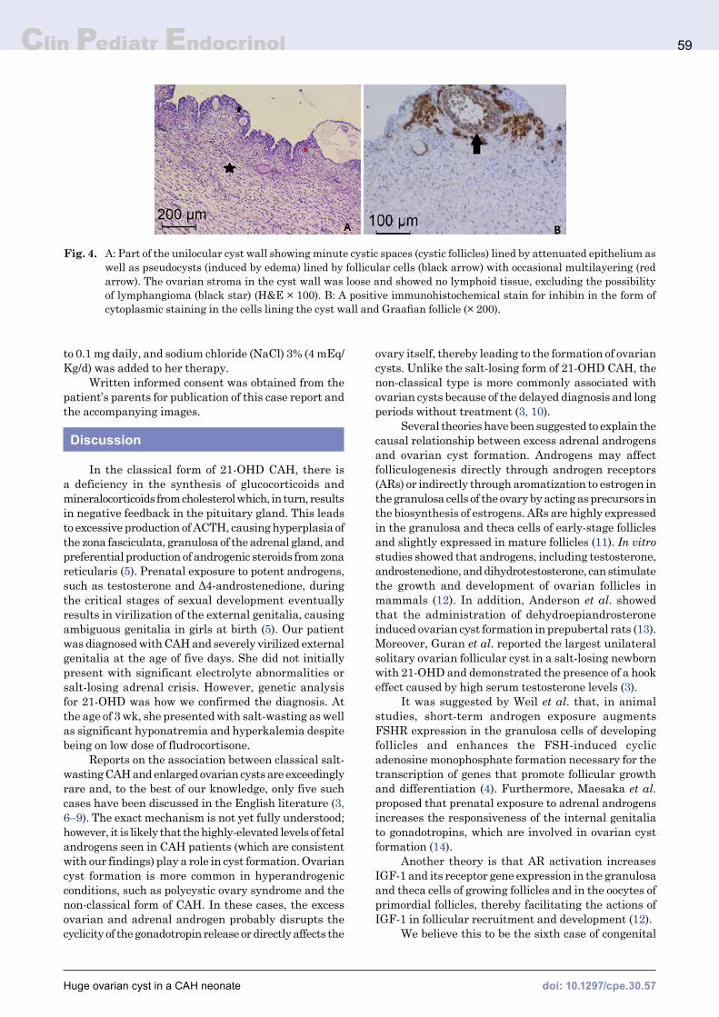
to exteriorize the cyst outside of the abdomen. We then found a unilocular right ovarian cyst with a diameter of approximately 6 cm and a very small amount of normal ovarian tissue, which we opened and drained. Near-total excision of the cyst was attempted, leaving only the part of the wall adjacent to the adnexa in order to preserve any remaining ovarian tissues. The histopathological examination using both hematoxylin and eosin (H&E) and immunohistochemical staining for inhibin revealed a functional (follicular) cyst (Fig. 4). Postoperatively, the patient was given a stress-dose of intravenous hydrocortisone consisting of 100 mg/m2/d. This dose was divided across 6-h intervals and reduced to maintenance doses when her levels were stable and she could tolerate oral feeding. The maintenance doses included 10 mg/m2/d of hydrocortisone and one daily tablet of fludrocortisone 0.025 mg tablet daily. She was discharged on the sixth postoperative day after an uneventful course.
One week after discharge, the patient presented with salt-losing crisis at follow up. This confirmed the diagnosis of classical salt-wasting CAH. Her serum sodium and potassium were 124 mmol/L and 6.5 mmol/L, respectively. The dose of fludrocortisone was increased
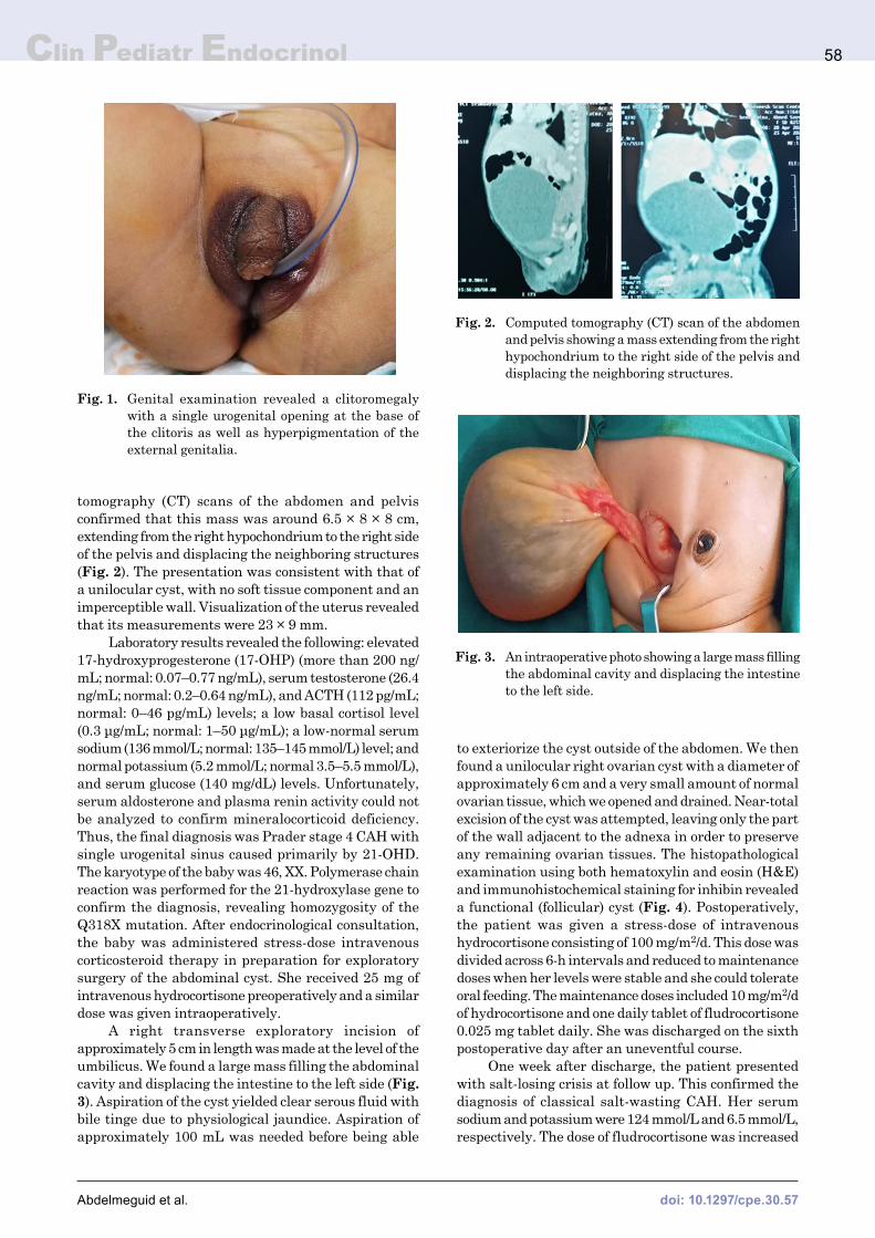
Fig. 1. Genital examination revealed a clitoromegaly with a single urogenital opening at the base of the clitoris as well as hyperpigmentation of the external genitalia.
Fig. 2. Computed tomography (CT) scan of the abdomen and pelvis showing a mass extending from the right hypochondrium to the right side of the pelvis and displacing the neighboring structures.
Fig. 3. An intraoperative photo showing a large mass filling the abdominal cavity and displacing the intestine to the left side.
Clin Pediatr EndocrinolClin Pediatr Endocrinol
Huge ovarian cyst in a CAH neonate
59
doi: 10.1297/cpe.30.57
to 0.1 mg daily, and sodium chloride (NaCl) 3% (4 mEq/Kg/d) was added to her therapy.
Written informed consent was obtained from the patient’s parents for publication of this case report and the accompanying images.
Discussion
In the classical form of 21-OHD CAH, there is a deficiency in the synthesis of glucocorticoids and mineralocorticoids from cholesterol which, in turn, results in negative feedback in the pituitary gland. This leads to excessive production of ACTH, causing hyperplasia of the zona fasciculata, granulosa of the adrenal gland, and preferential production of androgenic steroids from zona reticularis (5). Prenatal exposure to potent androgens, such as testosterone and Δ4-androstenedione, during the critical stages of sexual development eventually results in virilization of the external genitalia, causing ambiguous genitalia in girls at birth (5). Our patient was diagnosed with CAH and severely virilized external genitalia at the age of five days. She did not initially present with significant electrolyte abnormalities or salt-losing adrenal crisis. However, genetic analysis for 21-OHD was how we confirmed the diagnosis. At the age of 3 wk, she presented with salt-wasting as well as significant hyponatremia and hyperkalemia despite being on low dose of fludrocortisone.
Reports on the association between classical salt-wasting CAH and enlarged ovarian cysts are exceedingly rare and, to the best of our knowledge, only five such cases have been discussed in the English literature (3, 6–9). The exact mechanism is not yet fully understood; however, it is likely that the highly-elevated levels of fetal androgens seen in CAH patients (which are consistent with our findings) play a role in cyst formation. Ovarian cyst formation is more common in hyperandrogenic conditions, such as polycystic ovary syndrome and the non-classical form of CAH. In these cases, the excess ovarian and adrenal androgen probably disrupts the cyclicity of the gonadotropin release or directly affects the
ovary itself, thereby leading to the formation of ovarian cysts. Unlike the salt-losing form of 21-OHD CAH, the non-classical type is more commonly associated with ovarian cysts because of the delayed diagnosis and long periods without treatment (3, 10).
Several theories have been suggested to explain the causal relationship between excess adrenal androgens and ovarian cyst formation. Androgens may affect folliculogenesis directly through androgen receptors (ARs) or indirectly through aromatization to estrogen in the granulosa cells of the ovary by acting as precursors in the biosynthesis of estrogens. ARs are highly expressed in the granulosa and theca cells of early-stage follicles and slightly expressed in mature follicles (11). In vitro studies showed that androgens, including testosterone, androstenedione, and dihydrotestosterone, can stimulate the growth and development of ovarian follicles in mammals (12). In addition, Anderson et al. showed that the administration of dehydroepiandrosterone induced ovarian cyst formation in prepubertal rats (13). Moreover, Guran et al. reported the largest unilateral solitary ovarian follicular cyst in a salt-losing newborn with 21-OHD and demonstrated the presence of a hook effect caused by high serum testosterone levels (3).
It was suggested by Weil et al. that, in animal studies, short-term androgen exposure augments FSHR expression in the granulosa cells of developing follicles and enhances the FSH-induced cyclic adenosine monophosphate formation necessary for the transcription of genes that promote follicular growth and differentiation (4). Furthermore, Maesaka et al. proposed that prenatal exposure to adrenal androgens increases the responsiveness of the internal genitalia to gonadotropins, which are involved in ovarian cyst formation (14).
Another theory is that AR activation increases IGF-1 and its receptor gene expression in the granulosa and theca cells of growing follicles and in the oocytes of primordial follicles, thereby facilitating the actions of IGF-1 in follicular recruitment and development (12).
We believe this to be the sixth case of congenital
Fig. 4. A: Part of the unilocular cyst wall showing minute cystic spaces (cystic follicles) lined by attenuated epithelium as well as pseudocysts (induced by edema) lined by follicular cells (black arrow) with occasional multilayering (red arrow). The ovarian stroma in the cyst wall was loose and showed no lymphoid tissue, excluding the possibility of lymphangioma (black star) (H&E × 100). B: A positive immunohistochemical stain for inhibin in the form of cytoplasmic staining in the cells lining the cyst wall and Graafian follicle (× 200).
Clin Pediatr Endocrinol
Abdelmeguid et al.
60
doi: 10.1297/cpe.30.57
adrenal hyperplasia accompanied by a huge ovarian cyst in the neonatal period in the scientific literature. Of these, three presented with the classical salt-wasting 21-OHD (3, 7, 9), one with 11ß-hydroxylase deficiency (6), and one with congenital lipoid adrenal hyperplasia, which is considered the most severe form of congenital adrenal hyperplasia (8). Bilaterality was reported in two cases (6, 7). In general, when the size of the cyst exceeds 5 cm, it becomes liable to mass effect, torsion, rupture, and hemorrhage (15). In almost all of the reported cases, including ours, the large size of the ovarian cyst was the main indication for surgical intervention, especially if nearby structures were being compressed. However, in a single case, ovarian torsion was the main indication (8).
When surgery is planned in CAH, 5 to 10 times the daily maintenance dose of hydrocortisone is needed, as patients are unable to produce sufficient cortisol in response to stress (16). A preoperative and intraoperative stress-dose of hydrocortisone should be given to prevent serious metabolic abnormalities associated with acute
adrenal insufficiency (5). This should be followed by high doses of hydrocortisone during the first 24–48 postoperative hours, after which maintenance therapy can be used. Glucocorticoid replacement should consist of thrice-daily doses of oral hydrocortisone totaling 10–15 mg/m2/d in addition to 0.05 to 0.2 mg/d of fludrocortisone and 1–2 g/d of supplemental NaCL (17–34 mEq/d) (5). Finally, the measurement of serum levels of 17-OHP, androstenedione, serum testosterone, ACTH, plasma renin, and aldosterone should be included in follow-up to ensure that the doses administered during glucocorticoid and mineralocorticoid therapy are adequate (16).
Although uncommon, giant ovarian cysts should be suspected in neonates with CAH presenting with abdominal distention. In some cases, it could be indicative of classical salt-wasting CAH when the typical manifestations of the condition are absent.
Conflict of interests: The authors declare that they have no conflicts of interest.
References
1. GattiJM,Dowlut-McelroyT,WilligL.Differencesofsexualdevelopment.In:HolocombIIIGW,MurphyJP,StPeterSD,editors.HolocombandAschcraft’sPediatricsurgery.7thed.Philadelphia:Elsevier;2020.p.953-65.
2. DabasA,VatsP,SharmaR,SinghP,SethA,JainV,et al.Managementofinfantswithcongenitaladrenalhyperplasia.IndianPediatr2020;57:159–64.[Medline] [CrossRef]
3. GüranT,YeşilG,GüranÖ,CesurS,BosnalıO,CelayirA,et al.Agiantovariancystinaneonatewithclassical21-hydroxylasedeficiencywithveryhightestosteronelevelsdemonstratingahigh-dosehookeffect.JClinResPediatrEndocrinol2012;4:151–3.[Medline] [CrossRef]
4. WeilS,VendolaK,ZhouJ,BondyCA.Androgenandfollicle-stimulatinghormoneinteractionsinprimateovarianfollicledevelopment.JClinEndocrinolMetab1999;84:2951–6.[Medline] [CrossRef]
5. PierettiRV,DonahoePK.Disordersofsexualdifferentiation.In:CoranAG,CaldamoneA,AdzickNS,KrummelTM,LabergeJM,ShambergerR,editors.PediatricSurgery.7thed.Philadelphia:Elsevier;2012.p.1565-90.
6. TopalogluAK,VadeA,ZellerWP.Congenitaladrenalhyperplasiaandbilateralovariancystsinaneonate.ClinPediatr(Phila)1997;36:719–20.[Medline] [CrossRef]
7. ShankarR,MahajanJK,KhannaS,RaoKL.Bilateralovariancystsinaneonatewithsalt-wastingcongenitaladrenalhyperplasia.JPediatrSurg2010;45:e19–21.[Medline] [CrossRef]
8. JinHY,ChoiJH,LeeBH,KimGH,KimHK,YooHW.Ovariancysttorsioninapatientwithcongenitallipoidadrenalhyperplasia.EurJPediatr2011;170:535–8.[Medline] [CrossRef]
9. ŞahinNM,BayramoğluE,ÇetinkayaS,ErdeveŞŞ,KaramanA,AkdoğanMP,et al.Vaginalbleedingandagiantovariancystinaninfantwith21-hydroxylasedeficiency.JPediatrEndocrinolMetab2018;31:229–33.[Medline] [CrossRef]
10. GilJuniorAB,RezendeAP,doCarmoAV,DuarteEI,deMedeirosMM,deMedeirosSF.Adrenalandrogenparticipationinthepolycysticovarysyndrome.RevBrasGinecolObstet2010;32:541–8(inPortuguese).[Medline] [CrossRef]
11. FranksS,HardyK.Androgenactionintheovary.FrontEndocrinol(Lausanne)2018;9:452.[Medline] [CrossRef]12. GervásioCG,BernuciMP,Silva-de-SáMF,Rosa-E-SilvaAC.Theroleofandrogenhormonesinearlyfolliculardevelopment.
ISRNObstetGynecol2014;2014:818010.[Medline] [CrossRef]13. AndersonE,LeeMT,LeeGY.Cystogenesisoftheovarianantralfollicleoftherat:ultrastructuralchangesandhormonal
profilefollowingtheadministrationofdehydroepiandrosterone.AnatRec1992;234:359–82.[Medline] [CrossRef]14. MaesakaH,SuwaS,TachibanaK,KatsumataN.Prolongedactivationofhypothalamo-pituitary-ovarianaxisduringearly
infancyinfemalepatientswithsalt-losing21-hydroxylasedeficiency.PediatrRes1985;19:1258–62.[Medline] [CrossRef]15. FallatME,IgnacioJrRC.Ovariantumors.In:PuriP,HöllwarthM,editors.Pediatricsurgery–Diagnosisandtreatment.
Heidelberg:Springer;2009.p.745-54.16. SpeiserPW,ArltW,AuchusRJ,BaskinLS,ConwayGS,MerkeDP,et al.Congenitaladrenalhyperplasiaduetosteroid
21-hydroxylasedeficiency:AnEndocrineSocietyClinicalPracticeGuideline.JClinEndocrinolMetab2018;103:4043–88.[Medline] [CrossRef]
Clin Pediatr EndocrinolClin Pediatr Endocrinol