How to recognize symptoms of diseases caused by Phytophthora
15
Matteo Garbelotto UC Berkeley, Cooperative Extension David M. Rizzo UC Davis Jennifer M. Davidson USDA Forest Service, Research Susan J. Frankel USDA Forest Service, State & Private Forestry How to recognize symptoms of diseases caused by causal agent of Sudden Oak Death Phytophthora ramorum Photo: Keith Parker Photo: David M. Rizzo
Transcript of How to recognize symptoms of diseases caused by Phytophthora
Matteo GarbelottoUC Berkeley,Cooperative Extension
David M. RizzoUC Davis
Jennifer M. DavidsonUSDA Forest Service,Research
Susan J. FrankelUSDA Forest Service,State & Private Forestry
������������� ������ ����� �� � ���� �����
��� ������
������������������
�����������������
Photo: Keith Parker
Photo: David M. Rizzo

�����
Page
Background 3
The Pathogen 3
How to detect Phytophthora ramorum 5
Gallery of symptoms 6
Tanoak 7
Coast Live Oak, California Black Oak, Shreve’s Oak 9
Rhododendron 10
Huckleberry 10
California Bay Laurel 11
Pacific Madrone 11
California Buckeye 12
Big Leaf Maple 12
Toyon, Honeysuckle, Manzanita, and Coffeeberry 13
Viburnum 14
Table 1 4
List of Figures 15

�
Recognizing symptoms of Phytophthora ramorum, a newly described microbe and cause of“Sudden Oak Death,” can be difficult so we have assembled pictures and information to guidefield identification.
��������
Phytophthora ramorum, cause of Sudden Oak Death (SOD), is responsible for widespread treemortality in Central and Northern California. This common name “Sudden Oak Death” refers tothe apparent rapid decline of the entire tree crown; the foliage of trees affected often turns froman apparently healthy green color to brown in only a few weeks. Despite this graphic commonname, not all trees affected by the disease incur in this apparent sudden death. There is nothing“sudden” in SOD and it may take several months or years for the pathogen to kill a tree. Re-searchers are still trying to understand how rapidly the disease may progress; it is expected thatdisease progression may vary based on different tree individuals, different tree species, differentgeographic regions and other factors.
Three species of native California oaks (Quercus spp.) and a close relative of oaks, called tanoakor tanbark oak (Lithocarpus densiflorus), may be killed by the disease. To date, only oaks in theblack or red oak group have been found to be susceptible to the disease, white oaks are currentlynot known to be hosts. At least 11 other unrelated plant species may serve as hosts forP. ramorum (Table 1). On many of these hosts,infection by the pathogen may not lead to death ofthe whole plant, but rather to leaf spots and/or twigand branch dieback. The Latin name of the pathogenmeans “destructor of branches” reflecting its discov-ery by plant pathologists in Europe in 1993 as thecause of a new disease of leaves and branches ofornamental rhododendrons. Foliar hosts can at timesbe killed, especially if host plants are shrubby orsmall. Progressive dieback of branches can also leadto plant mortality, normally if conditions conduciveto the disease persist for several years in a row.
���� �����
There are about 60 known species closely related toP. ramorum, and only specialists can differentiateamongst them. The reproductive, dispersal andsurvival structures of the pathogen are only visibleunder a microscope. Sporangia (Fig. 1) are reproduc-
��������������
������ ������ �� �
��� �����
�����������������!
��� ���������
���������������Pho
to: K
eith
Par
ker
Fig. 1. Sporangia of P. ramorum. (Rizzo)

"
������#$�%����� � ����� ������ ��������������������������
�����������$
�������������������������� ���������
���� � ��������� � �������� �&��������� '�(���� �������� �������
Quercus agrifolia Coast live oak main stem YES NO(Fagaceae)
Q. kellogii California Black oak main stem YES NO(Fagaceae)
Q. parvula var. shrevei Shreve’s oak main stem YES NO(Fagaceae)
Lithocarpus densiflorus Tanoak main stem, branches, leaves YES YES(Fagaceae)
Arbutus menzesii Madrone branches, leaves PROBABLY YES(Ericaceae)
Vaccinium ovatum Evergreen main stem, branches, leaves YES YES(Ericaceae) huckleberry
Arctostaphylos manzanita Manzanita branches, leaves UNKNOWN UNKNOWN
Rhododendron sp. (Ericaceae) Ornamental branches, leaves YES YES(Ericaceae) rhododendron
Rhododendron macrophyllum Rhododendron leaves, branches YES YES (Ericaceae)
Umbellaria californica Bay laurel, leaves NO NO(Lauraceae) Oregon myrtle
Acer macrophyllum Big leaf maple leaves NO NO(Aceraceae)
Heteromeles arbutifolia Toyon branches, leaves UNKNOWN UNKNOWN(Rosaeceae)
Aesculus californica Buckeye branches, leaves NO NO(Hippocastanaceae)
Rhamnus californica Coffeeberry leaves UNKNOWN UNKNOWN(Rhamnaceae)
Lonicera hispidula Honeysuckle leaves UNKNOWN UNKNOWN(Caprifoliaceae)
)
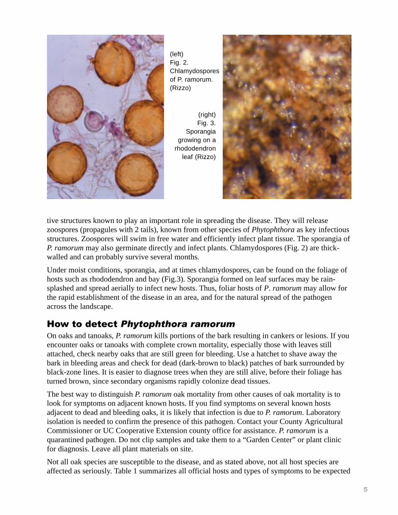
tive structures known to play an important role in spreading the disease. They will releasezoospores (propagules with 2 tails), known from other species of Phytophthora as key infectiousstructures. Zoospores will swim in free water and efficiently infect plant tissue. The sporangia ofP. ramorum may also germinate directly and infect plants. Chlamydospores (Fig. 2) are thick-walled and can probably survive several months.
Under moist conditions, sporangia, and at times chlamydospores, can be found on the foliage ofhosts such as rhododendron and bay (Fig.3). Sporangia formed on leaf surfaces may be rain-splashed and spread aerially to infect new hosts. Thus, foliar hosts of P. ramorum may allow forthe rapid establishment of the disease in an area, and for the natural spread of the pathogenacross the landscape.
�������������������������������
On oaks and tanoaks, P. ramorum kills portions of the bark resulting in cankers or lesions. If youencounter oaks or tanoaks with complete crown mortality, especially those with leaves stillattached, check nearby oaks that are still green for bleeding. Use a hatchet to shave away thebark in bleeding areas and check for dead (dark-brown to black) patches of bark surrounded byblack-zone lines. It is easier to diagnose trees when they are still alive, before their foliage hasturned brown, since secondary organisms rapidly colonize dead tissues.
The best way to distinguish P. ramorum oak mortality from other causes of oak mortality is tolook for symptoms on adjacent known hosts. If you find symptoms on several known hostsadjacent to dead and bleeding oaks, it is likely that infection is due to P. ramorum. Laboratoryisolation is needed to confirm the presence of this pathogen. Contact your County AgriculturalCommissioner or UC Cooperative Extension county office for assistance. P. ramorum is aquarantined pathogen. Do not clip samples and take them to a “Garden Center” or plant clinicfor diagnosis. Leave all plant materials on site.
Not all oak species are susceptible to the disease, and as stated above, not all host species areaffected as seriously. Table 1 summarizes all official hosts and types of symptoms to be expected
(left)Fig. 2.Chlamydosporesof P. ramorum.(Rizzo)
(right)Fig. 3.
Sporangiagrowing on a
rhododendronleaf (Rizzo)

*
on each host species. This list is incomplete; researchers are still studying the host and geo-graphic ranges of the pathogen.
+���������� ������
#,������ �� ������������ �������(��-���(����� ���$
Whether in wildlands or at the wildland-urban interface, the advanced stage of the disease resultsin high levels of mortality of tanoak, coast live oak, and California black oak (Figs. 4 and 5). Theentire crown of dead trees will initially turn brown (Fig. 6) and then turn gray as the foliage islost (Fig. 7). Note that California black oak (a host species) and other non-host species such asValley oak, appear gray or leafless during the winter because they are deciduous. Tree mortalitynormally appears in patches. California buckeyes are summer deciduous and naturally drop theirleaves in the summer. Other diseases (for example, the root rots Armillaria mellea, Phytophthoracinnamomi) can cause patches of dead trees, but their symptoms are quite different. Fires andchemical girdling may also cause patches of dead trees. Look for burnt litter and bark or forchemical injections to differentiate between diseases and other causes of tree mortality.
Fig. 4. SOD in coastlive oak in theurban-wildlandinterface (KeithParker)
Fig. 5. Dyingtanoaks in a
redwood-tanoakforest in
Marin County(Kent Julin)

.
/,������ �� ������������ �������(��-������� ��� $
Although not fully understood yet, it is believed that at first the pathogen will colonize leaves offoliar hosts (non oaks) and of tanoaks. The presence of suspicious spots and blotches on leavesof two or more hosts may be an indicator that the disease has arrived in an area and has not yetcaused lethal cankers on oaks or tanoak.
�,������ �� ����(����� ��� �� ����� $
P. ramorum has been discovered relatively recently; for this reason this list (Table 1, page 4)should not be considered complete or exhaustive. New symptoms and more hosts may be discov-ered. Due to the limited extent of scientific observations, some of the images shown here are theresult not of natural infection, but of artificial inoculations in the laboratory.
Most of the symptoms described below are not exclusively caused by P. ramorum, they may becaused by other pathogens, sunburn, frost or other abiotic agents. Beware of look-alikes and lookfor the clustering of symptoms on several hosts in an area.
TANOAK (Lithocarpus densiflorus): Tanoak is highly susceptible to P. ramorum infectionthrough a combination of foliar blight, branch dieback, and stem cankers. Plants of all sizes maybe killed. Early symptoms of the disease include wilting of apical shoots (“Shepard’s crook ”)
Fig. 6. The crown of an understory tanoak killed byP. ramorum (Garbelotto lab)
Fig. 7. The gray trees are coast live oaks killedby P. ramorum more than a year before thepicture was taken (Garbelotto lab)

0
(Fig. 8), light brown leaf spots and blotches(Fig. 9), and dead leaves. As the diseaseprogresses, branches die back due to formationof branch cankers. Stem cankers develop atvarious heights from the ground, most com-monly just above the soil line.
Cankers appear as brown water-soaked lesionsin the bark and the cambium of infected trees,often progressing into the outer part of thexylem (Fig. 10). Black lines are often, but notalways, seen at the canker margins (Fig.11).Seeping of viscous sap, black to amber incolor, can at times be seen, creating the typicalSOD symptom referred to as “bleeding”(Fig.12).
Stem cankers may girdle the tree causing theentire tree crown to turn yellow then brown.Crown decline is generally rapid after a periodof months when the stem is dying. Infectedtrees will often resprout at the base but eventu-ally all, or most of these basal sprouts, die aswell.
Once the tree is seriously compromised byadvanced infection of P. ramorum, opportunis-tic organisms may become established. Theseinclude the sapwood decay fungus Hypoxylonthouarsianum (Fig. 13); this fungus produces
Fig. 8. Wilting of tanoak shoot (Pavel Svihra)
Fig. 9. Tanoak leaves showing initial foliarsymptoms of SOD (Garbelotto lab)
Fig. 10. Cankers appear as brown water-soakedlesions in the bark. (Garbelotto lab)
Fig. 11. Close up of a black linebetween infected tissues (right) and
healthy tissue (Rizzo)

1
charcoal-black globose fruiting bodies on the bark. Ambrosia and bark beetles, tunneling into thesapwood and bark respectively, will produce sawdust expelled from the beetle galleries. It shouldbe noted that these secondary organisms are not necessarily associated with SOD, but can beseen when trees decline and die because of a variety of reasons.
COAST LIVE OAK, CALIFORNIA BLACK OAK, SHREVE’S OAK (Quercus agrifolia,Quercus kelloggii, and Quercus parvula var. shrevei). These three oak species can effectively bekilled by stem cankers caused by P. ramorum. Leaves and branches generally do not appear to besusceptible. Cankers are mostly seen on the lowerpart of the stem just above the root collar, but do notapparently extend into the roots. This feature differ-entiates P. ramorum cankers from those caused byother Phytophthoras (e.g., P. cinnamomi). Cankerscan also develop high on the stem, but they are rarelyseen on portions of the stem smaller than 10 cm.Infection of seedlings is unreported in nature, andinfection of saplings appears to be extremely rare.Cankers are water-soaked regions of dead bark oftenextending into the outer portion of the xylem. Theyare generally demarcated by visible black zone lines(Fig. 11). Seeping is commonly associated withcankers. Seeping ooze is generally black to amber incolor and viscous. At the early stages of the canker,seeping will occur through the intact bark withoutany noticeable physical wounding. In later stages, thebark can fracture and seeping keeps occurring boththrough broken and intact bark. Often seeps dry outand can be identified by their brownish tinge on thedarker bark (Fig. 14).
(left)Fig. 12.Seeping throughbark of aninfected oak tree(Garbelotto lab)
(right)Fig. 13.Fruiting
bodies ofHypoxylon
Fig. 14. Brown dried seep on the bark of acoast live oak (Garbelotto lab)

#2
Once trees are girdled by the pathogen they will eventually die. In general, the foliage of infectedtrees will change from green to brown over a period of several weeks; however, this browningmay occur from several months to more than a year after the pathogen has effectively girdled thewhole stem. It is not uncommon to observe the loss of a significant number of leaves aftercankers have girdled the tree but before the whole crown browns.
Seeping can be caused by other pathogens such as Armillaria or other Phytophthora spp. or byphysical wounding caused by people and insects. Wetwood, a bacterial infection often initiatedby major wounding and tree aging, can also result in streaks of blackish seeps on the bark sur-face. Wetwood seeps are always dark, not viscous, and often associated with a physical openingin the bark to surface. Wetwood seeps also have a foul odor as compared to a wine smell ofseeping caused by P. ramorum infection. Common secondary organisms are the same as thosedescribed for tanoaks.
RHODODENDRON. Rhododendron spp. Not all species and ornamental varieties are equallysusceptible, but precise information on host range is still unavailable. Both California nativespecies (R. macrophyllum and R. occidentale) can succumb to the pathogen. Leaves are readily
infected by P. ramorum,and brown-blacklesions often developon the leaf portionwhere water accumu-lates. These lesionsgenerally have “fuzzy”borders while lesionscaused by sun damagetypically have clearblack contour lines(Fig. 15). At times,though, P. ramorumconcentric ring growthpattern is evident whenexamining the lesions.
P. ramorum can alsoinfect and kill branches, and
may progress to kill entire plants.The pathogen readily sporulates onthe leaf surface of Rhododendronspp. (Fig. 3).
HUCKLEBERRY. The Californiaor Pacific huckleberry (Vacciniumovatum) can be infected by P.ramorum. Although individual leafspots have been observed, the mostcommon symptom includes branchdieback. Lesions can be observed onthe stems (canes), and the wholebranch upward from the lesion willdie and brown. Although entire
Fig. 15. Comparative damage caused by sunburn and byP. ramorum on rhododendron leaf (Tim Tidwell)
Fig. 16. Branch dieback of a huckleberry plant. (Davidson)

##
plants can be killed, it is more common to observepatches of dead and live branches (Fig. 16).
CALIFORNIA BAY LAUREL (Umbellulariacalifornica). P. ramorum damage on this plant isvirtually undistinguishable from symptoms caused byanother common disease known as bay anthracnose.For both diseases, the most striking symptom consistsof a black tip, turning gray in time. It is often, but notalways, delimited by a chlorotic (e.g., yellow) zone(Fig. 17). Lesions are normally on the tip since wateris most likely to accumulate there. On leaves that areflat or carried with the tip upwards, lesion can developin the middle section, especially around the edges orat the base of the petiole. Often infection results in anumber of clearly distinct black spots that are visibleon the green portion of the leaf. At times, only theblack spots may be visible without the presence of anextensive blotch. Some branch dieback has beenobserved in trees infected by P. ramorum, but it is stilluncertain whether P. ramorum is responsible or not forthe development of this symptom (Fig. 18). Mortalityof bay laurel due to infection by P. ramorum has neverbeen reported.
PACIFIC MADRONE. Leaf spots, leaf death, andbranch dieback are commonly associated with infec-tion by P. ramorum on this species. A precise analysisof the effects of this disease on pacific madrone iscomplicated by its high susceptibility to other com-mon pathogens that produce similar symptoms.P. ramorum’s destructive effect on madrone is sug-gested by the observation that in areas infested byP. ramorum, levels of tree dieback and mortalityamong the regenerating madrone/juveniles and seed-ling madrone are higher than in comparable siteswhere P. ramorum is not present. Discrete leaf spot-ting, often with a purplish tinge, is one of the firstsymptoms of infection by P. ramorum (Fig. 19). Spotsexpand into larger botches that often turn to gray andbrown. Lesions also may develop on the stems ofsmall plants and on branches. Although not always areliable trait, infection by other foliar and branchpathogens may lead to the formation of small blackreproductive structures barely visible by the nakedeye. At times though, several pathogens may bepresent on the same leaf and diagnosis must be madeby a professional.
Fig. 17. Necrotic tips and spotson bay laurel (Garbelotto lab)
Fig. 19. Madrone leaf withspots caused by P. ramorum(Garbelotto lab)
Fig. 18. Dead branches on baytrees attributed toP. ramorum (Garbelotto lab)

#/
CALIFORNIA BUCKEYE. P. ramorum can infectthe leaves, the petioles and the twigs of Californiabuckeye trees. Early symptoms start as roundedindividual spots that tend to coalesce later in theseason (Figs. 20 and 21). The symptoms are verysimilar to those caused by the buckeye anthracnosepathogen, Guignardia aesculi. Darkened lesions onpetioles and twigs (Fig. 22) indicate presence of P.ramorum. Early foliar senescence (buckeyes aredeciduous trees and drop the foliage unusually earlyin hot and dry areas) causes leaf distortions similarto those caused by P. ramorum.
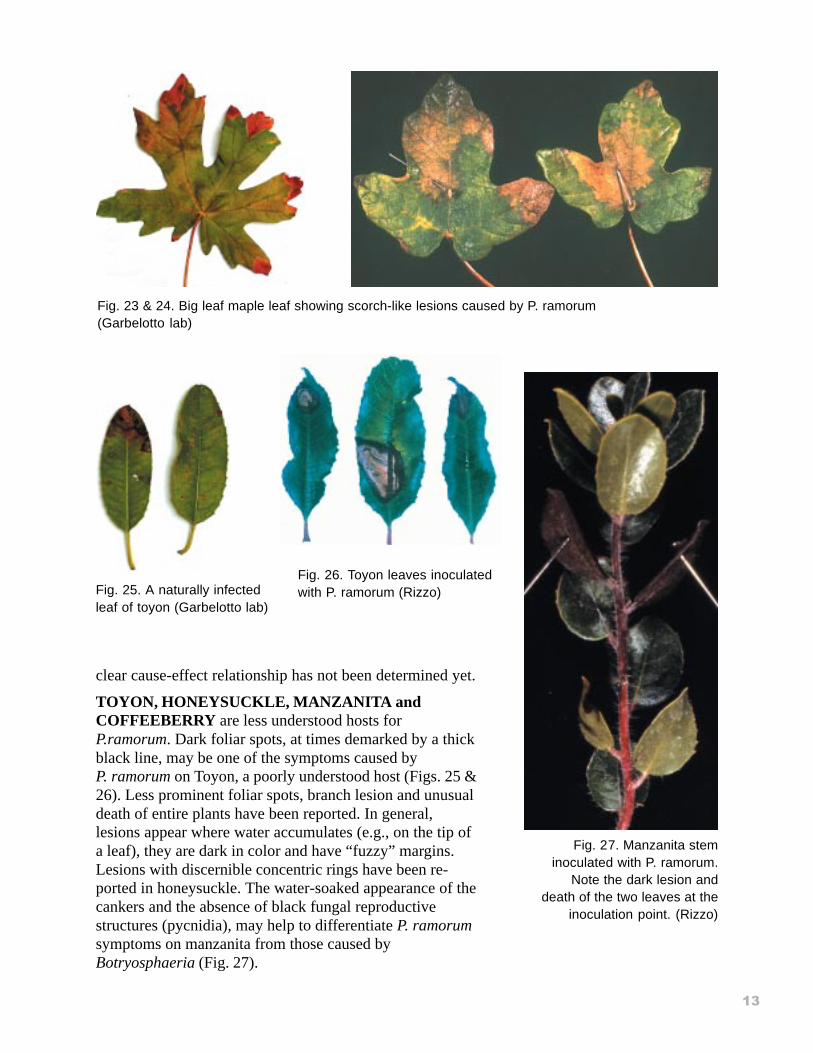
BIG LEAF MAPLE. In this tree species, P.ramorum infection appears more like a scorch,normally starting from the edges of the leaf (Fig.23). In general, the scorching will have irregularborders that do not precisely follow leaf contour.The discoloration can be variable in color with huesranging from orange to brown (Fig. 24). Once theleaves senesce and yellow, the discoloration shouldstill be visible for some time. Branch dieback hasbeen observed in areas infested by P. ramorum, but a
Figs. 21. Fig. 22. Petiole infection by P. ramorum onbuckeye. (Garbelotto lab)
Fig. 20 & 21. Different stages of infectionby P. ramorum on buckeye (Garbelotto lab)
#�
clear cause-effect relationship has not been determined yet.
TOYON, HONEYSUCKLE, MANZANITA andCOFFEEBERRY are less understood hosts forP.ramorum. Dark foliar spots, at times demarked by a thickblack line, may be one of the symptoms caused byP. ramorum on Toyon, a poorly understood host (Figs. 25 &26). Less prominent foliar spots, branch lesion and unusualdeath of entire plants have been reported. In general,lesions appear where water accumulates (e.g., on the tip ofa leaf), they are dark in color and have “fuzzy” margins.Lesions with discernible concentric rings have been re-ported in honeysuckle. The water-soaked appearance of thecankers and the absence of black fungal reproductivestructures (pycnidia), may help to differentiate P. ramorumsymptoms on manzanita from those caused byBotryosphaeria (Fig. 27).
Fig. 23 & 24. Big leaf maple leaf showing scorch-like lesions caused by P. ramorum(Garbelotto lab)
Fig. 26. Toyon leaves inoculatedwith P. ramorum (Rizzo)Fig. 25. A naturally infected
leaf of toyon (Garbelotto lab)
Fig. 27. Manzanita steminoculated with P. ramorum.
Note the dark lesion anddeath of the two leaves at the
inoculation point. (Rizzo)
#"
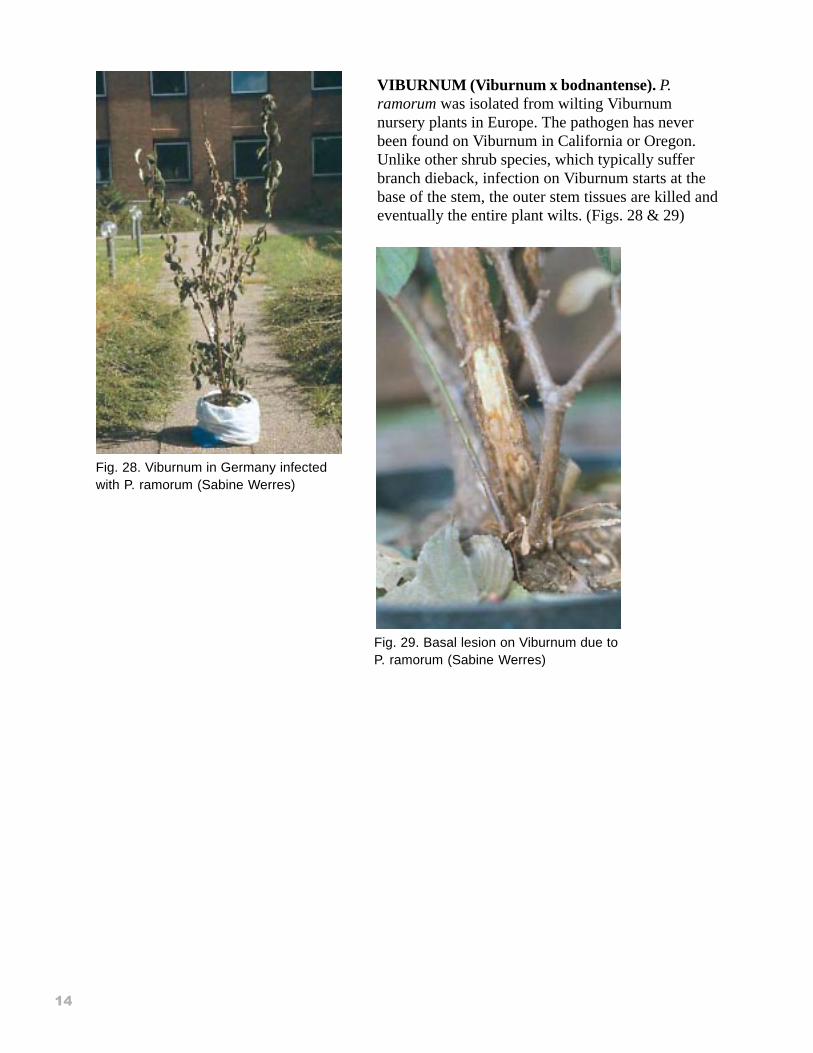
VIBURNUM (Viburnum x bodnantense). P.ramorum was isolated from wilting Viburnumnursery plants in Europe. The pathogen has neverbeen found on Viburnum in California or Oregon.Unlike other shrub species, which typically sufferbranch dieback, infection on Viburnum starts at thebase of the stem, the outer stem tissues are killed andeventually the entire plant wilts. (Figs. 28 & 29)
Fig. 28. Viburnum in Germany infectedwith P. ramorum (Sabine Werres)
Fig. 29. Basal lesion on Viburnum due toP. ramorum (Sabine Werres)

#)
Fig. 1. Sporangia of P. ramorum. (Rizzo)
Fig. 2. Chlamydospores of P. ramorum. (Rizzo)
Fig. 3. Sporangia growing on a rhododendron leaf (Rizzo)
Fig. 4. SOD in coast live oak in the urban-wildland interface (Keith Parker)
Fig. 5. Dying tanoaks in a redwood tanoak forest in Marin County (Kent Julin)
Fig. 6. The crown of an understory tanoak killed by P. ramorum (Garbelotto lab)
Fig. 7. The gray trees are coast live oaks killed by P. ramorum more than a year before the picture wastaken (Garbelotto lab)
Fig. 8. Wilting of tanoak shoot (Pavel Svihra)
Fig. 9. Tanoak leaves showing initial foliar symptoms of SOD (Garbelotto lab)
Fig. 10. Cankers appear as brown water-soaked lesions in the bark. (Garbelotto lab)
Fig. 11. Close up of a black line between infected tissues (right) and healthy tissue (Rizzo)
Fig. 12. Seeping through bark of an infected oak tree (Garbelotto lab)
Fig. 13. Fruiting bodies of Hypoxylon
Fig. 14. Brown dried seep on the bark of a coast live oak (Garbelotto lab)
Fig. 15. Comparative damage caused by sunburn and by P. ramorum on rhododendron leaf (TimTidwell)
Fig. 16. Branch dieback of a huckleberry plant. (Davidson)
Fig. 17. Necrotic tips and spots on bay laurel (Garbelotto lab)
Fig. 18. Dead branches on bay trees attributed to P. ramorum (Garbelotto lab)
Fig. 19. Madrone leaf with spots caused by P. ramorum (Garbelotto lab)
Figs. 20 & 21. Different stages of infection by P. ramorum on buckeye (Garbelotto lab)
Fig. 22. Petiole infection by P. ramorum on buckeye. (Garbelotto lab)
Fig. 23 & 24. Big leaf maple leaf showing scorch-like lesions caused by P. ramorum (Garbelotto lab)
Fig. 25. Manzanita stem inoculated with P. ramorum. Note the dark lesion and death of the two leavesat the inoculation point. (Rizzo)
Fig. 26. A naturally infected leaf of toyon (Garbelotto lab)
Fig. 27. Toyon leaves inoculated with P. ramorum (Rizzo)
Fig. 28. Viburnum in Germany infected with P. ramorum (Sabine Werres)
Fig. 29. Basal lesion on Viburnum due to P. ramorum
ACKNOWLEDGEMENTS. Dr. Doug Schmidt and Tami Harnik were instrumental in preparingthe photographic material and the text for this document. Thanks also to Donna Dell’Ario,Roxane Scales and Ervin Castle, USDA Forest Service, Pacific Southwest Region, for design,layout and production of this publication.
3� �����4����