For a dialogical approach of the Trinitarian indwelling Jean-Baptiste ...
How to Place an Indwelling Chest Tube forDrainage and Lavage of the Pleural Cavity inHorses Affected with Pleuropneumonia
M. Keith Chaffin, DVM, MS; G. Kent Carter, DVM, MS;Robin M. Dabareiner, DVM, MS, PhD
Placement of indwelling chest tubes in horses with pleuropneumonia is an easy and safe procedure tofacilitate continuous drainage of pleural fluid. Pleural lavage via indwelling chest tubes is aneffective procedure to promote drainage of thick exudate, bacteria, and necrotic material from thepleural cavity. Authors’ address: Department of Large Animal Medicine and Surgery, College ofVeterinary Medicine, Texas A&M University, College Station, TX 77843-4475. © 2000 AAEP.
Introduction
Bacterial pleuropneumonia (i.e., pleuritis) involvesbacterial colonization of the lung, resulting in pneu-monia and/or lung abscessation, followed by exten-sion to the pleural space and accumulation ofpleural effusion (parapneumonic effusion).1 Inhorses, pleuropneumonia has been associated withstress, long-distance transport, and strenuous exer-cise, likely because of suppressive effects on pulmo-nary defenses.1–6 Pulmonary aspiration has alsobeen associated with pleuropneumonia, likely due tooverwhelming of pulmonary defense mechanisms.1
Clinical signs of pleuropneumonia are variableand include lethargy, anorexia, fever, nasal dis-charge, cough, exercise intolerance, dyspnea, respi-ratory distress, flared nostrils, weight loss, andsternal edema.7–11 Acutely affected horses oftenexhibit pleurodynia, evident as pawing, stiff gait,abducted elbows, and reluctance to move orcough.7 Diagnosis is made based on thoracic aus-cultation and percussion, hematology, thoracic radi-
ography and ultrasonography, thoracocentesis, andcytologic examination and microbiologic culture ofpleural fluid and tracheobronchial aspirates.7–11
Treatment of pleuropneumonia varies depend-ing on the severity and duration of the diseaseprocess; the causative organisms; the presence,character, and volume of pleural effusion; the extentof fibrin deposition; the microenvironment within thepleural space; and the development of sequelae.12
Therapeutic modalities include anti-inflammatory andantimicrobial medications, pleural drainage, pleurallavage, thoracostomy, rest, supportive care, and man-agement of sequlae.8,10,12,13
An important decision in the management ofhorses with pleuropneumonia is determiningwhether pleural drainage is indicated. Parapneu-monic effusions have been classified based on theneed for pleural drainage. Uncomplicated effu-sions are those that resolve spontaneously withoutdrainage, whereas complicated effusions generallyrequire drainage in addition to antimicrobial ther-
AAEP PROCEEDINGS / Vol. 46 / 2000 145
HOW-TO SESSION
NOTES
Reprinted in the IVIS website with the permission of AAEP Close window to return to IVIS
Proceedings of the Annual Convention of the AAEP 2000
apy for resolution.1,12 Indications for pleuraldrainage include a poor response to conservativetherapy and/or pleural fluid with one or more of thefollowing characteristics: sufficient volume tocause respiratory distress, empyematous character,putrid odor, cytologically-visible bacteria, positivemicrobiologic cultures, low concentration of glucose(, 40 mg/dl), low pH (, 7.1) and high LDH concen-tration (. 1,000 IU/L).8,12–14 When indicated, pleu-ral drainage should be initiated as early in the diseaseprocess as possible, because when delayed, fibrin loc-ulation can hinder adequate drainage.12
Pleural fluid can be drained using either intermit-tent thoracocentesis or indwelling chest tubes. In-termittent thoracocentesis may be considered inhorses with small volumes of pleural fluid that donot have a putrid odor or contain thick cellulardebris.12 In horses that are accumulating pleuralfluid rapidly or in which pleural fluid is thick andexudative, indwelling chest tubes enhance drain-age and facilitate continual drainage throughouttreatment.
In some affected horses, pleural lavage is helpfulto remove fibrin, debris, and necrotic tissue; to dilutethick, viscous pleural fluid; and to facilitatedrainage.12 Lavage is probably most effective insubacute stages before fibrin loculated pockets ofeffusion develop.
The purpose of this paper is to describe the tech-niques for placement of indwelling chest tubes andfor pleural lavage. Our experience has been thatpractitioners are often reluctant to perform pleuraldrainage and lavage, likely due to unfamiliaritywith the procedure.
Materials and Methods
The materials we use for placement of an indwell-ing chest tube include 20 ml of local anesthetic,fenestrated chest tubes of various size (16 –32
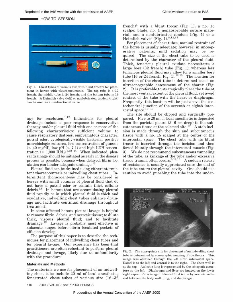
french)a with a blunt trocar (Fig. 1), a no. 15scalpel blade, no. 1 nonabsorbable suture mate-rial, and a nonlubricated condom (Fig. 1) or aHeimlich valveb (Fig. 1).8,12,13
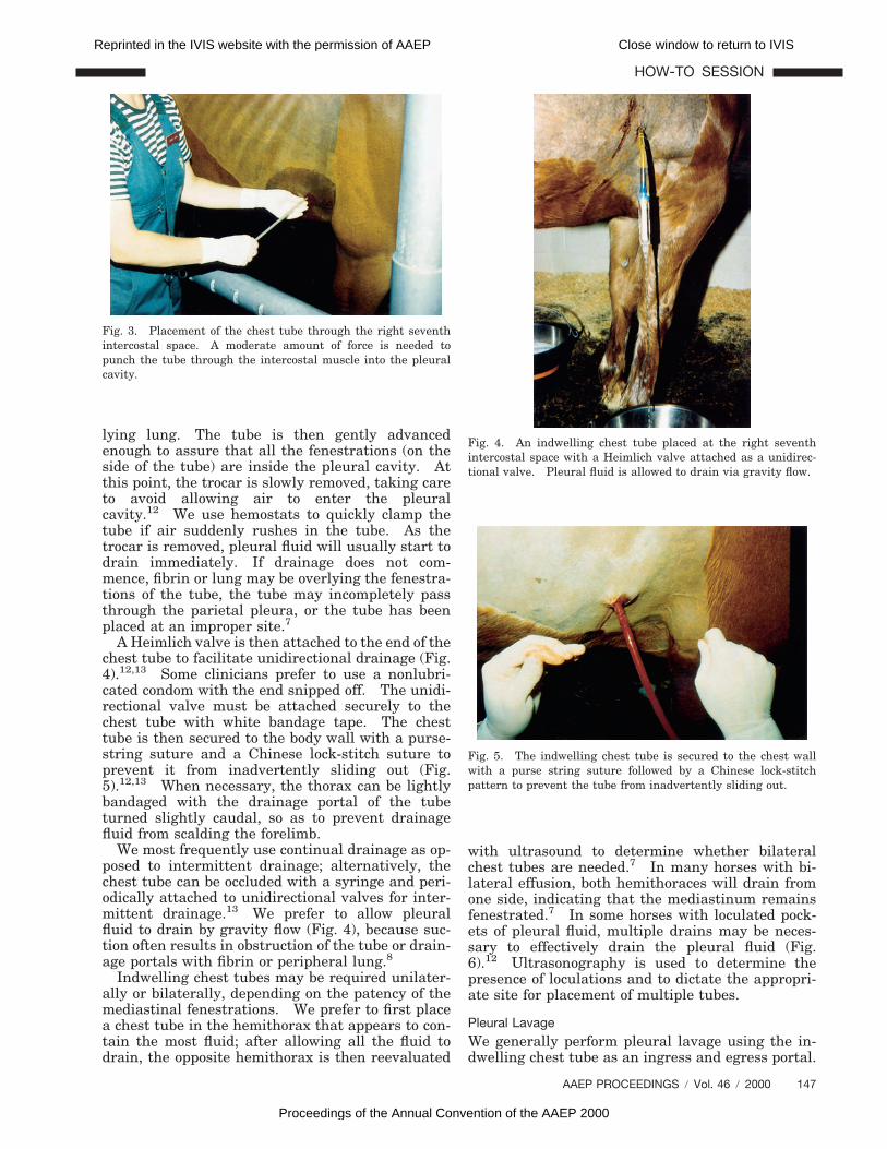
For placement of chest tubes, manual restraint ofthe horse is usually adequate; however, in uncoop-erative patients, mild sedation may be re-quired. The size of the chest tube to be used isdetermined by the character of the pleural fluid.Thick, tenacious pleural exudate necessitates alarge bore (32 french) tube (Fig. 1); whereas lesstenacious pleural fluid may allow for a smaller boretube (16 or 24 french; Fig. 1).12,13 The location forinsertion of the chest tube is determined based onultrasonographic assessment of the thorax (Fig.2). It is preferable to strategically place the tube atthe most ventral extent of the pleural fluid, yet avoidcontact of the tube with the heart or diaphragm.Frequently, this location will be just above the cos-tochondral junction of the seventh or eighth inter-costal space.10–13
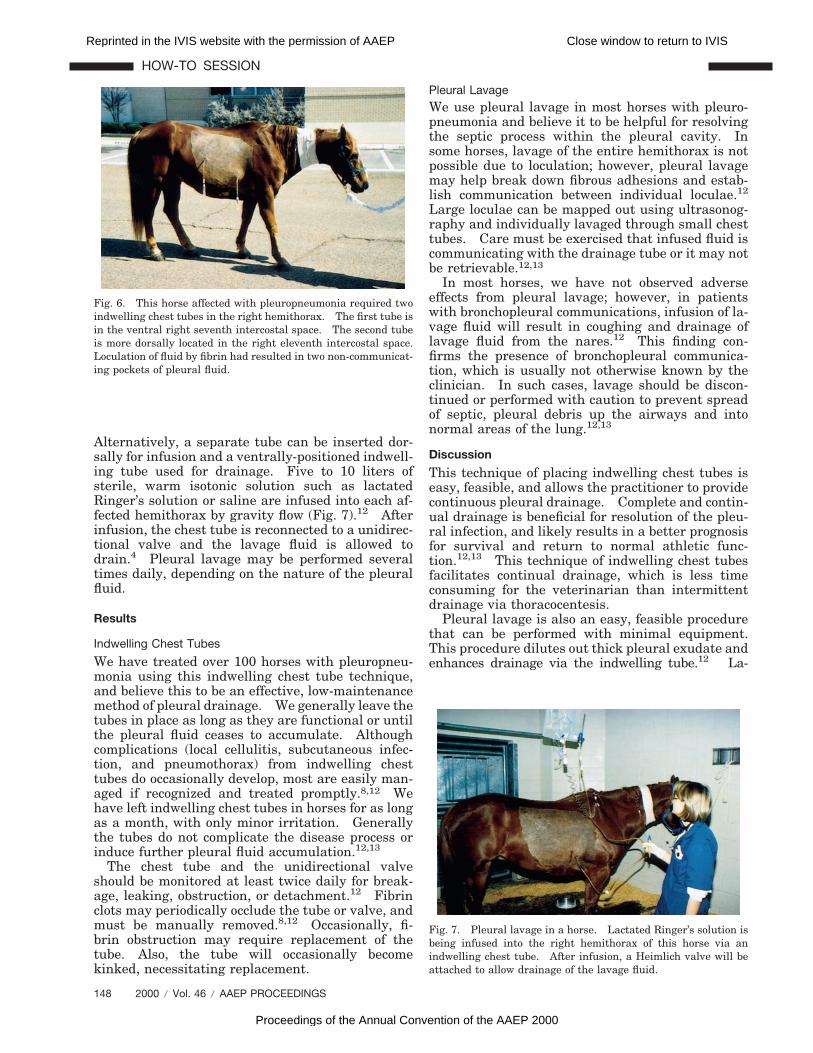
The site should be clipped and surgically pre-pared. Five to 20 ml of local anesthetic is depositedfrom the parietal pleura (3–6 cm deep) to the sub-cutaneous tissue at the selected site.10 A stab inci-sion is made through the skin and subcutaneoustissue with a no. 15 scalpel at the center of theintercostal space. The chest tube with its blunttrocar is inserted through the incision and thenforced bluntly through the intercostal muscle (Fig.3). We do not recommend subcutaneous tunnelingof the tube, as kinkage of the tube and/or excessivetissue trauma often occurs.8,12,13 A sudden releaseof resistance is usually appreciated once the end ofthe tube enters the pleural cavity. One should usecaution to avoid punching the tube into the under-
Fig. 1. Chest tubes of various size with blunt trocars for place-ment in horses with pleuropneumonia. The top tube is a 32french, the middle tube is 24 french, and the bottom tube is 16french. A Heimlich valve (left) or nonlubricated condom (right)can be used as a unidirectional valve.
Fig. 2. The appropriate site for placement of an indwelling chesttube is determined by sonographic imaging of the thorax. Thisimage was obtained through the left ninth intercostal space.Dorsal is to the left and ventral is to the right. The chest wall isat the top. Atelectic lung is represented by the echogenic struc-ture on the left. Diaphragm and liver are imaged on the lowerright aspect of the image. Pleural fluid is the hypoechoic mate-rial between the body wall, lung, and diaphragm.
146 2000 / Vol. 46 / AAEP PROCEEDINGS
HOW-TO SESSION
Reprinted in the IVIS website with the permission of AAEP Close window to return to IVIS
Proceedings of the Annual Convention of the AAEP 2000
lying lung. The tube is then gently advancedenough to assure that all the fenestrations (on theside of the tube) are inside the pleural cavity. Atthis point, the trocar is slowly removed, taking careto avoid allowing air to enter the pleuralcavity.12 We use hemostats to quickly clamp thetube if air suddenly rushes in the tube. As thetrocar is removed, pleural fluid will usually start todrain immediately. If drainage does not com-mence, fibrin or lung may be overlying the fenestra-tions of the tube, the tube may incompletely passthrough the parietal pleura, or the tube has beenplaced at an improper site.7
A Heimlich valve is then attached to the end of thechest tube to facilitate unidirectional drainage (Fig.4).12,13 Some clinicians prefer to use a nonlubri-cated condom with the end snipped off. The unidi-rectional valve must be attached securely to thechest tube with white bandage tape. The chesttube is then secured to the body wall with a purse-string suture and a Chinese lock-stitch suture toprevent it from inadvertently sliding out (Fig.5).12,13 When necessary, the thorax can be lightlybandaged with the drainage portal of the tubeturned slightly caudal, so as to prevent drainagefluid from scalding the forelimb.
We most frequently use continual drainage as op-posed to intermittent drainage; alternatively, thechest tube can be occluded with a syringe and peri-odically attached to unidirectional valves for inter-mittent drainage.13 We prefer to allow pleuralfluid to drain by gravity flow (Fig. 4), because suc-tion often results in obstruction of the tube or drain-age portals with fibrin or peripheral lung.8
Indwelling chest tubes may be required unilater-ally or bilaterally, depending on the patency of themediastinal fenestrations. We prefer to first placea chest tube in the hemithorax that appears to con-tain the most fluid; after allowing all the fluid todrain, the opposite hemithorax is then reevaluated
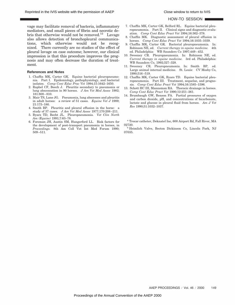
with ultrasound to determine whether bilateralchest tubes are needed.7 In many horses with bi-lateral effusion, both hemithoraces will drain fromone side, indicating that the mediastinum remainsfenestrated.7 In some horses with loculated pock-ets of pleural fluid, multiple drains may be neces-sary to effectively drain the pleural fluid (Fig.6).12 Ultrasonography is used to determine thepresence of loculations and to dictate the appropri-ate site for placement of multiple tubes.
Pleural LavageWe generally perform pleural lavage using the in-dwelling chest tube as an ingress and egress portal.
Fig. 3. Placement of the chest tube through the right seventhintercostal space. A moderate amount of force is needed topunch the tube through the intercostal muscle into the pleuralcavity.
Fig. 4. An indwelling chest tube placed at the right seventhintercostal space with a Heimlich valve attached as a unidirec-tional valve. Pleural fluid is allowed to drain via gravity flow.
Fig. 5. The indwelling chest tube is secured to the chest wallwith a purse string suture followed by a Chinese lock-stitchpattern to prevent the tube from inadvertently sliding out.
AAEP PROCEEDINGS / Vol. 46 / 2000 147
HOW-TO SESSION
Reprinted in the IVIS website with the permission of AAEP Close window to return to IVIS
Proceedings of the Annual Convention of the AAEP 2000
Alternatively, a separate tube can be inserted dor-sally for infusion and a ventrally-positioned indwell-ing tube used for drainage. Five to 10 liters ofsterile, warm isotonic solution such as lactatedRinger’s solution or saline are infused into each af-fected hemithorax by gravity flow (Fig. 7).12 Afterinfusion, the chest tube is reconnected to a unidirec-tional valve and the lavage fluid is allowed todrain.4 Pleural lavage may be performed severaltimes daily, depending on the nature of the pleuralfluid.
Results
Indwelling Chest Tubes
We have treated over 100 horses with pleuropneu-monia using this indwelling chest tube technique,and believe this to be an effective, low-maintenancemethod of pleural drainage. We generally leave thetubes in place as long as they are functional or untilthe pleural fluid ceases to accumulate. Althoughcomplications (local cellulitis, subcutaneous infec-tion, and pneumothorax) from indwelling chesttubes do occasionally develop, most are easily man-aged if recognized and treated promptly.8,12 Wehave left indwelling chest tubes in horses for as longas a month, with only minor irritation. Generallythe tubes do not complicate the disease process orinduce further pleural fluid accumulation.12,13
The chest tube and the unidirectional valveshould be monitored at least twice daily for break-age, leaking, obstruction, or detachment.12 Fibrinclots may periodically occlude the tube or valve, andmust be manually removed.8,12 Occasionally, fi-brin obstruction may require replacement of thetube. Also, the tube will occasionally becomekinked, necessitating replacement.
Pleural LavageWe use pleural lavage in most horses with pleuro-pneumonia and believe it to be helpful for resolvingthe septic process within the pleural cavity. Insome horses, lavage of the entire hemithorax is notpossible due to loculation; however, pleural lavagemay help break down fibrous adhesions and estab-lish communication between individual loculae.12
Large loculae can be mapped out using ultrasonog-raphy and individually lavaged through small chesttubes. Care must be exercised that infused fluid iscommunicating with the drainage tube or it may notbe retrievable.12,13
In most horses, we have not observed adverseeffects from pleural lavage; however, in patientswith bronchopleural communications, infusion of la-vage fluid will result in coughing and drainage oflavage fluid from the nares.12 This finding con-firms the presence of bronchopleural communica-tion, which is usually not otherwise known by theclinician. In such cases, lavage should be discon-tinued or performed with caution to prevent spreadof septic, pleural debris up the airways and intonormal areas of the lung.12,13
Discussion
This technique of placing indwelling chest tubes iseasy, feasible, and allows the practitioner to providecontinuous pleural drainage. Complete and contin-ual drainage is beneficial for resolution of the pleu-ral infection, and likely results in a better prognosisfor survival and return to normal athletic func-tion.12,13 This technique of indwelling chest tubesfacilitates continual drainage, which is less timeconsuming for the veterinarian than intermittentdrainage via thoracocentesis.
Pleural lavage is also an easy, feasible procedurethat can be performed with minimal equipment.This procedure dilutes out thick pleural exudate andenhances drainage via the indwelling tube.12 La-
Fig. 7. Pleural lavage in a horse. Lactated Ringer’s solution isbeing infused into the right hemithorax of this horse via anindwelling chest tube. After infusion, a Heimlich valve will beattached to allow drainage of the lavage fluid.
Fig. 6. This horse affected with pleuropneumonia required twoindwelling chest tubes in the right hemithorax. The first tube isin the ventral right seventh intercostal space. The second tubeis more dorsally located in the right eleventh intercostal space.Loculation of fluid by fibrin had resulted in two non-communicat-ing pockets of pleural fluid.
148 2000 / Vol. 46 / AAEP PROCEEDINGS
HOW-TO SESSION
Reprinted in the IVIS website with the permission of AAEP Close window to return to IVIS
Proceedings of the Annual Convention of the AAEP 2000
vage may facilitate removal of bacteria, inflammatorymediators, and small pieces of fibrin and necrotic de-bris that otherwise would not be removed.12 Lavagealso allows detection of bronchopleural communica-tions, which otherwise would not be recog-nized. There currently are no studies of the effect ofpleural lavage on case outcome; however, our clinicalimpression is that this procedure improves the prog-nosis and may often decrease the duration of treat-ment.
References and Notes1. Chaffin MK, Carter GK. Equine bacterial pleuropneumo-
nia. Part I. Epidemiology, pathophysiology, and bacterialisolates. Comp Cont Educ Prac Vet 1994;15:1642–1650.
2. Raphel CF, Beech J. Pleuritis secondary to pneumonia orlung abscessation in 90 horses. J Am Vet Med Assoc 1982;181:808–810.
3. Mair TS, Lane JG. Pneumonia, lung abscesses and pleuritisin adult horses: a review of 51 cases. Equine Vet J 1989;21:175–180.
4. Smith BP. Pleuritis and pleural effusion in the horse: astudy of 37 cases. J Am Vet Med Assoc 1977;170:208–211.
5. Byars TD, Becht JL. Pleuropneumonia. Vet Clin NorthAm (Equine) 1991;7:63–78.
6. Foreman JH, Austin SM, Hungerford LL. Risk factors forthe development of post-transport pneumonia in horses, inProceedings. 8th Am Coll Vet Int Med Forum 1990;509–511.
7. Chaffin MK, Carter GK, Relford RL. Equine bacterial pleu-ropneumonia. Part II. Clinical signs and diagnostic evalu-ation. Comp Cont Educ Pract Vet 1994;16:362–378.
8. Chaffin MK. Diagnostic assessment of pleural effusion inhorses. Comp Cont Educ Pract Vet 1994;16:1035–1039.
9. Chaffin MK, Carter GK. Bacterial pleuropneumonia. In:Robinson NE, ed. Current therapy in equine medicine. 4thed. Philadelphia: WB Saunders Co 1997;449–452.
10. Sweeney CR. Pleuropneumonia. In: Robinson NE, ed.Current therapy in equine medicine. 3rd ed. Philadelphia:WB Saunders Co, 1992;327–328.
11. Sweeney CR. Pleuropneumonia. In: Smith BP, ed.Large animal internal medicine. St. Louis: CV Mosby Co,1990;516–519.
12. Chaffin MK, Carter GK, Byars TD. Equine bacterial pleu-ropneumonia. Part III. Treatment, sequelae, and progno-sis. Comp Cont Educ Pract Vet 1994;16:1585–1596.
13. Schott HC III, Mannsman RA. Thoracic drainage in horses.Comp Cont Educ Pract Vet 1990;12:251–261.
14. Brumbaugh GW, Benson PA. Partial pressures of oxygenand carbon dioxide, pH, and concentrations of bicarbonate,lactate and glucose in pleural fluid from horses. Am J VetRes 1990;51:1032–1037.
a Trocar catheter, Deknatel Inc, 600 Airport Rd, Fall River, MA02720.
b Heimlich Valve, Becton Dickinson Co, Lincoln Park, NJ07035.
AAEP PROCEEDINGS / Vol. 46 / 2000 149
HOW-TO SESSION
Reprinted in the IVIS website with the permission of AAEP Close window to return to IVIS
Proceedings of the Annual Convention of the AAEP 2000