How to control and prevent pesky parasites -...
120
How to control and prevent pesky parasites: basics on worms, coccidia and other internal parasites Dr. Christine Petersen DVM, PhD Department of Veterinary Pathology- Global Parasitology section, Iowa State University (515) 294-9013, [email protected] May 2008
Transcript of How to control and prevent pesky parasites -...

How to control and prevent pesky parasites: basics on worms, coccidia and other internal parasites
Dr. Christine PetersenDVM, PhDDepartment of Veterinary Pathology- Global Parasitology section, Iowa State University(515) 294-9013, [email protected] 2008

Where are you currently located?

Zoonosis
Pl. ZoonosesA disease such as rabies that can be transmitted from animals to peopleZoo- from Gk zoio- living being, animalindicates animals or animal formsNosos- illness (Gk)Adj- zoonotic

Overview- what will cover
HookwormsRoundwormsCoccidia (Isospora particularly-dogs/cats)GiardiaTritrichomonas
Parasitic diseases that are zoonotic

Do you see hookworms at your shelter/clinic?
Yes
No

How often do you diagnose hookworms?
All the time (daily to weekly)Sometimes (weekly to monthly)Only in the summertimeOccasionally (monthly)Rarely (a couple times a year)Never

Hookworms- scientific nameAncylostomatoidea
Anterior end is bent (hooked) Have highly cuticularized buccal capsule
Used to pierce GI wall and obtain blood
Adults occur in small intestine of dog/cat
Attach to host intestinal GI wall & suck blood


Buccal cavity of Uncinaria
showing cutting plates

Hookworms of dogs and cats
Ancylostoma caninumcommon hookworm of dogsvery rare in catsmost pathogenic hookworm in dogs/cats
A. tubaeformecommon hookworm of felids rare in dogs
Hookworms of dogs and cats
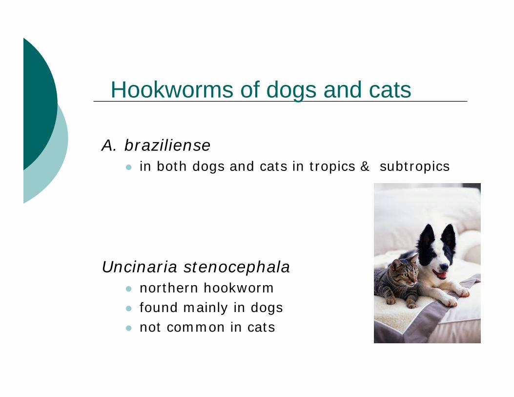
A. braziliensein both dogs and cats in tropics & subtropics
Uncinaria stenocephalanorthern hookworm found mainly in dogs not common in cats

Eggs of Ancylostoma (top) & Uncinaria (bottom)
•thin shell, oval to oblong, pale• non-embryonated in fresh feces (2-8 cell stage)Diagnosis:
Signalment, age and clinical signsthis is the only basis for ante-mortem diagnosis in per-acute neonatal ancylostomiasis
Detection of eggs in feces and/or worms at necropsy
Fecal flotation to detect eggsWorms grossly visible

Ancylostoma caninum
Very common in dogs of all ages in midwest
More significant problem in south & central US
Infectious larval stage (L3) survive best in moist, sandy-loam soils at moderate temperatures
do not survive freezing do not survive at temps >37 ºC
Clinical cases more common during warm weather

Uncinaria stenocephala
More prevalent in northern US
less common & less pathogenic than A. caninum because it sucks less blood
Structureeggs slightly larger than A. caninum
Lactogenic transmissionimportantNo prenatal transmission

Hookworm transmission
L3 (infective larvae) can be acquired by:1. Skin penetration 2. Ingestion
In food or waterLactogenic transmission: major source of infection of puppiesIngestion of arrested larvae in paratenic hosts
3. Transplacental route ~unimportant

Tracheal vs. somatic migration
Tracheal- Penetrate capillary and alveolar walls
Enter lumen of alveoli and migrate up airway to tracheaGet coughed up and swallowed Develop to adults in small intestine
Somatic- Remain in alveolar capillaries Blood flow carries L3 to musclesExit muscle capillaries and become hypobioticor arrested larvae

L3 acquired by ingestion
Undergo mucosal migrationEnter gastric glands or SI mucosa Most emerge in a few days & develop to adults
pre-patent period = 2-3 weeks
Some enter bloodstream from SI mucosa and undergo either tracheal or somatic migration
in older or infected dogs, most undergo a somatic migration to become arrested larvae in muscles

Fate of hypobiotic larvaein pregnant dog
Larvae are “activated”L3 occur in milk up to 20 days post-whelpingThese larvae develop directly to adults in puppy small intestine
blood-lung migration already completed in dam
Undergo lactogenic transmission to nursing pups
Possible transplacental transmission to fetal lung

Persistent hookworm infection
Source of infection: hypobiotic larvae in SI mucosa or muscles 1. Larvae “leak” out of these sites
~continuously and reach the SI lumenif animal already has adults in SI: probably shed into lumen and expelled if no hookworms present: develop to adults
2. If adult hookworms killed by an anthelmintic, arrested larvae can be activated and quickly repopulate the SI

Pathogenesis
Blood sucking & plug-feeding activities
attach to mucosa and pull a plug of intestinal mucosa into the buccal capsule use teeth to lacerate mucosasecrete proteolytic enzymes & anticoagulantchange sites frequently so old sites continue to bleed Leads to blood loss- anemia

Clinical signsVary from asymptomatic to rapidly fatal-not enough red blood cells (anemia)Severity depends upon:
dosage of infecting L3 age resistance and acquired immunity iron reserves (low in puppies) nutritional status
Clinical signs
↓ stamina with weaknesspale mucous membranesmelena or possible bloody feces unthrifty appearance (thin, rough hair coat)

Hookworm infection in neonatal pups
Results from massive milk-based transmissionSigns develop as early as 4 days of
ageworms start to suck blood after molting from L4 to L5 (immature adult stage)Can be fatal by 10-24 days of age even with treatment

Pups: extremely pale with bloody feces, diarrheaNO eggs in feces (yet):
Adults begin egg-laying at 16 DPIClinical signs usually occur ~7-14-days of age
Necropsy findings: blood & immature worms in intestinal tract
Hookworm infection in neonatal pups

Hookworm in older pups
Results from acquisition of large numbers of infectious larvae over short period of time
usually seen in older pupsgeneral pallor due to severe anemiaBlack feces- digested bloodoften die unless treated promptlyeggs in fecesadults worms and blood in SI
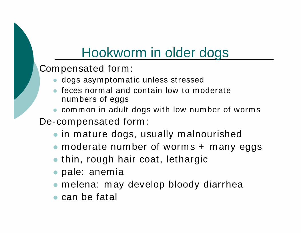
Hookworm in older dogsCompensated form:
dogs asymptomatic unless stressedfeces normal and contain low to moderate numbers of eggscommon in adult dogs with low number of worms
De-compensated form: in mature dogs, usually malnourishedmoderate number of worms + many eggsthin, rough hair coat, lethargicpale: anemiamelena: may develop bloody diarrhea can be fatal

Small intestine showing hemorrhagic lesions caused by hookworms

Treatment
Peracute neonatal ancylostomiasis AnthelminticSupportive care including blood transfusionPoor prognosis
Acute and chronic compensated form
AnthelminticChronic decompensated form
Anthelmintic plus nutritional support

Treatment
Anthelmintics effective against adult worms
butamisol dichlorvosdisophenol febantelfenbendazole* mebendazolemilbemycin* pyrantel pamoate
(Strongid)ivermectin* - effective dose is extra-label!*also effective against larvae

Prevention and control
Identify and treat infected dogs Sanitation: remove feces daily + cleaningGood flooring with no cracks
Coated concrete avoid gravel, sand, dirt, grass, damp shady areas Spray with 1% bleach (3c/gal)
Apply sodium borate (10 lbs/100 ft2) and rake inL3 killed by hard frost
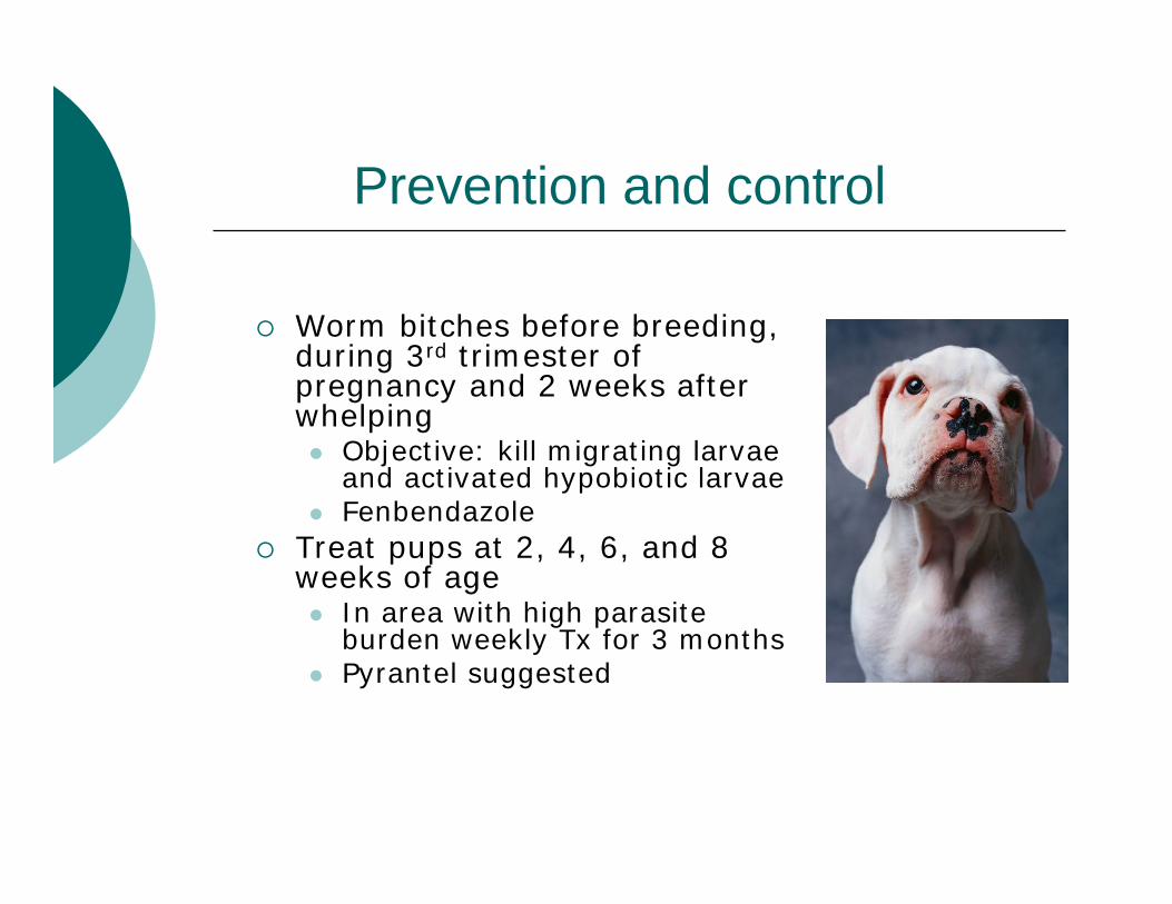
Prevention and control
Worm bitches before breeding, during 3rd trimester of pregnancy and 2 weeks after whelping
Objective: kill migrating larvae and activated hypobiotic larvaeFenbendazole
Treat pups at 2, 4, 6, and 8 weeks of age
In area with high parasite burden weekly Tx for 3 monthsPyrantel suggested

Anthelmintics for prevention
Heartworm/hookworm preventatives:
ivermectin + pyrantelmilbemycinstyrid/caracideoxibendazole + diethylcarbamazine
Hookworms: Public health importance
Cause cutaneous larval migrans (CLM) linear, tortuous, erythematous and intensely pruritic eruptions caused by migration of nematode larvae in humans
Most commonly caused by A. brazilienseA. caninum, U. stenocephala, Bunostomum spp & Strongyloides spp. can also cause CLMKnown as “barnyard itch”, “creeping eruption”or “ground itch”

Cutaneous Larval Migrans
Hookworms (Ancylostoma and Uncinaria) can produce cutaneous larval migrans
Eggs are passedLarva develop and penetrate human skin- cases of walking barefoot in yard- AustraliaIngestion of worms leads to intestinal problems

Cutaneous larval migrans
Migration tunnels of larvae appear as ridges on skin surface
larvae want into bloodstream!!!cause intense iching that can persist for weeks to monthssecondary bacterial infection commonMore severe and persistent in persons hypersensitized by prior exposure
Rare cases of intestinal infection of humans with adult A. caninum


Hookworms
Prevention in humans involvesTreatment of infected animalsRoutine de-worming (CDC) Daily fenbendazole treatment of pregnant dogs from 40th day of gestation through 14th day of lactation shown to inhibit T. canis larvae in tissues- prevents or greatly reduces incidence of infection in puppiesCleaning the environment (pick up yard!!)Keeping pets from defecating in child play areas
Treatment in humansHandouts available from CDC for lobby area use, fast facts from CFSPH website
www.cfsph.iastate.edu

Questions?

Roundworms
Dogs, catsToxocara sp., Toxascaris
HorsesParascaris equorum
SwineAscaris suum
CattleNeoascaris (Toxocara) vitulorum
Ascarids of dogs and cats
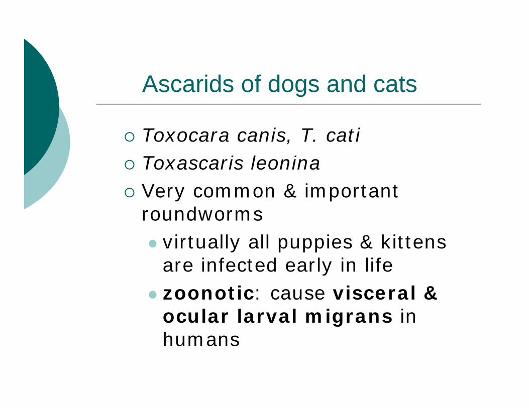
Toxocara canis, T. catiToxascaris leoninaVery common & important roundworms
virtually all puppies & kittens are infected early in lifezoonotic: cause visceral & ocular larval migrans in humans

Toxocara canisCommon dog roundwormHosts
wild & domestic canids rarely in felids
Many animals can be paratenic hosts
rodents, rabbits, cattle, sheep, goats, birds, earthworms, etc

Do you see roundworms at your shelter/clinic?
Yes
No

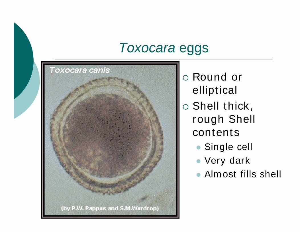
Toxocara eggs
Round or ellipticalShell thick, rough Shell contents
Single cellVery darkAlmost fills shell

Eggs of Toxocara cati are similarto T. canis eggs, but smaller
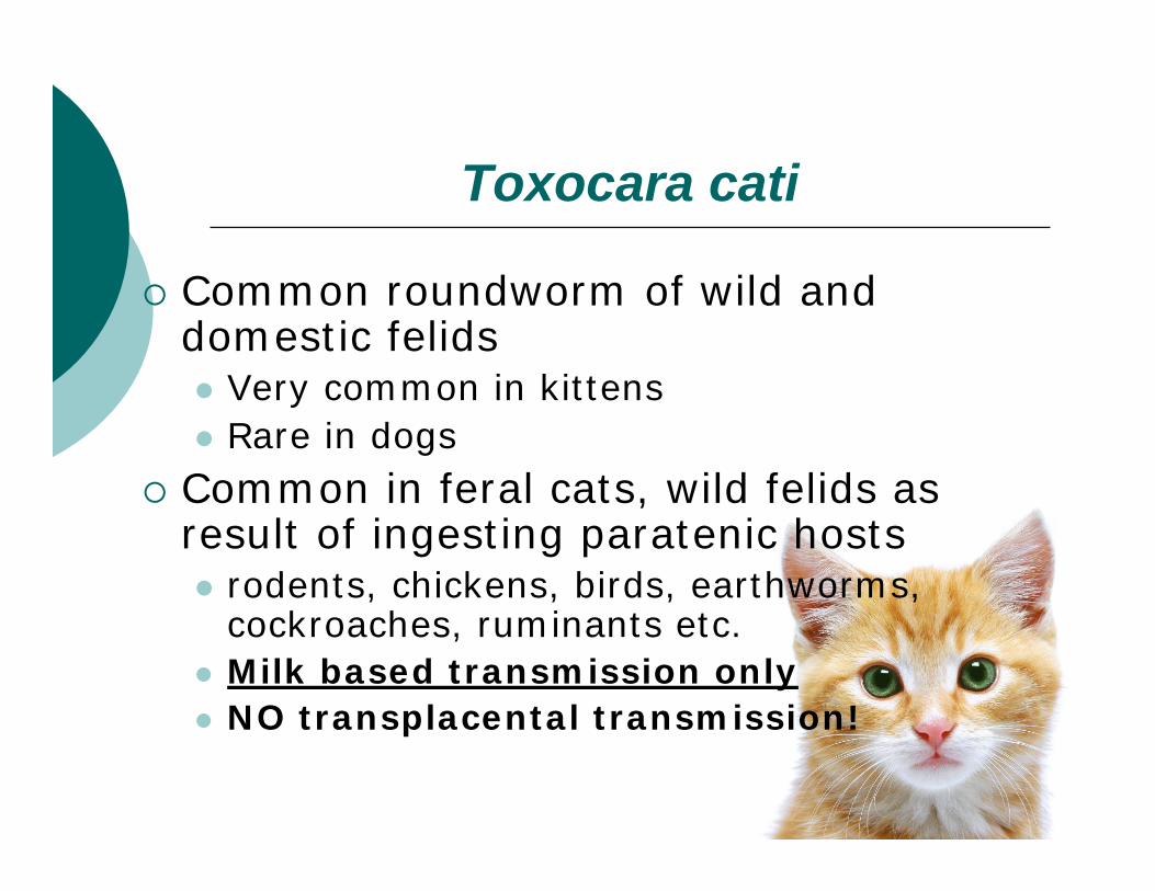
Toxocara cati
Common roundworm of wild and domestic felids
Very common in kittens Rare in dogs
Common in feral cats, wild felids as result of ingesting paratenic hosts
rodents, chickens, birds, earthworms, cockroaches, ruminants etc.Milk based transmission onlyNO transplacental transmission!

Toxascaris leonina
Occurs in both dogs and cats of all ages
More common in dogs than cats in the Midwest
Not as prevalent as ToxocaraMixed infections with Toxocara occur
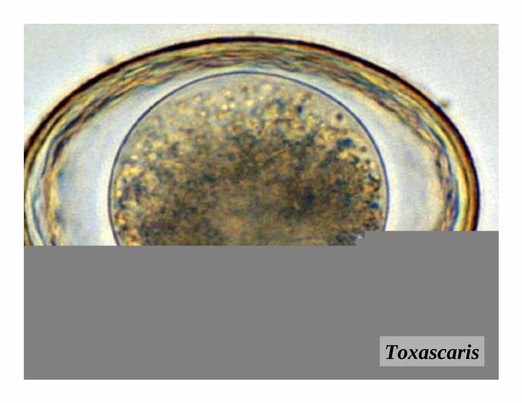
Toxascaris

Eggs of Toxocara andToxascaris

Life cycle of T. canisAdults in small intestine → eggs (lots!) in fecesInfective larva develops within eggin ~2-4 weeksEgg with larva ingestedLarva hatches in duodenumLarval migration:
pathway & fate of larvae varies with host age and susceptibilityyoung hosts<12 weeks: liver-lung-tracheal migrationolder hosts > 12 weeks: liver-lung-somatic migration

T. canis in pregnant dogs
Worm can cross placenta before pups born
Activation of hypobiotic larvae in last trimester Enter uterine circulation, cross placenta → fetal liver + lungsLarvae complete migration after birth
liver→ lung → trachea → GI track

T. canis in pregnant dogs
milk transmission to pupsHypobiotic larvae activated in late gestation & early lactation Migrate to mammary glandsPups ingest larvae in colostrum & milkIngested larvae mature in GI track

Life cycle of T. canisMales, non-pregnant or spayed females
Granulomas = dead end for hypobiotic larvae
Small rodents, prey species (paratenic hosts)
larvae from ingested egg undergo somatic migration hypobiotic in tissuessurvive in granulomas for several yearsdevelop directly to adults in ~3 weeks if dog ingests paratenic host

Epidemiology
Transplacental transmission is most common route of infection
Very common in puppies & young dogs
Patent infections more common in males than femalesSmall rodents reservoir of infection
may be common source of infection for rural and feral dogs

Epidemiology
Adult worms survive ~4 monthsPups expel most worms by 6 mos of age
irritation of intestine or “self-cure”??
Eggs infective in >2 weeks in environment
Very hardy - resist environmental extremes
Persist in soil for yearsKilled by direct sunlight and heat

Summary: T. canis transmission
Transplacental transmission to fetal liver via activated hypobiotic larvae in bitchIngestion of:
Egg in environment with infective larvaLarvae from colostrum or milk of dam Hypobiotic larvae in paratenic host tissuesEggs, larvae or immature worms in puppy vomit or feces

Clinical signs: T. canis
Due to transplacental transmission
In utero (fetal pups)migrating larvae can damage liver cause fetal death or stillbirths
Newborn puppiesWeak pups due to liver & lungdamage by migrating larvae Lung hemorrhages, pneumoniaMay die at 2-3 days of age
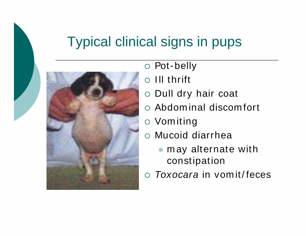
Typical clinical signs in pupsPot-bellyIll thrift Dull dry hair coatAbdominal discomfortVomitingMucoid diarrhea
may alternate with constipation
Toxocara in vomit/feces

Clinical signs in pupsworst case scenarios
Aspiration pneumonia (vomiting)Obstruction of bile or pancreatic ductsObstruction of SI with possible ruptureCNS signs can occur in heavy infections
Cause?Recover with anthelmintic treatment
Death ~2-3 wks of age
Clinical signs older dogs
Adults usually asymptomaticfew adult ascarids present in SIingestion of large number of eggs can result in vomiting with bloody enteritis
Encapsulated larvae in tissues rarely cause clinical signs
possible CNS problems (convulsions) associated with larval death in brain

Clinical signs of T. cati infection
Type & severity of signs determined by:
host ageinfecting parasite stage infecting dose of parasitelocation of infecting stage
Not as pathogenic as T. canisAdult cats usually asymptomatic

Toxocaracati in the small intestineof a cat.

How often do you diagnose roundworms?
All the time (daily to weekly)Sometimes (weekly to monthly)Only in the summertimeOccasionally (monthly)Rarely (a couple times a year)Never

Diagnosis of Toxocara infection
History plus clinical signsprevious infection in litter, kennel etc.
Presence of Toxocara in feces and/or vomitusDetection of typical eggs in feces
Fecal flotation

Treatment of Toxocara infection
For dogs: Anthelmintics for treatment of intestinal stages:
ivermectin @ 0.1-0.2 mg/kg orally NOT for collies, collie like breeds (shelties, border collies, etc.)!Extra-label at dosages >0.006 mg/kg
ivermectin plus pyrantel (Heartguard Plus)milbemycin piperazinepyrantel pamoate dichlorvos
fenbendazole
Treatment of T. canis infection
Anthelmintics with activity against larvae
fenbendazole for pregnant bitchkills only larvae activated during pregnancyneed ~continuous treatment
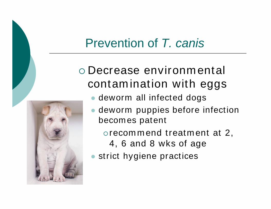
Prevention of T. canis
Decrease environmental contamination with eggs
deworm all infected dogsdeworm puppies before infection becomes patent
recommend treatment at 2, 4, 6 and 8 wks of age
strict hygiene practices

Treatment: cats
Similar to treatment of Toxocara in dogs
Fewer anthelmintics approved for catsFenbendazole (10 mg/kg for 3 days orally)Febantel approved for cats and effective
Treat kittens at 3, 5, 7 and 9 wks of ageEvaluate cats of all ages for eggs in feces and treat as needed
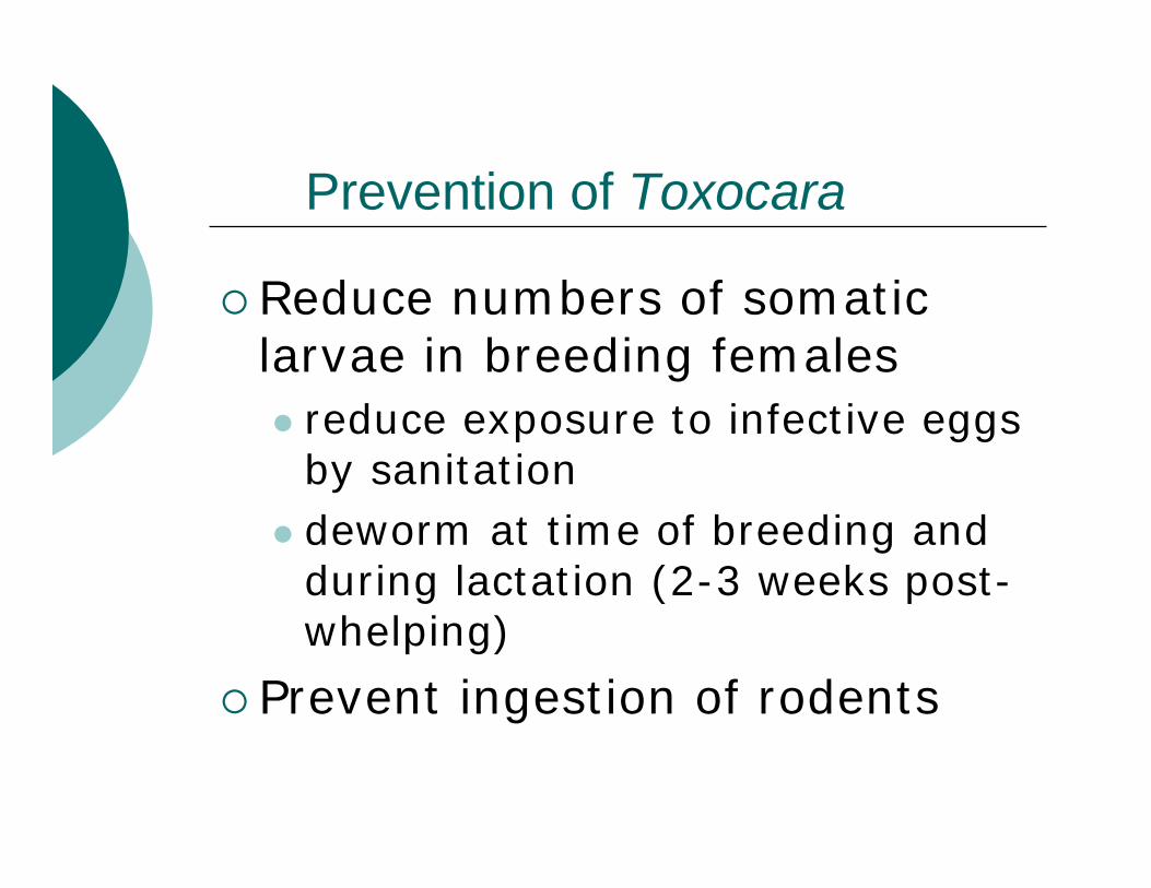
Prevention of Toxocara
Reduce numbers of somatic larvae in breeding females
reduce exposure to infective eggs by sanitationdeworm at time of breeding and during lactation (2-3 weeks post-whelping)
Prevent ingestion of rodents

Baylisascaris procyonisRaccoon roundworm
Very common in raccoons in North AmericaAdults and egg similar to ToxocaraIngestion egg with infective larva Undergo liver-lung-tracheal migrationNonpathogenic in raccoons, can also infect dogs
Paratenic hosts have arrested larvae in tissues

Baylisascaris fromthe small intestine of a raccoon

Eggs of hookworm, Toxocara, and Baylisascaris (B)
T
B

Do you see raccoon roundworms at your shelter/clinic?
Yes
No
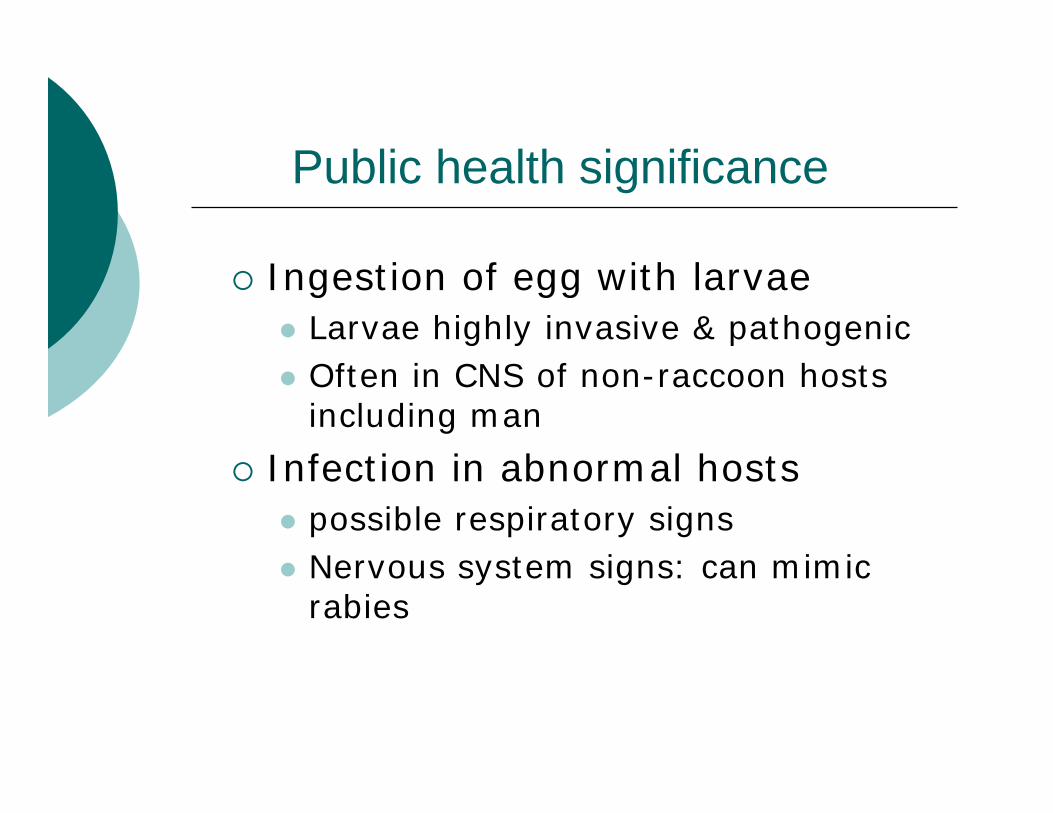
Public health significance
Ingestion of egg with larvaeLarvae highly invasive & pathogenic Often in CNS of non-raccoon hosts including man
Infection in abnormal hostspossible respiratory signsNervous system signs: can mimic rabies

Prevention and controlTreat captive raccoons with anthelmintic
Wormers effective against dog and cat ascarids should be effective against B. procyonis
Keep wild raccoons out of barns, sheds Pick up & destroy raccoon fecesWash hands after handling raccoons

Visceral larval migrans (VLM)
a single female roundworm can produce more than 100,000 eggs/dayOnce eggs become infective can remain infective in the soil for yearsMost commonly caused by T. canis in humansCan also be caused by: Baylisascaris procyonisLess frequently caused by Toxocara cati, Toxascaris leonina, Ascaris suum

Pathogenesis
Sandboxes, playgrounds etc. frequently contaminated with dog/cat feces
children ingest dirt/sand with infective ascarid eggslarvae hatch in intestine & attempt normal migration pathwayinvade lungs, liver, eyes, brain etc. and cause disease

Human VLM
Most common in children <6 yrs oldClinical signs and lab findings include:
Irritability, intermittent fever, loss of appetite, weight lossAching muscles, nausea, swelling, itching Enlarged liver, inflamed lungs Immune responses to parasites (eosinophils, IgE)

Diagnosis
History of picaClinical signsDetection of larvae by biopsy (liver or lung)Serology
ELISA

Ocular larval migrans
Sequel to VLMLarvae of T. canisinvade human eyeCause unilateral inner eye lesion similar to retinoblastomaTreatable
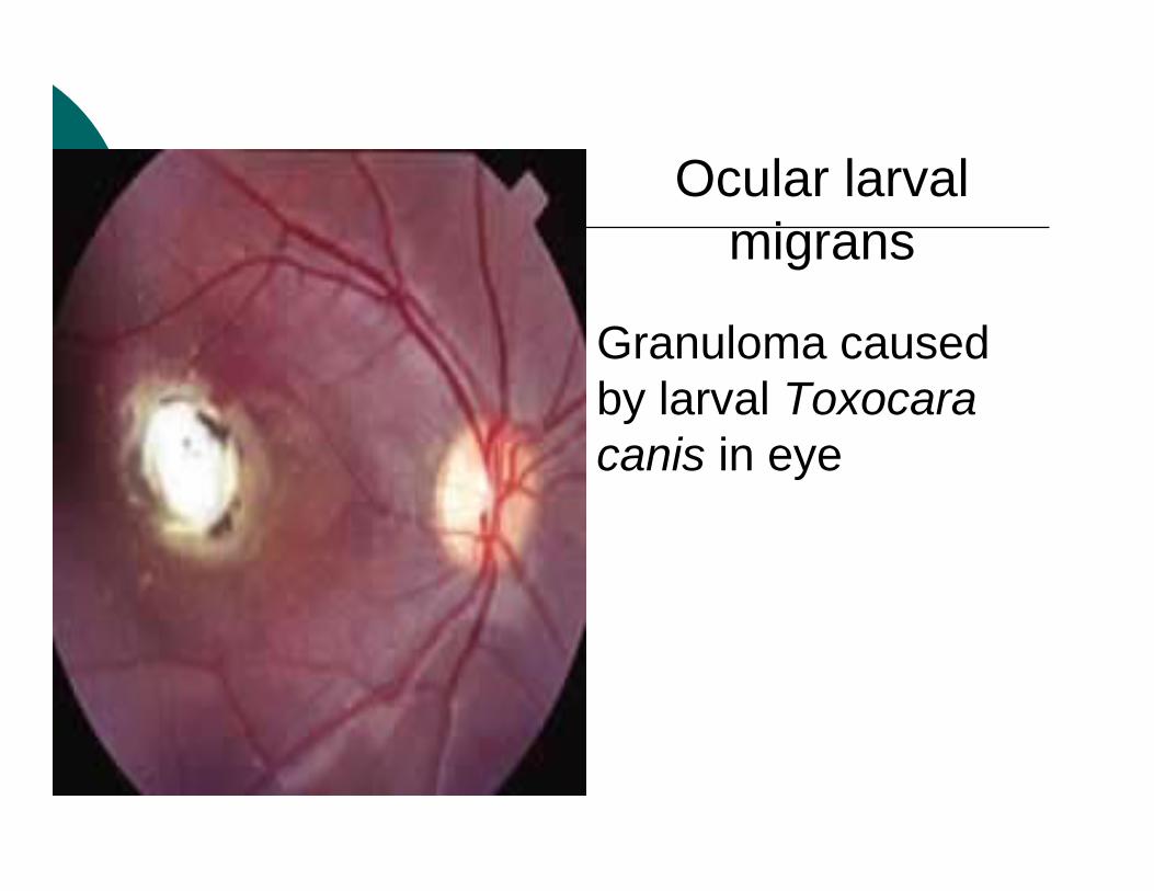
Ocular larval migrans
Granuloma caused by larval Toxocara canis in eye

Questions?
Coccidia- Isospora spp.
Disease: isosporosis; coccidiosisVery common parasitesLife cycle fecal-oral transmission of sporulated oocyst
Addition of rodents in dog and cat spp.

Rodent paratenic hosts
dog and cat Isospora spp. Rodent ingests sporulated oocysts
sporozoites released in GI tractpenetrate gut wall, enter extra-intestinal cellseach sporozoite forms a monozoic cystsporozoite remains viable but does not develop
definitive host ingests rodentsporozoite released from monozoic cystinitiates merogony in cat/dog intestine

Isosporosis of cats and dogs
Each host parasitized by >1 Isospora spp.Location of life cycle stagesLocation of life cycle stages
Unsporulated oocysts shed in fecesUnsporulated oocysts shed in fecesSporogony in environmentSporogony in environmentMerogony & gametogony in small Merogony & gametogony in small intestineintestine

Isosporosis in cats and dogs
Usually non- or mildly pathogenicCan cause severe enteritis (pups & kittens)
Signsanorexia, weight lossmucoid to bloody diarrhea possible vomitingdehydration possible anemiacan be fatal
Significant problem in kennels and catteries
Diagnosis: isosporosis in cats and dogs
Age, history and clinical signs A differential in cases of diarrhea, esp. puppies and kittensdetect oocysts in feces
non-sporulated in fresh feces
clinical signs precede oocyst shedding

Sporulated oocyst ofIsospora rivolta

Treatment of isoporosis
Sulfonamides Sulfadimethoxine (Albon)Trimethoprim-sulfadiazine (Tribrissen)Amprolium (Corid) – extra-label
Supportive care
Control of isosporosis
Sanitationsteam cleaning, boiling water, ammonium hydroxide, bleach
Post-partum treatment of cat or dogProphylactic treatment of offspring
extra-label use of coccidiostatamprolium (thiamine antagonist) or decoquinate

Questions?

Giardia lamblia
Trophozoites
Giardia lamblia
Synonyms: G. duodenalis, G. intestinalisInfects wide range of mammalian hosts Comprised of several “Assemblages”
Some assemblages zoonoticHuman to human transmission probably more common

Giardia lamblia genotypes
Assemblage A : zoonoticHumans, livestock, dog, cat, beaver, guinea pig, deer, prairie dog, bobcat, others
Assemblage B: zoonoticHumans, cattle, dog, cat, beaver, muskrats, rat, others
Assemblages C & D: primarily infect dogsAssemblage E: artiodactyl speciesAssemblage F: cats

Prevalence of Giardia
Most common human intestinal parasite~7% of humans
~10% of household dogs and catsGreatest prevalence in young and in kennels or catteries Japan survey of dog breeding kennels (2005)
100% of kennels positive for Giardia54.5% of pups vs. 30.9% of adults
7.3% of cats <1 year old in NY in 20017.2% of 1216 dogs seen in vet clinics in Canada

Prevalence of Giardia
Increasing reports from calves, lambs, kids, foals
Ruminants from 2 weeks - 2 months oldRecent US study in calves
began shedding cysts at 4 days of age peak numbers at 14 days of age
Foals 2 weeks to ~6 months old
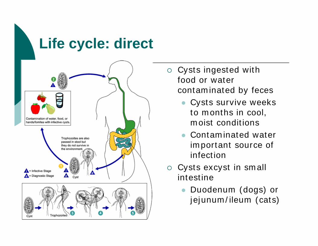
Life cycle: directCysts ingested with food or water contaminated by feces
Cysts survive weeks to months in cool, moist conditionsContaminated water important source of infection
Cysts excyst in small intestine
Duodenum (dogs) or jejunum/ileum (cats)

Giardiatrophozoite showing flagellaand two nuclei

Trophozoite of GiardiaWhat characteristicof the diplomonadsis evident?

Pathogenesis
Trophozoites attach/detach from gut epithelial cellsInfection results in:
Villous atrophy Diffuse loss of microvillous brush borderDisaccharidase insufficiencyImpaired absorption
Electrolytes, nutrient, waterResults in diarrhea

Giardiasis
Spectrum varies from asymptomatic carriage to severe diarrhea and malabsorption.
Acute giardiasis-develops after 5-6 day incubation period -usually lasts 1 to 3 weeks-diarrhea, abdominal pain, bloating, nausea, and
vomiting.
Chronic giardiasis -acute symptoms are recurrent -and malabsorption and debilitation may occur.
Clinical features-humans
Clinical signs-animals
Persistent diarrheaCharacter of feces
Liquid to semi-formedPale & malodorous↑ mucus (especially in cats)Steatorrhea
↑ Borborygmi

Clinical signs-animals cont
AfebrileBright & alertNot anorexic Rarely see melena, vomiting, serious dehydrationChronic giardiasis can result in weight loss, retarded growth

Diagnosis
Clinical signs are relatively non-specific
Numerous differentials
Always do a fecal exam on an animal with diarrhea to obtain a definitive diagnosis of giardiasis

Definitive diagnosis-fecal flotation
Trophozoites more likely in loose stoolsNeed fresh feces kept cool and examined asap or preserved promptly
Wet mount of fresh sample -”falling leaf”Stained smear (Dif-Quik works well)Can also find trophozoites in small intestinal samples collected by endoscopy or laparotomy

Definitive diagnosis-fecal flotationCysts more likely in semi-formed or formed feces Best method: centrifugal flotation in zinc sulfate
sugar distorts cystsCysts shed intermittently
1 exam detects ~77% of positive dogs3 exams detect ~95% of positive dogs
Recommend fecal exams every other day X3
Definitive diagnosis-other methods
IFA - detects cysts using fluorescent antibodyELISA – detects antigen in feces
SNAP Giardia, IDEXXProSpecT Giardia Rapid Assay, Alexon
Check with lab for sample submission Submit fresh chilled or formalin-fixed fecesDilute feces in 5-10% NBF and/or PVA
Definitive diagnosisComparative studies: IFA and ELISA tests comparableBoth are more sensitive and specific than zinc sulfate flotation method
NOTE: cysts disappear temporarily after barium enema, laxatives, kaopectate...
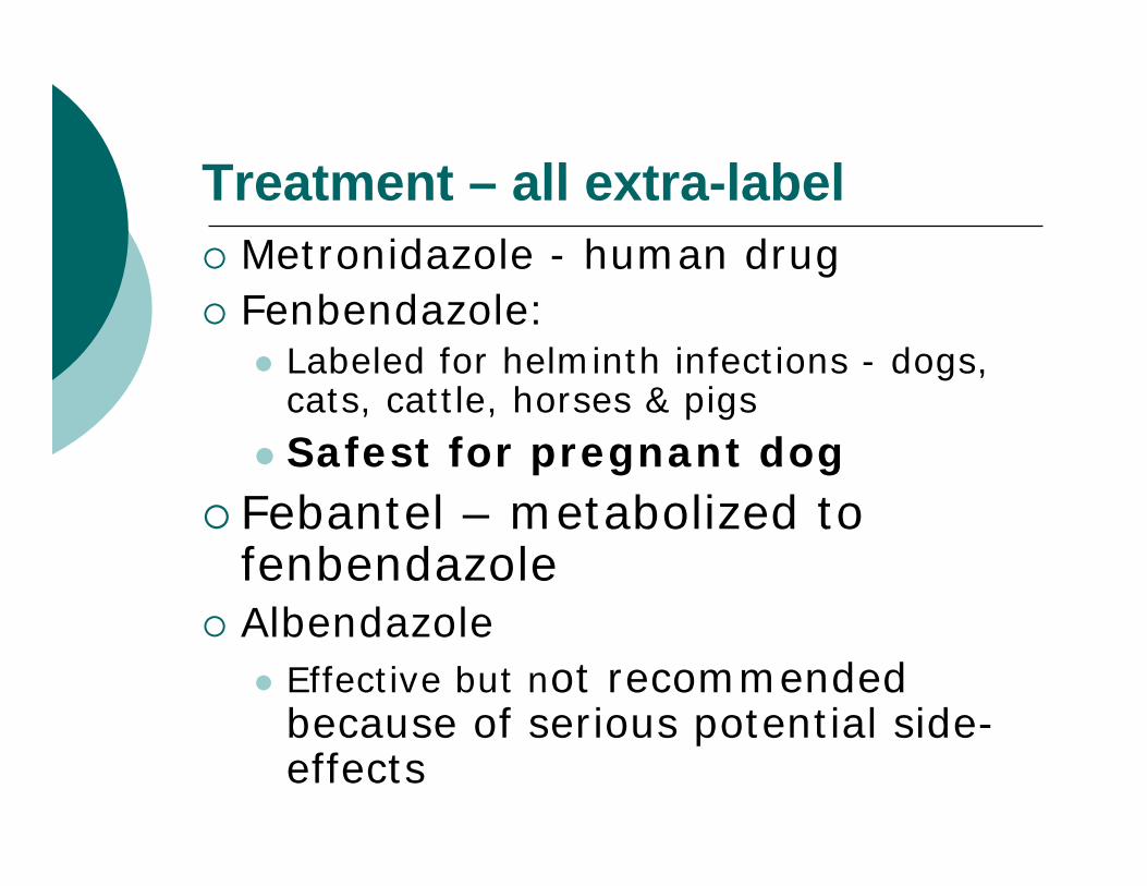
Treatment – all extra-labelMetronidazole - human drugFenbendazole:
Labeled for helminth infections - dogs, cats, cattle, horses & pigsSafest for pregnant dog
Febantel – metabolized to fenbendazoleAlbendazole
Effective but not recommended because of serious potential side-effects

Treatment & control
Refractory casesIs re-infection occurring? Repeat treatment with same, different or a combination of drugs
Sanitation important Vaccine available
GiardiaVax – Ft. DodgeAdditional tool for use in treatment and control

Questions?

Tritrichomonas foetus
Worldwide venereal disease of cattle
Bovine trichomoniasis
Higher prevalence in older bulls (vs. young)
bulls >4 yrs old = permanent carriersfunction of preputial epithelium structure
More common in beef herds than dairyFinding in shelter cats in IA, CO- why?

Transmission- cows
Trophozoite stage only (No cyst)Mechanical transmission during coitusIatrogenic
AI: bull semen to cow or cow to cow (repro exam)
Bull to bull transmission possible
Use of same artificial vagina by infected and clean bull within short time frame
Photo courtesy of Dr. Diane Addie

Diagnostic characteristics of Tritrichomonas foetus

Feline trichomoniasis
Large bowel diarrhea caused by a trichomonad indistinguishable from T. foetus
Morphology: light, transmission, and scanning electron microscopy rRNA gene sequence analysisRFLP-PCR

Feline trichomoniasis
Cases reported from 12 states Alaska to FloridaAlso reported in England, Scotland, Germany
More prevalent in shelters, cat colonies and multi-cat householdsMost commonly affects cats <1 yr oldCat to cat spread believed to occur via direct contact

Questions?

Resources on this topic:
Georgis’ parasitology for veterinarians, 8th ed, 2003CFSPH Technical Fact Sheets- for instance, Baylisascariasis, at http://www.cfsph.iastate.edu/DiseaseInfo/default.htmCDC web site. Baylisascariasis at http://www.cdc.gov/ncidod/dpd/parasites/baylisascaris/default.htmCAPC web site (although sponsored by Pharmacompanies, so very pro-year round preventatives), for example, ascarids at http://www.capcvet.org/?p=Guidelines_Ascarid&h=0&s=0


![[Health care book] 1-4.how to prevent parasites](https://static.fdocuments.net/doc/165x107/568c49291a28ab4916931bc6/health-care-book-1-4how-to-prevent-parasites.jpg)