How to approach a patient with bleeding? · 2018-04-02 · low platelet count consumption ......
100
How to approach a patient with bleeding? ISTH Advanced training course, Portugal March 2014
Transcript of How to approach a patient with bleeding? · 2018-04-02 · low platelet count consumption ......

How to approach
a patient with bleeding?
ISTH Advanced training course, Portugal March 2014

Differential diagnosis
Platelet disorders
Hemophilia
vWD
hyper-fibrinolysis
factor deficiencies
bleeding
liver failure TIC
anticoagulanttreatment
suspected bleedingdisorder
w/o acutebleeding
acute bleeding
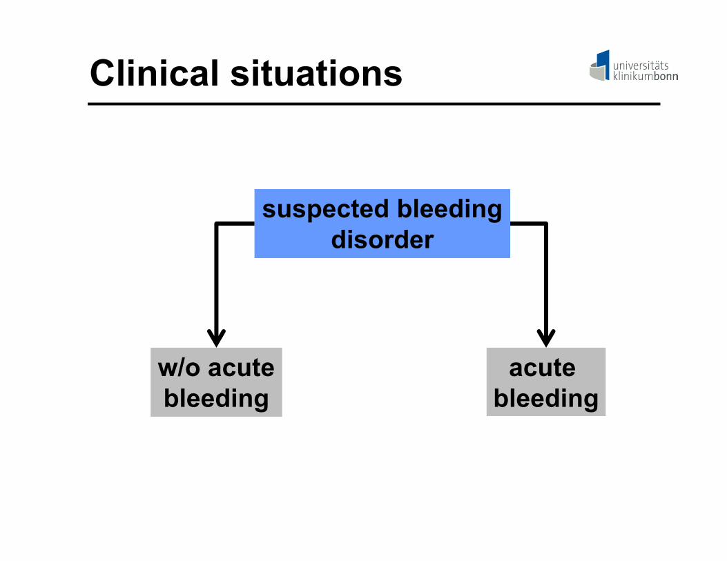
Clinical situations

suspected bleedingdisorder
w/o acutebleeding
acute bleeding
Clinical situations

Initial question
Suspectedbleeding disorder
bleeding disorderlikely or unlikely?

Initial work-up (I)Suspected bleeding
disorder
bleeding history
bleeding disorderlikely or unlikely?

Bleeding history
- type and frequency of bleeding
- provoked or unprovoked
- type of treatment
- family history (family tree)
- drug history

Bleeding history
- usually clear in patients with severe bleeding disorders
- in patients with mild/moderate bleeding symptoms a standardized questionnaire is helpful
- standardized scores to quantitate bleeding symptoms

Bleeding history: scoring keySymptom 0 1 2 3
Epistaxis no/trivial < 5/y
> 5/y > 10 min
Packing/cauterization
transfusion, replacement, DDAVP
Cutaneous no/trivial < 1 cm
> 1 cm w/h trauma
- -
Minor wounds
no/trivial < 5/y
> 5/y or > 5 min
Surgicalhemostasis
Hemostatic treatment
Oral cavity no Reported at least 1
Surgical hemostasis
Hemostatic treatment
Gastro-intestinal
tract
no Identified cause
Surgical hemostasis
Hemostatictreatment

Bleeding history: scoring keySymptom -1 0 1 2 3
Tooth extraction
No bleeding in 2
None done or no bleeding in 1
reported Resuturing, repacking or antifibrinolytics
Transfusion, replacement, DDAVP
Surgery No bleeding in 2
None done or no bleeding in 1
reported Surgical hemostasis or antifibrinolytics
Transfusion, replacement, DDAVP
Muscle hematoma
- never Post-trauma, no therapy
Spontaneous, no therapy
Spontaneousrequiring treatment
Hemarthrosis - never Post-trauma, no therapy
Spontaneous
CNS - never - - Subdural, intracerebral

Validated questionnaire1,2
- includes 13 bleeding symptoms
- provides a summative score
- mean bleeding scores:in healthy individuals: 0.5abnormal: 2
1Biss TT, et al. J Thromb Haemost 2010; 8: 9502Biss TT, et al. J Thromb Haemost 2010; 8: 1416

Initial work up (II)
Suspectedbleeding disorder
bleedinghistory
physical examination

Physical examination
- inspection for any bleeding signs
- joint abnormalities
- lymphadenopathy
- organomegalies
- in children: signs of nonaccidental trauma!

Initial work up (III)
Suspectedbleeding disorder
bleedinghistory
physicalexamination
laboratoryscreening
?

Decision pathway (I)Suspected bleeding
disorder
bleeding history: normal
physical examination:no signs of bleeding
laboratory screeningnot recommended

Take home message (I)
- Thorough personal and family histories are the best screening tests for identifying potential hemostatic problems.
- Properly obtained histories eliminate the need for laboratory screening procedures.

Decision pathway (II)Suspected
bleeding disorder
bleeding history:abnormal
physical examination:signs of bleeding
bleeding disorderpossible/likely
laboratory screeningrecommended

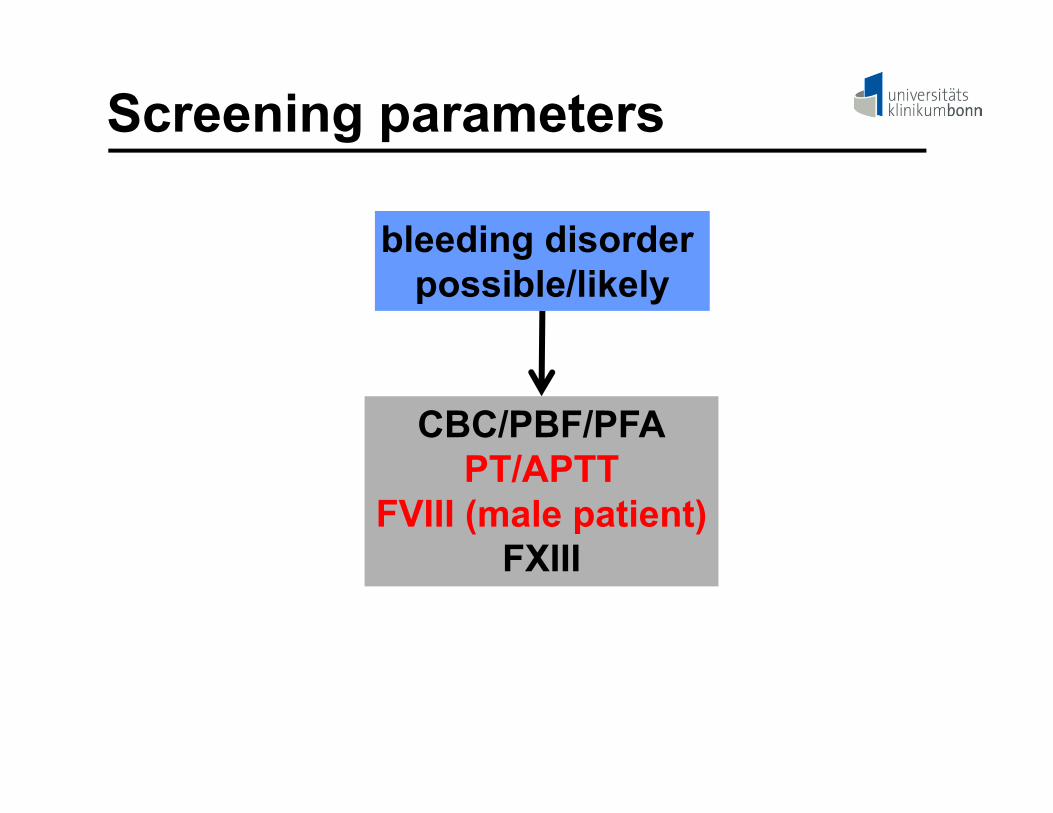
Screening parameters
bleeding disorderpossible/likely
CBC/PBF/PFAPT/APTT
FVIII (males)FXIII
CBC, complete blood count; PBF, peripheral blood film; PFA, platelet functionanalyser; PT, prothrombin time; APTT, activated partial thromboplastin time

Screening parameters
bleeding disorderpossible/likely
CBC/PBF/PFAPT/APTT
FVIII (male patient)FXIII

CBC/PBF/PFACBC/PBF:- platelet count, size and morphology
- leukocyte morphology
- other cytopenias
PFA:- axis subendothelium-vWF-platelet
- platelet-platelet interaction
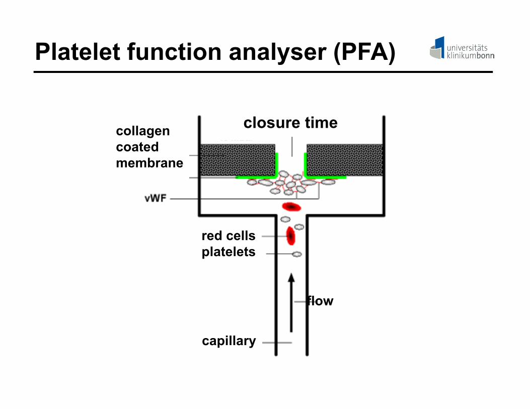
Platelet function analyser (PFA)
collagencoatedmembrane
flow
capillary
closure time
red cellsplatelets

CBC/PBF/PFA: pitfalls
CBC:- pseudothrombocytopenia
PFA:- hematocrit < 35

Pseudothrombocytopenia
- EDTA-inducedagglutinationof platelets
- w/o clinical relevance
- confirmed by platelet counting using citrate anticoagulated blood
ASH et al. Blood 2011;117:4168-4168

Decision finding (III)
platelet count> 150.000/µl
blood smear: normal
platelet disorder/vWD unlikely
PFA normal

Decision finding (IV)
abnormalplatelet morphology
platelet disorderlikely

DD: Platelet disorder
mean platelet volume
micro platelets
Wiskott-Aldrich-syndrome/XLT
giant platelets
< 6 fl > 15 fl
DD: BSS/MYH9-related disorders
DD: granule disorders/
GT
7 - 11 fl

Confirmatory procedures
abnormalplatelet morphology
platelet disorderlikely
genetictesting
flow cytometry
aggregation/secretion

Decision finding (V)
platelet count:< 100.000/µl
blood smear/mean platelet volume:
normal
quantitative platelet disorder likely

synthesis
low platelet count
consumption degradation
Isolated thrombocytopenia

synthesis
low platelet count
consumption degradation
Isolated thrombocytopenia
BM-failure infection/DIC splenomegalie/immune response

Additional information
- isolated versus combined
- new onset or chronic
- signs of organomegalie
- drug history
- previous infections

Additional information- isolated versus combined
- new onset or chronic
- no signs of organomegalie
- no drug intake
- previous infections
immune thrombocytopenia suspected

Decision finding (VI)
platelet count> 150.000/µl
blood smear: normal
vWD suspected
PFA abnormal
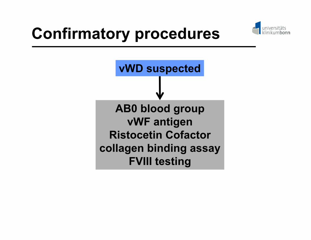
Confirmatory procedures
vWD suspected
AB0 blood groupvWF antigen
Ristocetin Cofactorcollagen binding assay
FVIII testing

Confirmatory procedures
vWD suspected
AB0 blood groupvWF antigen
Ristocetin Cofactorcollagen binding assay
FVIII testing
genetic testingvWF-multimeric analysis
propeptide analysis

Screening parameters
bleeding disorder possible/likely
CBC/PBF/PFAPT/APTT
FVIII (male patient)FXIII

PT versus APTT
FXa/FVa
FIIa
fibrin
contact factors
FXIa
FIXa/FVIIIaFVIIa/TF

DD: single factor deficiencies
FVII FX/FV/FIIfibrinogen
APTT APTT
prolonged normal
HK/PK/FXIIFXIFIXFVIII
prolonged
factor deficiency
unlikelyexcept FXIII
normal prolonged normal
PT

Screening parameters
bleeding disorder possible/likely
CBC/PBF/PFAPT/APTT
FVIII (male patient)FXIII

Hemophilia A pattern
FVII FX/FV/FIIfibrinogen
APTT APTT: 80s (25-35s)
prolonged normal
HMWKPKKFXIIFXI/FIXFVIII: 3%
prolonged
factor deficiency
unlikelyexcept FXIII
normal prolonged normal
PT: 12 s (10.7-12.9s)

20
25
30
35
40
45
50
55
60
0 10 20 30 40 50 60 70 80 90 100 110 120 130 140
aPTT
(s)
FVIII activity (%)
healthy volunteers
hemophiliac patients
25,5 - 34,3 s
48 - 124 %
APTT versus FVIII (one stage)
Screening parameters
bleeding disorder possible/likely
CBC/PBF/PFAPT/APTT
FVIII (male patient)FXIII

Confirmatory procedures
hemophilia A suspected
amidolytic FVIIItwo-stage FVIII
TGAvWD type N
genetic testing
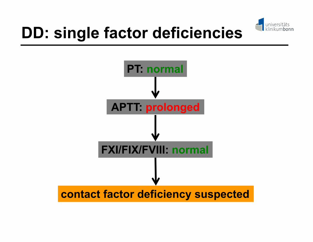
DD: single factor deficiencies
APTT: prolonged
FXI/FIX/FVIII: normal
PT: normal
contact factor deficiency suspected

Confirmatory procedurescontact factor deficiency
suspected
single factor analysis:HK/PK/FXII
contact factor deficiency:clinically not relevant
except ACT-/APTT-monitoring

Patients : initial screens
Patient Platelet count (/µl)
PT (s) APTT (s) FXIII (%)
#1 234,000 61 > 150 89
#2 172,000 11 > 150 91

DD: single factor deficiencies
FVII FX/FV/FIIfibrinogen
APTT APTT
prolonged normal
HMWKPKK
FXIIFXI/FIX/FVIII
prolonged
factor deficiency
unlikelyexcept FXIII
normal prolonged normal
PT
patient #1 patient #2

Patients: single factor analysis
Patient FII (%)
FX (%)
FV (%)
FIX (%)
FVIII (%)
FXI (%)
#1 87 2 89 - - -
#2 - - - 83 < 1 91
Suspected diagnosis:Patient #1: FX deficiencyPatient #2: FVIII deficiency (severe hemophilia)

Patient #1: 68-y old male
- severe hematoma after minimal trauma
- he reported no personal or familial history of bleeding

Patient #2: 56-y old male- severe postoperative bleeding
after hernia operation
- haematothorax after centralvenous support
- massive transfusion24 RBC, 32 FFP4 platelet conc.
- referred to Bonn via helicopter

- large hematomas
- extensive ecchymoses
- severe mucosal bleeding
- gastrointestinal bleeding
- gross hematuria
- w/o a bleeding history
Acquired hemophilia

Laboratory approach
inhibitor suspected
inhibitor screen(mixing test)

- patient plasma is mixed with increasingconcentrations of normal human plasma
- clotting factor activity measured afterincubation for 1 and 2 hours at 37°C
Inhibitor screen

0
20
40
60
80
100
0 25 50 75 100
Normal human plasma (%)
FVIII
-act
ivity
(%)
Inhibitor screen: negative
1 hour
2 hours

0
20
40
60
80
100
0 25 50 75 100 125
normal human plasma (%)
FVIII
-act
ivity
(%)
Inhibitor screen: positive
2 hours
1 hour

Patients: inhibitor screen
Patient FX (%)
FV (%)
FVIII (%)
Mixing test
#1 2 89 - neg
#2 - - < 1 pos
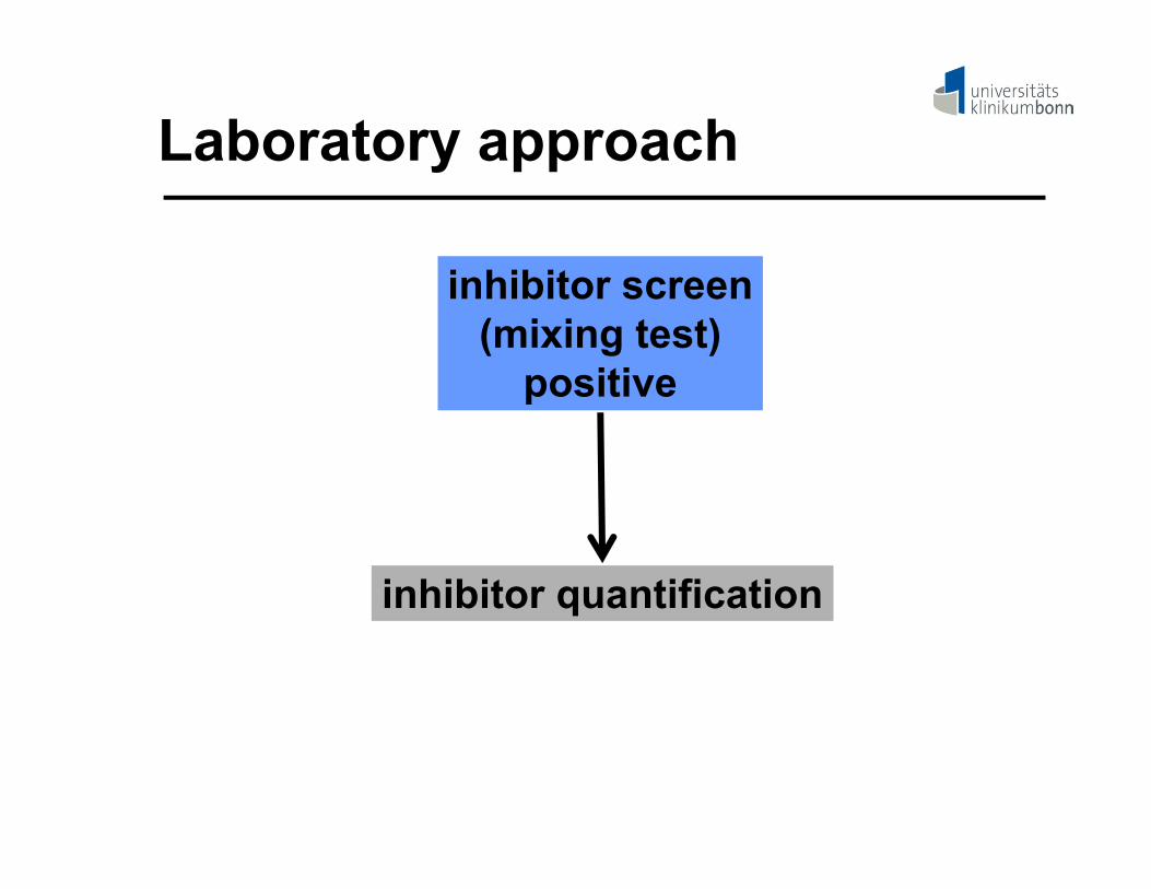
Laboratory approach
inhibitor screen(mixing test)
positive
inhibitor quantification

- serial dilutions of patient plasma are incubated for two hours at 37°C with normalhuman plasma
- factor activity is then measured using aclotting assay
- 1 Bethesda unit (BU) is defined to reducethe activity of a clotting factor in normalhuman plasma to 50%
Bethesda assay

Bethesda units (BU)
0
25
50
75
100
0 25 50 75 100 125 150
FVIII
-act
ivity
(%)
Dilution factor 1/n

Patients: Bethesda units (BU)
Patient FX (%)
FV (%)
FVIII (%)
Mixing test
BU
#1 2 89 - neg -
#2 - - < 1 pos 37

Patient #2: 56-y old male- severe postoperative bleeding
after hernia operation
- haematothorax after centralvenous support
acquired hemophilia A caused byhigh titer FVIII-autoantibodies

Acquired inhibitors: frequencies
Molecular target Estimated frequency
Factor VIII 1 – 1.5 x 10⁶ in non-hemophiliacs
Factor II few case reports only
Factor V 105 cases described
Factors VII, IX, X, XI few case reports only
Factor XIII 20 cases described
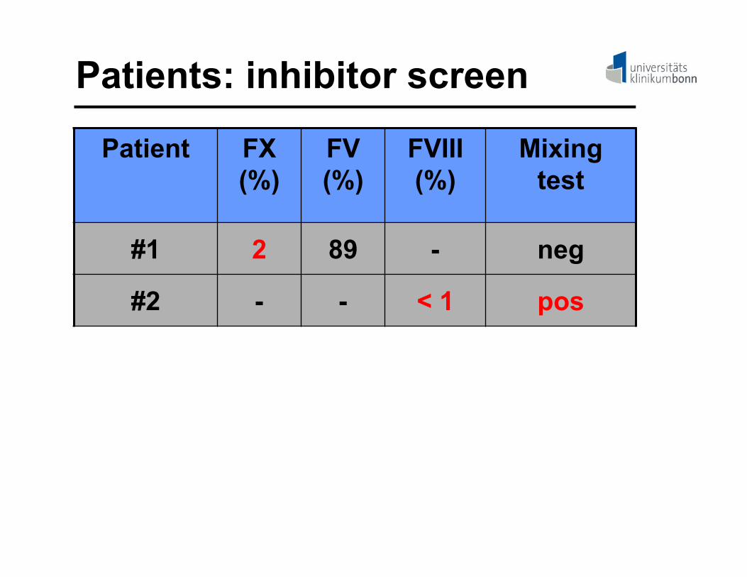
Patients: inhibitor screen
Patient FX (%)
FV (%)
FVIII (%)
Mixing test
#1 2 89 - neg
#2 - - < 1 pos

Acquired inhibitors
- The majority of acquired inhibitors are
antibodies that either inhibit the activity or
increase the clearance of a clotting factor.

Laboratory approach
inhibitor suspected
screen forprecipitating antibodies
inhibitor screennegative

Acquired inhibitor: ELISA
purified clottingfactor
microtiter plate
*-IgG/M

Patients: laboratory data
Patient FX (%)
FV (%)
Mixing test
BU APA ELISA
#1 2 89 neg - neg neg
#2 - - pos 37 neg -

Patient #1: 68-y old male
- severe hematoma after minimal trauma
- skin biopsy reveals amyloidosis
amyloid-associated FX-deficiency

Screening parameters
bleeding disorder possible/likely
CBC/PBF/PFAPT/APTT
FVIII (male patient)FXIII
normal?

Extended screeningbleeding disorder possible/likely
initial screen:normal
α2-antiplasminplatelet function testing/vWF-testing
vascular bleeding disorderrepeat testing during active bleeding

Clinical decision finding
bleeding disorder possible/likely
extended screeningw/o abnormal results
suspected diagnosis:bleeding disorder of unknown reason

Take home message (II)
- If relatively simple screening procedures are used, the vast majority of hemorrhagic problems can be identified.
- Confirmatory tests are subsequently usedto establish an appropriate differential dia-gnosis.

suspected bleedingdisorder
w/o acutebleeding
acute bleeding
Clinical situations

Acute Bleeding: Grading
- Life-threatening (WHO grade 4)Trauma or critical organ bleeding
- Severe (WHO grade 3)gross blood loss, requires transfusion
- Mild blood loss but clinically significant (WHO grade 2)

severe trauma
massive blood loss
loss of clottingfactors/platelets
coagulopathy
TIC-cascade (I)

severe trauma
consumption ofclotting factors/platelets
massive blood loss
activation of thecoagulation system
loss of clottingfactors/platelets
coagulopathy
TIC-cascade (II)
thrombin burst

severe trauma
generation of APC
consumption ofclotting factors/platelets
massive blood loss
activation of thecoagulation system
loss of clottingfactors/platelets
systemic anticoagulation
coagulopathy
TIC-cascade (III)
thrombin burst

TIC: APC-formation
Chesebro BB, et al. Shock 2009; 32: 659-665
C = control, T = trauma, H = hemorrhage, TH = trauma + hemorrhage

severe trauma
hypoperfusion/anoxia
generation of APC
consumption ofclotting factors/platelets
massive blood loss
activation of thecoagulation system
loss of clottingfactors/platelets
systemic anticoagulation
coagulopathy
TIC-cascade (IV)
thrombin burst

Ca2+ cAMP
PAR? V2-R ?
epinephrinprostacyclinevasopressin
thrombinacidosis
t-PA-secretion
t-PA: tissue-type plasminogen activator
t-PA/PAI

severe trauma
hypoperfusion/anoxia
generation of APC
consumption ofclotting factors/platelets
massive blood loss
activation of thecoagulation system
loss of clottingfactors/platelets
PA-release from ECs
hyperfibrinolysis systemic anticoagulation
coagulopathy
TIC-cascade (V)
thrombin burst

severe trauma
hypoperfusion/anoxia
generation of APC
consumption ofclotting factors/platelets
massive blood loss
activation of thecoagulation system
loss of clottingfactors/platelets
PA-release from ECs
hyperfibrinolysis systemic anticoagulation
coagulopathy
TIC: treatment options
thrombin burst
surgery
transfusion transfusionTXA FFP?

Tranexamic acid (TXA)
plasminogen plasmin
t-PA
fibrin
TXA
fibrin
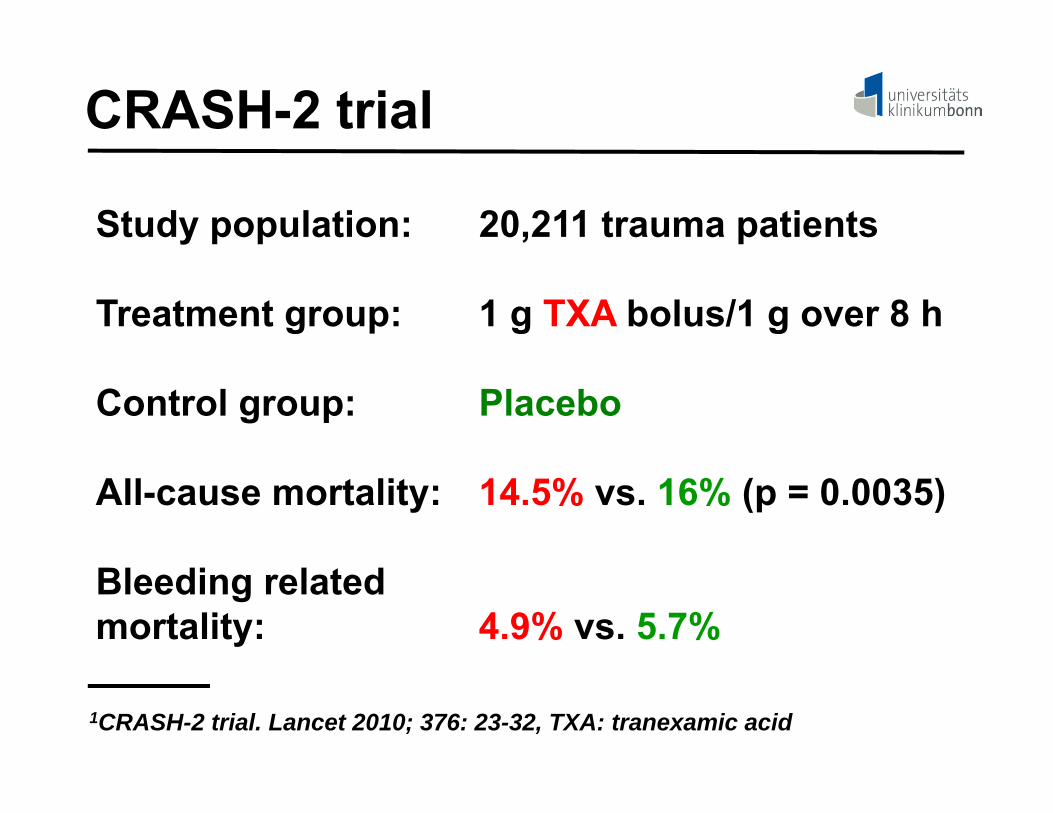
CRASH-2 trial
Study population: 20,211 trauma patients
Treatment group: 1 g TXA bolus/1 g over 8 h
Control group: Placebo
All-cause mortality: 14.5% vs. 16% (p = 0.0035)
Bleeding relatedmortality: 4.9% vs. 5.7%
1CRASH-2 trial. Lancet 2010; 376: 23-32, TXA: tranexamic acid

TIC: treatment triggers
1. Clinical probability of TIC
1. Laboratory values

High TIC risk: indicators*- hemorrhagic shock on admission
- pelvic fracture/multiple bone fractures
- rupture of liver/spleen/positive FAST
- brain damage
- BE < -6
- use of antithrombotic drugs
* TASH-Score, German Society of Trauma Surgeons

TIC: hemostatic support (I)
severetrauma
hemostasis screen:CBC/PT/APTT/
fibrinogen
TXA 1g Bolusfollowed by
1g/h1
TIC-risk: high
1CRASH-2 trial. Lancet 2010; 376: 23-32, TXA: tranexamic acid

TIC: hemostatic support (II)
3g fibrinogenFFP/PRC 1:1
parameter-adjustedtreatment
High TIC risk?
yes no

Parameter-adjusted treatment
Fibrinogen:- < 1.5 g/dl with ongoing blood loss/ICB- < 0.5 g/dl→ 3 g fibrinogen concentrate
Prothrombin time:- > 25s with ongoing blood loss/ICB- < 50s→ 50 IE/kg b.w. PCC
ICB, intracranial bleeding;
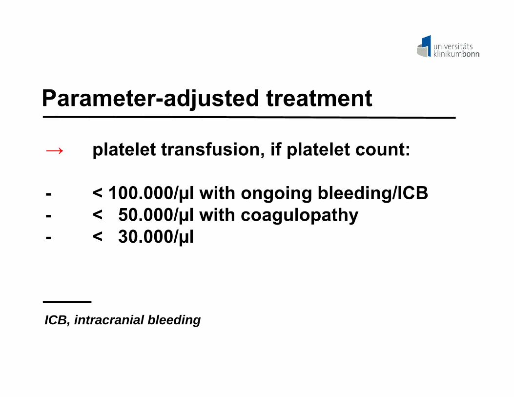
Parameter-adjusted treatment
→ platelet transfusion, if platelet count:
- < 100.000/µl with ongoing bleeding/ICB- < 50.000/µl with coagulopathy- < 30.000/µl
ICB, intracranial bleeding

Parameter-adjusted treatment
Signs of hyperfibrinolysis
→ bolus injection of 1 g TXA followed by 20 mg/kg b.w./h
→ 3 g fibrinogen concentrate

time axis
max. amplitude
r-time k-time
TEG: fibrinolysis

Take home message (III)
- TIC is frequent in severe trauma patients.
- TIC is caused by consumption and loss ofcoagulation factors and platelets and bysecondary hyperfibrinolysis and APC for-mation.
- TIC can be successfully treated by TXA treat-ment and transfusion of blood products including fibrinogen, fresh frozen plasmaand platelets.

Acute Bleeding: Grading
- Life-threatening (WHO grade 4)Trauma or critical organ bleeding
- Severe (WHO grade 3)gross blood loss, requires transfusion
- Mild blood loss but clinically significant (WHO grade 2)

Critical organ bleeding
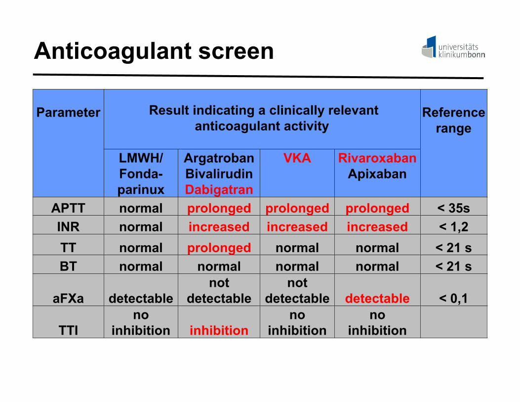
Suspected anticoagulant treatment
antithrombotic screen:APTT/INR/TT/BT/aFXa/TTI
APTT, activated partial thromboplastin time;INR, international normalized ratio; TT, thrombin time;BT, batroxabin time; aFXa, anti-FXa-activity; TTI, thrombininhibition time

Anticoagulant screen
Parameter Result indicating a clinically relevant anticoagulant activity
Reference range
LMWH/Fonda-parinux
ArgatrobanBivalirudinDabigatran
VKA RivaroxabanApixaban
APTT normal prolonged prolonged prolonged < 35sINR normal increased increased increased < 1,2TT normal prolonged normal normal < 21 sBT normal normal normal normal < 21 s
aFXa detectablenot
detectablenot
detectable detectable < 0,1
TTIno
inhibition inhibitionno
inhibitionno
inhibition

Take home message (IV)
- If six relatively simple screening procedures are used, patients showing clinically relevant plasma levels of anticoagulants can be identified.