How Cells Harvest Chemical Energy Chapter 6. Overview Photosynthesis Aerobic respiration Anaerobic...
78
How Cells Harvest Chemical Energy Chapter 6
-
date post
19-Dec-2015 -
Category
Documents
-
view
222 -
download
1
Transcript of How Cells Harvest Chemical Energy Chapter 6. Overview Photosynthesis Aerobic respiration Anaerobic...

How Cells Harvest Chemical Energy
Chapter 6
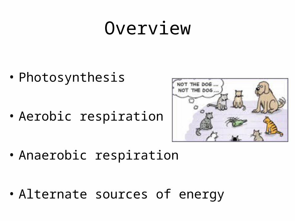
Overview
• Photosynthesis
• Aerobic respiration
• Anaerobic respiration
• Alternate sources of energy
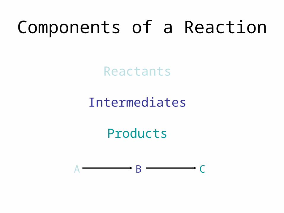
Components of a Reaction
Reactants
Intermediates
Products
A CB

Endergonic vs. Exergonic ReactionsEndergonic
= Energy-requiring
Exergonic
= Energy-releasing

Redox Reactions
One molecule gives up electrons = oxidized
One molecule gains electrons = reduced
H+ atoms released simultaneously(are attracted to negative charge of electrons)
Coenzymes pick up e-s & H+ from substrates & deliver to e- transfer chains
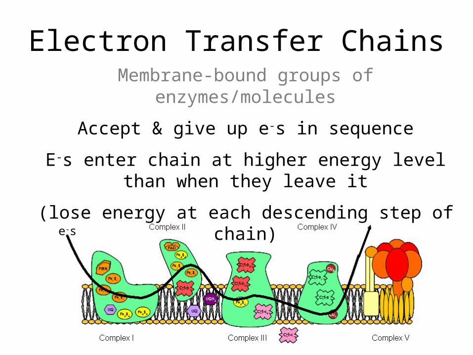
Electron Transfer ChainsMembrane-bound groups of enzymes/molecules
Accept & give up e-s in sequence
E-s enter chain at higher energy level than when they leave it
(lose energy at each descending step of chain)
e-s

Substrate-Level Phosphorylation
Formation of ATP by direct transfer of Pi group to ADP from intermediate

NAD+ & FAD
Coenzymes
1. Accept e-s & H+ from intermediates that form during glucose catabolism
Become reduced = NADH & FADH2
2. NADH and FADH2 give up e-s & H+ to e- transfer chains during final stages of
aerobic respirationBecome oxidized = NAD+ & FAD
Autotrophs
“Self-nourishing”
Synthesize own food
Obtain energy & organic compounds (e.g. C) from the physical environment

Chemoautotrophs
Have no enzymes to allow for complex metabolic reactions
Obtain energy & C from simple inorganic & organic compounds e.g. CH4, H2S

Photoautotrophs
Contain light-sensitive molecules
Can split H2O & use electrons
Process releases lots of oxygen, which reacts rapidly with metals & creates toxic
free radicals

Early photoautotrophs existed when there was lots of Fe & metals everywhere
Released O2 oxidized these metals & rusted them out
O2 could then be released freely
Over a few thousand years, O2 levels in sea & atmosphere increased
Survival of the fittest:
Most anaerobes died out because couldn’t neutralize toxic O2 radicals
Chemoautotrophs with little or no O2 tolerance restricted to extreme & anoxic environments

As O2 accumulated in the atmosphere, O atoms combined to form O3
= ozone layer(protects against lethal UV radiation from sun)
Life was able to move out from the “darks” & live under open sky
= diversification= evolution

Photosynthesis
The process by which photoautotrophs use light energy from the sun to make glucose,
which can then be converted into ATP
12H2O + 6CO2 6O2 + C6H12O6 + 6H2O

Respiration
= breathing
Cellular respiration
= getting energy from food

Organisms need usable energy in order to survive
Obtained energy is converted into ATP chemical bond energy
Can be used to do work e.g. metabolism

Anaerobes
Can’t tolerate O2
Make ATP via fermentation
1 glucose → 2 ATP
e.g. first organisms, some prokaryotes & eukaryotes
Clostridium difficile
Aerobes
Require O2
Make ATP via aerobic respiration
(many also use anaerobic pathways)
1 glucose → 36+ ATP(vital for survival of large organisms)
e.g. most eukaryotes, some prokaryotes
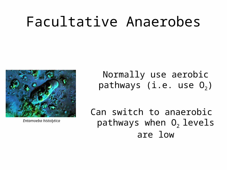
Facultative Anaerobes
Normally use aerobic pathways (i.e. use O2)
Can switch to anaerobic pathways when O2 levels are lowEntamoeba histolytica
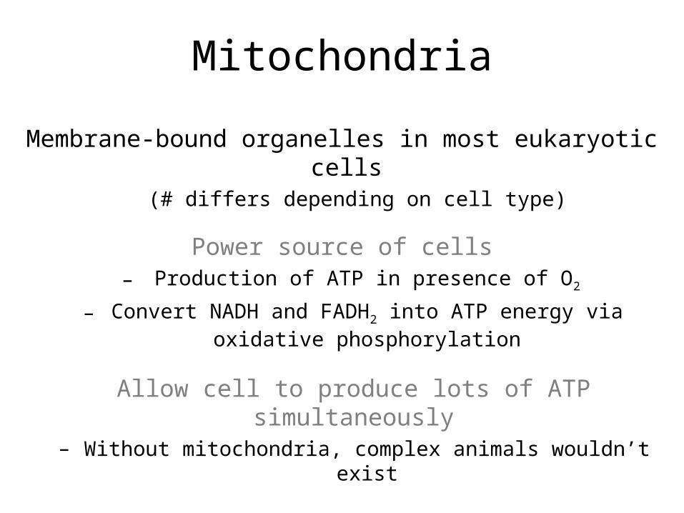
Mitochondria
Membrane-bound organelles in most eukaryotic cells (# differs depending on cell type)
Power source of cells– Production of ATP in presence of O2
– Convert NADH and FADH2 into ATP energy via oxidative phosphorylation
Allow cell to produce lots of ATP simultaneously– Without mitochondria, complex animals wouldn’t exist

Mitochondrion Structure
Outer membrane
Selectively permeable
Inner membrane
Highly impermeable Contains ATP synthase Has membrane potential
Cristae
↑ surface area of inner membrane, which ↑ capacity to generate ATP
Matrix
Contains 100s of enzymes which oxidize pyruvate and fatty acids, and control the Krebs cycle

Cellular Respiration
The oxidation of food molecules (e.g. glucose) into CO2 & H2O
Energy released is captured as ATPUsed for all endergonic activities of cell
Enzymes catalyze each step
Intermediates formed at one step become substrates for enzyme at next step

2 phases of cellular respiration:
Glycolysis:Glucose → 2 pyruvates
Occurs in all cells
Oxidation of pyruvate into CO2 and H2O:
Energy-releasing pathways differ depending on cell & its
needs

40% of energy from glucose is harvested
Rest (60%) is lost as heat
A working muscle uses 10 million ATP per second!
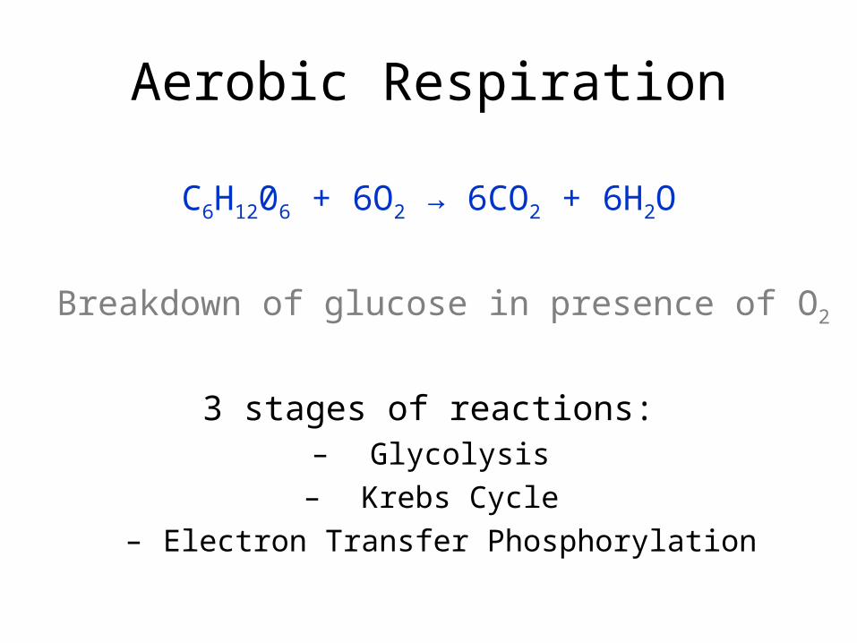
Aerobic Respiration
C6H1206 + 6O2 → 6CO2 + 6H2O
Breakdown of glucose in presence of O2
3 stages of reactions:– Glycolysis
– Krebs Cycle– Electron Transfer Phosphorylation

Glycolysis– Glucose → 2 pyruvates
– Occurs in cytosol
Krebs Cycle– Pyruvate → CO2 + H2O + e-s
– Occurs in mitochondria
Electron Transfer Phosphorylation– Formation of lots of ATP

Stage I: Glycolysis
Glucose → 2 pyruvates
“Universal energy-harvesting process of life”
Initial energy-releasing mechanism for all cells
Occurs in cytosol
Coupled endergonic & exergonic reactions

Endergonic Steps of Glycolysis
Requires input of 2 ATP
ATP #1 phosphorylates glucoseGlucose → intermediate
ATP #2 transfers Pi to intermediate
Intermediate → PGAL + DHAP
DHAP converts into PGAL= 2 PGAL enter next stage


Exergonic Steps of Glycolysis
Each PGAL gives 2 e-s + H+ to NAD+ 2 NAD+ → 2 NADH
Intermediates each give Pi to ADP2 ADP → 2 ATP
(substrate-level phosphorylation)
Pays back 2 ATP used in endergonic steps
Intermediates each release H+ + OH2 intermediates → 2 PEP
Each PEP gives Pi to ADP2 ADP → 2 ATP
(substrate-level phosphorylation)
2 PEP → 2 pyruvate
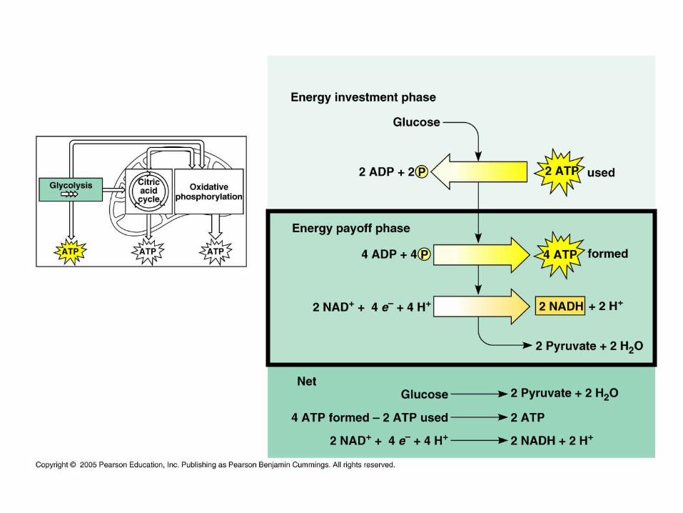

Sum Total of GlycolysisGlucose → 2 pyruvate + 2 NADH + 2 ATP
From here, pyruvate can enter:– Aerobic pathway (Krebs cycle)
– Anaerobic pathway (fermentation)
(depends on cell & environmental conditions)

Stage II: Krebs Cycle
Pyruvate → CO2 + H2O (+ e-s)
a.k.a. citric acid cycle
Occurs in mitochondria
Main function is to supply Stage III with e-s
(in order to reduce NAD+ & FAD in stage III)

Mitochondrial membrane proteins transport pyruvate into inner compartment
Enzymes take 1 C from pyruvateC + O2 → CO2
Intermediates + coenzyme A → acetyl-CoA
NAD+ is reduced into NADH

Acetyl-CoA enters Krebs cycle
Transfers 2 Cs to oxaloacetate → citrate
Rearrangement of intermediates occurs
2 C released → 2CO2
3 NAD+ + H+ + e-s → 3 NADH
ADP + Pi → ATP
FAD + H+ + e-s → FADH2
Oxaloacetate regenerates so that cycle can run again

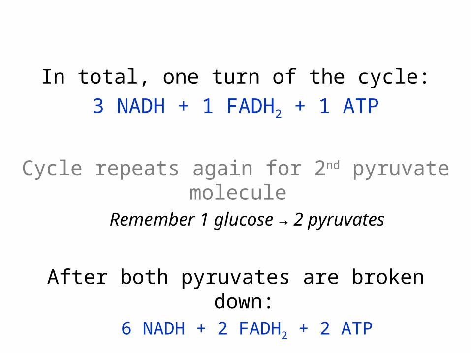
In total, one turn of the cycle:
3 NADH + 1 FADH2 + 1 ATP
Cycle repeats again for 2nd pyruvate molecule Remember 1 glucose → 2 pyruvates
After both pyruvates are broken down:6 NADH + 2 FADH2 + 2 ATP

Sum Total of the Krebs Cycle
With 2 NADH from acetyl-CoA formation:
2 pyruvate → 8 NADH + 2 FADH2 + 2 ATP + 6CO2
CO2 released into surroundings
NADH & FADH2 deliver e-s and H+ to 3rd stage

Stage III: Electron Transfer Phosphorylation
H+ + e-s → H2O + ATP
E-s delivered to electron transfer chains (ETCs) in inner mitochondrial membrane
E- flow in ETCs drives phosphorylation of ADP → ATP (lots of it!)

a.k.a. oxidative phosphorylation
ATP formed by oxidation of NADH & FADH2
Responsible for high ATP yield

NADH & FADH2 give e-s to ETCsSimultaneous release of H+
Energy released at each transfer of ETCAt 3 transfers, released energy pumps H+ across mitochondrial membrane into outer compartment
Concentration & electric gradients result across inner membrane= membrane potential

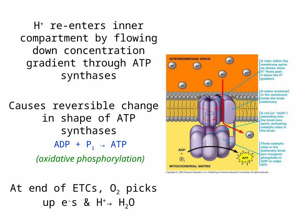
H+ re-enters inner compartment by flowing
down concentration gradient through ATP
synthases
Causes reversible change in shape of ATP synthases
ADP + Pi → ATP
(oxidative phosphorylation)
At end of ETCs, O2 picks up e-s & H+→ H2O


Sum Total of ET Phosphorylation
H+ + e-s → H2O + 32 ATP
In liver, heart, and kidney:
– e-s from NADH delivered to different ETC entry point
– H+ gradient makes 3 ATP (instead of 1)
– Results in 34 ATP total

Animation of ET Phosphorylation
• http://vcell.ndsu.nodak.edu/animations/etc/movie.htm
• http://highered.mcgraw-hill.com/sites/0072437316/student_view0/chapter9/animations.html#

In oxygen-starved cells, e-s have nowhere to go so get gridlocked
No e- flow = no H+ gradients = no ATP forms
Results in cell death because not enough ATP to sustain metabolic processes

3 different categories of poisons interfere with cellular respiration:
• ETC Blockers
• Inhibitors
• Uncouplers

ETC Blockers
Block ETC at various steps of chain
Starves cells of energy by prohibiting ATP
synthesis
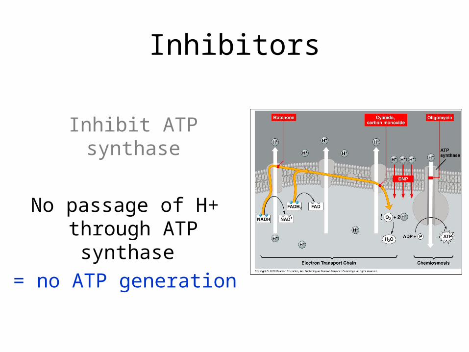
Inhibitors
Inhibit ATP synthase
No passage of H+ through ATP synthase
= no ATP generation

Uncouplers
Make mitochondrial membrane leaky to H+
Electron transport & O2 consumption continue but lack of H+ gradient
= no ATP synthesis

Sum Total of Aerobic Respiration
GlycolysisGlucose → 2 pyruvate + 2 NADH + 2 ATP
Krebs Cycle2 pyruvate → 8 NADH + 2 FADH2 + 2 ATP + 6CO2
ET PhosphorylationH+ + e-s (from coenzymes) → 32 ATP + 6H2O
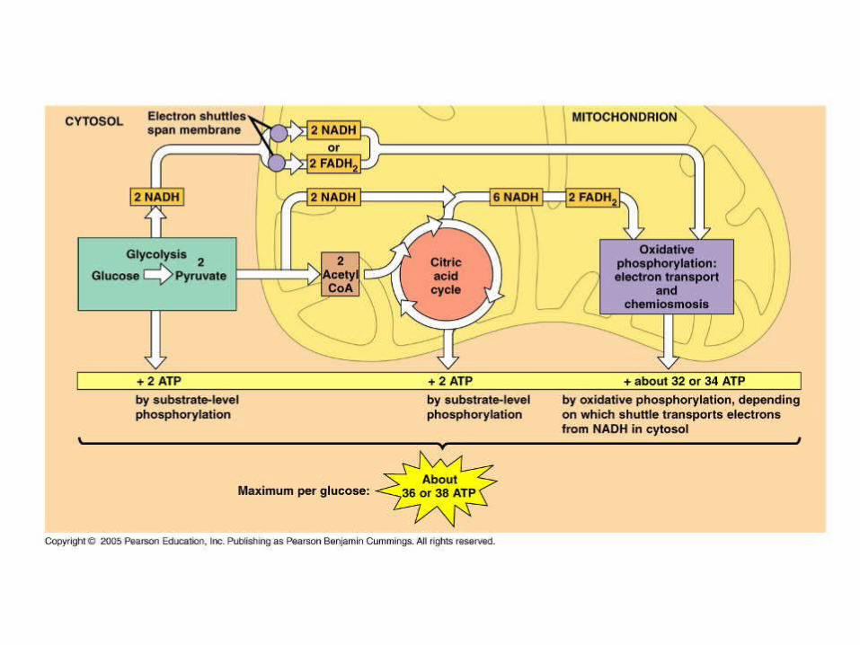
Glucose + 6O2 → 6CO2 + 6H2O + 36 ATP
(38 ATP in liver, heart, kidney)

Cellular Respiration Video
• http://video.google.com/videoplay?docid=1463788471587082686&q=respiration+ninja&total=2&start=0&num=10&so=0&type=search&plindex=0

Anaerobic Respiration
Oxidation of molecules in absence of O2
Requires different electron acceptor at the end of it
2 stages:
– Glycolysis
– Various Energy-Releasing Pathways:• Alcoholic Fermentation• Lactate Fermentation
• Anaerobic Electron Transfers

Stage I: Glycolysis
Glucose → 2 pyruvate + 2 NADH + 2 ATP
Glucose is not broken down any further into CO2 or H2O
All ATP comes from glycolysis(not enough energy to sustain large multicellular
organisms)

After Glycolysis
Final stages in fermentation pathways do not generate more ATP
Instead, they regenerate NAD+ so that it can act as an electron acceptor

Pyruvate not moved into mitochondria
(stays in cytosol & is converted into waste products that can be transported out of cells)
Waste product depends on type of celle.g. ethanol in yeast
e.g. lactate in skeletal muscles, bacteria
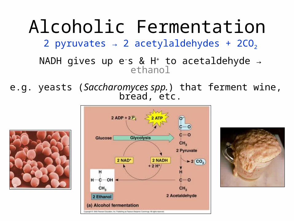
Alcoholic Fermentation2 pyruvates → 2 acetylaldehydes + 2CO2
NADH gives up e-s & H+ to acetaldehyde → ethanol
e.g. yeasts (Saccharomyces spp.) that ferment wine, bread, etc.

Lactate FermentationNADH gives e-s and H+ to pyruvate → lactate
e.g. bacteria (Lactobacillus spp. and others) in cheese, yoghurt, etc.

Certain organisms can couple aerobic & anaerobic respiration or switch from one
mode to the other
e.g. skeletal muscles associated with bonesVariety of cell types within muscle fibres
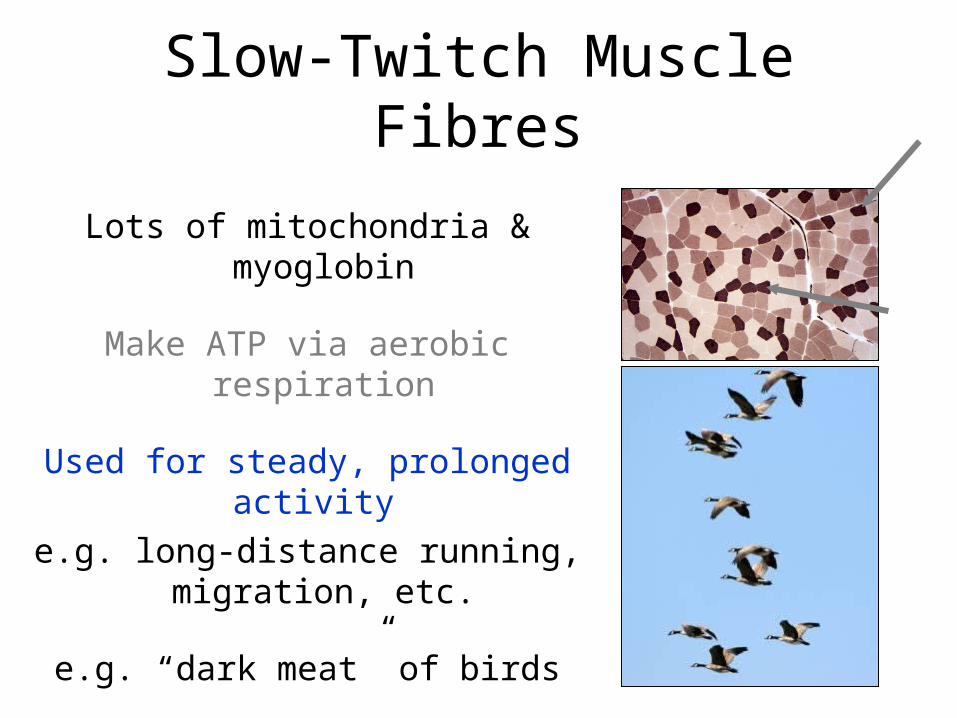
Slow-Twitch Muscle Fibres
Lots of mitochondria & myoglobin
Make ATP via aerobic respiration
Used for steady, prolonged activity
e.g. long-distance running, migration, etc.
e.g. “dark meat” of birds

Fast-Twitch Muscle Fibres
Few mitochondria & no myoglobin
Make ATP via lactate fermentation
Used for short bursts of intense activity
e.g. sprints, weight-training
e.g. “white meat” of birds

Anaerobic Energy Transfers
Pathway of some archaeans & bacteria
Inorganic compounds used as final e- acceptors rather than O2
Aids in cycling of elements through biosphere
Energy yield varies but is small
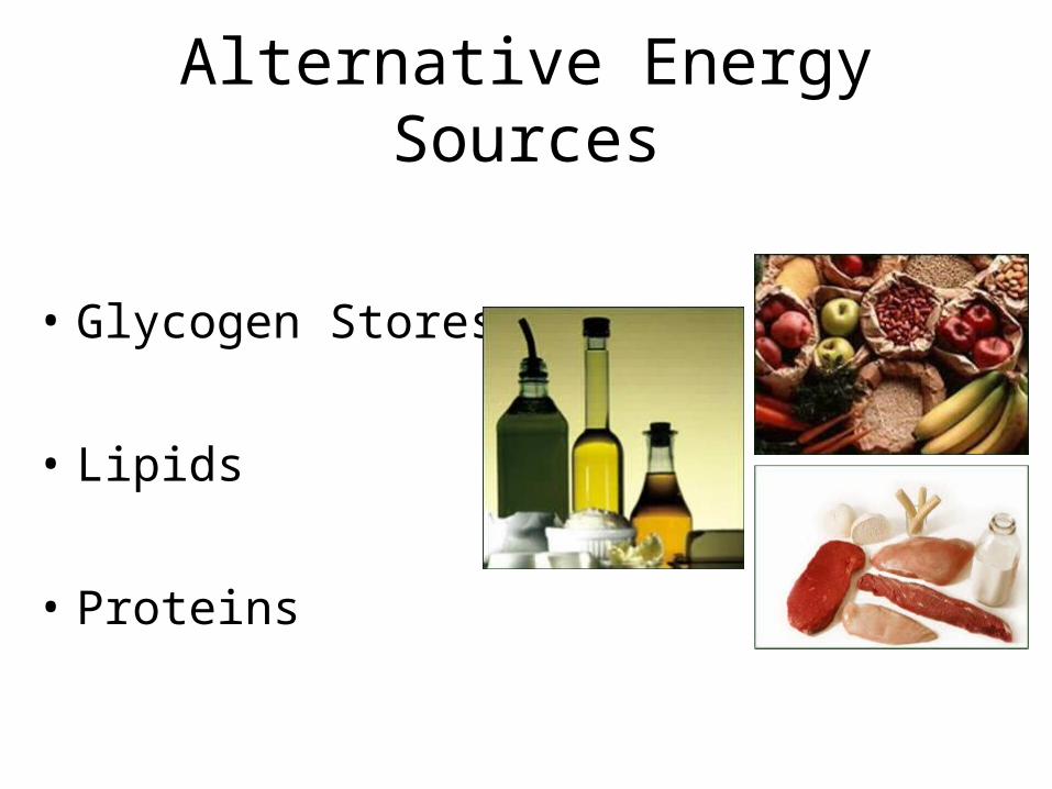
Alternative Energy Sources
• Glycogen Stores
• Lipids
• Proteins

The Fate of Glucose
When food is ingested, glucose is absorbed into the bloodstream
Pancreas secretes insulin to make cells take up glucose faster
Cells convert glucose to glucose-6-phosphate (intermediate of glycolysis)
Can’t leave cell once phosphorylated
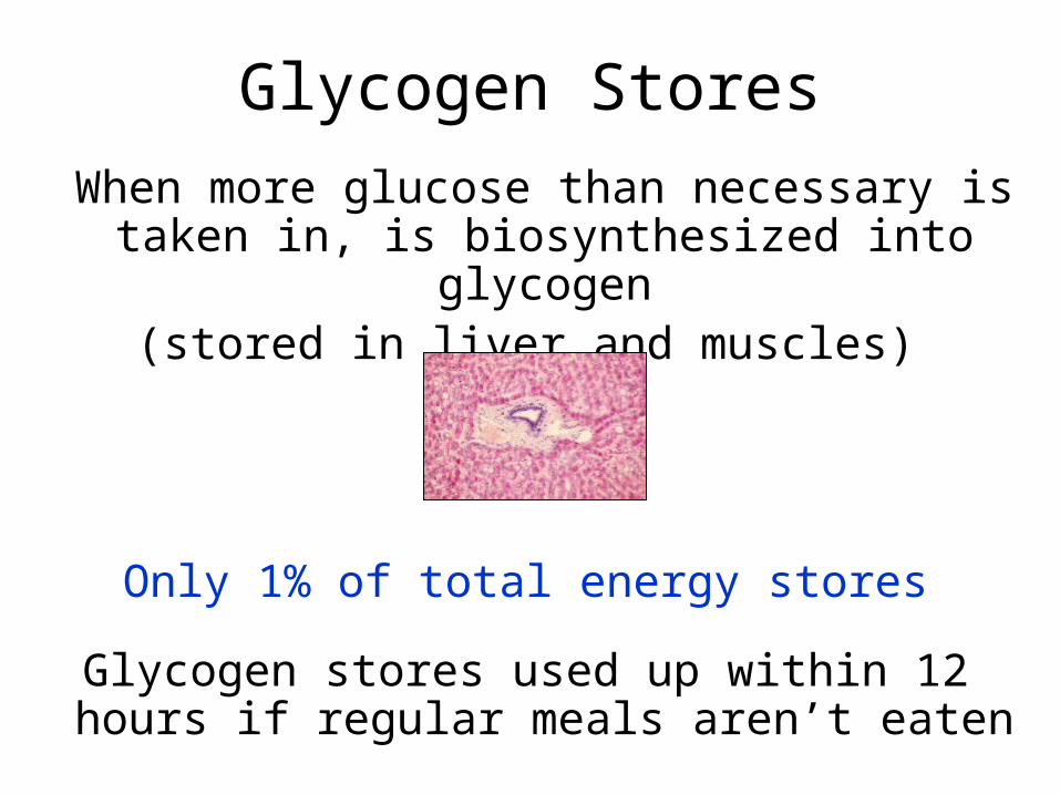
Glycogen Stores
When more glucose than necessary is taken in, is biosynthesized into glycogen
(stored in liver and muscles)
Only 1% of total energy stores
Glycogen stores used up within 12 hours if regular meals aren’t eaten

When blood glucose drops, pancreas secretes glucagon
Liver cells convert stored glycogen → glucose & send back to blood
Can then enter glycolysis pathway
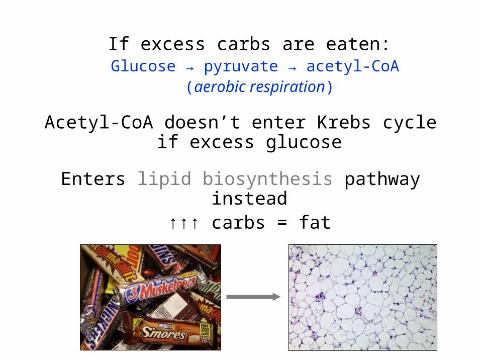
If excess carbs are eaten:Glucose → pyruvate → acetyl-CoA
(aerobic respiration)
Acetyl-CoA doesn’t enter Krebs cycle if excess glucose
Enters lipid biosynthesis pathway instead↑↑↑ carbs = fat

Fat Stores
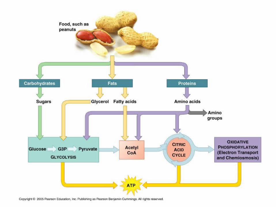
Fat stored as triglycerides in adipose cellsTriglyceride = glycerol + 3 fatty acid tails
Between meals or during sustained exercise, fatty acids yield half of ATP needed by
muscle, liver, kidney cells

When blood glucose ↓, enzymes in adipose cells separate glycerol & fatty acids and
release into blood
Glycerol → PGALUsed in glycolysis
Fatty acids → acetyl-CoAUsed in Krebs cycle

Protein Stores
When proteins ingested, are broken down into amino acids
Absorbed into bloodstream and taken up by cells to make more proteins, etc.
If excess protein eaten, amino acids broken down
Form acetyl-CoA, pyruvate, or intermediates of Krebs cycle
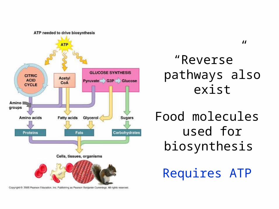
“Reverse” pathways also exist
Food molecules used for biosynthesis
Requires ATP

Summary
Energy is required by all living organisms to sustain life
Because energy flow is one-directional, constant energy
inputs are needed
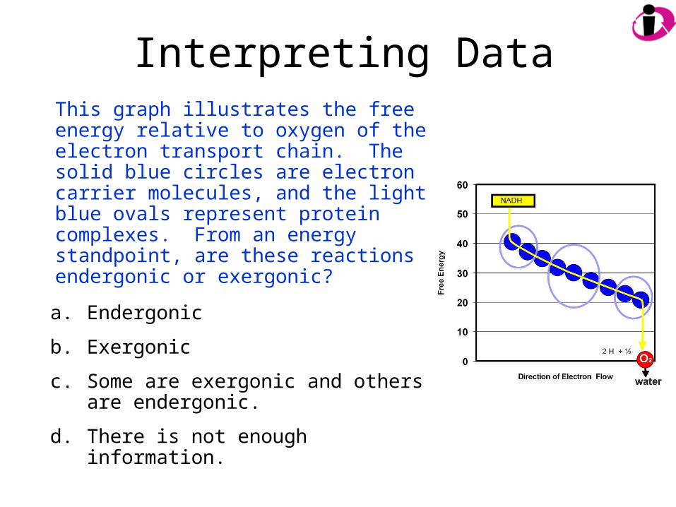
Interpreting DataThis graph illustrates the free energy relative to oxygen of the electron transport chain. The solid blue circles are electron carrier molecules, and the light blue ovals represent protein complexes. From an energy standpoint, are these reactions endergonic or exergonic?
a. Endergonic
b. Exergonic
c. Some are exergonic and others are endergonic.
d. There is not enough information.

Interpreting Data
What would happen to the flow of electrons if oxygen were not present?
a. The flow of electrons would continue but at a slower rate.
b. The flow would cease and ATP production would stop.
c. The presence of oxygen would have no effect.