Homeostatic plasticity and spike-time-dependent plasticity ... · Computational and dynamical...
9
Abstract—Tinnitus is considered as an auditory perception in one ear, both ears or in the head without any external source. A very effective method of tinnitus management is referred to as sound therapy. Computational and dynamical models with plasticity using a neural oscillator or neuronal networks have been proposed by our team in order to investigate mechanisms of tinnitus generation and the clinical effects of sound therapy. In the present paper, two models are proposed, a neuronal network model with homeostatic plasticity (HP) and another model with both HP and spike-time-dependent plasticity (STDP). The results are compared in reference to their effects on inhibition of oscillations as a model of tinnitus management. The outcome data show that the model with both HP and STDP is more robust than the model with STDP only or HP only in the sense that oscillation can be inhibited in a larger range of the intensity of external constant input. Keywords— tinnitus, neuronal network model, sound therapy, spike-time-dependent plasticity, homeostatic plasticity, oscillation, inhibition I. INTRODUCTION INNITUS is considered as an auditory perception in one ear, both ears or in the head without any external source [1]. Tinnitus is not a real sound; it is an actual brain electrical activity. This annoying auditory phenomenon is generated by many factors such as noise exposure and/or chemical and medicinal exposure. Other contributing factors include aging, metabolic and endocrine disorders, neurologic atypicalities, and cardiovascular disturbances. For many years, tinnitus has been considered as a Manuscript received September 29, 2013. This work was supported in part by Japan Society of Promotion of Science Grant-in-Aid for Scientific Research (C) Grant Number 21560429 and 24560498. H. Nagashino is with Department of Biomedical Information Science, Institute of Health Biosciences, The University of Tokushima, Tokushima 770-8509 Japan (phone: 088-633-9025; fax: 088-633-9025; e-mail: [email protected]). Y. Kinouchi is with Institute of Technology and Science, The University of Tokushima, Tokushima 770-8506 Japan (e-mail: [email protected]. ac.jp). A. A. Danesh is with Departments of Communication Sciences & Disorders, and Biomedical Sciences, Florida Atlantic University, Boca Raton, FL 33431 USA (e-mail: [email protected]). A. S. Pandya is with Department of Computer Science and Electrical and Computer Engineering, College of Engineering and Computer Science, Florida Atlantic University, Boca Raton, FL 33431 USA (e-mail: [email protected]). difficult-to-manage clinical condition. Throughout the history many scientists and clinicians have attempted to find ways to help those who suffer from this condition. Tinnitus generation is still a mystery. There are many proposed theories that have attempted to explain its generation. Some of these theories relate the percept of tinnitus to hyperactivity of the auditory cortex and some attribute the changes in inhibitory and excitatory neurotransmitters of the auditory system. The most favored tinnitus generation theory describes tinnitus as a product of brain reorganization as a consequence of hearing loss [2]. Based on the tonotopic organization maps of the auditory cortex, it has been shown that those cortical areas that represent the corresponding frequency region of hearing loss are “invaded” by adjacent frequencies. This reorganization and neuroplasticity has been credited in generating tinnitus. Additionally, the mechanisms of tinnitus generation have been described based on neurophysiological models [3], [4]. The role of neural plasticity to explain the neural correlates of tinnitus also has been reported [5]-[11]. Auditory electrophysiological recordings have addressed the thalamic plasticity via top down modulation [12]. A scientific literature review showed that cochlear damage decreases auditory nerve activity and this change leads to plastic adjustments, a shift in the balance of excitation and inhibition, and increase of spontaneous firings in the central auditory system [8], [9]. Neuroimaging studies such as magnetic resonance imaging (MRI) have shown structural brain changes in individuals with tinnitus [13]. Computational modeling has been applied for better understanding of tinnitus [14]-[17]. There are many areas in the brain that contribute to tinnitus generation; however, it has been shown that the thalamo-cortical network is important for its generation [13], [18]. A neural network model of thalamo-cortical correlates with plasticity toward understanding of the tinnitus has been reported [14]. A tinnitus model based on the neurophysiological model of Jastreboff [3], combined with the adaptive resonance theory of cognitive sensory processing [19] has been proposed for identification of neural correlates of tinnitus [20]. Using models of corticothalamic feedback dynamics, the effect of auditory selective attention on the tinnitus decompensation has also been investigated [20], [21]. A variety of therapeutic approaches for tinnitus have been used for the management of tinnitus [22]-[24]. These include use of medications, supplemental vitamins and Homeostatic plasticity and spike-time-dependent plasticity in computational modeling of tinnitus generation and its management by sound therapy Hirofumi Nagashino, Yohsuke Kinouchi, Ali A. Danesh, and Abhijit S. Pandya T INTERNATIONAL JOURNAL OF BIOLOGY AND BIOMEDICAL ENGINEERING Volume 8, 2014 ISSN: 1998-4510 6
Transcript of Homeostatic plasticity and spike-time-dependent plasticity ... · Computational and dynamical...

Abstract—Tinnitus is considered as an auditory
perception in one ear, both ears or in the head without any external source. A very effective method of tinnitus management is referred to as sound therapy. Computational and dynamical models with plasticity using a neural oscillator or neuronal networks have been proposed by our team in order to investigate mechanisms of tinnitus generation and the clinical effects of sound therapy. In the present paper, two models are proposed, a neuronal network model with homeostatic plasticity (HP) and another model with both HP and spike-time-dependent plasticity (STDP). The results are compared in reference to their effects on inhibition of oscillations as a model of tinnitus management. The outcome data show that the model with both HP and STDP is more robust than the model with STDP only or HP only in the sense that oscillation can be inhibited in a larger range of the intensity of external constant input. Keywords— tinnitus, neuronal network model, sound therapy,
spike-time-dependent plasticity, homeostatic plasticity, oscillation, inhibition
I. INTRODUCTION
INNITUS is considered as an auditory perception in one ear, both ears or in the head without any external source [1]. Tinnitus is not a real sound; it is an actual brain
electrical activity. This annoying auditory phenomenon is generated by many factors such as noise exposure and/or chemical and medicinal exposure. Other contributing factors include aging, metabolic and endocrine disorders, neurologic atypicalities, and cardiovascular disturbances.
For many years, tinnitus has been considered as a
Manuscript received September 29, 2013. This work was supported in
part by Japan Society of Promotion of Science Grant-in-Aid for Scientific Research (C) Grant Number 21560429 and 24560498.
H. Nagashino is with Department of Biomedical Information Science, Institute of Health Biosciences, The University of Tokushima, Tokushima 770-8509 Japan (phone: 088-633-9025; fax: 088-633-9025; e-mail: [email protected]).
Y. Kinouchi is with Institute of Technology and Science, The University of Tokushima, Tokushima 770-8506 Japan (e-mail: [email protected]. ac.jp).
A. A. Danesh is with Departments of Communication Sciences & Disorders, and Biomedical Sciences, Florida Atlantic University, Boca Raton, FL 33431 USA (e-mail: [email protected]).
A. S. Pandya is with Department of Computer Science and Electrical and Computer Engineering, College of Engineering and Computer Science, Florida Atlantic University, Boca Raton, FL 33431 USA (e-mail: [email protected]).
difficult-to-manage clinical condition. Throughout the history many scientists and clinicians have attempted to find ways to help those who suffer from this condition. Tinnitus generation is still a mystery. There are many proposed theories that have attempted to explain its generation. Some of these theories relate the percept of tinnitus to hyperactivity of the auditory cortex and some attribute the changes in inhibitory and excitatory neurotransmitters of the auditory system. The most favored tinnitus generation theory describes tinnitus as a product of brain reorganization as a consequence of hearing loss [2]. Based on the tonotopic organization maps of the auditory cortex, it has been shown that those cortical areas that represent the corresponding frequency region of hearing loss are “invaded” by adjacent frequencies. This reorganization and neuroplasticity has been credited in generating tinnitus.
Additionally, the mechanisms of tinnitus generation have been described based on neurophysiological models [3], [4]. The role of neural plasticity to explain the neural correlates of tinnitus also has been reported [5]-[11]. Auditory electrophysiological recordings have addressed the thalamic plasticity via top down modulation [12]. A scientific literature review showed that cochlear damage decreases auditory nerve activity and this change leads to plastic adjustments, a shift in the balance of excitation and inhibition, and increase of spontaneous firings in the central auditory system [8], [9]. Neuroimaging studies such as magnetic resonance imaging (MRI) have shown structural brain changes in individuals with tinnitus [13].
Computational modeling has been applied for better understanding of tinnitus [14]-[17]. There are many areas in the brain that contribute to tinnitus generation; however, it has been shown that the thalamo-cortical network is important for its generation [13], [18]. A neural network model of thalamo-cortical correlates with plasticity toward understanding of the tinnitus has been reported [14]. A tinnitus model based on the neurophysiological model of Jastreboff [3], combined with the adaptive resonance theory of cognitive sensory processing [19] has been proposed for identification of neural correlates of tinnitus [20]. Using models of corticothalamic feedback dynamics, the effect of auditory selective attention on the tinnitus decompensation has also been investigated [20], [21].
A variety of therapeutic approaches for tinnitus have been used for the management of tinnitus [22]-[24]. These include use of medications, supplemental vitamins and
Homeostatic plasticity and spike-time-dependent plasticity in
computational modeling of tinnitus generation and its management by sound therapy
Hirofumi Nagashino, Yohsuke Kinouchi, Ali A. Danesh, and Abhijit S. Pandya
T
INTERNATIONAL JOURNAL OF BIOLOGY AND BIOMEDICAL ENGINEERING Volume 8, 2014
ISSN: 1998-4510 6

micronutrients, psychotherapy and biofeedback, electrical stimulation, transcranial magnetic stimulation, and more importantly and least invasively sound therapy or acoustic therapy. Tinnitus has many types and subcategories depending on what caused it. Attempts have been made to categorize tinnitus based on its characteristics which in turn can facilitate the selection of management methods [25].
The process of sound therapy is one of the most effective methods. The tinnitus patients who have gone under sound therapy protocol report diminished annoyance from tinnitus [26]. Potentially, patients may perceive a reduction in tinnitus loudness following acoustical stimulation through sound therapy. This cessation of tinnitus following the use of sound therapy has been termed “residual inhibition”. Sound therapy employs a variety of stimuli such as music, white noise, narrow band noise and environmental sounds to facilitate the habituation process to tinnitus. The mechanisms of tinnitus management by sound therapy; however, have not been thoroughly clarified. Some attribute the success with sound therapy to brain plasticity [27] while others consider it a habituation process [28].
Previously we proposed computational and dynamical models employing a neural oscillator [15], [30], [31] or a neuronal network [32]-[35] in order to replicate tinnitus and its management by sound therapy. We have demonstrated that those models conceptually imitate tinnitus perception and exhibit tinnitus inhibition with sound. This inhibition is provided by applying a variety of input with constant amplitude, sinusoidal waveform or noise that represent the role of acoustic stimuli which are used for treatment of tinnitus. By employing these models we could inhibit the oscillations (i.e., tinnitus). This was accomplished by incorporating neural plasticity through parameters in a way that their values can be modified. By hypothesizing that the oscillation and the equilibrium in the model correspond to perception and inhibition of tinnitus, respectively, we reported that these phenomena could explain the fact that the habituated human auditory system temporarily halts perception of tinnitus following sound therapy. However, a model that has larger range of input intensity for inhibition of oscillation is preferable. In order to explore it, we propose a model with different plasticity in the present paper.
For plasticity of our previous models we employed Hebbian hypothesis [15], [30]-[33], or spike-timing- dependent plasticity (STDP) [34], [35] in one of the couplings between the components.
Hebbian hypothesis [36] has been adopted in a number of neural network models for many years. As a newer and biologically plausible hypothesis for synaptic plasticity in the nervous system, “spike-timing-dependent plasticity (STDP)”, has been proposed [37]. It does not replace the idea of Hebbian hypothesis; however, it describes Hebbian synaptic plasticity more specifically. This hypothesis has been adopted in a number of computational models of neuronal networks [38].
As another hypothesis for the plasticity in the nervous system, homeostatic plasticity (HP) was proposed [39]. The HP is applied to nervous systems that require stability of the activities and its role has been widely investigated [40]. The role of HP in hearing loss-induced tinnitus has been investigated [41]. A computational model with HP for tinnitus with hearing loss has been proposed [17], [42]. That model, however, is not a dynamical system. Further
modeling of a dynamical system for tinnitus with HP [43]-[45] is required.
In the present paper, we propose a dynamical model with HP. The current model has the same structure as the previous one [35]. It is composed of the model neurons described by simplified Hodgkin-Huxley equations [46]-[48]] as we employed in the previous studies [32]-[35]. The plasticity is given to inhibitory coupling [49] between neurons, which is based on the neurophysiological consideration [18].
We show the results of analysis of a neuronal network model with HP only and another model with both HP and STDP. We demonstrate the results of computer simulation of this model. The results show that the present model is more robust than the model with STDP only, which was reported in [35] and the model with HP only in the sense that oscillation can be inhibited in a larger range of the intensity of external constant input.
II. A NEURONAL NETWORK MODEL The neuronal network model that we analyze in this paper
is shown in Fig. 1. In the model the firing sequences in the nervous system are simulated. The present model only replicates the inhibition of tinnitus by external sound stimulation. Modeling the habituation would need much larger network configuration. The present model is a conceptually simplified system of a tinnitus generation network. However, we believe that the neural mechanism proposed here could form components of models involving large-scale neural correlates for providing a neurophysiological framework [2].
The model is composed of two excitatory neurons and one inhibitory neuron as shown in Fig. 1. This mechanism includes a positive feedback loop of the excitatory neurons E1 and E2 mutually coupled, and a negative feedback loop with the excitatory neuron E1 and the inhibitory neuron I that are also mutually coupled. The negative feedback loop controls the firing rate. The mechanism can be bistable with a sustained firing state and a non-firing state.
The coupling strength between neurons is denoted by Cij (
€
i, j ∈ 1, 2, 3{ }). The neuron E1 receives external stimuli S that is afferent signal due to the acoustic stimuli that are employed in sound therapy.
We express the dynamics of the model by a simplified version of Hodgkin-Huxley equations (HH) [46]-[48]. We employed it instead of HH to reduce the computational complexity and the related simulation time by reducing the number of state variables for each neuron from four to two.
Fig. 1 A neuronal network model.
INTERNATIONAL JOURNAL OF BIOLOGY AND BIOMEDICAL ENGINEERING Volume 8, 2014
ISSN: 1998-4510 7

A. Formulation of the model without plasticity
We describe the basic dynamics of the model as
€
dv1dt
=G (v1, m1, n1, h1) +C12z2 −C13z3 +D + S
Cm
,
(1)
€
dh1dt
=αh (v1)(1− h1) + βh (v1)h1 , (2)
€
dv2dt
=G (v2, m2, n2, h2) +C21z1
Cm
, (3)
€
dh2dt
=αh (v2)(1− h2) + βh (v2)h2 , (4)
€
dv3dt
=G (v3, m3, n3, h3) +C31z1 +C32z2
Cm
, (5)
and
€
dh3dt
=αh (v3)(1− h3) + βh (v3)h3 . (6)
where v is the membrane potential, m, n and h are the variables associated with activation of sodium ion channel, inactivation of sodium ion channel and activation of potassium ion channel in the neuron E1, E2 or I. The functions
€
G (v,m, n, h) , m and n are expressed as
€
G(v,m,n,h) = g Nam 3h(VNa − v) +
g K n4 (VK − v) + g l (Vl − v) (7)
{ })()()( vvvm mmm βαα += (8)
and
)1(8.0 hn −= (9)
respectively. In the original HH model [41] m and n are expressed by differential equations. In the simplified version that we employ in the present study, m is expressed by the function of the membrane potential v, as Eq. (8), and n is expressed by the function of the variable h, as Eq. (9), since the change of m and n rapidly converges compared with v and h. The functions
€
αm (v) and
€
βm (v) in Eq. (8) are expressed respectively as
€
αm (v) = 0.1(25− v) e(25−v) 10−1{ } (10)
and
€
βm (v) = 4 e−v 18 (11)
Functions
€
αh (v) and
€
βh (v) in Eq. (2), (4), (6) are expressed respectively as
€
αh (v) = 0.07 e−v 20 (12)
and
€
βh (v) = 1 e(30−v) 10+1{ } . (13)
The parameters of the neuron model were fixed as
Cm=1[µF/cm2],
€
g Na = 120[mS /cm2 ] ,
€
g K = 36[mS /cm2 ] ,
€
g l = 0.3[mS /cm2 ] , VNa=115[mV],
€
VK = −12 [mV], Vl=10.6 [mV], based on the values in the original HH model [41].
The output of the neuron j to its postsynaptic neurons is denoted by zj and expressed as function of the membrane potential vj as
€
z j = {1 (v j ≥ 6)0 (v j < 6)
. (14)
Moreover, a bias term D is introduced in the equation of the membrane potential v1 of the neuron E1, Eq. (1) in order to enable the neurons to elicit sustained firings keeping zj at 0 when the neurons are not firing.
B. Introduction of plasticity All the couplings in the model could have plasticity.
Based on the physiological consideration in [xx] and for simplicity of the modeling, we assume in the current model that only single coupling of inhibition between neurons has plasticity. In the present model both HP and STDP are introduced.
C. Formulation of HP We incorporate HP in the present model as a dynamical
process. We assume that the plastic coupling coefficient C13 changes depending on the activity of the neuron E1. The dynamics of C13 is modeled in such a way that the higher the activity of E1 is, the larger C13 grows. When E1 does not fire, C13 converges to CS. The change of the synaptic coefficient due to HP is expressed as
dC13dt
=−C13 +CS + pz1
τ , (15)
where CS is the stationary value of C13 when E1 does not fire, p is a parameter that gives the quantity of the modification of C13, and τ is the time constant of C13.
D. Formulation of spike-time-dependent plasticity (STDP) Secondly we incorporate spike-time-dependent plasticity
(STDP) in the present model. We assume that the inhibitory coupling, the coupling strength from the neuron I to the neuron E1, C13, also has STDP. The key idea of this hypothesis on inhibitory synapses is that when the postsynaptic neuron fires before the presynaptic neuron, the synaptic strength becomes stronger (long term depression), and when the presynaptic neuron fires before the postsynaptic neuron fires, the synaptic strength becomes weaker (long term potentiation). Hence, C13 decreases when E1 fires after I fires, and increases when I fires after E1 fires. The time difference between firings of neuron I and neuron E1, t31, is defined as
€
t31 = t3 − t1 (16)
where t1 and t3 are the latest firing times of E1 and I, respectively as shown in Fig. 2. The value of coupling strength with plasticity C13 at time t +Δt, C13(t +Δt), is given by addition of the value at time t, C13(t), and the change of C13, ΔC13,
INTERNATIONAL JOURNAL OF BIOLOGY AND BIOMEDICAL ENGINEERING Volume 8, 2014
ISSN: 1998-4510 8

€
C13 (t + Δt) = C13 (t) + ΔC13 , (17)
where Δt is the time step of calculation, and ΔC12 is given as
€
ΔC13 = −dC13MAXT1
t31 + dC13MAX (18)
when
€
0 < t31 <T1 ,
€
ΔC13 = −dC13MINT2
t31 − dC13MIN (19)
when
€
−T2 < t31 ≤ 0 , and
€
ΔC13 = 0 , (20) when
€
t31 ≤ −T2 or
€
t31 ≥T1.
Fig. 2 Definition of firing time.
Fig. 3 Modeling of STDP in inhibitory coupling strength C13.
III. RESULTS We demonstrate the results of computer simulation of the
model. Throughout the simulation the parameter values
€
D = 18
€
[µA/cm2 ] ,
€
C21 =10 ,
€
C31 = 10 ,
€
C32 = 20 were employed.
A. Analysis of the model without input or plasticity Without stimulation or plasticity, the model has two
stable solutions, an oscillatory state by sustained firings and a non-firing state. They are bistable for a parameter region.
First, we performed the simulation changing the value of the coupling coefficient C12 by one in the range
€
0 <C12 ≤ 30 with the value C13=10. The non-firing state exists for any value of C12 in the range. On the other hand, the oscillatory state exists when
€
C12 ≥ 23. That is, the two solutions coexist when
€
C12 ≥ 23 . It corresponds to the clinical fact that a number of patients of tinnitus claim that they do not always hear sound when there is no external sound. The larger C12 brings the larger basin of the oscillatory solution in the state space of the model in the region.
Secondly, we performed the simulation changing the
value of the coupling coefficient C13 by one in the range
€
0 < C13 ≤ 30 with the value C12=25. The non-firing state exists for any value of C12 in the range. On the other hand, the oscillatory state exists when
€
0 ≤ C13 ≤ 22 and
€
27 ≤ C13 ≤ 30 . Also in this case the two solutions coexist when
€
0 ≤ C13 ≤ 22 and
€
27 ≤ C13 ≤ 30 .
B. Analysis of the model with input and HP only The inhibition of oscillation by constant input with
amplitude I as stimulus S to neuron E1 was examined with plasticity. The parameter values
€
CS = 15 and
€
τ = 50 [ms] were employed for plasticity. The time scale of the change of the synaptic strength is much smaller than the clinical process. It was arranged so that the simulation is completed in a reasonable time. The initial value of the coupling strength C13 is denoted by
€
C0 . Simulations were performed where the parameter
€
C0 = 25, in which only non-firing solution exists stably. The amplitude I of the input was changed by 1
€
µA/cm2 in the range of
€
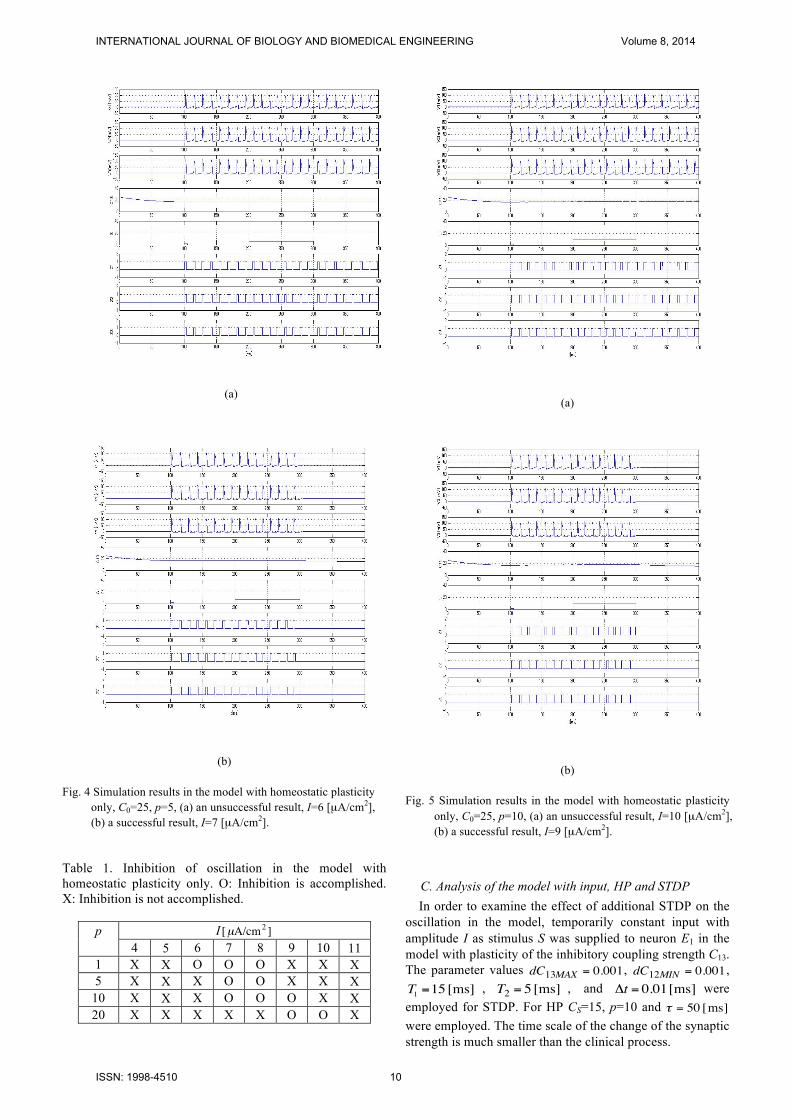
0 < I ≤ 15[µA/cm2 ] . Fig. 4 and Fig. 5 show the examples of simulation results. In the figures, the rows illustrate the membrane potentials v1, v2, v3, the coupling strength C13, input S, output of the neurons z1, z2 and z3, and time difference between firings of neuron I and neuron E1, t31, respectively from the top.
At first the neurons do not fire since the model is in the parameter region where only non-firing solution exists stably. The coupling strength C13 decreases towards its stationary value
€
CS = 15. The model enters the parameter region where both non-firing and firing solutions. However, the non-firing state is sustained since the state of the model system is in the basin of the non-firing solution in the state space of variables vj and hj. For a short period of time from
€
t = 100 [ms], appropriate input S constant with time whose amplitude is appropriate is applied. Then the neurons start firing since the state variables move to the basin of the firing solution. From
€
t = 200 to 300 [ms], input S constant with time whose amplitude is I is applied to the neuron E1 for 100ms. The neurons continue to fire for the period. After the input is removed at
€
t = 300[ms], the behavior of the model depends upon the amplitude of the input which is applied from
€
t = 200 to 300 [ms]. As shown in Fig. 4, when p=5, the input with I=7
[µA/cm2] for 100ms makes the network stop the oscillation after the input is removed, while the input with I=6 [µA/cm2] fails to stop the oscillation. For p=5, the amplitude I=7 or 8 [µA/cm2] was required for inhibition of oscillation. When p=10, the input with I=9 [µA/cm2] for 100ms makes the network stop the oscillation after the input is removed, while the input with I=10 [µA/cm2] fails to stop the oscillation, which is shown in Fig. 5. For p=10, the input with I=7, 8 or 9 [µA/cm2] was required for inhibition of oscillation. When p=1, the input with I=6, 7 or 8 [µA/cm2], and when p=20, the input with I=9 or 10 [µA/cm2], respectively, for 100ms was required to make the network stop the oscillation after the input is removed. These results are summarized in Table 1.
INTERNATIONAL JOURNAL OF BIOLOGY AND BIOMEDICAL ENGINEERING Volume 8, 2014
ISSN: 1998-4510 9
(a)
(b)
Fig. 4 Simulation results in the model with homeostatic plasticity only, C0=25, p=5, (a) an unsuccessful result, I=6 [µA/cm2], (b) a successful result, I=7 [µA/cm2].
Table 1. Inhibition of oscillation in the model with homeostatic plasticity only. O: Inhibition is accomplished. X: Inhibition is not accomplished.
p
I
€
[µA/cm2 ] 4 5 6 7 8 9 10 11
1 X X O O O X X X 5 X X X O O X X X
10 X X X O O O X X 20 X X X X X O O X
(a)
(b)
Fig. 5 Simulation results in the model with homeostatic plasticity only, C0=25, p=10, (a) an unsuccessful result, I=10 [µA/cm2], (b) a successful result, I=9 [µA/cm2].
C. Analysis of the model with input, HP and STDP In order to examine the effect of additional STDP on the
oscillation in the model, temporarily constant input with amplitude I as stimulus S was supplied to neuron E1 in the model with plasticity of the inhibitory coupling strength C13. The parameter values
€
dC13MAX = 0.001,
€
dC12MIN = 0.001 ,
€
T1 =15 [ms] ,
€
T2 = 5 [ms] , and
€
Δt = 0.01[ms] were employed for STDP. For HP CS=15, p=10 and
€
τ = 50 [ms] were employed. The time scale of the change of the synaptic strength is much smaller than the clinical process.
INTERNATIONAL JOURNAL OF BIOLOGY AND BIOMEDICAL ENGINEERING Volume 8, 2014
ISSN: 1998-4510 10

(a)
(b)
Fig. 6 Simulation results in the model with both homeostatic plasticity and spike-time-dependent plasticity,
€
C0 = 25 , p=1, (a) an unsuccessful result, I=2 [µA/cm2], (b) a successful result, I=3 [µA/cm2].
It was arranged so that the simulation is completed in a reasonable time. Simulations were performed with the initial value of the coupling strength C13,
€
C0=25, at which no oscillation occurs without external input. The amplitude I of the input was changed by 1
€
µA/cm2 in the range of
€
0 < I ≤ 15[µA/cm2]. Fig. 6 and Fig. 7 show examples of simulation results. In
the figures, the rows illustrate the membrane potentials v1, v2, v3, the coupling strength C13, input S, output of the neurons z1, z2 and z3, and time difference between firings of neuron I
(a)
(b)
Fig. 7 Simulation results in the model with both homeostatic plasticity and spike-time-dependent plasticity,
€
C0 = 25 , p=10, (a) an unsuccessful result, I=8 [µA/cm2], (b) a successful result, I=7 [µA/cm2].
Table 2. Inhibition of oscillation in the model with both homeostatic plasticity and spike-time-dependent plasticity. O: Inhibition is accomplished. X: Inhibition is not accomplished.
p I
€
[µA/cm2 ] 2 3 4 5 6 7 8 9 1 X O O O X X X X 5 X O O O X X X X
10 X X O O O O X X 20 X X X X O O O X
INTERNATIONAL JOURNAL OF BIOLOGY AND BIOMEDICAL ENGINEERING Volume 8, 2014
ISSN: 1998-4510 11

and neuron E1, t31, respectively from the top. At first from time t=0[ms] to t=200[ms] input S=0. The coupling strength C13 decreases according to HP so that the firing of neuron E1 is easier to occur. It decays to the value in which oscillatory solution also exists. At t=200[ms] a trigger input with very short duration and with intensity It is given to E1. The constant input I was applied to E1 for 100[ms] from t=400[ms] to 500[ms]. The neurons fire with higher rate for this period. Consequently it gives an effect of plasticity that is different from the one given while no input is applied. From t=500[ms] to t=600[ms] input is not applied. As shown in Fig. 4 and Fig. 5, when It=1.3[µA/cm2], the input with I=4 [µA/cm2] for 100ms makes the network stop the oscillation after the input is removed, while the input with I=3 [µA/cm2] fails to stop the oscillation. For the inhibition of oscillation the input amplitude of a suitable range is required.
As shown in Fig. 6, when p=5, the input with I=3 [µA/cm2] for 100ms makes the network stop the oscillation after the input is removed, while the input with I=4 [µA/cm2] fails to stop the oscillation. For p=5, the amplitude I=3, 4 or 5 [µA/cm2] was required for inhibition of oscillation. When p=10, the input with I=7 [µA/cm2] for 100ms makes the network stop the oscillation after the input is removed, while the input with I=8 [µA/cm2] fails to stop the oscillation, which is shown in Fig. 5. For p=10, the input with I=4, 5, 6 or 7 [µA/cm2] was required for inhibition of oscillation. When p=1, the input with I=3, 4 or 5 [µA/cm2], and when p=20, the input with I=6, 7 or 8 [µA/cm2], respectively, for 100ms was required to make the network stop the oscillation after the input is removed. Table 2 summarizes these results.
For It=1.3, the amplitude I=5, 6 or 7 [µA/cm2] was required for inhibition of oscillation. Table 1 demonstrates the inhibition is accomplished or not with different values of It and I. With larger It, smaller range of I was appropriate for inhibition.
The plastic coupling coefficient C13 increases slightly during the stimulation.
D. Discussion In summary, it was observed that the model succeeds in
demonstrating the effect of the introduction of the external stimulus S. This leads to termination of firing of the neurons.
Tables 1 and 2 show that the range of input intensity in which the oscillation is inhibited in the model with STDP and HP is wider than the range of input intensity in the model with HP only for some values of the parameter p.
Comparing the results in the model with STDP and HP and those in the model with STDP only described in [xx], we can see that the range of input intensity in which the oscillation is inhibited in the model with both STDP and HP is wider than the range of input intensity in the model with STDP only.
It can be stated that that the model with both STDP and HP is more robust than the model with STDP only or HP only in the sense that oscillation can be inhibited by a larger
range of the intensity of external input. In the models with STDP only, HP only, and both STDP
and HP, the plastic coupling coefficient does not change to the value in which the firing solution does not exist during the stimulation. The oscillation stops due to the change of the state of the model as well as the change of the coupling coefficient by the input. Hence, further investigation of simulation or different modeling is required in order to reproduce the inhibition of oscillation by synaptic plasticity only.
IV. CONCLUSION The results of computer simulation of a computational
and dynamical neuronal network model with HP only and the one with both HP and STDP for tinnitus generation and its management by sound therapy were described in this paper. The structure of the models is the same as that of the model with STDP only that was previously proposed.
It has been shown through computer simulations that the model with both STDP and HP is more robust than the model with STDP only or HP only in the sense that oscillation can be inhibited by a larger range of the intensity of external input that can be hypothesized as activation by sound stimulus in sound therapy.
In the present model, the inhibition of the oscillation was realized by both the change of the plastic coupling strength and the change of the state of the model by supplying the input. More investigation for improvement of the model is required in order to demonstrate that only the synaptic plasticity brings the inhibition of oscillation and the model is more robust in the input amplitude for inhibition of oscillation.
Our future work will expand this model so that it can more effectively explain underlying physiology of tinnitus, and explore better stimulation for its inhibition through sound therapy techniques.
ACKNOWLEDGMENT Authors thank Hidemitsu Otsu and Yamato Maeki for
their help with computer simulation.
REFERENCES [1] J. Vernon, S. Griest and L. Press,”Attributes of tinnitus and the
acceptance of masking,” Am. J. Otolaryngol., vol. 11, no. 1, 1990, pp. 44–50.
[2] J. A. Kaltenbach, “Tinnitus: Models and mechanisms,” Hearing Research, vol. 276, no.1-2, 2011, pp. 52-60.
[3] P. J. Jastreboff, “Phantom auditory perception (tinnitus): mechanisms of generation and perception,” Neuroscience Research, vol. 8, no. 4, 1990, pp. 221-254.
[4] H. P. Zenner, M. Pfister and N. Birbaumer, “Tinnitus sensation: sensory and psychophysical aspects of a new pathway of acquired centralization of chronic tinnitus,” Otol. Neurotol., vol. 8, 2006, pp. 1054-1063.
[5] J. J. Eggermont and L. E. Roberts, “The neuroscience of tinnitus,” Trends in Neurosciences, vol. 27, no. 11, 2004, pp. 676-682.
[6] A. R. Moller, Neural plasticity and disorders of the nervous system, Cambridge: Cambridge University Press, 2006.
[7] T. Tzounopoulos, “Mechanisms of synaptic plasticity in the dorsal cochlear nucleus: plasticity-induced changes that could underlie tinnitus,” American J. of Audiology, vol. 17, Dec. 2008, pp. S170-S175.
[8] L. E. Roberts, J. J. Eggermont, D. M. Caspary, S. E. Shore, J. R. Melcher, J. A. Kaltenbach, “Ringing ears: the neuroscience of tinnitus,” The Journal of Neuroscience, vol. 30, no. 45, 2010, pp. 14972-14979.
INTERNATIONAL JOURNAL OF BIOLOGY AND BIOMEDICAL ENGINEERING Volume 8, 2014
ISSN: 1998-4510 12

[9] S. E. Shore, “Plasticity of somatosensory inputs to the cochlear nucleus—implication for tinnitus,” Hearing Research, vol. 281, no. 1-2, 2011, pp. 38-46.
[10] N. D. Engineer, J. R. Riley, J. D. Seale, W. A. Vrana, J. A. Shetake, S. P. Sudanagunta, M. S. Borland, M. P. Kilgard, “Reversing pathological neural activity using target plasticity,” Nature, vol. 470, 2011, pp. 101-106.
[11] L. E. Roberts, D. J. Bosnyak and D. C. Thompson, “Neural plasticity expressed in central auditory structures with and without tinnitus,” Frontiers in Systems Neuroscience, vol. 6, Article 40, 2012.
[12] N. Suga and X. Ma, “Multiparametric corticofugal modulation and plasticity in the auditory system,” Nat. Rev. Neurosci., vol. 4, 2003, pp. 783-794.
[13] M. Muhlau, J. P. Rauschecker, E. Oestreicher, C. Gaser, M. Rottinger, A. M. Wohlshlager, F. Simon, T. Etgen, B. Conrad and D. Sander, “Structural brain changes in tinnitus,” Cereberal Cortex, vol. 16, Sept 2006, pp. 1283-1288.
[14] M. Dominguez, S. Becker, I. Bruce and H. Read, “A spiking neuron model of cortical correlates of sensorineural hearing loss: spontaneous firing, synchrony, and tinnitus,” Neural Computation, vol. 18, 2006, pp. 2942-2958.
[15] K. Fujimoto, H. Nagashino, Y. Kinouchi, A. A. Danesh and A. S. Pandya, “Oscillation and its inhibition in a neural oscillator model for tinnitus,” in Proc. of the 28th IEEE EMBS Annual International Conference, New York, USA, 2006, pp. 5547-5550.
[16] D. J. Strauss, W. Delb, R. D’Amelio, Y. F. Low and P. Falkai, “Objective quantification of the tinnitus decompensation by synchronization measures of auditory evoked single sweeps,” IEEE Trans. Neural Systems and Rehabilitation Eng., vol. 16, Feb. 2008, pp. 74-81.
[17] R. Schaette and R. Kempter, “Development of tinnitus-related neuronal hyperactivity through homeostatic plasticity after hearing loss: a computational model,” European Journal of Neuroscience, vol. 23, 2006, pp. 3124-3138.
[18] X. Yu, X. Xu, S. He and J. He, “Change detection by thalamic reticular neurons,” Nature Neuroscience, vol. 12, 2009, pp. 1165-1170.
[19] S. Grossberg, “Linking attention to learning, expectation, and consciousness,” in Neurobiol. Attention, L. Itti and J. Tsotsos, Eds., 2005, pp. 652-662.
[20] C. Trenado, L. Haab, W. Reith and D. J. Strauss, “Biocybernetics of attention in the tinnitus decompensation: an integrative multiscale modeling approach,” J. Neurosci. Methods, vol. 178, 2009, pp. 237-247.
[21] C. Trenado, L. Haab and D. J. Strauss, “Corticothalamic feedback dynamics for neural correlates of auditory selective attention,” IEEE Trans. Neural Systems and Rehabilitation Eng., vol. 17, Feb. 2009, pp. 46-52.
[22] R. S. Tyler Ed., Tinnitus Treatment: Clinical protocols, New York: Thieme, 2006.
[23] R. S. Tyler, J. Rubinstein, T. Pan, S. A. Chang, S. A. Gogel, A. Gehringer and C. Coelho, “Electrical stimulation of the cochlea to reduce tinnitus,” Semin. Hear., vol. 29, no. 4, 2008, pp. 326-332.
[24] R. S. Tyler, B. Noble, C. B. Coelho and H. Ji, “Tinnitus retraining therapy: Mixing point and total masking are equally effective,” Ear Hear., May 17, 2012 [Epub ahaed of print].
[25] R. Tyler, C. Coelho, P. Tao, H. Ji, W. Noble, A. Gehringer, S. Gogel. “Identifying tinnitus subgroups with cluster analysis,” American Journal of Audiolology, vol. 17, no. 2, Dec. 2008, pp. S176-184.
[26] J. A. Henry, M. A. Schechter, T. L. Zaugg, S. Griest, P. J. Jastreboff, J. A. Vernont, C. Kaelin, M. B. Meikle, K. S. Lyons and B. J. Stewart, “Outcomes of clinical trial: tinnitus masking versus tinnitus retraining therapy,” J. Am. Acad. Audiol., vol. 17, no. 2, 2006, pp. 104-132.
[27] P. B. Davis, “Music and the acoustic desensitization protocol for tinnitus,” in Tinnitus Treatment: Clinical protocols, R. S. Tyler Ed. New York: Thieme, 2006, pp. 146-160.
[28] R. S. Hallam and L. McKenna, “Tinnitus habituation therapy,” in Tinnitus Treatment: Clinical protocols, R. S. Tyler Ed. New York: Thieme, 2006, pp. 65-80.
[29] P. J. Jastreboff and M. M. Jastreboff, “Tinnitus retraining therapy: a different view on tinnitus,” ORL J Otorhinolaryngol. Relat Spec, vol. 68, 2006, pp. 23-30.
[30] H. Nagashino, K. Fujimoto, Y. Kinouchi, A. A. Danesh and A. S. Pandya, “A neural oscillator model for tinnitus and its management by sound therapy,” International Journal of Modern Engineering, vol. 11, no. 1, 2010, pp. 58-66.
[31] H. Nagashino, K. Fujimoto, Y. Kinouchi, A. A. Danesh and A. S. Pandya, “Inhibition of oscillation in a neural oscillator model for sound therapy of tinnitus,” International Journal of Modelling and Simulation, vol. 32, issue 4, 2012, pp. 279-285.
[32] H. Nagashino, K. Fujimoto, Y. Kinouchi, A. A. Danesh, A. S. Pandya and J. He, “Oscillation and its inhibition in a neuronal network model for tinnitus sound therapy,” in Proc. of the 30th Annual International Conference of the IEEE EMBS, Vancouver, Canada, 2008, pp. 311-314.
[33] H. Nagashino, Y. Kinouchi, A. A. Danesh and A. S. Pandya, “A neuronal network model for tinnitus and its management by sound therapy,” International Journal of Biology and Biomedical Engineering, vol. 3, issue 4, 2009, pp. 43-50.
[34] H. Nagashino, Y. Kinouchi, A. A. Danesh and A. S. Pandya, “A plastic neuronal network model with STDP for tinnitus management by sound therapy,” International Journal of Mathematical Models and Methods in Applied Sciences, vol. 6, issue 1, 2012, pp. 90-97.
[35] H. Nagashino, Y. Kinouchi, A. A. Danesh and A. S. Pandya, “Spike-time-dependent plasticity of excitation and inhibition in a neuronal network model for tinnitus relief with sound therapy,” International Journal of Biology and Biomedical Engineering, vol. 6, issue 3, 2012, pp. 165-173.
[36] D. O. Hebb, The Organization of behavior: A neuropsychological theor. New York: John Wiley & Sons, 1949.
[37] W. B. Levy and O. Steward, “Temporal contiguity requirements for long-term associative potentiation/depression in the hippocampus,” Neuroscience, vol. 8, Issue 4, 1983, pp. 791-797.
[38] M. Gilson, A. N. Burkitt, D. B. Grayden, D. A. Thomas and J. L. van Hemmen, “Emergence of network structure due to spike-timing-dependent-plasticity in recurrent neuronal networks. I. Input selectivity-strengthening correlated input pathways,” Biol. Cybern., vol. 101, 2009, pp. 81-102.
[39] G. G. Turrigiano and S. B. Nelson, “Homeostatic plasticity in the developing nervous system,” Nature Reviews Neuroscience, vol. 5, 2004, pp. 97-107.
[40] B. Chandler and S. Grossberg, “Joining distributed pattern processing and homeostatic plasticity in recurrent on-center off-surround shunting networks: noise, saturation, short term memory, synaptic scaling, and BDNF,” Neural Networks, vol. 25, 2012, pp. 21-29.
[41] S. Yang, S. D. Weiner, L. S. Zhang, S. Cho and S. Bao, “Homeostatic plasticity drives tinnitus perception in an animal model,” Proceedings of the National Academy of Sciences in the USA, vol. 108, no. 36, 2011, pp. 14974-14979.
[42] R. Schaette and D. M. McAlpine, “Tinnitus with a normal audiogram: physiological evidence for hidden hearing loss and computational model,” J. Neurosci., vol. 31, 2011, pp. 13452-13457.
[43] H. Nagashino, Y. Kinouchi, A. A. Danesh and A. S. Pandya, “A neuronal network model with homeostatic plasticity for tinnitus generation and its management by sound therapy,” Proceedings of 2012 IEEE EMBS International Conference on Biomedical Engineering and Sciences, Langkawi, Malaysia, 2012, pp. 43859:1-5.
[44] H. Nagashino, Y. Kinouchi, A. A. Danesh and A. S. Pandya, “A neuronal network model with STDP and homeostatic plasticity for tinnitus generation and its management by sound therapy,” Proceedings of The 4th International Conference on Bioscience and Bioinformatics, 2013, pp. 135-139.
[45] L. Haab, M. Scheerer, J. Ruckert, R. Hannenmann and D. J. Strauss, “Support of a patient-specific therapeutical acoustic stimulation in tinnitus by numerical modeling,” Proc. of the 34th Annual International Conference of the IEEE EMBS, 2012, pp.5578-5581.
[46] H. Kawakami, Dynamics of biological rhythmic phenomina— Nonlinear dynamics applied to ME. Tokyo: Corona, 2001, ch. 7.
[47] J. Rinzel, “Excitation dynamics: Insights from simplified membrane models,” Fed. Proc., vol. 15, no. 44, 1985, pp. 2944-2946.
[48] A. L. Hodgkin and A. F. Huxley, “A quantitative description of membrane current and its application to conduction and excitation in nerve,” The Journal of Physiology, 1952, vol. 117, pp. 500-544.
[49] K. J. Bender and L. O. Trussell, “Synaptic plasticity in inhibitory neurons of the auditory brainstem,” Neuropharmacology, vol. 60, 2011, pp. 774-779.
Hirofumi Nagashino (Born in Tokushima, Japan, March 1, 1950) received the Bachelor of Engineering and Master of Engineering degrees in Electrical Engineering from The University of Tokushima, Japan in 1972 and 1974, respectively. He received the Doctor of Engineering degree in 1982 from Osaka University, Japan. In 1974 he joined Department of Electrical Engineering, Faculty of Engineering, The University of Tokushima as an assistant professor and was promoted to associate professor in Department of Electrical and Electronic Engineering, Faculty of Engineering, The University of Tokushima. Since 2002 he has been a professor in Department of Radiologic Science and Engineering, School of Health Sciences, Faculty of Medicine, The University of Tokushima. Since 2008 he also has been a
INTERNATIONAL JOURNAL OF BIOLOGY AND BIOMEDICAL ENGINEERING Volume 8, 2014
ISSN: 1998-4510 13

professor in Subdivision of Biomedical Information Science, Division of Health Sciences, Institute of Health Biosciences, The University of Tokushima. His research interest includes biocybernetics, neural networks and its application to biomedical engineering, particularly neural network models for oscillatory activities, signal source identification, pattern recognition, etc. Dr. Nagashino is a member of IEEE Engineering in Medicine and Biology Society, System, IEEE Man and Cybernetics Society, IEEE Computational Intelligence Society, Japanese Society for Medical and Biological Engineering, Institute of Electronics, Information and Communication Engineers, Japan, The Society of Instrument and Control Engineers, Japan, Japanese Neural Networks Society, and Japanese Society of Magnetic Applications in Dentistry. Yohsuke Kinouchi (Born in Tokushima, Japan, November 1943) received Bachelor of Engineering and Master of Engineering degrees in Electrical Engineering from The University of Tokushima, Tokushima, Japan in 1966 and 1968, respectively, and Doctor of Engineering degree in 1975 from Kyoto University, Kyoto, Japan. In 1968 he joined Department of Electrical Engineering, Faculty of Engineering, The University of Tokushima as an assistant professor, and was promoted to associate professor and then professor in Department of Electrical and Electronic Engineering, Faculty of Engineering, The University of Tokushima. Currently he is a Professor Emeritus and Deputy Director, The University of Tokushima, Tokushima, Japan and is also a Guest Professor at Harbin Institute of Technology, Shenzhen Graduate School, Shenzhen, China. His current research interests include magnetic dentistry, biological effects of magnetic fields, bioimpedance, blood flow measurement, mobile telemedicine, medical applications of neural networks, physiological inverse problems and medical applications of LED. Dr. Kinouchi is a member of IEEE Engineering in Medicine and Biology Society, Japanese Society for Medical and Biological Engineering, Institute of Electronics, Information and Communication Engineers, Japan, The Society of Instrument and Control Engineers, Japan, and Japanese Society of Magnetic Applications in Dentistry. Ali A. Danesh (Born in the city of Khoy, Western Azerbaijan province, Iran, January 1964) received Bachelor of Science degree in Audiology from College of Rehabilitation Sciences, Iran University of Medical Sciences, Tehran, Iran in 1987 and Master of Science in Audiology from Idaho State University, Pocatello, Idaho, USA in 1994. He completed his PhD in Audiology with an emphasis on Auditory Electrophysiology from the University of Memphis, Memphis, Tennessee, USA in 1998. In 1998 he joined the Department of Health Sciences at Florida Atlantic University, Boca Raton, Florida, USA as an assistant professor and was promoted to professor in the Department of Communication Sciences and Disorders in 2013. He also has joint appointment at the College of Medicine at Florida Atlantic University and is an adjunct faculty at Department of Audiology, Nova Southeastern University, Fort Lauderdale, Florida, USA. Additionally, he serves as a voluntary faculty, Department of Otolaryngology at the Miller School of Medicine, University of Miami, Miami, Florida. His research interests include auditory electrophysiology, tinnitus, auditory processing and vestibular disorders. Dr. Danesh is a board certified audiologist and is a member of American Academy of Audiology, American Speech-Language and Hearing Association, International Audiology Society, American Tinnitus Association and American Auditory Society. Abhijit S. Pandya (Born in the city of Mumbai, India 1958) received his undergraduate education at the Indian Institute of Technology, Bombay and graduated with a M.Sc. in Physics (specialization in Electronics) in 1977. He earned his M.S. and Ph.D. in Computer Science from the Syracuse University, New York in 1985 and 1988 respectively. In 1988 Dr. Pandya joined the Center for Complex System and the Computer Science and Engineering Department at Florida Atlantic University, Boca Raton, Florida, USA. He was promoted to Associate Professor in 1994 and Professor in 1999. He also has joint appointment at the Department of Communication Sciences and Disorders at Florida Atlantic University. He is a member of the Board of Trusties at the Mahatma Gandhi Medical College and Hospital, Jaipur, India. Dr. Pandya consults for several industries including IBM, Motorola, Coulter industries and the U.S. Patent Office. He has worked as a visiting Professor in various countries including Japan, Korea, India, etc. His areas of research include VLSI implementable algorithms, Applications of AI and Neural Networks, Image analysis in Medicine and Electronic Health Records.
INTERNATIONAL JOURNAL OF BIOLOGY AND BIOMEDICAL ENGINEERING Volume 8, 2014
ISSN: 1998-4510 14