Hiv
77
HIV Dr Mallum Senior Registrar, Dept of Internal Medicine LUTH
-
Upload
chindo-mallum -
Category
Health & Medicine
-
view
56 -
download
0
Transcript of Hiv

HIV
Dr MallumSenior Registrar,
Dept of Internal MedicineLUTH

INTRODUCTION
• Human immunodeficiency virus (HIV) is a blood-borne virus typically transmitted via sexual intercourse, shared intravenous drug paraphernalia, and mother-to-child transmission (MTCT), which can occur during the birth process or during breastfeeding.
• There are two types: HIV1 and HIV2.

SUBTYPES OF HIV 1
• Ten genetically distinct subtypes: Major group (Group M) contains subgroups A to K. Group O (Outliers) contains distinct group of heterogeneous viruses. Found only in West Africa. Group N (interaction between group M and group O viruses

HIV-2
• HIV2 was isolated in West Africa and is confined primarily to West Africa. Compared to HIV1, HIV2 is less transmissible and affected patients are less infectious. Develops more slowly. Associated with less viral burden. Has slower rate of cell decline and clinical progress. Does not respond to non nucleoside analogues.

HISTORY OF HIV IN NIGERIA
• The first two cases of HIV and AIDS in Nigeria were identified in 1985
• HIV prevalence rose from 3.8 percent in 1993 to 5.4 percent in 1999. Following a peak of 5.8 percent in 2001, HIV prevalence then declined steadily throughout the decade.

HIV2
• Less transmissible , has 5 – 9 fold lower probability of transmission per sexual act.
• Associated with Less viral burden • Shower rate of cell decline and clinical
progression rates of progression to HIV – related disease, AIDS and Mortality
• . Vertical transmission .lesser• . NRTls Less active • . NNRls not active • .Pls active
EPIDEMIOLOGY
• Worldwide, Nigeria has the second highest number of new infections reported each year
• Approximately 80 percent of HIV infections in Nigeria are a result of heterosexual sex.
• Women are particularly affected by HIV; in 2009 women accounted for 56 percent of all adults aged 15 and above living with the virus

MODES OF TRANSMISSION
• Sexual: heterosexual male to female or female to male; homosexual male to male or female to female.
• Parenteral: Blood transfusions, use of intravenous injections by intravenous drug users, needle stick accidents.
• Mother to child transmission in utero and during labour, post partum in breast milk.

EPIDEMIOLOGY
• Risk of Occupational Transmission
• 1. percutaneous exposure to infected blood - 0.3%
• 2. mucus membrane exposure - 0.09%
• 3. skin exposure <0.09%
• Most-at-risk groups in Nigeria include brothel and non-brothel based female sex workers (FSW), men-who-have-sex-with-men (MSM), injecting drug users (IDUs), transport workers, members of the Armed Forces and Police.

Structure of HIV
• HIV is a Retrovirus and belongs to the family of Lentivirus.
• Surrounded by a lipoprotein membrane
• Gp120 is an external glycoprotein
• Gp41 is a transmembrane protein
• P24 is the core antigen
• P17 is anchored inside the viral lipoprotein membrane

VIRAL GENOME
• Gag, pol and env genes
• Gag = Group AntiGen
• Pol= POLymerase
• Env= ENVelope
Gag codes for p7,p17,p24
Pol codes for p11 protease,p66 reverse transcriptase,p32 integrase
Env codes for Gp41 and Gp120

HIV REPLICATION CYCLE
• Fusion – Gp 120
• Uncoating
• Reverse transcription – reverse transcriptase
• Integration of proviral DNA- integrase
• Transcription
• Cleavage of precursor molecules- protease
• Assembly
• Budding + maturation

IMMUNOPATHOPHYSIOLOGY
• The Gp120 receptor on the HIV membrane binds to the CD4 receptor and CCR5 or CXCR4 receptors co receptors.
• The Gp41 receptor initiates cell membrane fusion.
• Upon entry, the enzyme reverse transcriptase produces

IMMUNOPATHOPHYSIOLOGY OF HIV
• The Virus migrates to lymphoid tissue early in infection and actively replicates rapidly in lymphoid tissue,CD4 cells and follicular dendritic cells.

IMMUNOPATHOPHYSIOLOGY OF HIV
• There is loss of CD4 T lymphocytes due to :
-Syncytial cell formation
-Cytotoxic and antibody dependent killing of both infected and unifected cells
-Damage to CD4 membranes from budding of newly formed
-Domination of host cell protein synthesis by viral RNA and protein synthesis

IMMUNOPATHOPHYSIOLOGY OF HIV
• In addition to loss of CD4 lymphocytes, there is dysfunction of:
Monocytes,cytotoxic T-cells,T suppressor cells,TNatural killer cells,B lymphocytes.

IMMUNOPATHOPHYSIOLOGY OF HIV
• CYTOTOXIC T CELLS
• Dysfunction is implicated in causation of:
-Herpes zoster,herpes simplex types 1,2 and 8
-CMV,EBV,Hepatitis
T-suppressor cells
-Heightened hypersensitivity reactions
-HIV nephropathies and arthropathies from immune complex disorder

IMMUNOPATHOPHYSIOLOGY OF HIV
• Natural killer cells:
-Predisposition to cancers such as Kaposi’s Sarcoma,Lymphoma,CA cervix.
B cells:
Dysfunction causes bacterial infections like Skin Sepsis,Pharyngitis,Pneumonia,UTI,Septicaemia,PID,Malaria
HIV mutation
• HIV has potential for mutation because reverse transcriptase is an error prone enzyme.
• Mutation may be responsible for change in viral infectivity,fitness and ARV resistance, and escape from CD8 cells and neutralizing antibodies.

PATHOGENESIS OF ACUTE HIV-1 INFECTION.
• The progression is usually:
-Infection of CD4 cells and macrophages at site of infection,Dissemination to lymph nodes,Viraemia resulting from viral replication, development of HIV specific antibodies(Humoralimmunity), and then HIV specific CD4 and CD8 cells(Cellular immunity).
PATHOGENESIS OF CHRONIC HIV-1 INFECTION.
• High turnover of CD4 cells
• Viral load reaches set point
• Immune activation results in immune dysfunction
• Viral reservoirs result from sanctuary sites like the brain and genitourinary tract,persistencein lymphoid tissue.
IMMUNE RESPONSE IN CHILDREN
• Absolute CD4 is age-dependent, therefore CD4% is used
• Viral set point is higher.
• 15-20% of children develop AIDS or die within 1 year
• 10% survive for 5-6 years.

CLINICAL FEATURES
• Acute seroconversion illness : flu like illness -fever,malaise,generalised rash
• Generalised lymphadenopathy
• AIDS presents as recurrent,severe infections or opportunistic malignancies
• The risk of non-AIDS morbidity and mortality like cardiovascular disease,liver disease, cancer is higher.

Non-AIDS related complications that may be more common in patients with HIV
• Hypertension
• Diabetes mellitus and insulin resistance
• Cardiovascular disease
• Pulmonary hypertension
• Cancer- lung cancer, skin cancer, colorectal cancer, prostate cancer
• Osteopenia and osteoporosis

Non-AIDS related complications that may be more common in patients with HIV
• Liver failure- chronic viral hepatitis and alcohol misuse
• Kidney failure
• Peripheral neuropathy
• Frailty
• Cognitive decline and dementia

HIV and TB co-infection
• M. tuberculosis
↑HIV replication and ↑HIV
progression
• HIV ↑ Risk of active TB

HISTORY
• Risk factors for possible exposure to HIV-Unprotected sexual intercourse especially receptive anal intercourse-Large number of sexual partners-Previous or current STDS-Sharing of IV drug paraphernalia-Receipt of blood products-Mucosal contact with infected blood or needle-stick injuries-Maternal HIV infection
PHYSICAL EXAMINATION
• Generalised lymphadenopathy,weight loss
• Anaemia,
• Evidence of opportunistic infections such as herpetic lesions,oral candidiasis

DIAGNOSIS OF HIV/AIDS
• Historically,
• presumptive diagnosis 1981 to 1987
• definitive diagnosis 1987-1993
• Definitive diagnosis with
- Severity at presentation
- Response to ARVS
from 1993- date

DEFINITIVE DIAGNOSIS
• This combines clinical manifestation with laboratory evidence.
• The CDC published the diagnostic criteria in 1987 which included 20 clinical conditions which were expanded to 23 in 1993.

Fungal Infections
• Candidiasis of oesophagus
• Coccidiomycosis- disseminated or extrapulmonary
• Cryptococcosis-extrapulmonary
• Histoplasmosis-Disseminated or extrapulmonary
• Pneumocystis carinii pneumonia

VIRAL INFECTIONS
• CMV of organs other than liver or spleen
• CMV retinitis
• HIV encephalopathy
• Herpes simplex: chronic ulcers> 1 month
• Progressive Multifocal Encephalopathy(JC virus)

Protozoal & Bacterial Infections
Protozoal infections:
• Isosporiasis Chronic intestinal >1 month
• Cryptosporidiasis Chronic intestinal > 1month
• Toxoplasmosis of the brain > 1 month

Bacterial Infections:
• Mycobacterium Avium complex or M . Kansaii
• Recurrent salmonella infection
• Mycobacterium TB(Pulmonary) (added in 1993)
• Recurrent bacterial pneumonia (added in 1993)

Opportunistic cancers:
-Kaposi Sarcoma
-Burkitt’s Lymphoma
-Immunoblastic lymphoma
-Primary lymphoma of the brain
-Invasive Cervical Ca (added in 1993)

HIV wasting syndrome:
• Persistent fever
• Temp >38.5 for 1 month
• Loss of weight in excess of 10% of pre illness weight
• Chronic diarrhoea for >1 month
• Chronic fatigue
CDC CLASSIFICATION
• Category A: Asymptomatic HIV infection without symptoms or AIDS-defining illness
• Category B: HIV infection with symptoms attributable to ,or complicated by , HIV infecton
-Bacillary angiomatosis-Oropharyngeal candidiasis-Vulvovaginal candidiasis-Pelvic inflammatory disease-Cervical dysplasia(moderate or severe)/cervical CA in situ-Oral hairy leukoplakia-Idiopathic Thrombocytopenic purpura-
CDC CLASSIFICATION
• Category B:
- Constitutional symptoms : fever >38.5 degrees C, or diarhoea > 1 month
- Peripheral neuropathy
- Herpes zosters (shingles) , involving 2 or more dermatomes or 1 or more dermatome.
• Category C:HIV infection with AIDS defining illnesses.

CDC CLASSIFICATION
• CD4 counts are further used to subdivide the categories:
-CD4 counts >500/uL : categories A1,B1,C1
-CD4 counts 200-400/uL: categories A2,B2,C2
-CD4 counts <200/uL: categories A3,B3,C3
Once an HIV infection is staged into a higher category it stays in that category permanently.
WHO CLINICAL STAGING
• Stage 1:
-Asymptomatic
-Generalised lymphadenopathy
Performance scale 1: normal activity.

WHO CLINICAL STAGE 2
• Weight loss < 10% body weight• Mucocutaneous manifestations:-seborrheic dermatitis-prurigo-fungal nail infections-recurrent oral ulcerations-angular stomatitis• Herpes zoster in last 5 years• Recurrent upper respiratory tract infections Performance scale 2: symptomatic,normal activity.
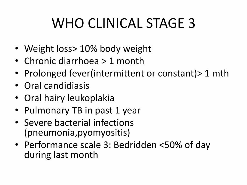
WHO CLINICAL STAGE 3
• Weight loss> 10% body weight• Chronic diarrhoea > 1 month• Prolonged fever(intermittent or constant)> 1 mth• Oral candidiasis• Oral hairy leukoplakia• Pulmonary TB in past 1 year• Severe bacterial infections
(pneumonia,pyomyositis)• Performance scale 3: Bedridden <50% of day
during last month
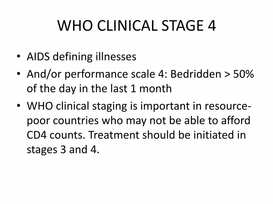
WHO CLINICAL STAGE 4
• AIDS defining illnesses
• And/or performance scale 4: Bedridden > 50% of the day in the last 1 month
• WHO clinical staging is important in resource-poor countries who may not be able to afford CD4 counts. Treatment should be initiated in stages 3 and 4.
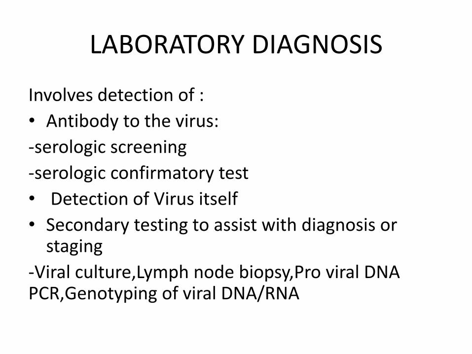
LABORATORY DIAGNOSIS
Involves detection of :
• Antibody to the virus:
-serologic screening
-serologic confirmatory test
• Detection of Virus itself
• Secondary testing to assist with diagnosis or staging
-Viral culture,Lymph node biopsy,Pro viral DNA PCR,Genotyping of viral DNA/RNA
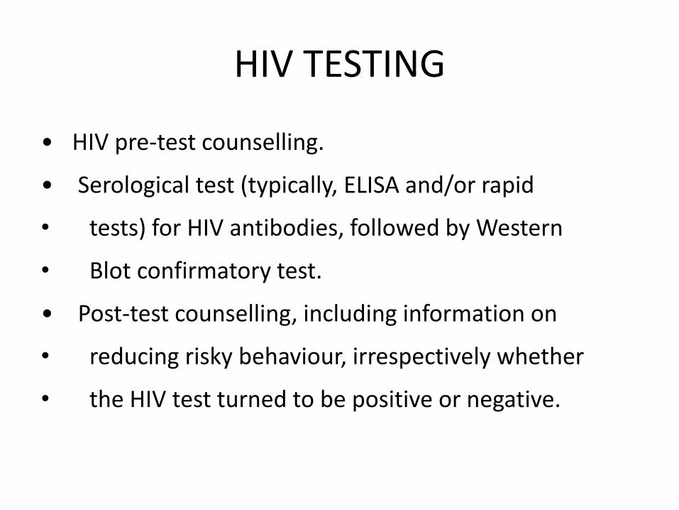
HIV TESTING
• HIV pre-test counselling.
• Serological test (typically, ELISA and/or rapid
• tests) for HIV antibodies, followed by Western
• Blot confirmatory test.
• Post-test counselling, including information on
• reducing risky behaviour, irrespectively whether
• the HIV test turned to be positive or negative.
SEROLOGIC TESTING
• All testing and counselling services must include the five C's recommended by WHO: informed Consent, Confidentiality, Counselling, Correct test results and linkage to Care, treatment and other services.
• Antibodies appear 6-8 weeks after exposure.
• This is called the window period or serologic latency period.

Screening test:Enzyme linked Immunoadsorbent Assay(ELISA)
• ELISA is cheap, easy to perform, highly reproducible and is the most common diagnostic test available
• 3 types of ELISA:-Direct -Indirect-SandwichDrawbacks include high false positive and negative rates,failure to diagnose neonatal infection and those in window period
ELISA
• ELISA’s can detect HIV1 types M,N,O and HIV2.
• Results typically reported as positive,negative, indeterminate.
• HIV2 should be tested for in patients from an HIV2 endemic region or those with indeterminate testing in HIV1.
• Early detection is facilitated by combination screens, and additional detection of p24 antigen or viral RNA.
HIV SCREENING RECOMMENDATIONS
• -All adolescents and adults at increased risk for HIV, and pregnant women(USPSTF)
• -Persons at high risk to be screened at least annually (CDC)
• -

Confirmatory test
• Antibodies to viral antigens are detected:
• Viral antigens are produced from 3 main genes gag,env,pol
• Gag gene products :p56,p27,p17
• Env: p160,gp120,gp41
• Pol: p66-RT heavy chain,p51-RT Light chain,P-32 integrase enzyme, p10 protease enzyme

WESTERN BLOT TEST
• Commonest confirmatory test
• The red cross criteria is unequivocal evidence of antibody to at least one product of each of 3 viral genes
• CDC recommends that presence of any two of the following is a positive result:
-p160,gp120,gp41,p24

CD4 T cell count
• Reference range for CD4 counts is 500-2000 cells/uL.
• After seroconversion,CD4 tends to decrease(700/uL) and continues to decline over time
• A CD4 count < 200/uL is considered AIDS-defining.
• There may be discordance between absolute CD4 T cell counts and CD4 T cell percentages esp in those with active Hepatic C infection and advanced Liver disease.
• In children, CD4 % <25 % consider starting therapy

VIRAL LOAD
• Viral load is a surrogate marker of viral replication.
• The test is conducted using Nucleic acid sequence based analysis(NASBA) or Reverse transcriptase PCR for amplification of viral RNA.
• Patients with viral loads >30,000/uL are more likely to die of AIDS.
• Optimal viral suppression is to an undetectable level (<20-75cells/mls)
• Virologic failure= viral load >200 cells/mls

SECONDARY HIV TESTING
• Viral culture: less sensitive in patients with low viral loads, may be performed as part of phenotypic drug resistance testing.
• Lymph node architecture: HIV DNA,RNA,andproteins may be detected with molecular techniques.
• Pro viral DNA PCR: Performed in newborns because maternal antibodies persist for 9 months.Two negative results seperated by a month considered negative.

SECONDARY HIV TESTING
• Genotyping of viral DNA/RNA.
-sequencing of viral genome to detect mutations that result in resistance to drugs/drug classes.

BASE LINE TESTING
• Testing for other infections :-Purified protein derivative(PPD) skin testing for Tuberculosis + Chest X-ray-CMV testing: anti-CMV IgG indicates previous infxn-Syphilis testing:Rapid plasma reagin-Rapid amplification testing for chlamydia,gonorrhea-Hepatitis A,B,C testing:may need vaccination or RX-Antitoxoplasma antibody: Prophylaxis if CD4<100-Ophthalmologic examination for CMV retinitis

BASE LINE TESTING
• Base line tests prior to commencing ARVs-Liver function tests-serum chemistries-Blood Urea nitrogen/serum creatinine-Fasting Lipid Panel-Vitamin B12 and folate levels-Thyroid function -Urinalysis: to evaluate HIV-associated nephropathy-Drug screen

MANAGEMENT
• The current Department of Human and Health services (DHHS) recommendations on initiation of anti retroviral therapy(ART) are:
-ART should be initiated in all patients with history of AIDS-defining illness or CD4 count > 350 cells/uL
-ART should be commenced regardless of CD4 in : pregnant women,patients with HIV-associated nephropathy, those with HBV co-infection.

MANAGEMENT
• CD4 counts 350-500/uL : 55% strong recommendation, 45% moderate recommendation.
• CD4 counts > 500/ul

ANTI RETROVIRAL THERAPY
• Classes of antiretroviral agents:-Nucleoside reverse transcriptase inhibitors(NRTIs)-Non-nucleoside reverse transcriptase inhibitors(NNRTIs)-1997-protease inhibitors(PIs)-1995-Fusion inhibitors -2003-CCR5 Co receptor antagonists (entry inhibitors)-HIV integrase strand transfer inhibitors -2007There are now more than 20 approved antiretroviral drugs.

ANTI RETROVIRAL THERAPY
• Both NRTIs and NNRTIs interfere with viral replication by inhibiting reverse transcriptase
• PIs inhibit Protease, an enzyme involved in replication
• Fusion or entry inhibitors prevent HIV from binding to or entering cells.
• Integrase inhibitors interfere with integrasethe enzyme HIV needs to insert genetic material into cells.

Nucleoside reverse transcriptase inhibitors(NRTIs)
• Lamivudine 3TC
• Abacavir ABC
• Zidovudine AZT or ZDV
• Stavudine d4T
• Didanosine ddI
• Emtricitabine FTC
• Tenofovir TDF

Non-nucleoside reverse transcriptase inhibitors(NNRTIs)
• Delavirdine DLV
• Efavirenz EFV
• Etravine ETR
• Nevirapine NVP
• Rilpivirine
PROTEASE INHIBITORS
• Amprenavir AMP• FOS- APV Fosamprenavir Telzir• Atazanavir ATV• Darunavir DRV• Indinavir IDV• Lopinavir + ritonavir LPV/RTV• Nelfinavir NFV• Ritonavir RTV• Saquinavir SQV• Tipranavir TPV

FUSION OR ENTRY INHIBITORS/INTEGRASE INHIBITORS
• Enfuvirtide T-20- binds to GP41
• Maraviron MVC- binds to CCR5 receptor
• Integrase inhibitors:
Raltegravir RAL

Highly active anti retroviral therapy(HAART)
• Taking a combination of three or more anti retroviral agents is called HAART.
• This vastly reduces the rate at which resistance occurs.
• The most common combination is 2 NRTIs with either a NNRTI or “boosted” protease inhibitor.
• Fixed dose combinations reduce the number of pills to be taken daily.

Highly active anti retroviral therapy(HAART)
• Regimen selection is based on the following:
-Virologic efficacy
-Toxicity
-pill burden
-dosing frequency
-Drug-drug interaction profile
-Drug resistance testing results
-co morbid conditions
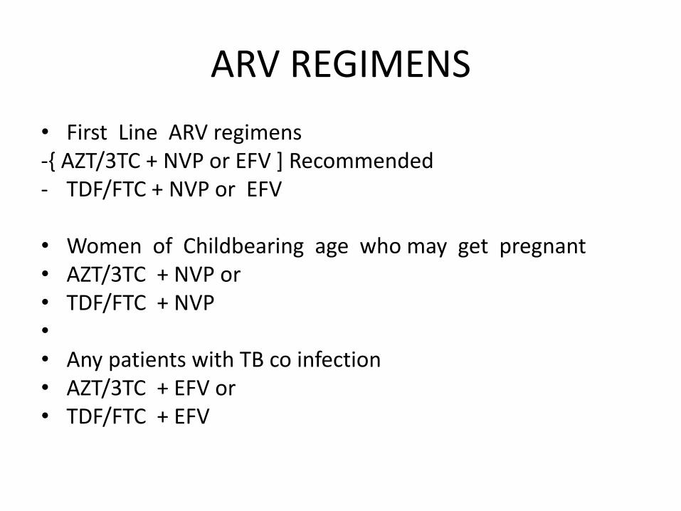
ARV REGIMENS
• First Line ARV regimens -{ AZT/3TC + NVP or EFV ] Recommended- TDF/FTC + NVP or EFV
• Women of Childbearing age who may get pregnant • AZT/3TC + NVP or• TDF/FTC + NVP•
• Any patients with TB co infection • AZT/3TC + EFV or • TDF/FTC + EFV

ADDITIONAL TREATMENTS
• Prophylaxis is indicated for :
-pneumocystis jiroveci
-Toxoplasma
-Mycobacterium Avium complex
- Fungal infections : fluconazole to prevent candidal or cryptococcal infections at CD4< 50/uL
- Viral infections: oral ganciclovir for CMV in advanced AIDS

ADDITIONAL TREATMENTS
• Treatment of HIV lipodystrophy
• Suppressive therapy for HSV-2 : acyclovir
• Treatment of HIV associated diarrhoea.

TREATMENT OF HIV AND TB CO
INFECTION
Criteria Anti-TB treatment ART
Extrapulmonary TB
(Irrespective of CD4
count)
Start immediately Start ART as soon as TB
treatment is tolerated
(between 2 weeks and 2
months)
Pulmonary TB CD4 <
200/mm3
Start immediately Start ART as soon as TB
treatment is tolerated
(between 2 weeks and 2
months)
Pulmonary TBCD4
between 200-350/mm3
Start immediately Start ART after completion
of initial TB treatment
phase (if severely
compromised start earlier)
Pulmonary TBCD4
>350/mm3
Start immediately Monitor CD4
countConsider ART if CD4
cell count drops below
350/mmm3

Side effects of ARVS: NNRTIs
-Nevirapine can cause a severe skin rash. It has also been found to be hepatotoxic
-Efavirenz can cause CNS manifestations like mood alterations.
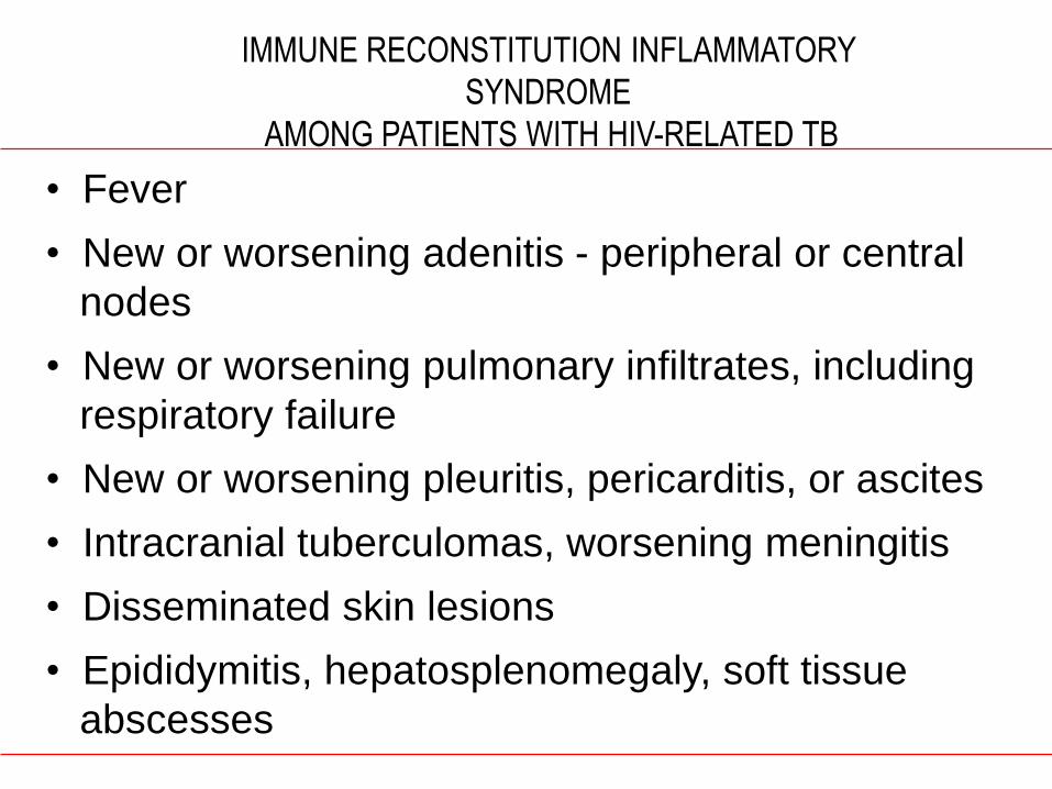
IMMUNE RECONSTITUTION INFLAMMATORY
SYNDROME
AMONG PATIENTS WITH HIV-RELATED TB
• Fever
• New or worsening adenitis - peripheral or central
nodes
• New or worsening pulmonary infiltrates, including
respiratory failure
• New or worsening pleuritis, pericarditis, or ascites
• Intracranial tuberculomas, worsening meningitis
• Disseminated skin lesions
• Epididymitis, hepatosplenomegaly, soft tissue
abscesses

NRTIs
• Abacavir causes a hypersensitivity reaction
• Stavudine- neuropathy
• Lamivudine- anaemia
• Didanosine- pancreatitis
• Lactic acidosis
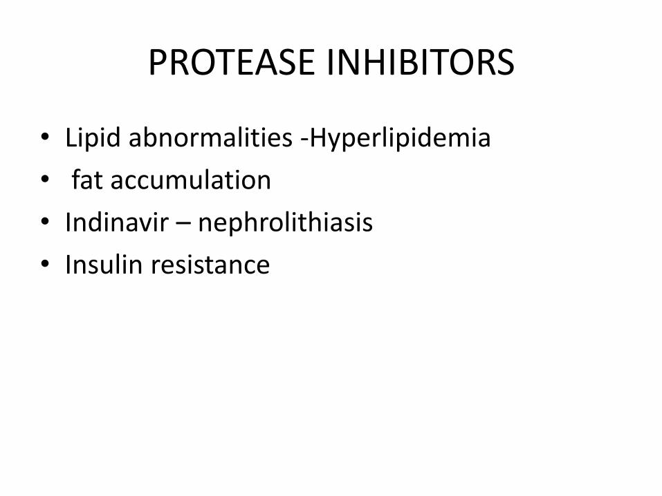
PROTEASE INHIBITORS
• Lipid abnormalities -Hyperlipidemia
• fat accumulation
• Indinavir – nephrolithiasis
• Insulin resistance

DRUG INTERACTIONS
• Rifampicin and Nevirapine both induce CYP3A4
• Avoid combination of Rifampicin and Nevirapine,replace with Efavirenz
• Rifabutin can be combined with Nevirapine
• Avoid combination of zidovudine and stavudine due to antagonism
• Also avoid tenofovir and didanosine due to antagonism

PREVENTION
• Education of risk groups
• Practising safer sex and abstinence
• Rigorous testing of donor blood
• Institution of measures to prevent MTCT(mother to child transmission) during pregnancy,labour,delivery & post partum period.
• Post exposure prophylaxis for health workers