HIV-1 tuberculosis-associated immune reconstitution ... · HIV-1 tuberculosis-associated immune...
14
REVIEW HIV-1 tuberculosis-associated immune reconstitution inflammatory syndrome Rachel P. J. Lai 1 & Graeme Meintjes 2,3,4 & Robert J. Wilkinson 1,2,3,4 Received: 1 July 2015 /Accepted: 21 September 2015 /Published online: 30 September 2015 # The Author(s) 2015. This article is published with open access at Springerlink.com Abstract Patients co-infected with HIV-1 and tuberculosis (TB) are at risk of developing TB-associated immune reconsti- tution inflammatory syndrome (TB-IRIS) following com- mencement of antiretroviral therapy (ART). TB-IRIS is charac- terized by transient but severe localized or systemic inflamma- tory reactions against Mycobacterium tuberculosis antigens. Here, we review the risk factors and clinical management of TB-IRIS, as well as the roles played by different aspects of the immune response in contributing to TB-IRIS pathogenesis. Keywords HIV-1 infection . Tuberculosis . Immune reconstitution inflammatory syndrome . Drug therapy-complications HIV-tuberculosis co-infection Human immunodeficiency virus type 1 (HIV-1) and Myco- bacterium tuberculosis are two major infectious diseases and each was responsible for causing approximately 1.5 million deaths in 2013 [1, 2]. There were an estimated 9.0 million cases of active tuberculosis (TB) in 2013, among which about 1.1 million (13 %) were HIV-1 co-infected [2]. The prevalence of HIV-1 co-infection among TB cases is the highest in Africa, accounting for around 78 % of the total HIV-TB cases. HIV-1 infection and active TB disease individually lead to severe immune impairment and they can also exacerbate the disease progression and pathology of one another. Tuberculo- sis can cause lymphocytopenia in the absence of HIV-1 or other cause of immunodeficiency [3, 4]. When CD4 + T cells counts were controlled for, HIV-1 patients with active TB disease still have more a rapid course of disease progression and poorer survival rate than those without active TB [5]. Furthermore, people living with HIV-1 are 26–31 times more likely to develop TB than HIV-uninfected individuals [6]. HIV-1 causes functional disruption of the immune response and impairs the host’ s ability to control M. tuberculosis infec- tion via several mechanisms (reviewed in [7]). Firstly, HIV-1 replication is higher in stimulated macrophages at the site of M. tuberculosis infection [8, 9], and higher viral loads in bron- choalveolar lavage and in pleural fluid have been observed [10, 11]. Secondly, HIV-1-infected macrophages have reduced secretion of TNF-α that associates with reduced apoptosis in response to M. tuberculosis [12, 13]. While expression of TLR2 and TLR4 are preserved in HIV-1-infected macro- phages, TNF-α secretion in response to specific TLR ago- nists, as well as signal transduction through IRAK-1 and NF-κB nuclear translocation, are significantly reduced [13]. Finally, HIV-1 infects and depletes CD4 + T cells, including those that are M. tuberculosis-specific, resulting in decreased production of IFN-γ, TNF-α and other cytokines that are im- portant in controlling both pathogens [14–18]. Several interventions have been implemented to address the syndemics of HIV-1 and TB. These include routine HIV This article is a contribution to the Special Issue on Immunopathology of Mycobacterial Diseases - Guest Editor: Stefan Kaufmann * Robert J. Wilkinson [email protected] 1 The Francis Crick Institute, Mill Hill Laboratory, London NW7 1AA, UK 2 Institute of Infectious Disease and Molecular Medicine, Faculty of Health Sciences, University of Cape Town, Room 3.03.05, Wolfson Pavilion, Anzio Road, Observatory, Cape Town 7925, South Africa 3 Department of Medicine, Imperial College London, London W2 1PG, UK 4 Department of Medicine, University of Cape Town, Observatory, Cape Town 7925, South Africa Semin Immunopathol (2016) 38:185–198 DOI 10.1007/s00281-015-0532-2
Transcript of HIV-1 tuberculosis-associated immune reconstitution ... · HIV-1 tuberculosis-associated immune...

REVIEW
HIV-1 tuberculosis-associated immune reconstitutioninflammatory syndrome
Rachel P. J. Lai1 & Graeme Meintjes2,3,4 & Robert J. Wilkinson1,2,3,4
Received: 1 July 2015 /Accepted: 21 September 2015 /Published online: 30 September 2015# The Author(s) 2015. This article is published with open access at Springerlink.com
Abstract Patients co-infected with HIV-1 and tuberculosis(TB) are at risk of developing TB-associated immune reconsti-tution inflammatory syndrome (TB-IRIS) following com-mencement of antiretroviral therapy (ART). TB-IRIS is charac-terized by transient but severe localized or systemic inflamma-tory reactions against Mycobacterium tuberculosis antigens.Here, we review the risk factors and clinical management ofTB-IRIS, as well as the roles played by different aspects of theimmune response in contributing to TB-IRIS pathogenesis.
Keywords HIV-1 infection . Tuberculosis . Immunereconstitution inflammatory syndrome . Drugtherapy-complications
HIV-tuberculosis co-infection
Human immunodeficiency virus type 1 (HIV-1) and Myco-bacterium tuberculosis are two major infectious diseases and
each was responsible for causing approximately 1.5 milliondeaths in 2013 [1, 2]. There were an estimated 9.0 millioncases of active tuberculosis (TB) in 2013, among which about1.1million (13%)were HIV-1 co-infected [2]. The prevalenceof HIV-1 co-infection among TB cases is the highest in Africa,accounting for around 78 % of the total HIV-TB cases.
HIV-1 infection and active TB disease individually lead tosevere immune impairment and they can also exacerbate thedisease progression and pathology of one another. Tuberculo-sis can cause lymphocytopenia in the absence of HIV-1 orother cause of immunodeficiency [3, 4]. When CD4+ T cellscounts were controlled for, HIV-1 patients with active TBdisease still have more a rapid course of disease progressionand poorer survival rate than those without active TB [5].Furthermore, people living with HIV-1 are 26–31 times morelikely to develop TB than HIV-uninfected individuals [6].
HIV-1 causes functional disruption of the immune responseand impairs the host’s ability to controlM. tuberculosis infec-tion via several mechanisms (reviewed in [7]). Firstly, HIV-1replication is higher in stimulated macrophages at the site ofM. tuberculosis infection [8, 9], and higher viral loads in bron-choalveolar lavage and in pleural fluid have been observed[10, 11]. Secondly, HIV-1-infected macrophages have reducedsecretion of TNF-α that associates with reduced apoptosis inresponse to M. tuberculosis [12, 13]. While expression ofTLR2 and TLR4 are preserved in HIV-1-infected macro-phages, TNF-α secretion in response to specific TLR ago-nists, as well as signal transduction through IRAK-1 andNF-κB nuclear translocation, are significantly reduced [13].Finally, HIV-1 infects and depletes CD4+ T cells, includingthose that are M. tuberculosis-specific, resulting in decreasedproduction of IFN-γ, TNF-α and other cytokines that are im-portant in controlling both pathogens [14–18].
Several interventions have been implemented to addressthe syndemics of HIV-1 and TB. These include routine HIV
This article is a contribution to the Special Issue on Immunopathology ofMycobacterial Diseases - Guest Editor: Stefan Kaufmann
* Robert J. [email protected]
1 The Francis Crick Institute,Mill Hill Laboratory, LondonNW7 1AA,UK
2 Institute of Infectious Disease and Molecular Medicine, Faculty ofHealth Sciences, University of Cape Town, Room 3.03.05, WolfsonPavilion, Anzio Road, Observatory, Cape Town 7925, South Africa
3 Department of Medicine, Imperial College London, London W21PG, UK
4 Department of Medicine, University of Cape Town, Observatory,Cape Town 7925, South Africa
Semin Immunopathol (2016) 38:185–198DOI 10.1007/s00281-015-0532-2
testing among TB patients, increased coverage of co-trimoxazole and isoniazid preventive therapy in HIV-1-infected TB patients and earlier diagnosis of HIVand initiationof antiretroviral therapy (ART) [2]. The increased accessibilityof ART has significantly improved the clinical outcome ofHIV-1 infected patients and reduces the TB risk by 58–80 %[19]. The positive effects of ART have been described astriphasic, starting with an early and rapid rise of CD45RO+
memory T cells that redistribute from lymphoid tissues intothe plasma within the first month of therapy, followed by areduction in T cell activation and improved CD4+ reactivity torecall HIV-1 antigens, and finally the slower replenishment ofthe CD45RA+ naïve Tcells [20]. In a longitudinal study of co-infected patients sensitized byM. tuberculosis, ART led to anincrease in central memory (CD27+CD45RA− andCD27+CCR5−CD4+) T cells by 12 weeks post-treatment,followed by an increase in naïve (CD27+CD45RA+) T cellsat 36 weeks [21]. A separate longitudinal study confirmed thatthe polyfunctional effector memory (CD27−CD45RO+) andterminally differentiated memory (CD27−CD45RO−) CD4+
T cells responsive to mycobacterial purified protein derivative(PPD) were restored 12 months post-ART [22].
Immune reconstitution inflammatory syndrome
Although ART has positive effects through suppression ofHIV-1 viral load and restoring CD4+ Tcell numbers, this rapidrestoration of the immune system can also lead to an undesir-able complication known as immune reconstitution inflamma-tory syndrome (IRIS; also known as immune restoration dis-ease, IRD). IRIS is characterized by a transient but sometimessevere local and systemic inflammatory response directedagainst a known condition (e.g., opportunistic pathogens orautoimmune diseases) in HIV-1 infected patients shortly afterART initiation. It was first reported in 1992, where HIV-1 co-infected patients developed Mycobacterium avium-complex(MAC) disease with severe lymphadenopathy and high feverfollowing commencement of zidovudine monotherapy [23].IRIS was subsequently documented to be associated withmany different pathogens, including cytomegalovirus(CMV), hepatitis B and C viruses, Cryptococcus, andM. tuberculosis, as well as in cancer (Kaposi’s sarcoma andnon-Hodgkin’s lymphoma) and autoimmune diseases [24].
In a meta-analysis on 54 cohort studies, Muller et al.reviewed the incidence and mortality of IRIS associated withdifferent health conditions [25]. CMV-IRIS was found to havethe highest incidence (pooled estimates 37.7 %), while cryp-tococcal meningitis-IRIS had the highest mortality (pooledestimates 20.8 %). More recently, a meta-analysis on 40 stud-ies indicates the overall incidence of IRIS is ~18 % amongpatients with HIV-associated TB, withmortality attributable toTB-IRIS of ~2 % [26]. Patients with TB meningitis (TBM),
however, are at high risk of developing IRIS (47 %), with amortality rate of up to 30 % following development of IRIS[27–29].
Clinical definition of TB-IRIS
There is no diagnostic test for TB-IRIS and diagnosis there-fore relies on medical history, laboratory data, and clinicalpresentation. Prior to 2006, a number of TB-IRIS case defini-tions existed [30–34], but the lack of consensus between thesedefinitions hindered clinical management and research onIRIS. In response to the increase in incidence of this compli-cation in resource-limited settings, over 100 researchers gath-ered to agree consensus case definitions for ART-associatedTB and TB-IRIS [35], which have several times been re-investigated prospectively and validated [36–39].
There are two forms of TB-IRIS that share clinical featuresbut are distinct in their temporal relationships between tuber-culosis diagnosis and treatment and the initiation of ART:paradoxical and unmasking. In paradoxical TB-IRIS, HIV-1-patients are diagnosed with TB prior to ART commencementand TB pathology has stabilized or improved during antitu-bercular therapy. Following ART initiation, patients experi-ence new, recurrent or worsening features of TB, such aslymph node swelling and abscess formation, serositis, andradiographic deterioration [35]. In unmasking TB-IRIS, pa-tients with previously undiagnosed and untreated TB presentafter initiation of ART and with often marked inflammatoryfeatures of TB [35]. Exclusion criteria for both types of TB-IRIS include exacerbation of TB due to drug-resistantM. tuberculosis, presence of other opportunistic infections,poor drug adherence, or adverse drug reactions.
Risk factors for TB-IRIS
Although the underlying mechanisms of TB-IRIS pathogene-sis are incompletely elucidated, A few clinical risk factorshave been related to developing TB-IRIS. Firstly, patientswith low CD4+ T cell counts at the time of ART initiation,followed by a rapid increase of CD4 counts post-ART, aremore likely to develop TB-IRIS [40–43]. This contributes toan exaggerated T cell response directed to M. tuberculosisalong with overproduction of both pro- and anti-inflammatory cytokines [44]. The second risk factor is a shortinterval between starting antitubercular therapy and ART[45–47]. The CD4 counts should be taken into considerationwhen deciding the optimal time to initiate ART. For patientswith low CD4+ T cell counts (<50 cells/μl), the benefits ofearly initiation of ART in reducingmortality and opportunisticinfections outweigh the risk of IRIS and therefore ARTshouldnot be delayed more than 2 weeks. For those with CD4 counts>50 cells/μl, ART should be commenced between 2 and 12weeks after starting TB treatment. Furthermore, dissemination
186 Semin Immunopathol (2016) 38:185–198

of TB infection to extrapulmonary organs appears to increasethe risk of TB-IRIS by up to eight-fold, probably due to higherbacterial burden in such cases [48, 49]. TBM is the mostsevere form of extrapulmonary TB and in one seriesaccounted for 12 % of all TB-IRIS cases [27]. Finally, highHIV-1 viral load is another risk factor for TB-IRIS [26] andM. tuberculosis culture positivity in the cerebrospinal fluid(CSF) is a risk factor for TBM-IRIS [27].
Management
The inflammatory response associated with TB-IRIS can betransient and may resolve without specific intervention. How-ever, in many cases there is marked inflammation that, atworst, may result in death. Anecdotal reports of glucocorti-coids to reduce inflammation in TB-IRIS [50–52] encourageda randomized placebo-controlled trial to evaluate TB-IRIStreatment with prednisone [53]. Patients with paradoxicalTB-IRIS who received a 4-week course of prednisone(1.5 mg/kg per day for 2 weeks then 0.75 mg/kg per day for2 weeks) were found to have reduced need for hospitalization,quicker resolution of symptoms and a better quality of lifewithout excess of severe adverse events, compared to thosewho received placebo [53]. The beneficial effects of predni-sone in reducing the inflammatory reactions in TB-IRIS wereassociated with more rapid resolution of the elevated C-reactiveprotein (CRP) and suppression of pro-inflammatory cytokines
of innate immune origin [54]. Nevertheless, prednisone shouldbe prescribed and monitored with caution in patients with ad-vanced HIV-1 infection as (albeit outside the context of wide-spread ART) it associates in some studies with an increase inthe risk of HIV-1 related malignancy (such as Kaposi’s sarco-ma) and recurrent herpes zoster [55, 56]. Furthermore, as TB-IRIS may occur in patients with drug-resistantM. tuberculosis,prednisone therapy should be used with care pending satisfac-tory optimization of antimicrobial therapy [57].
In addition to glucocorticoids, other anti-inflammatoryagents have also been used to treat TB-IRIS or IRIS associatedwith other pathogens in isolated cases (Table 1). Non-steroidalanti-inflammatory drugs (NSAIDs) have been used to relievesymptoms in non-severe cases of paradoxical TB-IRIS,MAC-IRIS, and cryptococcal-IRIS with lymphadenitis withfavorable results [58]. Thalidomide is an immunomodulatorydrug with use in cancer and inflammatory diseases. It has beenused to treat patients with intractable tuberculomas [59, 60]and alleviated symptoms in steroid-refractory TB-IRIS, TB-and cryptococcal-lymphadenitis IRIS, as well as in pediatricneurological TB-IRIS [61–64]. Leukotriene antagonists (e.g.,montelukast and zileuton) are used in treatment in asthma andmost recently have been proposed as potential host-directedtherapy in active TB [65]. Montelukast has been used to treattwo cases of steroid-refractory IRIS associated with TB andsyphilis and also in a patient with urticarial IRIS vasculitis,with rapid clinical responses [66, 67]. Other drugs such as
Table 1 Drugs used to treat TB-IRIS
Drug/class of drugs Mechanism of action Potential side effects
Corticosteroids At gene level, activate the transcription of anti-inflammatorymediators and inhibit transcription of pro-inflammatorygenes (e.g., cyclooxygenase and cytokines). At cellularlevel, reduce the production of nitric oxide and inhibitsTCR signaling, thereby reducing cell migration,proliferation and effector function
Increased risk of Kaposi’s sarcoma, herpes simplexand zoster flare. General side effects includeincreased risk of infections, hypertension, diabetes,osteoporosis, ulcers and mental health problems
NSAIDs Relieve pain and reduce inflammation by inhibitingcyclooxygenase-1 and −2, thereby reducing the synthesisof inflammation mediator prostaglandins
Increased risk of gastrointestinal problems such asulcers; not recommended in patients with a historyof renal or liver disease
Thalidomide Modulates the production of cytokines and inflammatorymediators; also stimulates T cells and modulates NKcell cytotoxicity
Peripheral neuropathy, somnolence, hepatotoxicity,teratogenicity, skin reactions, constipation, tremor,mood changes and headache
Leukotriene receptorantagonists
Blocks pro-inflammatory leukotrienes by inhibiting the5-lipoxygenase pathway or by antagonizingcysteinyl-leukotriene type 1 receptors; inhibit leukocytestrafficking to the sites of antigen stimulation
Skin reaction, sinus pain, tremors, mood changes,gastrointestinal problems; may also interact withrifampicin and antiretroviral drugs
Pentoxifylline A non-selective adenosine receptor antagonist and alsonon-selectively inhibits phosphodiesterase, resulting in anincrease in cAMP activity and reduced inflammation. Inaddition, it improves erthyocytes deformability, decreasesblood viscosity and inhibits neutrophil adhesion andactivation
Hemorrhage, gastrointestinal problems, nausea,dizziness, blurred vision, flushing and chest pain
Hydroxychloroquine Blocks activation through TLR and interferes with MHC-IIprocessing; reduce synthesis of pro-inflammatory cytokines
Blurred vision, somnolence, gastrointestinal problems,skin rash and lost of appetite
Drugs with reported use in treating TB-IRIS, their mechanism of actions and side effects are listed
Semin Immunopathol (2016) 38:185–198 187
pentoxifylline and hydroxychloroquine had also been used totreat isolated cases of IRIS associated with different pathogenswith some reported benefits [68–71]. Nevertheless, these ther-apies lack clinical trial data to assess their effectiveness andpotential adverse effects in treating TB-IRIS and further rig-orous evaluations are required.
Finally, TNF-α inhibitors (infliximab, etanercept,adalimumab ,or certolizumab pegol) are effective in treatinginflammation in rheumatoid arthritis, spondyloarthropathies,and Crohn’s disease. While TNF-α inhibitors are to be pre-scribed with caution in patients with latent TB due to in-creased risk of reactivation, in one case withdrawal ofadalimumab led to a life-threatening paradoxical TB reactionand the symptoms resolved when the patient was restarted onadalimumab [72]. In another case study of steroid-refractoryTBM, the patient improved upon adalimumab therapy [73].Similar observations have been reported with another TNF-αinhibitor infliximab, where the patient suffered a debilitatingTB paradoxical reaction unresponsive to corticosteroids andcyclophosphamide yet showed a favorable response followingthree doses of infliximab given at monthly intervals [74]. In asingle case study with HIV-related inflammatory cerebralcryptococcoma, patient had improved disease symptoms fol-lowing treatment with adalimumab along with isoniazid andpyridoxine (to prevent tuberculosis reactivation) [75]. Sinceelevated levels of TNF-α are associated with TB-IRIS [76,77], TNF-α inhibitors may offer potential benefits as treat-ment for TB-IRIS.
Inflammatory reactions in TB-IRIS
TB-IRIS results from excessive inflammatory reactionsagainstM. tuberculosis antigen driven by ART-induced recon-stitution of the immune system [44, 78]. Numerous reportshave described aspects of the immune responses observed inTB-IRIS, which we will discuss in detail in the followingsections.
CD4+ T cells and TCRγδ T cells
Paradoxical TB-IRIS was initially ascribed to acute expansionof the mycobacteria-specific Th1 response following ARTcommencement [44]. The first patient cohort study on TB-IRIS consisted of 19 HIV-TB patients, 7 of whom developedIRIS during the first month of ART. Compared to the non-IRIS patients at 3 months, those with TB-IRIS were found tohave significantly higher number of IFN-γ-producing Th1cells specific tomycobacterial PPD, but not toM. tuberculosis-specific ESAT antigen or CMV [44]. Furthermore, peripheralblood mononuclear cells (PBMC) from a subset of four pa-tients (three TB-IRIS and one non-IRIS) were stimulated withPPD antigen and those with TB-IRIS secreted significantly
higher concentrations of Th1 (IL-2, IL-12, IFN-γ, and IP-10) and other (TNF-α, IL-1β, IL-6, IL-10, RANTES, andMCP-1) cytokines and chemokines, while Th2 cytokines(IL-4, IL-5, IL-13, and IL15) were not detected [44]. Thepresence of M. tuberculosis antigen-specific Th1 expansionsin TB-IRIS was confirmed in a separate study of 95 HIV-TBpatients (35 TB-IRIS, 29 non-IRIS and 31 ART-naïve) [79].However, not all cases of TB-IRIS were characterized by Th1expansions and conversely expansions occurred longitudinal-ly duringART inmany patients who did not develop TB-IRIS.The proportion of HLA-DR+CD71+ CD4+ and CD8+ T cellswas similar in TB-IRIS and non-IRIS controls [79]. Two ad-ditional studies by Tieu et al. [80] and Elliott et al. [81] utilizedinterferon-gamma release assay (IGRA) and did not find anydifference in IFN-γ response in whole blood between patientswith paradoxical TB-IRIS and the non-IRIS controls 12weekspost-ART, although the latter study observed an increase inIFN-γ response to PPD 24 weeks post-ART.
Since IRIS commonly occurs in patients who are severelylymphopenic following ART initiation, Sereti and colleaguesproposed that the syndrome is contributed by lymphopenia-induced T cell homeostatic mechanisms, and an imbalance inthe regulatory mechanisms occurs irrespective of antigenstimulus [82]. In a longitudinal study of 45 HIV-1-infectedpatients commencing ART, 16 developed IRIS associatedwith different etiological causes and were found to have ahighly activated, predominantly PD-1+ HLA-DR+ and Ki67+
CD4+ T cell phenotype prior to and during IRIS, compared tothe non-IRIS controls [82]. Moreover, these IRIS patients alsohad a Th1/Th17-skewed cytokine profile upon polyclonalstimulation and increased PD-1 and Ki67 expression in regu-latory T cells (Treg), suggesting increased activation of effec-tor T cells due to antigenic exposure. Furthermore, TB-IRISpatients were found to have a significantly higher serum con-centration of IL-7 and sCD25 compared to the non-IRIS con-trols [82, 83], further indicating that TB-IRIS is associatedwith exaggerated T cell activation and proliferation. In afollow-up study, it was noted that patients with IRIS relatedto different pathogens did not have a generalized T cell dys-function and the dysregulated CD4+ Tcell response represent-ed an upsurge of pre-existing responses that were bothpolyfunctional and targeted exclusively to the opportunisticpathogen, not to HIV-1 itself [84].
In addition to Th1, other T cell subsets have also beeninvestigated for their potential contribution to TB-IRIS. Asmentioned above, Foxp3+ CD4+ T cells (considered as Treg)cells are critical in maintaining T cell homeostasis [85]. Inmice infected with TB, Treg cells expanded and accumulatedat the site of infection and contribute to suppression of Th1-typeimmune responses [86]. Increased circulating Treg frequencywas also reported in TB patients, in which blood CD4+ T cellswere found to have increased cell surface expression ofCD25high and increased mRNA expression of Foxp3 [87].
188 Semin Immunopathol (2016) 38:185–198
Several studies have investigated whether a dysregulated Tregresponse may contribute to TB-IRIS and reported conflictingresults. Two of these studies found an expansion of Foxp3+
Tregs in M. avium and CMV-associated IRIS, although theability of these cells to secrete IL-10 appeared to be compro-mised in vitro [88, 89]. Conversely Meintjes et al. reported anoverall low level of Foxp3+ T cells among PBMC, with nodifference in the percentage of these cells in TB-IRIS andnon-IRIS patients [79]. Similar observations were also reportedbyAntonelli et al.: neither the number nor the frequency of Tregdiffered when comparing IRIS to non-IRIS patients [82].
The role of TCRγδ T cells in TB-IRIS has also been ex-plored. TCRγδ T cells are potent producers of IFN-γ andTNF-α, possess cytotoxic capacity and release granule-associated perforin and granzyme B to lyse infected cells uponantigen recognition [90]. They are activated in response toM. tuberculosis early after infection and may reduce intracel-lular bacterial growth [91]. The Vδ2+TCRγδ+ T cell subsetare ascribed a role in adaptive immunity to mycobacterialinfection [92, 93] and their numbers are restored followingsuccessful antitubercular treatment in HIV-1 infected persons[94]. Patients co-infected with HIV-1 and M. tuberculosis,however, have significantly reduced TCRγδ T cell responses[91]. Compared to non-IRIS patients, those with paradoxicalTB-IRIS were found to have significantly higher number ofkiller immunoglobulin receptor (KIR)−Vδ2+TCRγδ+ T cellsbut a lower number of KIR+TCRγδ+ T cells at all timepointsincluding baseline [95]. This observation indicates indepen-dent regulation of different TCRγδ T cell subsets during IRISdevelopment, with the suggestion that the KIR−Vδ2+TCRγδ+
subset amplifies the dysregulated immune response toM. tuberculosis, whereas the inhibitory KIR+Vδ2+TCRγδ+
subset was suppressed.Together, these data demonstrate a role for CD4+ T cells
and other T cell subsets in the dysregulated inflammatory re-sponse in TB-IRIS. Nevertheless, as some TB-IRIS cases arenot associated with T cell expansion, it remains conjecturalwhether the T cell response is the cause, rather than the con-sequence, of the syndrome.
CD8+ T cells
Compared to CD4+ T cells, there have been very limited stud-ies on the role of CD8+ T cells in TB-IRIS pathogenesis. Twostudies showed that the numbers and frequency of CD8+ Tcells did not differ between TB-IRIS and non-IRIS patientsprior to ART and during IRIS [35, 96]. In contrast, Reyes-Teran and colleagues reported that increased frequency of cir-culating CD8+ T cells is a risk factor for developing IRISassociated with M. tuberculosis and M. avium [97]. Further-more, the authors reported an expansion of the naïve CD8+
subpopulation, as well as the CD38+ HLA-DR+ CD8+ Tcells,during TB-IRIS episode [97]. No further phenotyping of the
CD8+ T cells in the study was performed, thus the precisesubpopulation associated with TB-IRIS development wasnot defined. Furthermore, the functionality of CD8+ T cellshas not been investigated in the context of TB-IRIS and theircontribution to the syndrome remains unclear.
Soluble mediators
A feature of TB-IRIS is increased production of both pro- andanti-inflammatory cytokines, which is in turn influenced bythe antigen load in patients [98, 99]. In the study by Tadokeraet al., PBMC from paradoxical TB-IRIS patients stimulatedwith heat-killed M. tuberculosis secreted significantly higherconcentrations of IL-1β, IL-2, IL-6, IL-8, IL-10, IL-12p40,IFN-γ, GM-CSF, and TNF-α, compared to those from thenon-IRIS controls [77]. Higher concentrations of IL-6, IL-8,IL-10, IL-12p40, IFN-γ, and TNF-αwere also detected in theserum of the same TB-IRIS patients. Similar observationshave been reported by others [100–102]. In a study comparingTB-IRIS patients randomized to prednisone or placebo, theserum concentration of IL-6, IL-10, IL-12p40, IFN-γ, IP-10,and TNF-α decreased during 4 weeks of prednisone therapy,but not in the placebo group [54], further implying a patho-logical role for hypercytokinemia in TB-IRIS. IL-6 has beensuggested to be a major pathological mediator in TB-IRIS as ahigher level of plasma IL-6 and CRP at baseline was associ-ated with subsequent development of TB-IRIS [103]. Barberet al. assessed whether inhibition of IL-6 can reduce pathologyin a murine model with M. avium-associated IRIS (seeBAnimal model^ section) and showed that neutralization ofIL-6 with a monoclonal antibody reduced CRP levels, allevi-ated disease pathology and extended survival [104]. Most re-cently, Ravimohan et al. reported that inflammatory markersincreased rapidly in HIV-TB patients with early deaths and inpatients who developed TB-IRIS, but the two groups havedifferential recovery of the adaptive immune system [76]. Pa-tients with early mortality were found to have increased pre-ART plasma concentrations of IL-6, IL-10, TNF-α, MCP-1,and eotaxin, but these increases were not accompanied byearly recovery of CD4+ T cells. In contrast, the significantincrease in the plasma concentrations of IL-6 and TNF-α fol-lowing ART initiation paralleled a marked increase in CD4counts in TB-IRIS patients. Together, the CD4 counts prior toand following ART, along with the concentrations of inflam-matory markers such as IL-6 and TNF-αmight have potentialuse as biomarkers to assist TB-IRIS diagnosis.
Furthermore, plasma or serum concentrations of IL-17 andIL-22 have also been reported to be higher in both paradoxicaland unmasking TB-IRIS [100, 105]. Both IL-17 and IL-22arise from distinct lineages of T cells and are critical in bridg-ing innate and adaptive immunity in host defense against path-ogens at mucosal sites (reviewed in [106]). More recently,elevation of plasma IL-18 at baseline (pre-ART) and during
Semin Immunopathol (2016) 38:185–198 189
TB-IRIS has also been reported [107, 108]. In addition to theexcessive release of both pro- and anti-inflammatory cyto-kines, IP-10 and CCL4 chemokines were also found to beelevated in the plasma of TB-IRIS patients longitudinally overthe course of ART [107, 109]. Similar findings were reportedin patients with TBM-IRIS, where the concentrations of anarray of chemokines (CXCL1-3, CCL2/3/4, and IP-10) wereall significantly higher in the CSF of TBM-IRIS patients com-pared to non-IRIS TBM patients [110]. The induction of thesechemokines suggests an increased chemoattraction ofmonocytes/macrophages and other immune cells to the siteof inflammation, thereby contributing to the pathogenesis ofTB-IRIS.
Other inflammatory mediators have also been reported tobe elevated in TB-IRIS.
Matrix metalloproteinases (MMPs) are zinc-dependent en-dopeptidases involved in tissue repair, remodeling and modu-lation of the immune response [111, 112]. The concentrationof MMP-1 was significantly higher in the lung of TB patientswith or without HIV-1 infection [113]. In the context of TB-IRIS, PBMC from paradoxical TB-IRIS patients were foundto have significantly higher mRNA expression and proteinsecretion of MMP-1, -3, -7 and -10 than in non-IRIS controls[114]. Following prednisone therapy, the serum concentrationof MMP-7 showed a modest reduction, suggesting a specificcontribution to pathogenesis. Similarly, in the CSF of TBM-IRIS patients, the concentration of MMP-9 was significantlyhigher compared to non-IRIS TBMpatients both pre-ARTandpost-ART [110]. However, the CSF concentration of MMP-9did not decrease following antitubercular and corticosteroidtherapy and continued to rise following ART initiation. This,together with the poor clinical outcomes, suggests that morepotent and specific therapy may be needed for the manage-ment of TBM-IRIS.
Finally, deficiency of vitamin D3 is associated with activeTB disease [115]. Adjunctive vitamin D3 supplementation canhelp resolve TB pathology by suppressing antigen-stimulatedpro-inflammatory response and inhibiting secretion of MMP-1, -7, -9 and -10 [116, 117]. While deficient plasma levels ofvitamin D3 did not predict the development of TB-IRIS [118],serum concentrations of IL-1β, IL-6, and IL-8 were signifi-cantly higher at both baseline and during TB-IRIS in vitaminD3-deficient patients who experienced the syndrome [109].
In patients with TBM, concentrations of inflammatory me-diators were significantly higher in the CSF than in bloodregardless of IRIS status [110]. Individuals who eventuallydeveloped TBM-IRIS had elevated CSF concentrations ofan array of cytokines, chemokines, MMPs, as well asneutrophil-associated mediators (e.g., S100A8/A9) at bothbaseline and the time of symptom presentation [110]. Com-pared to CSF, representative of the neurological compartment,only subtle differences in the concentrations of these analyteswere detected in the corresponding blood samples between
TBM-IRIS and non-IRIS. Further studies conducted with ma-terials from IRIS disease sites are likely to be informative.
The potential genetic influence in IRIS predisposition hasbeen investigated in two studies by Price and colleagues.While the small sample size rendered these studies underpow-ered and inconclusive, they nevertheless provided somepointers that genetic factors might play a role in TB-IRIS. Ina study that included nine patients with MAC-associated IRISand two with TB-IRIS, the frequency of the IL6-174*C poly-morphism (36%) was significantly lower than in the non-IRIScontrols (61–71 %) [119]. Furthermore, none of the IRIS pa-tients carried the TNFA-308*2 polymorphism, while the fre-quency was 23–52 % in the non-IRIS group. In a cohort studywith 17 Cambodian and 19 Indian patients, TB-IRIS was as-sociated with higher frequencies of TNFA-1031*T andSLC11A1 D543*G in Cambodian patients, while higher fre-quencies of IL18-607*G andVDR Fokl(F/f)*Twere observedin Indian patients [120]. Larger-scale studies would be re-quired to determine with confidence if specific polymor-phisms relate to the risk of TB-IRIS, although recruitment tosuch a study might be difficult.
B cell response
TB-IRIS is regarded as a cell-mediated disease and little isknown about the role played by humoral immunity.Simmoney et al. examined longitudinal antibody responsesin a cohort of 24 HIV-TB co-infected patients starting onART by measuring circulating free and immune-complexedantibodies against M. tuberculosis antigens (ManLAM,ESAT-6/CFP10, and PGL-Tb1) [121]. Compared to non-IRIS patients, those with TB-IRIS had a significantly lowerlevel of anti-PGL-Tb1 antibody prior to the episode regardlessof CD4 counts or presence of complexed antibodies. Therewas no difference in the antibody levels against ManLAM orESAT-6/CFP-10 between TB-IRIS and non-IRIS. A similarresult was also reported in another study where the levels ofanti-PPD, anti-ManLAM, and anti-38-kDa antigen antibodiesdid not differ between TB-IRIS and non-IRIS patients prior toART, or at the time of IRIS [122]. Furthermore, the lineage ofM. tuberculosis did not appear to be associated with develop-ment of TB-IRIS [121], although this interesting issue is notresolved with certainty.
More recently, a regulatory role for B cells has been de-scribed in TB granulomas in primates, which is mediated viasecretion of IL-10 and M. tuberculosis-specific antibodies[123]. Antibodies contribute to immune defense through threemechanisms: neutralization, activation of the complement sys-tem, or opsonization. In opsonization, phagocytes expressingFcγ receptors recognize antibody-bound antigens (e.g.M. tuberculosis) and take up the complex into the phagosomesfor elimination. Increased expression of Fcγ receptors(FCGR1/2/3) has been reported in almost all transcriptional
190 Semin Immunopathol (2016) 38:185–198
studies on active TB disease [124] and was also identified inmicroarray profiling of TB-IRIS patients during the IRIS ep-isode [125]. Together, these data suggest that humoral immu-nity may also contribute to TB-IRIS pathogenesis, althoughthe precise mechanisms remain poorly defined.
Innate immune response
The role of innate immunity in TB-IRIS pathogenesis hasattracted increased attention in recent years. Myeloid cellsare the main cells targeted byM. tuberculosis and their pivotalrole in antimicrobial defense has been extensively described.Colebunders and colleagues speculated that macrophages inHIV-1 patients with advanced disease, particularly those co-infected with M. tuberculosis, are inappropriately activated[126]. Previous studies have reported that stimulation ofHIV-1 gp120 alone is sufficient to induce dysfunction andaberrant gene expression in monocytes or monocytes-derived macrophages [127–129]. During ART, these immu-nosuppressive phenotypes are reversed and the functional re-covery could result in excessive activation of the macrophagesby M. tuberculosis antigens [126], subsequently contributingto development of IRIS. Lawn et al. later reported a fatal caseof unmasking TB-IRIS, in which CD68+ macrophages wereidentified as the predominant inflammatory cells in postmor-tem staining of lung tissue sections [130].
Human monocytes can be categorized into three differentsubsets: classical CD14++CD16− and non-classicalCD14+CD16+ and CD14dimCD16+. The roles played by thesesubsets in TB-IRIS have recently been explored. PeripheralCD14+CD16+ monocytes were isolated from TB-IRIS andnon-IRIS patients and transcriptional profiling was performed.Genes associated with the complement system and patternrecognition receptors were found to be differentially abundantin TB-IRIS [131, 132]. The authors further investigated therole of complement in TB-IRIS and reported significantlyhigher levels of C1q and C1-inhibitor of the classical pathwayat baseline and an imbalance in their ratio during TB-IRISonset at 2 weeks post-ART. Activation of the complementsystem is known to trigger opsonization and killing of patho-gens as well as recruitment of inflammatory cells [133]. Inanother study that examined TB-IRIS patients from SouthIndia and South Africa, TB-IRIS was associated with in-creased plasma concentrations of sCD14, sCD163, and solu-ble tissue factor, all of which are markers of monocyte activa-tion [134]. Furthermore, patients with TB-IRIS lackedCD14dimCD16+ monocytes while the frequency ofCD14++CD16− monocytes were significantly higher than innon-IRIS patients. CD14++CD16− monocytes have high ex-pression of the activation marker CD163 and were found to beclosely associated with plasma levels of systemic pro-inflammatory markers CRP, IL-6, TNF-α, and soluble tissuefactor (CD142) prior to ART initiation and during TB-IRIS
[134]. Together, these data point towards a pathogenic role forboth classic and non-classical monocytes in TB-IRIS and themechanisms mediated by these cells should be investigatedfurther.
In addition to macrophages/monocytes, natural killer (NK)cells and invariant natural killer T (iNKT) cells may also con-tribute to TB-IRIS pathogenesis. Activated NK cells can pro-tect against M. tuberculosis either directly by lysing infectedmonocytes and antigen-specific Treg [135, 136], or indirectlyby restoring the frequency of M. tuberculosis-specificCD8+IFN-γ+ T cells [137]. Patients with unmasking TB-IRIS were found to have increased NK cell activation andplasma concentrations of CRP and IL-8, compared to non-IRIS or HIV-1-monoinfected controls [138]. In another cohortof 128 co-infected patients from Cambodia, baseline NK celldegranulation capacity was significantly higher in those whoeventually developed paradoxical TB-IRIS, but this differencewas abrogated following ART initiation, possibly due to reg-ulatory feedback mechanisms or the internalization of NKreceptors following ligand binding [139]. Finally, microarrayanalysis performed onM. tuberculosis-stimulated PBMC iso-lated from patients identified an overabundance of granzymeB and perforin transcripts in TB-IRIS, validated at the proteinlevel [96]. The increased secretion of perforin appeared to becontributed by an increased number of CD3+Vα24+ iNKTcells. Activation of CD1d-restricted iNKTcells has previouslybeen shown to protect mice fromM. tuberculosis infection viaa yet unknown mechanism [140]. Further studies are neededto address the role of cytotoxic cells in TB-IRIS.
Neutrophils also appear to have a significant role in thepathogenesis of the highly compartmentalized TBM-IRIS.Active TB disease in HIV-1 uninfected persons is character-ized by a type I interferon-inducible, neutrophil-driventranscriptomic signature [141]. Mice lacking IFN1 receptorwere protected from TB-induced death with reduced recruit-ment of neutrophils and inflammatory macrophages to the siteof infection [142]. In a cohort of HIV-1-infected South Africanpatients diagnosed with TBM, those who did not developTBM-IRIS but who were TB culture positive in their CSFshowed similar inflammatory response as the TBM-IRIS pa-tients both at the time of TBM diagnosis and 2 weeks post-ART [88]. However, TBM-IRIS was specifically associatedwith elevated neutrophil counts in CSF and increased expres-sion of the neutrophil mediators S100A8/9 [27, 110].
Most recently, we have employed transcriptomic profilingof whole blood to investigate potential mechanisms that un-derlie TB-IRIS pathogenesis. In a longitudinal study whoseendpoint was the development of TB-IRIS, we closely trackedresponses at various timepoints prior to and following ARTcommencement (within days of starting ART and up to themedian time of IRIS occurrence at 2 weeks). We identifiedtranscripts of TLR signaling and activation of inflammasomesto be prominent in the blood of TB-IRIS patients (Table 2)
Semin Immunopathol (2016) 38:185–198 191

[125]. Both TLR2 and TLR4 have been shown to recognizeM. tuberculosis antigen. Mice lacking TLR4 have reducedcapacity to eliminate M. tuberculosis from the lungs andsuccumbed to disease sooner than wild-type controls [143].A separate mouse study showed that in high-dose aerosolinfection, TLR2 is critical in mediating innate defense againstM. tuberculosis infection [144]. Furthermore, TLR2 was
previously reported to express at higher baseline levels inmyeloid dendritic cells and monocytes in patients with para-doxical and unmasking TB-IRIS, compared to non-IRIS, andits expression remained higher even at 24 weeks post-ARTinitiation [145]. This observation suggests that dysregulatedsignaling via TLR2 mediates the inflammatory reactions ob-served in TB-IRIS. Indeed, when we inhibited MyD88 (thedownstream adaptor of TLR2) in patient’s PBMC in vitro,secretion of pro-inflammatory cytokines was markedly re-duced specifically in TB-IRIS patients [125]. The productionof IL-1 in TB-IRIS appeared to be dependent on
Table 2 Canonical pathways associated with TB-IRIS
Top canonical pathways associatedwith TB-IRIS
p value Regulation
Inflammasome activation 2.01E−04 Up
Toll-like receptor signaling 2.01E−04 Up
Endothelin-1 signaling 3.84E−04 Up
Role of pattern recognition receptorsin bacteria and virus recognition
8.83E−04 Up
IL-1 signaling 8.83E−04 Up
Microarray profiling using whole blood from a cohort of TB-IRIS and TBnon-IRIS patients was performed and differentially abundant transcriptsassociated with TB-IRIS were identified. Functional analysis by Ingenu-ity Pathway Analysis (IPA) indicates that these differentially abundanttranscripts overrepresented innate signaling pathways, includinginflammasome activation, toll-like receptor signaling and IL-1 signaling,suggesting that innate immunity plays a significant role in TB-IRISpathogenesis
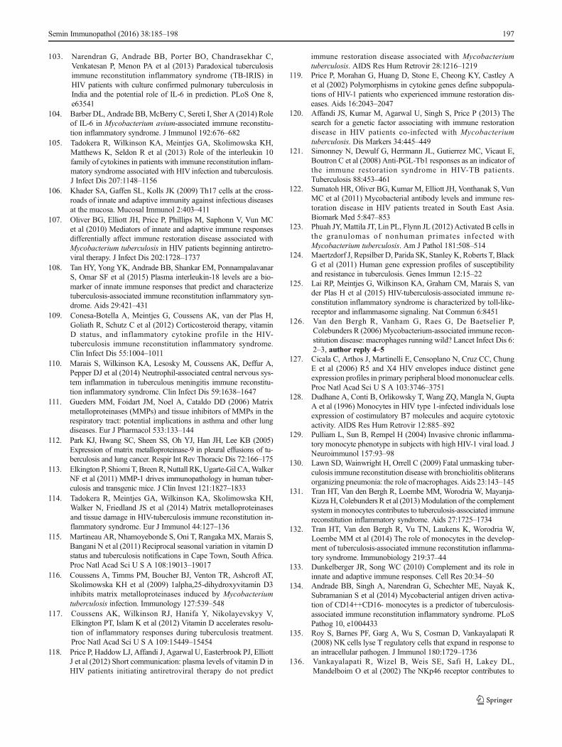
Fig. 1 Contrast-enhanced axial computed tomography (CT) image ofTBM-IRIS. CT image showing multiple ring-enhancing lesions withsurrounding edema and hydrocephalus in a 41-year-old woman. Thepatient previously presented with TBM (CSF TB culture was positivefor M. tuberculosis susceptible to rifampicin and isoniazid) 10 weeksprior to this presentation. At that time, she was started on TB treatmentand ART was initiated 9 weeks later. One week after initiating ART, thepatient developed recurrent headaches and this CT was performed2 weeks later. The recurrent symptoms and these CT findings wereascribed to TBM-IRIS and the patient was treated with corticosteroidswith symptom improvement. Image provided by Dr. Suzaan Marais
Fig. 2 A model of innate receptor signaling in mediating TB-IRIS path-ogenesis. Microarray profiling revealed that TLR signaling andinflammasome activation are critical in mediating TB-IRIS pathogenesis(Table 2) [125]. Our proposed model begins withM. tuberculosis antigenrecognition by surface-expressing TLRs, which triggers the downstreamsignaling cascade with adaptor molecules such as MyD88 and IRAK4 toactivate IRF7, thereby triggering the production of type I IFN. Paracrinesignaling of Type I IFN to IFNAR recruits and phosphorylates STAT1/2dimers, leading to further recruitment of IRF9 and the formation ofISGF3, thereby inducing pro-caspase-11 (caspase-4/5 in human) andAIM-2 inflammasome (caspase-1). Caspase-11 cleaves IL-1α into itsmature form and can lead to pyroptosis. The noncanonical inflammasome(caspase-11) can also activate the canonical inflammasome (caspase-1),which cleaves IL-1β and IL-18 into their mature form. Alternatively,TLR signaling via MyD88 can also activate NF-κb via the TAK1/IKKcomplex. Activation of NF-κb triggers the production of an array ofcytokines, including TNF-α, IL-6 and IL-12. In addition, NF-κb can alsoactivates NLRP1/3 inflammasomes and subsequently leads to the produc-tion of IL-1β and IL-18
192 Semin Immunopathol (2016) 38:185–198

inflammasome activation. In vitro inhibition of Caspase-1/4/5in PBMC fromTB-IRIS patients reduced IL-1 secretion, prob-ably due to disruption in the cleavage from immature to ma-ture form. Together, these data demonstrate the central roleplayed by innate receptor signaling in mediating TB-IRISpathogenesis and offer some mechanistic insights on the dis-ease (Figs. 1 and 2).
Animal model
Since HIV-1 does not readily infect non-primates, studyingmycobacteria-associated IRIS in small animal models posesa challenge. Thus far, only one murine model on IRIS hasbeen reported using a TCRα knockout mouse chronically in-fected M. avium infection [146]. To recapitulate the CD4+ Tcell reconstitution observed in TB-IRIS patients, purifiedCD4+ T cells from naïve mice were injected into T cell-deficient M. avium-infected TCRα knockout mice, whichled to impaired lung function, severe wasting symptoms andincreased mortality within 3 weeks of T cell transfer. Incontrast, transfer of CD4+ T cells into wild-type chroni-cally infected mice or TCRα knockout mice prior toM. avium infection did not cause any disease. Unexpect-edly, the nonlymphopenic OT-II mice were also found tobe susceptible to developing reconstitution disease in-duced by transfer of M. avium-specific CD4+ T cells, in-dicating that the reconstitution disease did not require alymphopenic environment. In addition, there were two- tofive-fold fewer donor CD4+ T cells recovered from infect-ed compared with naïve recipients. Instead of spontaneousproliferation of CD4+ T cells in T cell-deficient environ-ment, the model was associated with impaired, rather thanexaggerated, T cell expansion. Furthermore, both antigenrecognition and secretion of IFN-γ by the grafted CD4+ Tcells were necessary for disease induction, which correlat-ed with the large increase in the population of CD11b+
myeloid cells in the lungs and blood. While this modelrecapitulates the CD4 restoration characteristic in IRIS, itrequires a high-dose of M. avium infection leading to bac-terial burden that is very high. Furthermore, the modeldoes not capture the immunosuppressive phenotype ofmonocytes and macrophages induced by HIV-1. Severalhumanized mouse models have been developed for HIV-1, some of which are able to support persistent viral in-fection and gradual CD4+ T cells decline that mimics hu-man infection [147]. Non-human primates which shareclose physiology to humans have also been extensivelyused to study HIV-1 and TB infection, but not yet appliedto TB-IRIS. While these models may better recapitulatethe overall immune aspects of TB-IRIS, the high cost ofperforming such studies is a deterrent.
Concluding remarks
Although TB-IRIS was first described almost 20 years agowith many subsequent case reports, the underlying mecha-nisms that mediate the disease pathology remain to be fullyelucidated. There remains a lack of biomarkers to accuratelydiagnose and track the syndrome, particularly in resource-limited settings. The difficulty in studying the underlyingmechanisms of TB-IRIS pathogenesis is further complicatedby the absence of an animal model that can recapitulate dif-ferent aspects of the immune defects observed in TB-IRIS, aswell as challenges, both practically and ethically, to recruitappropriate controls groups: those who are infected only withHIV-1 or TB, treated and untreated with ART and antituber-cular drugs and healthy controls. While a set of consensusguidelines adopted in recent years has helped to standardizeTB-IRIS diagnosis and treatment for systematic comparison,our understanding on the pathogenesis remains incomplete.Finally, although corticosteroids such as prednisone canalleviate the symptoms and accelerate clinical improve-ment, they also have undesirable side effects and drugstargeting specific pathological pathways should be ex-plored. Our group and others have recently utilizedtranscriptomic approaches to study the underlying mech-anisms mediating TB-IRIS and the results from thesestudies offered insights on some of the immunologicalpathways involved that may guide future directions forhost-directed therapy.
Acknowledgments We would like to thank Dr. Suzaan Marais for pro-viding us with CT image on TBM-IRIS. This work was supported by theUK MRC (U1175.02.002.00014.01), Wellcome Trust (references081667, 098316, 097254, 104803); the European Union (PIRSES-GA-2011–295214 and FP7 HEALTH-F3-2012-305578); the National Re-search Foundation of South Africa (96841, 64787) and the Medical Re-search Council of South Africa.
Open Access This article is distributed under the terms of theCreative Commons Attribution 4.0 International License (http://creativecommons.org/licenses/by/4.0/), which permits unrestricted use,distribution, and reproduction in any medium, provided you give appro-priate credit to the original author(s) and the source, provide a link to theCreative Commons license, and indicate if changes were made.
References
1. WHO (2014) Global Health Observatory (GHO) data. http://www.who.int/gho/hiv/en/
2. WHO (2014) World Health Organization: Global TuberculosisReport 2014. http://www.who.int/tb/publications/global_report/en/
3. Kony SJ, Hane AA, Larouze B, Samb A, Cissoko S, Sow PS et al(2000) Tuberculosis-associated severe CD4+ T-lymphocytopeniain HIV-seronegative patients from Dakar. SIDAK ResearchGroup. J Infect 41:167–171
4. Pilheu JA, De Salvo MC, Gonzalez J, Rey D, Elias MC, RuppiMC (1997) CD4+ T-lymphocytopenia in severe pulmonary
Semin Immunopathol (2016) 38:185–198 193
tuberculosis without evidence of human immunodeficiency virusinfection. Int J Tuberc Lung Dis 1:422–426
5. Whalen C, Horsburgh CR, Hom D, Lahart C, Simberkoff M,Ellner J (1995) Accelerated course of human immunodeficiencyvirus infection after tuberculosis. Am J Respir Crit Care Med 151:129–135
6. WHO (2015) A guide to monitoring and evaluation for collabo-rative TB/HIV activities. http://apps.who.int/iris/bitstream/10665/150627/1/9789241508278_eng.pdf?ua=1
7. Diedrich CR, Flynn JL (2011) HIV-1/Mycobacterium tuberculosiscoinfection immunology: how does HIV-1 exacerbate tuberculo-sis? Infect Immun 79:1407–1417
8. Hoshino Y, Hoshino S, Gold JA, Raju B, Prabhakar S, Pine R et al(2007) Mechanisms of polymorphonuclear neutrophil-mediatedinduction of HIV-1 replication in macrophages during pulmonarytuberculosis. J Infect Dis 195:1303–1310
9. HoshinoY, Nakata K, Hoshino S, HondaY, TseDB, Shioda Tet al(2002) Maximal HIV-1 replication in alveolar macrophages dur-ing tuberculosis requires both lymphocyte contact and cytokines. JExp Med 195:495–505
10. Lawn SD, Pisell TL, Hirsch CS, Wu M, Butera ST, Toossi Z(2001) Anatomically compartmentalized human immunodeficien-cy virus replication in HLA-DR+ cells and CD14+ macrophagesat the site of pleural tuberculosis coinfection. J Infect Dis 184:1127–1133
11. Nakata K, Rom WN, Honda Y, Condos R, Kanegasaki S, Cao Yet al (1997) Mycobacterium tuberculosis enhances human immu-nodeficiency virus-1 replication in the lung. Am J Respir Crit CareMed 155:996–1003
12. Kumawat K, Pathak SK, Spetz AL, Kundu M, Basu J (2010)Exogenous Nef is an inhibitor of Mycobacterium tuberculosis-induced tumor necrosis factor-alpha production and macrophageapoptosis. J Biol Chem 285:12629–12637
13. Patel NR, Zhu J, Tachado SD, Zhang J, Wan Z, Saukkonen J et al(2007) HIV impairs TNF-alpha mediated macrophage apoptoticresponse to Mycobacterium tuberculosis. J Immunol 179:6973–6980
14. Geldmacher C, Schuetz A, Ngwenyama N, Casazza JP, Sanga E,Saathoff E et al (2008) Early depletion of Mycobacteriumtuberculosis-specific T helper 1 cell responses after HIV-1 infec-tion. J Infect Dis 198:1590–1598
15. Hertoghe T, Wajja A, Ntambi L, Okwera A, Aziz MA, Hirsch Cet al (2000) Tcell activation, apoptosis and cytokine dysregulationin the (co)pathogenesis of HIVand pulmonary tuberculosis (TB).Clin Exp Immunol 122:350–357
16. Kalsdorf B, Scriba TJ, Wood K, Day CL, Dheda K, Dawson Ret al (2009) HIV-1 infection impairs the bronchoalveolar T-cellresponse to mycobacteria. Am J Respir Crit Care Med 180:1262–1270
17. Mendonca M, Tanji MM, Silva LC, Silveira GG, Oliveira SC,Duarte AJ et al (2007) Deficient in vitro anti-mycobacterial im-munity despite successful long-term highly active antiretroviraltherapy in HIV-infected patients with past history of tuberculosisinfection or disease. Clin Immunol 125:60–66
18. Zhang M, Gong J, Iyer DV, Jones BE, Modlin RL, Barnes PF(1994) T cell cytokine responses in persons with tuberculosisand human immunodeficiency virus infection. J Clin Invest 94:2435–2442
19. Suthar AB, Lawn SD, del Amo J, Getahun H, Dye C, Sculier Det al (2012) Antiretroviral therapy for prevention of tuberculosis inadults with HIV: a systematic review and meta-analysis. PLoSMed 9, e1001270
20. Autran B, Carcelain G, Li TS, Blanc C,Mathez D, Tubiana R et al(1997) Positive effects of combined antiretroviral therapy onCD4+ T cell homeostasis and function in advanced HIV disease.Science 277:112–116
21. Wilkinson KA, Seldon R, Meintjes G, Rangaka MX, HanekomWA, Maartens G et al (2009) Dissection of regenerating T-Cellresponses against tuberculosis in HIV-infected adults sensitized byMycobacterium tuberculosis. Am J Respir Crit Care Med 180:674–683
22. Sutherland JS, Young JM, Peterson KL, Sanneh B, Whittle HC,Rowland-Jones SL et al (2010) Polyfunctional CD4(+) andCD8(+) T cell responses to tuberculosis antigens in HIV-1-infected patients before and after anti-retroviral treatment. JImmunol 184:6537–6544
23. French MA, Mallal SA, Dawkins RL (1992) Zidovudine-inducedrestoration of cell-mediated immunity to mycobacteria in immu-nodeficient HIV-infected patients. Aids 6:1293–1297
24. Lawn SD, Bekker LG, Miller RF (2005) Immune reconstitutiondisease associated with mycobacterial infections in HIV-infectedindividuals receiving antiretrovirals. Lancet Infect Dis 5:361–373
25. Muller M, Wandel S, Colebunders R, Attia S, Furrer H, Egger Met al (2010) Immune reconstitution inflammatory syndrome inpatients starting antiretroviral therapy for HIV infection: a system-atic review and meta-analysis. Lancet Infect Dis 10:251–261
26. Namale PE, Abdullahi LH, Fine S, KamkuemahM,Wilkinson RJ,Meintjes G (2015) Paradoxical TB-IRIS in HIV-infected adults: asystematic review and meta-analysis. Future Microbiol 10:1077–1099
27. Marais S, Meintjes G, Pepper DJ, Dodd LE, Schutz C, Ismail Zet al (2013) Frequency, severity, and prediction of tuberculousmeningitis immune reconstitution inflammatory syndrome. ClinInfect Dis 56:450–460
28. Asselman V, Thienemann F, Pepper DJ, Boulle A, Wilkinson RJ,Meintjes G et al (2010) Central nervous system disorders afterstarting antiretroviral therapy in South Africa. Aids 24:2871–2876
29. Pepper DJ, Marais S, Maartens G, Rebe K, Morroni C, RangakaMX et al (2009) Neurologic manifestations of paradoxicaltuberculosis-associated immune reconstitution inflammatory syn-drome: a case series. Clin Infect Dis 48:e96–e107
30. Colebunders R, John L, Huyst V, Kambugu A, Scano F, Lynen L(2006) Tuberculosis immune reconstitution inflammatory syn-drome in countries with limited resources. Int J Tuberc Lung Dis10:946–953
31. French MA, Price P, Stone SF (2004) Immune restoration diseaseafter antiretroviral therapy. Aids 18:1615–1627
32. Robertson J, Meier M, Wall J, Ying J, Fichtenbaum CJ (2006)Immune reconstitution syndrome in HIV: validating a case defini-tion and identifying clinical predictors in persons initiating antire-troviral therapy. Clin Infect Dis 42:1639–1646
33. Shelburne SA 3rd, Hamill RJ, Rodriguez-Barradas MC,Greenberg SB, Atmar RL, Musher DW et al (2002) Immune re-constitution inflammatory syndrome: emergence of a unique syn-drome during highly active antiretroviral therapy. Medicine 81:213–227
34. Shelburne SA, Montes M, Hamill RJ (2006) Immune reconstitu-tion inflammatory syndrome: more answers, more questions. JAntimicrob Chem 57:167–170
35. Meintjes G, Lawn SD, Scano F, Maartens G, French MA,Worodria W et al (2008) Tuberculosis-associated immune recon-stitution inflammatory syndrome: case definitions for use inresource-limited settings. Lancet Infect Dis 8:516–523
36. Eshun-Wilson I, Havers F, Nachega JB, Prozesky HW, Taljaard JJ,Zeier MD et al (2010) Evaluation of paradoxical TB-associatedIRIS with the use of standardized case definitions for resource-limited settings. J Int Assoc Phys AIDS Care 9:104–108
37. Haddow LJ, Moosa MY, Easterbrook PJ (2010) Validation of apublished case definition for tuberculosis-associated immune re-constitution inflammatory syndrome. Aids 24:103–108
38. Haddow LJ, Easterbrook PJ, MosamA, Khanyile NG, ParboosingR, Moodley P et al (2009) Defining immune reconstitution
194 Semin Immunopathol (2016) 38:185–198
inflammatory syndrome: evaluation of expert opinion versus 2case definitions in a South African cohort. Clin Infect Dis 49:1424–1432
39. Sharma SK, Dhooria S, Barwad P, Kadhiravan T, Ranjan S,Miglani S et al (2010) A study of TB-associated immune recon-stitution inflammatory syndrome using the consensus case-defini-tion. Indian J Med Res 131:804–808
40. French MA, Lenzo N, John M, Mallal SA, McKinnon EJ, JamesIR et al (2000) Immune restoration disease after the treatment ofimmunodeficient HIV-infected patients with highly active antire-troviral therapy. HIV Med 1:107–115
41. Jevtovic DJ, Salemovic D, Ranin J, Pesic I, Zerjav S, Djurkovic-Djakovic O (2005) The prevalence and risk of immune restorationdisease in HIV-infected patients treated with highly active antire-troviral therapy. HIV Med 6:140–143
42. Lawn SD, Myer L, Bekker LG, Wood R (2007) Tuberculosis-associated immune reconstitution disease: incidence, risk factorsand impact in an antiretroviral treatment service in South Africa.Aids 21:335–341
43. Ratnam I, Chiu C, Kandala NB, Easterbrook PJ (2006) Incidenceand risk factors for immune reconstitution inflammatory syn-drome in an ethnically diverse HIV type 1-infected cohort. ClinInfect Dis 42:418–427
44. Bourgarit A, Carcelain G, Martinez V, Lascoux C, Delcey V,Gicquel B et al (2006) Explosion of tuberculin-specific Th1-re-sponses induces immune restoration syndrome in tuberculosis andHIV co-infected patients. Aids 20:F1–F7
45. Abdool Karim SS, Naidoo K, Grobler A, Padayatchi N, Baxter C,Gray AL et al (2011) Integration of antiretroviral therapy withtuberculosis treatment. N Engl J Med 365:1492–1501
46. Blanc FX, Sok T, Laureillard D, Borand L, Rekacewicz C,Nerrienet E et al (2011) Earlier versus later start of antiretroviraltherapy in HIV-infected adults with tuberculosis. N Engl J Med365:1471–1481
47. Havlir DV, Kendall MA, Ive P, Kumwenda J, Swindells S, QasbaSS et al (2011) Timing of antiretroviral therapy for HIV-1 infectionand tuberculosis. N Engl J Med 365:1482–1491
48. Burman W, Weis S, Vernon A, Khan A, Benator D, Jones B et al(2007) Frequency, severity and duration of immune reconstitutionevents in HIV-related tuberculosis. Int J Tuberc Lung Dis 11:1282–1289
49. Manosuthi W, Kiertiburanakul S, Phoorisri T, Sungkanuparph S(2006) Immune reconstitution inflammatory syndrome of tuber-culosis among HIV-infected patients receiving antituberculousand antiretroviral therapy. J Infect 53:357–363
50. Benson CA, Kaplan JE, Masur H, Pau A, Holmes KK, Cdc et al(2004) Treating opportunistic infections among HIV-infectedadults and adolescents: recommendations from CDC, theNational Institutes of Health, and the HIV Medicine Association/Infectious Diseases Society of America. MMWR RecommendRep 53:1–112
51. Blumberg HM, Burman WJ, Chaisson RE, Daley CL, Etkind SC,Friedman LN et al (2003) American Thoracic Society/Centers forDisease Control and Prevention/Infectious Diseases Society ofAmerica: treatment of tuberculosis. Am J Respir Crit Care Med167:603–662
52. Breen RA, Smith CJ, Bettinson H, Dart S, Bannister B, JohnsonMA et al (2004) Paradoxical reactions during tuberculosis treat-ment in patients with and without HIV co-infection. Thorax 59:704–707
53. Meintjes G, Wilkinson RJ, Morroni C, Pepper DJ, Rebe K,Rangaka MX et al (2010) Randomized placebo-controlled trialof prednisone for paradoxical tuberculosis-associated immune re-constitution inflammatory syndrome. Aids 24:2381–2390
54. Meintjes G, Skolimowska KH, Wilkinson KA, Matthews K,Tadokera R, Conesa-Botella A et al (2012) Corticosteroid-
modulated immune activation in the tuberculosis immune recon-stitution inflammatory syndrome. Am J Respir Crit Care Med186:369–377
55. Elliott AM, Luzze H, Quigley MA, Nakiyingi JS, Kyaligonza S,Namujju PB et al (2004) A randomized, double-blind, placebo-controlled trial of the use of prednisolone as an adjunct to treat-ment in HIV-1-associated pleural tuberculosis. J Infect Dis 190:869–878
56. Volkow PF, Cornejo P, Zinser JW, Ormsby CE, Reyes-Teran G(2008) Life-threatening exacerbation of Kaposi's sarcoma afterprednisone treatment for immune reconstitution inflammatorysyndrome. Aids 22:663–665
57. Meintjes G, Rangaka MX, Maartens G, Rebe K, Morroni C,Pepper DJ et al (2009) Novel relationship between tuberculosisimmune reconstitution inflammatory syndrome and antituberculardrug resistance. Clin Infect Dis 48:667–676
58. AIDSInfo (2009) Guidelines for prevention and treatment of op-portunistic Infections in HIV-infected adults and adolescents
59. Roberts MT, Mendelson M, Meyer P, Carmichael A, Lever AM(2003) The use of thalidomide in the treatment of intracranialtuberculomas in adults: two case reports. J Infect 47:251–255
60. Schoeman JF, Fieggen G, Seller N, Mendelson M, Hartzenberg B(2006) Intractable intracranial tuberculous infection responsive tothalidomide: report of four cases. J Child Neurol 21:301–308
61. Brunel AS, Reynes J, Tuaillon E, Rubbo PA, Lortholary O,Montes B et al (2012) Thalidomide for steroid-dependent immunereconstitution inflammatory syndromes during AIDS. Aids 26:2110–2112
62. Fernandes GC, VieiraMA, LourencoMC,Gadelha AJ, Coura LC,Rolla VC (2002) Inflammatory paradoxical reaction occurring intuberculosis patients treatedwith HAARTand rifampicin. Rev InstMed Trop Sao Paulo 44:113–114
63. Lortholary O, Fontanet A, Memain N, Martin A, Sitbon K,Dromer F et al (2005) Incidence and risk factors of immune re-constitution inflammatory syndrome complicating HIV-associated cryptococcosis in France. Aids 19:1043–1049
64. van Toorn R, Rabie H, Dramowski A, Schoeman JF (2012)Neurological manifestations of TB-IRIS: a report of 4 children.Eur J Paediatr Neurol 16:676–682
65. Mayer-Barber KD, Andrade BB, Oland SD, Amaral EP, BarberDL, Gonzales J et al (2014) Host-directed therapy of tuberculosisbased on interleukin-1 and type I interferon crosstalk. Nature 511:99–103
66. Hardwick C,White D,Morris E, Monteiro EF, Breen RA, LipmanM (2006) Montelukast in the treatment of HIVassociated immunereconstitution disease. Sex Transm Infect 82:513–514
67. Lipman MC, Carding SK (2007) Successful drug treatment ofimmune reconstitution disease with the leukotriene receptorantagonist, montelukast: a clue to pathogenesis? Aids 21:383–384
68. Bell HC, Heath CH, French MA (2005) PulmonaryMycobacterium celatum immune restoration disease: immuno-pathology and response to corticosteroid therapy. Aids 19:2047–2049
69. Boelaert JR, Goddeeris KH, Vanopdenbosch LJ, Casselman JW(2004) Relapsing meningitis caused by persistent cryptococcalantigens and immune reconstitution after the initiation of highlyactive antiretroviral therapy. Aids 18:1223–1224
70. John M, French MA (1998) Exacerbation of the inflammatoryresponse to Mycobacterium tuberculosis after antiretroviral thera-py. Med J Aust 169:473–474
71. Wallis RS, Johnson JL, Okwera A, Nsubuga P, Whalen CC,Mugerwa RD et al (1998) Pentoxifylline in human immunodefi-ciency virus- positive tuberculosis: safety at 4 years. J Infect Dis178:1861
Semin Immunopathol (2016) 38:185–198 195

72. Wallis RS, van Vuuren C, Potgieter S (2009) Adalimumab treat-ment of life-threatening tuberculosis. Clin Infect Dis 48:1429–1432
73. Lee HS, Lee Y, Lee SO, Choi SH, Kim YS, Woo JH et al (2012)Adalimumab treatment may replace or enhance the activity ofsteroids in steroid-refractory tuberculous meningitis. J InfectChemother 18:555–557
74. Blackmore TK, Manning L, Taylor WJ, Wallis RS (2008)Therapeutic use of infliximab in tuberculosis to control severeparadoxical reaction of the brain and lymph nodes. Clin InfectDis 47:e83–e85
75. Sitapati AM, Kao CL, Cachay ER, Masoumi H, Wallis RS,Mathews WC (2010) Treatment of HIV-related inflammatory ce-rebral cryptococcoma with adalimumab. Clin Infect Dis 50:e7–e10
76. Ravimohan S, Tamuhla N, Steenhoff AP, Letlhogile R, NfanyanaK, Bellamy SL et al (2015) Immunological profiling oftuberculosis-associated immune reconstitution inflammatory syn-drome and non-immune reconstitution inflammatory syndromedeath in HIV-infected adults with pulmonary tuberculosis startingantiretroviral therapy: a prospective observational cohort study.Lancet Infect Dis 15:429–438
77. Tadokera R, Meintjes G, Skolimowska KH, Wilkinson KA,Matthews K, Seldon R et al (2011) Hypercytokinaemia accom-panies HIV-tuberculosis immune reconstitution inflammatorysyndrome. Eur Respir J 37:1248–1259
78. Breton G, Duval X, Estellat C, Poaletti X, Bonnet D, MvondoMvondo D et al (2004) Determinants of immune reconstitutioninflammatory syndrome in HIV type 1-infected patients with tu-berculosis after initiation of antiretroviral therapy. Clin Infect Dis39:1709–1712
79. Meintjes G, Wilkinson KA, Rangaka MX, Skolimowska K, vanVeen K, Abrahams M et al (2008) Type 1 helper T cells andFoxP3-positive T cells in HIV-tuberculosis-associated immune re-constitution inflammatory syndrome. Am J Respir Crit Care Med178:1083–1089
80. Tieu HV, Ananworanich J, Avihingsanon A, Apateerapong W,Sirivichayakul S, Siangphoe U et al (2009) Immunologic markersas predictors of tuberculosis-associated immune reconstitution in-flammatory syndrome in HIVand tuberculosis coinfected personsin Thailand. AIDS Res Hum Retrovir 25:1083–1089
81. Elliott JH, Vohith K, Saramony S, Savuth C, Dara C, Sarim C et al(2009) Immunopathogenesis and diagnosis of tuberculosis andtuberculosis-associated immune reconstitution inflammatory syn-drome during early antiretroviral therapy. J Infect Dis 200:1736–1745
82. Antonelli LR, Mahnke Y, Hodge JN, Porter BO, Barber DL,DerSimonian R et al (2010) Elevated frequencies of highly acti-vated CD4+ T cells in HIV+ patients developing immune recon-stitution inflammatory syndrome. Blood 116:3818–3827
83. Chakrabarti LA, Boucherie C, Bugault F, CumontMC, RoussillonC, Breton G et al (2014) Biomarkers of CD4+ T-cell activation asrisk factors for tuberculosis-associated immune reconstitution in-flammatory syndrome. Aids 28:1593–1602
84. Mahnke YD, Greenwald JH, DerSimonian R, Roby G, AntonelliLR, Sher A et al (2012) Selective expansion of polyfunctionalpathogen-specific CD4(+) T cells in HIV-1-infected patients withimmune reconstitution inflammatory syndrome. Blood 119:3105–3112
85. Sakaguchi S (2005) Naturally arising Foxp3-expressing CD25+CD4+ regulatory T cells in immunological tolerance to self andnon-self. Nat Immunol 6:345–352
86. Scott-Browne JP, Shafiani S, Tucker-Heard G, Ishida-Tsubota K,Fontenot JD, RudenskyAYet al (2007) Expansion and function ofFoxp3-expressing T regulatory cells during tuberculosis. J ExpMed 204:2159–2169
87. Guyot-Revol V, Innes JA, Hackforth S, Hinks T, Lalvani A (2006)Regulatory T cells are expanded in blood and disease sites inpatients with tuberculosis. Am J Respir Crit Care Med 173:803–810
88. Seddiki N, Sasson SC, Santner-Nanan B, Munier M, van BockelD, Ip S et al (2009) Proliferation of weakly suppressive regulatoryCD4+Tcells is associated with over-active CD4+ T-cell responsesin HIV-positive patients with mycobacterial immune restorationdisease. Eur J Immunol 39:391–403
89. Tan DB, Yong YK, Tan HY, Kamarulzaman A, Tan LH, Lim Aet al (2008) Immunological profiles of immune restoration diseasepresenting as mycobacterial lymphadenitis and cryptococcal men-ingitis. HIV Med 9:307–316
90. Matsuda JL, Mallevaey T, Scott-Browne J, Gapin L (2008) CD1d-restricted iNKT cells, the 'Swiss-Army knife' of the immune sys-tem. Curr Opin Immunol 20:358–368
91. Rojas RE, Chervenak KA, Thomas J, Morrow J, Nshuti L,Zalwango S et al (2005) Vdelta2+ gammadelta T cell function inMycobacterium tuberculosis- and HIV-1-positive patients in theUnited States and Uganda: application of a whole-blood assay. JInfect Dis 192:1806–1814
92. Shen Y, Zhou D, Qiu L, Lai X, Simon M, Shen L et al (2002)Adaptive immune response of Vgamma2Vdelta2+ T cells duringmycobacterial infections. Science 295:2255–2258
93. Kabelitz D, Bender A, Schondelmaier S, Schoel B, Kaufmann SH(1990) A large fraction of human peripheral blood gamma/delta+T cells is activated by Mycobacterium tuberculosis but not by its65-kD heat shock protein. J Exp Med 171:667–679
94. Gioia C, Agrati C, Casetti R, Cairo C, Borsellino G, Battistini Let al (2002) Lack of CD27-CD45RA-V gamma 9V delta 2+ T celleffectors in immunocompromised hosts and during active pulmo-nary tuberculosis. J Immunol 168:1484–1489
95. Bourgarit A, Carcelain G, Samri A, Parizot C, LafaurieM, AbgrallS et al (2009) Tuberculosis-associated immune restoration syn-drome in HIV-1-infected patients involves tuberculin-specificCD4 Th1 cells and KIR-negative gammadelta T cells. JImmunol 183:3915–3923
96. Wilkinson KA, Walker NF, Meintjes G, Deffur A, Nicol MP,Skolimowska KH et al (2015) Cytotoxic mediators in paradoxicalHIV-tuberculosis immune reconstitution inflammatory syndrome.J Immunol 194:1748–1754
97. Espinosa E, Ormsby CE, Vega-Barrientos RS, Ruiz-Cruz M,Moreno-Coutino G, Pena-Jimenez A et al (2010) Risk factorsfor immune reconstitution inflammatory syndrome under combi-nation antiretroviral therapy can be aetiology-specific. Int J STDAIDS 21:573–579
98. Lim A, D'Orsogna L, Price P, French MA (2008) Imbalancedeffector and regulatory cytokine responsesmay underliemycobac-terial immune restoration disease. AIDS Res Ther 5:9
99. Skolimowska KH, Rangaka MX, Meintjes G, Pepper DJ, SeldonR, Matthews K et al (2012) Altered ratio of IFN-gamma/IL-10 inpatients with drug resistantMycobacterium tuberculosis and HIV-Tuberculosis Immune Reconstitution Inflammatory Syndrome.PLoS One 7, e46481
100. Grant PM, Komarow L, Lederman MM, Pahwa S, Zolopa AR,Andersen J et al (2012) Elevated interleukin 8 and T-helper 1 andT-helper 17 cytokine levels prior to antiretroviral therapy in par-ticipants who developed immune reconstitution inflammatorysyndrome during ACTG A5164. J Infect Dis 206:1715–1723
101. Morlese JF, Orkin CM, Abbas R, Burton C, Qazi NA, NelsonMRet al (2003) Plasma IL-6 as a marker of mycobacterial immunerestoration disease in HIV-1 infection. Aids 17:1411–1413
102. Worsley CM, SuchardMS, StevensWS, Van Rie A,Murdoch DM(2010) Multi-analyte profiling of ten cytokines in South AfricanHIV-infected patients with Immune Reconstitution InflammatorySyndrome (IRIS). AIDS Res Ther 7:36
196 Semin Immunopathol (2016) 38:185–198
103. Narendran G, Andrade BB, Porter BO, Chandrasekhar C,Venkatesan P, Menon PA et al (2013) Paradoxical tuberculosisimmune reconstitution inflammatory syndrome (TB-IRIS) inHIV patients with culture confirmed pulmonary tuberculosis inIndia and the potential role of IL-6 in prediction. PLoS One 8,e63541
104. Barber DL, Andrade BB, McBerry C, Sereti I, Sher A (2014) Roleof IL-6 in Mycobacterium avium-associated immune reconstitu-tion inflammatory syndrome. J Immunol 192:676–682
105. Tadokera R, Wilkinson KA, Meintjes GA, Skolimowska KH,Matthews K, Seldon R et al (2013) Role of the interleukin 10family of cytokines in patients with immune reconstitution inflam-matory syndrome associated with HIV infection and tuberculosis.J Infect Dis 207:1148–1156
106. Khader SA, Gaffen SL, Kolls JK (2009) Th17 cells at the cross-roads of innate and adaptive immunity against infectious diseasesat the mucosa. Mucosal Immunol 2:403–411
107. Oliver BG, Elliott JH, Price P, Phillips M, Saphonn V, Vun MCet al (2010) Mediators of innate and adaptive immune responsesdifferentially affect immune restoration disease associated withMycobacterium tuberculosis in HIV patients beginning antiretro-viral therapy. J Infect Dis 202:1728–1737
108. Tan HY, Yong YK, Andrade BB, Shankar EM, PonnampalavanarS, Omar SF et al (2015) Plasma interleukin-18 levels are a bio-marker of innate immune responses that predict and characterizetuberculosis-associated immune reconstitution inflammatory syn-drome. Aids 29:421–431
109. Conesa-Botella A, Meintjes G, Coussens AK, van der Plas H,Goliath R, Schutz C et al (2012) Corticosteroid therapy, vitaminD status, and inflammatory cytokine profile in the HIV-tuberculosis immune reconstitution inflammatory syndrome.Clin Infect Dis 55:1004–1011
110. Marais S, Wilkinson KA, Lesosky M, Coussens AK, Deffur A,Pepper DJ et al (2014) Neutrophil-associated central nervous sys-tem inflammation in tuberculous meningitis immune reconstitu-tion inflammatory syndrome. Clin Infect Dis 59:1638–1647
111. Gueders MM, Foidart JM, Noel A, Cataldo DD (2006) Matrixmetalloproteinases (MMPs) and tissue inhibitors of MMPs in therespiratory tract: potential implications in asthma and other lungdiseases. Eur J Pharmacol 533:133–144
112. Park KJ, Hwang SC, Sheen SS, Oh YJ, Han JH, Lee KB (2005)Expression of matrix metalloproteinase-9 in pleural effusions of tu-berculosis and lung cancer. Respir Int Rev Thoracic Dis 72:166–175
113. Elkington P, Shiomi T, Breen R, Nuttall RK, Ugarte-Gil CA,WalkerNF et al (2011) MMP-1 drives immunopathology in human tuber-culosis and transgenic mice. J Clin Invest 121:1827–1833
114. Tadokera R, Meintjes GA, Wilkinson KA, Skolimowska KH,Walker N, Friedland JS et al (2014) Matrix metalloproteinasesand tissue damage in HIV-tuberculosis immune reconstitution in-flammatory syndrome. Eur J Immunol 44:127–136
115. Martineau AR, Nhamoyebonde S, Oni T, RangakaMX, Marais S,Bangani N et al (2011) Reciprocal seasonal variation in vitamin Dstatus and tuberculosis notifications in Cape Town, South Africa.Proc Natl Acad Sci U S A 108:19013–19017
116. Coussens A, Timms PM, Boucher BJ, Venton TR, Ashcroft AT,Skolimowska KH et al (2009) 1alpha,25-dihydroxyvitamin D3inhibits matrix metalloproteinases induced by Mycobacteriumtuberculosis infection. Immunology 127:539–548
117. Coussens AK, Wilkinson RJ, Hanifa Y, Nikolayevskyy V,Elkington PT, Islam K et al (2012) Vitamin D accelerates resolu-tion of inflammatory responses during tuberculosis treatment.Proc Natl Acad Sci U S A 109:15449–15454
118. Price P, Haddow LJ, Affandi J, Agarwal U, Easterbrook PJ, ElliottJ et al (2012) Short communication: plasma levels of vitamin D inHIV patients initiating antiretroviral therapy do not predict
immune restoration disease associated with Mycobacteriumtuberculosis. AIDS Res Hum Retrovir 28:1216–1219
119. Price P, Morahan G, Huang D, Stone E, Cheong KY, Castley Aet al (2002) Polymorphisms in cytokine genes define subpopula-tions of HIV-1 patients who experienced immune restoration dis-eases. Aids 16:2043–2047
120. Affandi JS, Kumar M, Agarwal U, Singh S, Price P (2013) Thesearch for a genetic factor associating with immune restorationdisease in HIV patients co-infected with Mycobacteriumtuberculosis. Dis Markers 34:445–449
121. Simonney N, Dewulf G, Herrmann JL, Gutierrez MC, Vicaut E,Boutron C et al (2008) Anti-PGL-Tb1 responses as an indicator ofthe immune restoration syndrome in HIV-TB patients.Tuberculosis 88:453–461
122. Sumatoh HR, Oliver BG, Kumar M, Elliott JH, Vonthanak S, VunMC et al (2011) Mycobacterial antibody levels and immune res-toration disease in HIV patients treated in South East Asia.Biomark Med 5:847–853
123. Phuah JY, Mattila JT, Lin PL, Flynn JL (2012) Activated B cells inthe granulomas of nonhuman primates infected withMycobacterium tuberculosis. Am J Pathol 181:508–514
124. Maertzdorf J, Repsilber D, Parida SK, Stanley K, Roberts T, BlackG et al (2011) Human gene expression profiles of susceptibilityand resistance in tuberculosis. Genes Immun 12:15–22
125. Lai RP, Meintjes G, Wilkinson KA, Graham CM, Marais S, vander Plas H et al (2015) HIV-tuberculosis-associated immune re-constitution inflammatory syndrome is characterized by toll-like-receptor and inflammasome signaling. Nat Commun 6:8451
126. Van den Bergh R, Vanham G, Raes G, De Baetselier P,Colebunders R (2006)Mycobacterium-associated immune recon-stitution disease: macrophages running wild? Lancet Infect Dis 6:2–3, author reply 4–5
127. Cicala C, Arthos J, Martinelli E, Censoplano N, Cruz CC, ChungE et al (2006) R5 and X4 HIV envelopes induce distinct geneexpression profiles in primary peripheral blood mononuclear cells.Proc Natl Acad Sci U S A 103:3746–3751
128. Dudhane A, Conti B, Orlikowsky T, Wang ZQ, Mangla N, GuptaA et al (1996) Monocytes in HIV type 1-infected individuals loseexpression of costimulatory B7 molecules and acquire cytotoxicactivity. AIDS Res Hum Retrovir 12:885–892
129. Pulliam L, Sun B, Rempel H (2004) Invasive chronic inflamma-tory monocyte phenotype in subjects with high HIV-1 viral load. JNeuroimmunol 157:93–98
130. Lawn SD, Wainwright H, Orrell C (2009) Fatal unmasking tuber-culosis immune reconstitution disease with bronchiolitis obliteransorganizing pneumonia: the role of macrophages. Aids 23:143–145
131. Tran HT, Van den Bergh R, Loembe MM, Worodria W, Mayanja-Kizza H, Colebunders R et al (2013)Modulation of the complementsystem inmonocytes contributes to tuberculosis-associated immunereconstitution inflammatory syndrome. Aids 27:1725–1734
132. Tran HT, Van den Bergh R, Vu TN, Laukens K, Worodria W,Loembe MM et al (2014) The role of monocytes in the develop-ment of tuberculosis-associated immune reconstitution inflamma-tory syndrome. Immunobiology 219:37–44
133. Dunkelberger JR, Song WC (2010) Complement and its role ininnate and adaptive immune responses. Cell Res 20:34–50
134. Andrade BB, Singh A, Narendran G, Schechter ME, Nayak K,Subramanian S et al (2014) Mycobacterial antigen driven activa-tion of CD14++CD16- monocytes is a predictor of tuberculosis-associated immune reconstitution inflammatory syndrome. PLoSPathog 10, e1004433
135. Roy S, Barnes PF, Garg A, Wu S, Cosman D, Vankayalapati R(2008) NK cells lyse T regulatory cells that expand in response toan intracellular pathogen. J Immunol 180:1729–1736
136. Vankayalapati R, Wizel B, Weis SE, Safi H, Lakey DL,Mandelboim O et al (2002) The NKp46 receptor contributes to
Semin Immunopathol (2016) 38:185–198 197

NK cell lysis of mononuclear phagocytes infected with an intra-cellular bacterium. J Immunol 168:3451–3457
137. Vankayalapati R, Klucar P, Wizel B, Weis SE, Samten B, Safi Het al (2004) NK cells regulate CD8+ T cell effector function inresponse to an intracellular pathogen. J Immunol 172:130–137
138. Conradie F, Foulkes AS, Ive P, Yin X, Roussos K, Glencross DKet al (2011) Natural killer cell activation distinguishesMycobacterium tuberculosis-mediated immune reconstitutionsyndrome from chronic HIV and HIV/MTB coinfection. JAcquir Immune Defic Syndr 58:309–318
139. Pean P, Nerrienet E, Madec Y, Borand L, Laureillard D, FernandezM et al (2012)Natural killer cell degranulation capacity predicts earlyonset of the immune reconstitution inflammatory syndrome (IRIS) inHIV-infected patients with tuberculosis. Blood 119:3315–3320
140. Chackerian A, Alt J, Perera V, Behar SM (2002) Activation ofNKT cells protects mice from tuberculosis. Infect Immun 70:6302–6309
141. Berry MP, Graham CM, McNab FW, Xu Z, Bloch SA, Oni T et al(2010) An interferon-inducible neutrophil-driven blood transcrip-tional signature in human tuberculosis. Nature 466:973–977
142. Dorhoi A, Yeremeev V, Nouailles G, Weiner J 3rd, Jorg S,Heinemann E et al (2014) Type I IFN signaling triggers
immunopathology in tuberculosis-susceptible mice by modulat-ing lung phagocyte dynamics. Eur J Immunol 44:2380–2393
143. Abel B, Thieblemont N, Quesniaux VJ, Brown N, Mpagi J,Miyake K et al (2002) Toll-like receptor 4 expression is requiredto control chronicMycobacterium tuberculosis infection in mice. JImmunol 169:3155–3162
144. Reiling N, Holscher C, Fehrenbach A, Kroger S, Kirschning CJ,Goyert S et al (2002) Cutting edge: Toll-like receptor (TLR)2- andTLR4-mediated pathogen recognition in resistance to airborne in-fection with Mycobacterium tuberculosis. J Immunol 169:3480–3484
145. Tan DB, Lim A, Yong YK, Ponnampalavanar S, Omar S,Kamarulzaman A et al (2011) TLR2-induced cytokine re-sponses may characterize HIV-infected patients experienc-ing mycobacterial immune restoration disease. Aids 25:1455–1460
146. Barber DL, Mayer-Barber KD, Antonelli LR, Wilson MS,White S, Caspar P et al (2010) Th1-driven immune reconsti-tution disease in Mycobacterium avium-infected mice. Blood116:3485–3493
147. Hatziioannou T, Evans DT (2012) Animal models for HIV/AIDSresearch. Nat Rev Microbiol 10:852–867
198 Semin Immunopathol (2016) 38:185–198