History - New Hampshire Musculoskeletal Institute Hip.pdf · • Brief history • Define FAI and...
8
9/2/2014 1 Conservative Management of FAI & Hip Labral Tears Steve Clark, PT, ATC, DPT, MS, CSCS Physical Therapist/Athletic Trainer Outline • Brief history • Define FAI and subtypes – Cause of LT • Etiology • Screening techniques • Non-invasive treatment ideas – Manual and therapeutic exercise strategies History • 1960s Murray suggested a deformity of proximal femur as cause of hip OA – Minor developmental deformity – Perhaps a mild untreated SCFE • 1970s and ‘80s Harris, Soloman • et al expanded on this theory – Additional data – “Pistol grip” deformity History • 2003 Ganz et al. coined “Femoracetabular Impingement” • Defined how these subtle deformities can cause OA • Introduced idea that correction of the deformity could reduce or slow development of OA • Access was an issue • Open surgical dislocation best option • Mid 2000’s – Byrd, Kelly, Philippon….etc. • Developed arthroscopic techniques Joint Preservation and Function – Depends on three biomechanical factors 1. Good femoral head-neck offset – For proper ROM of femoral head w/in acetabulum 2. Proper acetabular anteversion – Decreased anteversion increases external rotation 3. Correct acetabular coverage of the femoral head – amount of femoral head coverage Hip Labrum • Fibrocartilage • Deepens already stable FA joint • Helps contain femoral head in extreme ROMs • Flexion mostly • Maintains vacuum seal of joint space • ↑ Joint congruity
Transcript of History - New Hampshire Musculoskeletal Institute Hip.pdf · • Brief history • Define FAI and...

9/2/2014
1
Conservative Management of FAI
& Hip Labral Tears
Steve Clark, PT, ATC, DPT, MS, CSCS
Physical Therapist/Athletic Trainer
Outline
• Brief history
• Define FAI and subtypes
– Cause of LT
• Etiology
• Screening techniques
• Non-invasive treatment ideas
– Manual and therapeutic exercise strategies
History
• 1960s Murray suggested a deformity
of proximal femur as cause of hip OA
– Minor developmental deformity
– Perhaps a mild untreated SCFE
• 1970s and ‘80s Harris, Soloman
• et al expanded on this theory
– Additional data
– “Pistol grip” deformity
History
• 2003 Ganz et al. coined “Femoracetabular
Impingement”
• Defined how these subtle deformities can cause OA
• Introduced idea that correction of the deformity could
reduce or slow development of OA
• Access was an issue
• Open surgical dislocation best option
• Mid 2000’s – Byrd, Kelly, Philippon….etc.
• Developed arthroscopic techniques
Joint Preservation and Function
– Depends on three biomechanical factors
1. Good femoral head-neck offset– For proper ROM of femoral head w/in acetabulum
2. Proper acetabular anteversion– Decreased anteversion increases external rotation
3. Correct acetabular coverage of the
femoral head– amount of femoral head coverage
Hip Labrum
• Fibrocartilage
• Deepens already stable FA
joint
• Helps contain femoral
head in extreme ROMs
• Flexion mostly
• Maintains vacuum seal of
joint space
• ↑ Joint congruity

9/2/2014
2
Articular Cartilage
• Articular hyaline cartilage
• Collagen, chondroitin
sulfate…
• No innervation, avascular
• SHOCK ABSORPTION
• GLIDING of joint surfaces
What is Femoroacetabular Impingement?
• Simple!
– Subtle deformity of the bony structure of the FA
joint
• 3 types
1. CAM
2. Pincer
3. Mixed CAM/Pincer
Cam Impingement
• CAM = Abnormally shaped femoral head
– More common in males
– Symptoms reproduced w/ flexion/IR
• Bump at femoral head/neck junction
– Causes shearing of labrum off of acetabular rim
– …..damages cartilage underneath, then..
Why??
Abnormal extension of epiphyseal scar
-more about that later…
Cam lesion – 3D CT
Byrd 2010 – Sports Health
CAM - Alpha Angle
Johnston, TL et al 2008
Pincer Impingement
• More common in females
• Two types of Pincer
– Acetabular overcoverage
• Coxa profunda
– Acetabular retroversion
• Functionally, over coverage of the anterior/superior
femoral head
• “crushes” femoral head/neck into acetabular rim, destroying
labrum….then cartilage…then….

9/2/2014
3
Byrd 2010
Pincer lesion
(and cam)
Cam v. Pincer – type of tear
Ganz R 2008 Clin Orthop Relat Res
Who has FAI?Progression of Hip Labral Pathology
Excessive loading of the labrum
(FAI)
Fraying of the anterior labrum
Tearing of the anterior labrum
Delamination of the articular
cartilage
Global labral and articular cartilage
degeneration
McCarthy, JC et al. 2001
Etiology?
• Inconclusive body of evidence
• Congenital?
• Sure, lets just blame genetics
• Subacute SCFE?
• Mild slip over time untreated
• Growth of CAM over time?
• Deep flx/abd/IR causes and remodels osteophyte
• Enlargement head/neck junction due to loading during
adolescence.
CAM lesion growth?
Siebenrock et al., 2004

9/2/2014
4
“Flying Buttress”
RudmanKE et al. 2006
FAI in College FB players
Kapron et. al JBJS 2011
Radiologic Prevalence of FAI in College FB
• 67 players, avg. age 21
– Measured alpha angle, femoral head-neck offset, lateral
center-edge angle, acetabular index, crossover.
• 95% of 134 hips had at least one sign of cam or pincer
impingement, 77% > 1 sign
= hip morphologic changes common in highly trained
powerful athletes.
CAM FAI in Adolescents
• Philippon et all (2013 AJSM)
– 61 youth IH and 27 male youth skiers (ages 10-18)
– Clinical hip exam and MRI alpha angle compared
– Clinical exam findings did NOT differ
b/w groups
– but IH group had higher ᾳ than
skiiers
– AND, Alpha angles increased with
age
Is this as bilateral problem?
• Klingenstein et al. 2013 AJSM
• Reported 514 bilateral and 132 unilateral FAI
patients.
– Bilateral pts:
• higher alpha angles
• Sig. lower acetabular anteversion
– Younger pts had higher alpha angles, less
acetabular anteversion, and more likely for
bilateral FAI treatment (surgery).
Asymptomatic imaging
• Silvis et al, 2011 AJSM
– 21 pro & 18 male college hockey players
• Asymptomatic w/o hx pelvis/hip injury
– 77% (30/39) demonstrated hip or groin abnormalities on 3T MRI
• 64% hip pathology
• 56% labral tear
• 36% “common adductor/abdominal rectus dysfunction”
MRI must be adjunct to clinical evaluation of hip/groin pain
Relationship w/ Athletic Pubalgia
• Economopoulos et al. (2014 Sports Health)
– Retrospective review 43 pts w/ 56 athletic
pubalgia repairs
• 42 male, 1 female; 22.3 y/o; mostly college/HS athletes
• AP and frog leg lateral films evaluated
– FAI identified in 86% pts
– CAM lesions found in 83.7%
– Pincer lesions in 28% of hips

9/2/2014
5
Prevention Strategies
• Prevent FAI???
– Are kids pushed too hard, or specializing too
early?
• Can we identify those at risk?
• Can we slow progression of pathology?
So what do we do??
?
FAI/LT treatment
non- surgical
• Manual therapy
– Joint mobilizations
– A.R.T./IASTM, etc
• Injections
– Tx and dx in nature
• Postural control
• Neuromuscular re-ed
• Restore “joint centration”
surgical
• Arthroscopy
– Labral repair
– Capsular plication
– Chondroplasty/osteoplasty
– Rim trimming
– Microfx
• RTP ~ 4-6 months
Physical Evaluation
• Regardless of your evaluation techniques– Physical exam, SFMA/FMS..
• Your likely going to find some variation of the following:– Movement impairment
• Poor motor patterns (squat)
• Tight capsule -flexion, extension, internal rotation
• Tight/inhibited gluteals and deep external rotators
• Tight hip flexor complex– TFL, iliopsoas, adductors
– Increased lumbar lordosis• w/ limited thoracic extension/rotation
No Dysfunction
FAI &/or LT = Irreducible Dysfunction
Symptom Threshold
Glute weakness/postural control
Poor technique/postural awareness
Conservative Management Strategies:
What is our goal?
Hip capsule or anterior ms tightness
Thoracic mobility
Screening Athletes
FMS Overhead Squat

9/2/2014
6
• IR norm – 25-40 deg. �
• ER norm – 50-75 deg.
– Or FABER
PROM – hip flexion
• Clinician monitors lordosis
» Norm ~ 125˚
• Decreased ROM:
– Anterior block
» FAI - irreducible
– Posterior tightness
» Glutes, piriformis posterior capsule
Joint Mobilization
Inferior
Joint Mobilization
Anterior
Lateral MWM – w/ belt Lateral Mob - manual
9/2/2014
7
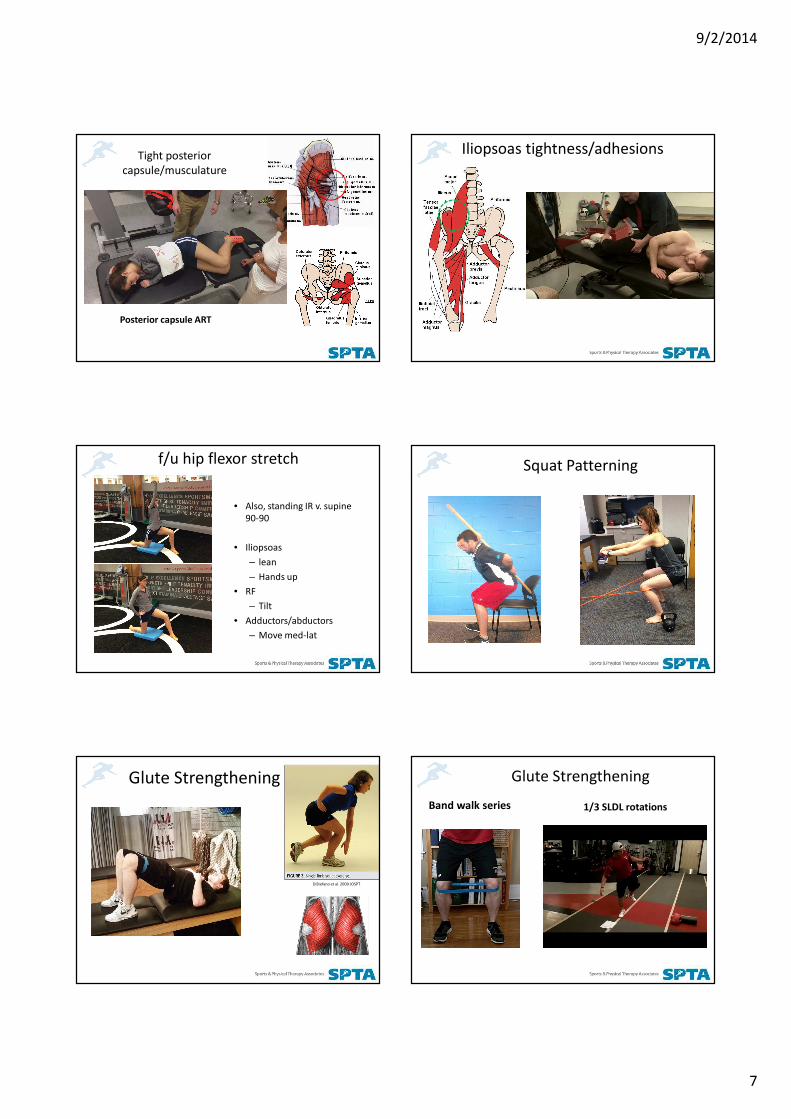
Tight posterior
capsule/musculature
Posterior capsule ART
Iliopsoas tightness/adhesions
f/u hip flexor stretch
• Also, standing IR v. supine
90-90
• Iliopsoas
– lean
– Hands up
• RF
– Tilt
• Adductors/abductors
– Move med-lat
Squat Patterning
Glute Strengthening
DiStefano et al. 2009 JOSPT
Glute Strengthening
1/3 SLDL rotationsBand walk series
9/2/2014
8
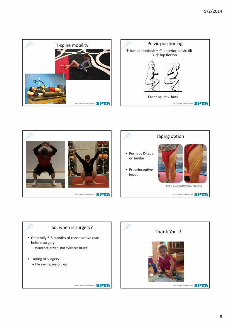
T-spine mobility Pelvic positioning
↑ lumbar lordosis = ↑ anterior pelvic Rlt = ↑ hip flexion
Front squat v. back
Taping option
• Perhaps K-tape
or similar
• Proprioceptive
input
Gelber & Dames, NATA News Feb. 2010
So, when is surgery?
• Generally 3-6 months of conservative care
before surgery
– Insurance driven; not evidence based
• Timing of surgery
– Life events, season, etc.
Thank You !!