History and Physical Examination of the Hand - Dartmouth-Hitchcock
104
Main Menu History and Physical Examination of the Hand
Transcript of History and Physical Examination of the Hand - Dartmouth-Hitchcock

Main Menu
History and Physical Examination of the Hand

Main Menu
Core Quest Goals (7/5/06)
• To discuss the components of a complete history for patients with hand disorders
• To review physical examination methods for patients with hand and upper extremity conditions
• Include understanding of anatomy and embryology• Apply this to specific examples of fingertip injuries
distal to the PIPJ ( Demas Requirements)• Cover topic in sufficient detail to be able to understand
and answer In-Service Exams that cover topic areas

Main Menu
History
• Can be the most important tool in developing an accurate diagnosis
• Must be both thorough and organized• Should document not only the current
complaint, but also relevant elements of past medical history

Main Menu
History: Five Main Categories
1. Patient demographics2. Current complaint (history of the present
illness)3. Medical History4. Allergies and Medications5. Social History

Main Menu
Patient Demographics
• Name• Age• Gender• Occupation• Hand dominance

Main Menu
Current Complaint: Ask About--
• Onset of the condition: timing and mechanism • Pain• Numbness• Tingling (paresthesias)• Weakness• Discoloration• Coldness• Clumsiness (coordination)• Clicking or snapping• Things that improve the condition• Things that worsen the condition

Main Menu
Distribution of numbness consistent with a radial digital nerve laceration of the index finger (at point “X”)
Regional Review Course 1998

Main Menu
Use Symptom Descriptors:
• Location• Intensity• Duration• Frequency• Radiation• Associated symptoms• What makes symptoms better or worse

Main Menu
Also Ask About Injury Details
• Mechanism (crush, sharp, etc.)• Time of occurrence• Location of occurrence (work, home, etc.)• Previous history of similar injuries• And don’t forget to ask about—
– Time of last meal (NPO status)– Tetanus immunization history

Main Menu
Medical History
• Ask about: diabetes, cardiac, pulmonary, or renal disease
• Also: rheumatologic and dermatologic problems
• Coagulation history• Anesthesia history (including any family
history)• Prior surgical history

Main Menu
Social History
• Tobacco use• Alcohol intake• Substance abuse• Hepatitis and HIV status• Patient’s hobbies and sports activities

Main Menu
Physical Examination: 8 Parts
• Inspection• Palpation• Range of motion• Stability• Muscle and Tendon Function• Nerve Assessment• Vascular Assessment• Integument Assessment

Main Menu
Inspection: Look For--
• Discoloration• Deformity• Muscular atrophy• Trophic changes (sweat pattern, hair growth)• Swelling• Wounds or scars• Also: compare to normal hand

Main Menu
Discoloration
• Redness: cellulitis• White: arterial blockage• Blue/purple: venous congestion• Patches of blue/purple: trauma• Black spots/lines: rule out melanoma• Other color producing processes: fungi,
viruses, psoriasis

Main Menu
Deformity
• Asymmetry, angulation, rotation, missing parts• Fractures: check angulation and rotation• Rotational alignment of fingers
– Must be checked with fingers extended and fully flexed
– Compare to opposite hand; some minor rotational malalignment may be normal
• Be sure to document missing parts; may be from a previous injury
Main Menu
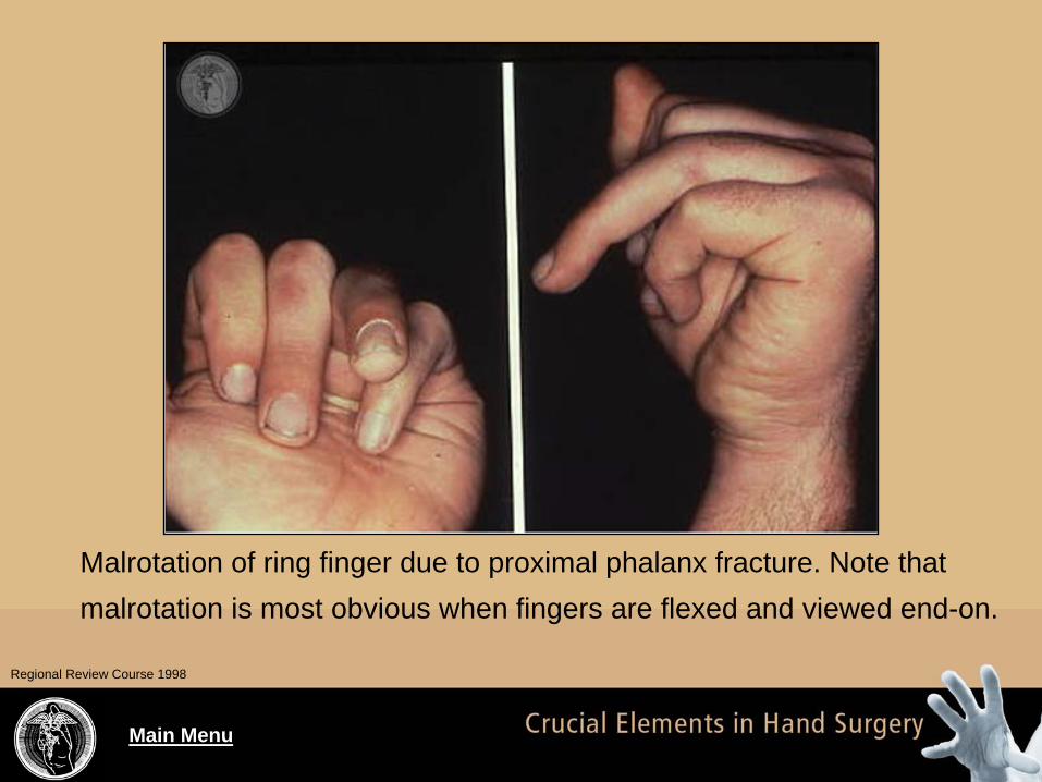
Malrotation of ring finger due to proximal phalanx fracture. Note that malrotation is most obvious when fingers are flexed and viewed end-on.
Regional Review Course 1998

Main Menu
Deformity
• Other processes that can produce deformity– Osteoarthritis– Rheumatoid arthritis– Psoriatic arthritis– Lupus– Scleroderma– Tumors

Main Menu
Muscle Atrophy
• Generalized: may indicate disuse• Specific muscle groups: suggest nerve
pathology– Thenar atrophy: carpal tunnel syndrome– Interossei atrophy: cubital tunnel or cervical spine problem
• Subcutaneous atrophy: often after local steroid injection

Main Menu
Muscle Strength
• Five basic grades of strength– Grade 5: normal strength– Grade 4: diminished strength to resistance– Grade 3: enough strength to overcome gravity– Grade 2: enough strength to contract but not to
overcome gravity– Grade 1: fibrillations or faintly palpable
contractions– Grade 0: no contractions
BRMC

Main Menu
A pinch strength dynamometer is being used to measure “key pinch” strength.
Regional Review Course 1998

Main Menu
Use of a hand grip dynamometer can sometimes help identify differences in hand strength.
Regional Review Course 1998

Main Menu
Trophic Changes
• Can represent a derangement of the sympathetic nervous system
• Increased hair growth or altered (usually increased) sweat production: may indicate sympathetically mediated pain syndrome

Main Menu
Swelling
• Compare to uninvolved extremity• Localized swelling: trauma or inflammation• Diffuse swelling: many causes
– Infection anywhere in the extremity– Venous or lymphatic obstruction– Fractures as far proximal as the shoulder
• Dorsal subcutaneous space in the hand frequently swells first, even if a palmar hand infection is present

Main Menu
Inspection for Wounds/Scars
• Note size and location of any acute wounds• Can often predict likelihood of nerve or
tendon damage just based upon location of wound
• Be particularly wary of wounds associated with fractures: must identify open fracture– 5th metacarpal fracture and open wound from
fist striking a tooth in a bar fight = open fracture

Main Menu
Note the location of this wound– associated with laceration of both the sublimus and profundus flexor tendons. The normal flexor tone is absent in the long finger.
Regional Review Course 1998

Main Menu
Wounds and Scars
• Carefully check nail areas and web spaces– May be site of tiny punctures that can cause
infections• Record any pre-existing scars to avoid
confusion later• Normal flexion creases of hand and palm are
useful reference points– Note MCP flexion crease is not over the joint but
overlies the shaft of the proximal phalanx

Main Menu
Palpation: Check for--
• Masses• Temperature abnormalities• Areas of tenderness• Crepitus• Clicking or snapping• Joint effusion

Main Menu
Palpation
• Mass: lymph node, ganglions• Heat: infection, inflammation• Cold: vascular pathology• Tender, crepitans: fracture• Clicking or snapping: tendonitis• Joint effusion: infection, inflammation, trauma
Main Menu
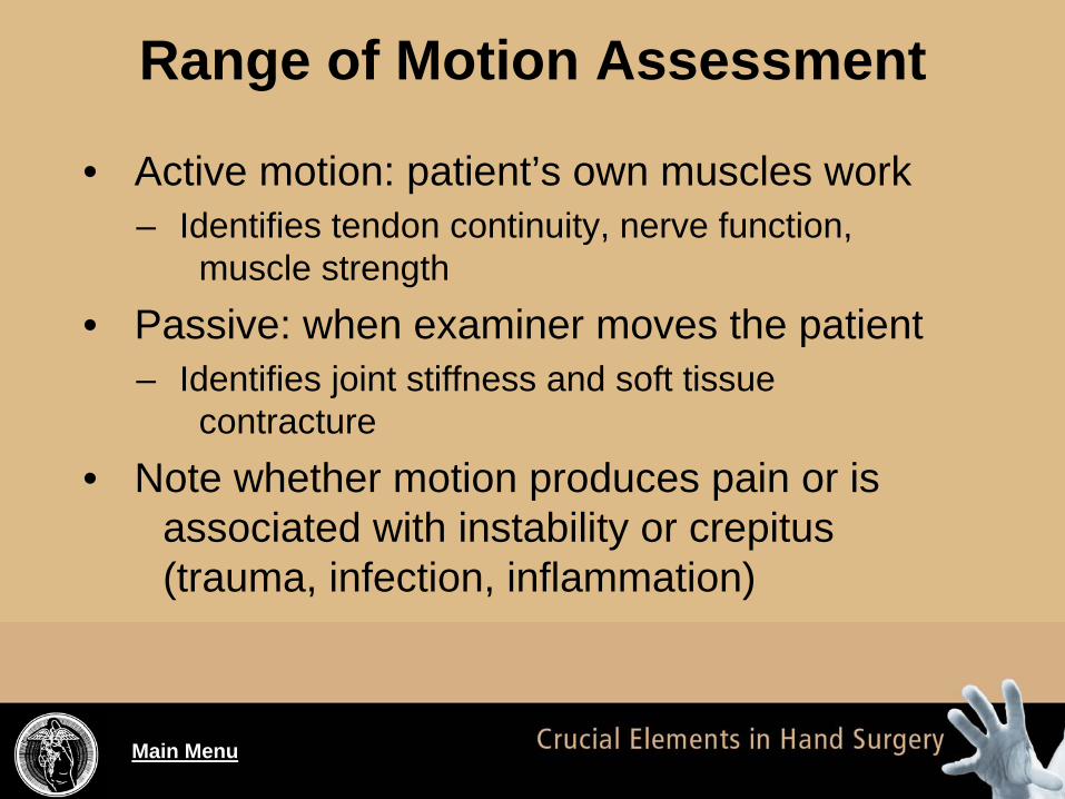
Range of Motion Assessment
• Active motion: patient’s own muscles work– Identifies tendon continuity, nerve function,
muscle strength• Passive: when examiner moves the patient
– Identifies joint stiffness and soft tissue contracture
• Note whether motion produces pain or is associated with instability or crepitus (trauma, infection, inflammation)

Main Menu
Normal values for finger flexionEssentials of Hand Surgery 2002

Main Menu
Normal planes of wrist motion.Regional Review Course 1998
Main Menu
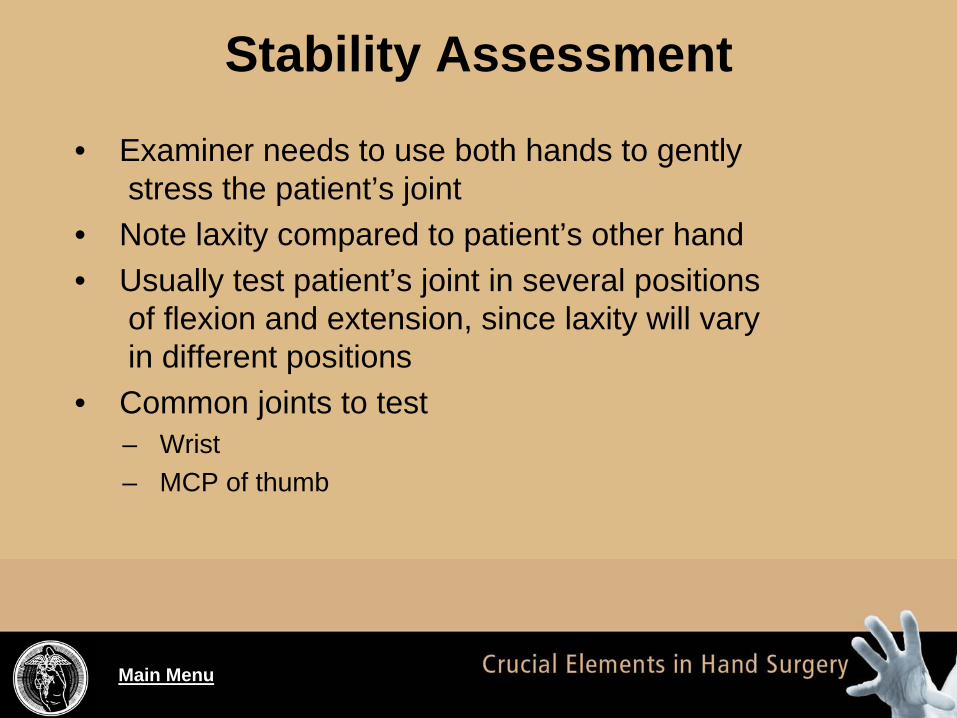
Stability Assessment
• Examiner needs to use both hands to gently stress the patient’s joint
• Note laxity compared to patient’s other hand• Usually test patient’s joint in several positions
of flexion and extension, since laxity will vary in different positions
• Common joints to test– Wrist– MCP of thumb

Main Menu
Muscle and Tendon Assessment
• Intrinsic muscles: origin and insertions are both within the hand
• Extrinsic muscles: myotendinous units span the forearm and hand
• In testing patient, resist the action of a particular muscle and consider both the:– Integrity of the tendon– Strength of the muscle

Main Menu
Specific Testing of Certain Muscles
• Flexor pollicis longus: flexion of thumb IP joint• Flexor digitorum profundus: flexion of finger
DIP joints while examiner holds PIP joint straight (blocks the FDS from bending digit)
• Flexor digitorum superficialis: flexion of PIP joint while examiner holds other fingers fully extended (block FDP from bending PIP joint)
Main Menu
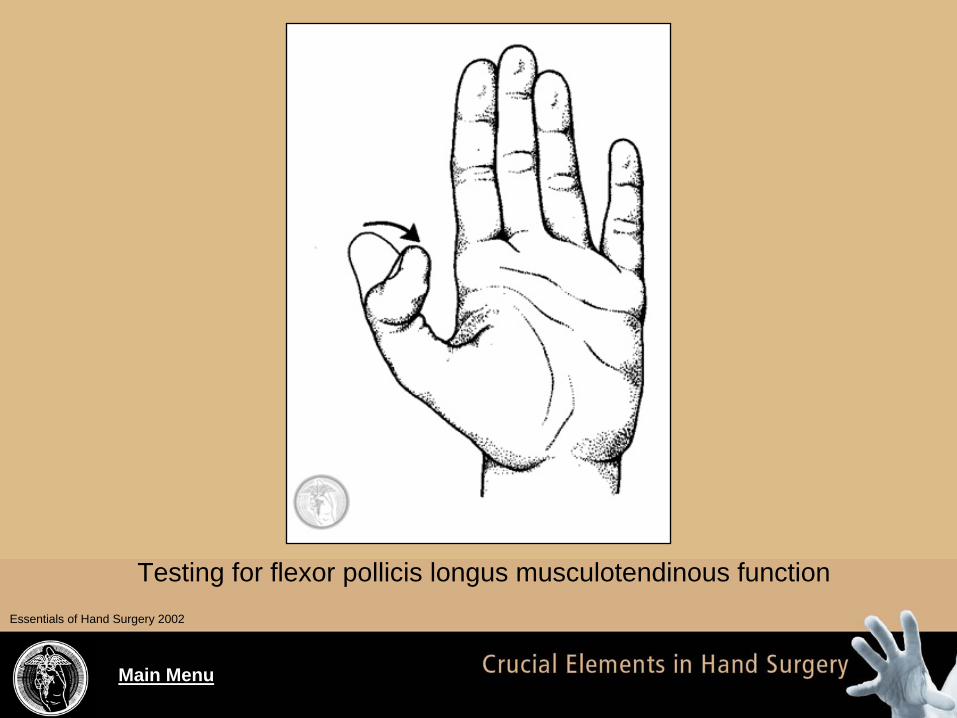
Testing for flexor pollicis longus musculotendinous functionEssentials of Hand Surgery 2002

Main Menu
Testing for flexor digitorum profundus musculotendinous function
Essentials of Hand Surgery 2002

Main Menu
Testing flexor digitorum superficialis musculotendonious function
Essentials of Hand Surgery 2002

Main Menu
Specific Testing of Certain Muscles
• Flexor carpi radialis: have patient volar flex wrist
• Flexor carpi ulnaris: have patient dorsiflex wrist
Main Menu
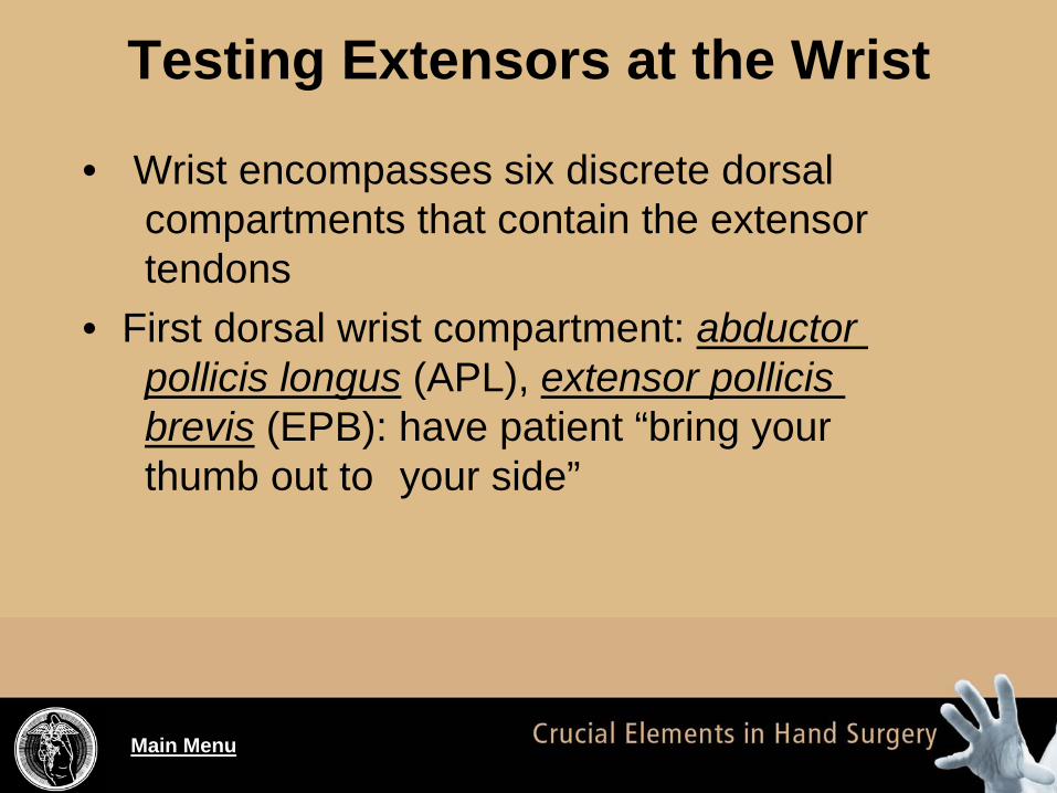
Testing Extensors at the Wrist
• Wrist encompasses six discrete dorsal compartments that contain the extensor tendons
• First dorsal wrist compartment: abductor pollicis longus (APL), extensor pollicis brevis (EPB): have patient “bring your thumb out to your side”

Main Menu
Arrangement of extensor tendons at the wrist into six compartments: dorsal and cross-sectional views
Essentials of Hand Surgery 2002

Main Menu
Pointer is overlying the first extensor compartment (containing the EPB and APL tendons).
Regional Review Course 1998

Main Menu
Testing for extensor pollicis brevis and abductor pollicis longus musculotendinous function
Essentials of Hand Surgery 2002

Main Menu
Testing Muscles at the Wrist
• Second dorsal wrist compartment: Extensor carpi radialis longus and brevis (ECRL and ECRB): have patient make a fist and then extend wrist backwards while holding a fist
• Third dorsal wrist compartment: Extensor pollicis longus (EPL): have patient hyperextend thumb while holding hand flat on a table

Main Menu
Testing for extensor carpi radialis longus and extensor carpi radialis brevis musculotendinous function
Essentials of Hand Surgery 2002

Main Menu
Pointer is overlying the third extensor compartment (containing the extensor pollicis longus tendon).
Regional Review Course 1998

Main Menu
Essentials of Hand Surgery 2002
Testing for extensor pollicis longus musculotendinous function

Main Menu
Testing Muscles at the Wrist
• Fourth dorsal wrist compartment: Extensor digitorum communis (EDC) and extensor indicis proprius (EIP): have patient straighten fingers and observe MCP motion
• To test EIP alone, have patient extend index finger while flexing all the other digits
• Fifth dorsal wrist compartment: Extensor digiti minimi (EDM): have patient extend small
finger while flexing all the other digits

Main Menu
Finger is overlying the fourth extensor compartment (containing the extensor digitorum communis tendons).
Regional Review Course 1998

Main Menu
Testing for extensor digitorum communis, extensor indicis proprius, and extensor digiti minimi musculotendinous function
Essentials of Hand Surgery 2002

Main Menu
Testing the EIP and EDM tendons. By having the patient flex the middle and ring fingers, the EDC tendons are prevented from being extensors.
Regional Review Course 1998

Main Menu
Testing Muscles at the Wrist
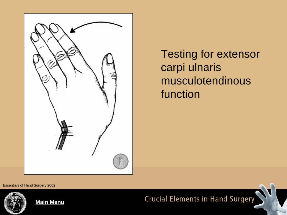
• Sixth dorsal wrist compartment: Extensor carpi ulnaris (ECU): ask patient to “pull your wrist up out and to the side”
Main Menu
Testing for extensor carpi ulnaris musculotendinous function
Essentials of Hand Surgery 2002

Main Menu
Testing: Extrinsic Extensor Tightness
• Extensor tendons can become adherent over
the dorsum of the hand or wrist (trauma)– Keep wrist in neutral position and test PIP flexion
passively: once with MCP joint extended, and once with MCP joint flexed
– If PIP joint can be passively flexed while MCP is extended but NOT when MCP is flexed, then “extrinsic tightness” is present

Main Menu
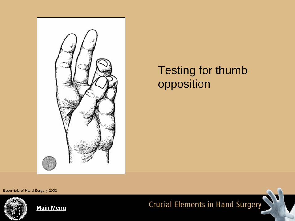
Testing: Thenar Muscles
• The muscles covering the thumb metacarpal– Abductor pollicis brevis (APB)– Opponens pollicis (OP)– Flexor pollicis brevis (FPB)
• Have patient touch thumb and small fingertips together so that nails are parallel: tests opposition
• Can also test by having patient hold dorsum of hand on a table and then raise thumb 90 degrees up from palm
Main Menu
Testing for thumb opposition
Essentials of Hand Surgery 2002

Main Menu
Testing: Adductor Pollicis
• Can test adductor pollicis by having patient hold a piece of paper sideways between thumb and index finger
• Pull the paper out while asking paper to hold it tight between sides of thumb and index: flexion of the thumb IP joint indicates weakness of adductor pollicis– When thumb IP joint flexes during this
maneuver, it is also called “Froment’s sign”

Main Menu
Essentials of Hand Surgery 2002
Froment’s sign is positive in hand B

Main Menu
Testing: Interossei and Lumbricals
• These muscles flex the MCP joints and extend the IP
• Interossei also abduct and adduct the fingers• Test interossei by having patient spread their
fingers apart and feel strength of index finger spreading (tests 1st dorsal interosseous)
• Can also have patient wiggle a hyper extended middle finger side-to-side while their hand is flat on a table

Main Menu
Essentials of Hand Surgery 2002
Testing for interosseous muscle function

Main Menu
Testing: Hypothenar Muscles
• Abductor digiti minimi (ADM)• Flexor digiti minimi (FDM)• Opponens digiti minimi (ODM)• Ask patient to separate away the small finger
from their other fingers– Can palpate the hypothenar muscle mass for
contraction and also see a dimpling of the hypothenar skin

Main Menu
Testing for hypothenar muscle function
Essentials of Hand Surgery 2002

Main Menu
Nerve Assessment: Motor Function
• Radial nerve: test thumb IP joint extension• Median
– Recurrent motor branch: palmar abduction of thumb
– Anterior interosseous branch: flexion of thumb IP joint and index DIP (“A-OK sign”)
• Ulnar: have patient attempt to cross fingers (tests interossei)

Main Menu
Nerve Assessment: Sensibility
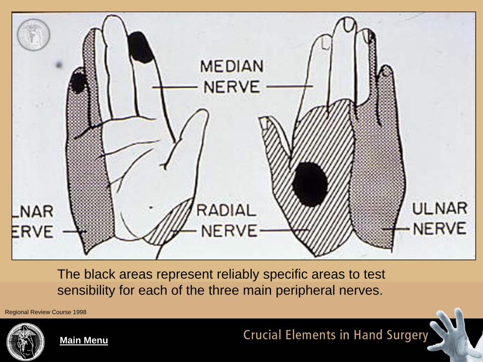
• Radial: test dorsal thumb-index web space• Median: test palmar surface of index or thumb• Ulnar: test palmar aspect of little finger• Digital nerves: test each the radial and ulnar
side of each fingertip on the palmar aspect
Main Menu
The black areas represent reliably specific areas to test sensibility for each of the three main peripheral nerves.
Regional Review Course 1998
Main Menu
Nerve Assessment: Sensibility
• Testing should include light touch perception
and also two-point discrimination– Two point is very specific for nerve integrity and
can be tested with a caliper or a bent paper-clip– Record the smallest distance that patient can still
distinguish between two contact points: normal is about 5 mm at palmar fingertip
• Other tests: Semmes-Weinstein monofilament (usually performed by therapist) and
temperature testing

Main Menu
Vascular Assessment
• Requires inspection and palpation skills• Arterial blockage: pale, white or gray fingers• Venous blockage: blue or purple discoloration• Other findings:
– Coolness– Loss of tissue pressure (turgor)– Capillary refill (normal is < 2 seconds at nail bed)– Subungual splinter hemorrhages: emboli

Main Menu
Special Tests:
• Palpation– Grind– Finkelstein’s
• Range of motion– Flexor profundus– Flexor sublimus– Intrinsic tightness (Bunnel’s)– Extrinsic tightness

Main Menu
Special Tests:
• Stability assessment– Scaphoid stability– Lunotriquetral ballottement (LT Shear or Shuck)– Midcarpal instability– Ulnar carpal abutment– Gamekeeper’s

Main Menu
Special Tests:
• Nerve assessment– Prune test– Tinel’s– Phalen’s– Froment’s sign– Jeanne’s sign– Wartenberg’s sign

Main Menu
Special Tests: Grind
• Used to test for pathology at the thumb carpometacarpal (CMC) joint
• Examiner applies an axial load to patient’s thumb metacarpal and rotates (“grinds”) it
• Positive findings: pain, crepitus, instability

Main Menu
A: Axial compression- adduction test
B: Axial compression rotation test.
Essentials of Hand Surgery 2002

Main Menu
Special Tests: Finkelstein’s
• Used to test for deQuervain’s tendonitis (inflammation of the EPB and APL tendons in the 1st extensor compartment)
• Patient is asked to make a fist with the fingers overlying the thumb
• Examiner then ulnarly deviates the wrist (gently)
• Positive findings: pain along the 1st
compartment

Main Menu
Finkelstein’s test for deQuervain’s disease
Essentials of Hand Surgery 2002

Main Menu
Special Tests: Flexor Profundus
• Tests the continuity of the FDP tendons• Each finger is tested separately• MCP and PIP joints are held in extension
while the patient is asked to flex the DIP• This isolates the FDP (from the FDS) as the
only tendon capable of bending the finger

Main Menu
Testing for flexor digitorum profundus function.Regional Review Course 1998
Main Menu
Special Tests: Flexor Sublimus
• Tests the continuity of the FDS tendons• Each finger is tested separately• The MCP, PIP, and DIP joints of all fingers
are held extended with the hand flat, palm up• The finger to be tested is allowed to flex at the
PIP joint• This isolates the FDS (from the FDP) as the
only tendon capable of bending the PIP

Main Menu
Testing for flexor digitorum sublimus function.Regional Review Course 1998

Main Menu
Special Tests: Bunnell’s
• Tests for intrinsic muscle tightness• The examiner passively flexes the patient’s
PIP joint -- twice– Once with the corresponding MCP in extension– Next with the MCP joint held in flexion
• INTRINSIC tightness is present if the PIP can be flexed easily when the MCP is flexed but NOT when the MCP is extended

Main Menu
Intrinsic muscle tightness
Essentials of Hand Surgery 2002

Main Menu
Special Tests: Extrinsic Tightness
• Tests for extrinsic muscle tightness• Procedure same as in Bunnell’s test:
– The examiner passively flexes the patient’s PIP joint twice: once with the corresponding MCP in extension and then with the MCP joint held in flexion
• EXTRINSIC tightness is present if the PIP can be flexed easily when the MCP is extended but NOT when the MCP is flexed

Main Menu
Special Tests: Scaphoid Stability
• Tests for scaphoid-lunate ligament tear– Examiner places thumb on patient’s scaphoid
distal pole, on palmar side of wrist; examiner then radially and ulnarly deviates patient’s wrist
– Scaphoid pole should feel palmarly more prominent with wrist radial deviation
– If scaphoid distal pole doesn’t seem to change with either radial or ulnar deviation of wrist,
suspect ligament disruption between scaphoid and lunate

Main Menu
Special Tests: LT Shear (Shuck)
• Tests for lunatotriquetral ligament tear• While holding the lunate between index and
thumb, the examiner tries to push the triquetrum dorsally with the other hand
• Positive test: pain or instability of the triquetrum

Main Menu
Special Tests: Midcarpal Instability
• Examiner stabilizes the distal radius and ulna with the nondominant hand, starting with the patient’s wrist in radial deviation
• The examiner then moves the patient’s wrist into ulnar deviation
• Positive test: a clunk is felt when the wrist is ulnarly deviated (as the midcarpal instability is reduced)

Main Menu
Special Tests: Ulnar Abutment
• Tests for TFCC tear or ulnar-carpal impingement
• The examiner ulnar deviates the patient’s wrist
• Positive test: if ulnar deviation reproduces patient’s pain or produces a pop or click

Main Menu
Special Tests: Gamekeeper’s
• Tests for ulnar collateral ligament tear at MCP of thumb
• Examiner stresses the thumb MCP joint into radial deviation– and should do this with the MCP joint both fully extended and also flexed
• Positive test: if more than 30 degrees of laxity is present in both positions (or gross laxity compared to patient’s normal thumb MCP)

Main Menu
Rupture of ulnar collateral ligament of the metacarpophalan geal joint of the thumb
Essentials of Hand Surgery 2002

Main Menu
Special Tests: Prune
• Tests sensory nerve function in the finger• The finger in question is held in a cup of water
for 5 to 10 minutes• Normally innervated glabrous skin will pucker
(or “prune”) after being submerged this long• Helpful for assessing sensory nerve continuity
in children or patients who can’t cooperate

Main Menu
The normal puckering of sensate skin after being soaked in water. Insensate skin will stay smooth.
Regional Review Course 1998
Main Menu
Special Tests: Tinel’s
• A provocative test for carpal tunnel syndrome• The examiner percusses with two fingers
directly over the distal palmar crease in the midline
• Positive test: patient reports paresthesias in the median distribution when the nerve is percussed

Main Menu
Tinel’s sign
Essentials of Hand Surgery 2002

Main Menu
Special Tests: Phalen’s
• A provocative test for carpal tunnel syndrome• The patient’s wrist is held in maximum flexion
for two minutes• Positive test: patient reports paresthesias in
the median distribution

Main Menu
Phalen’s sign (wrist flexion test)
Essentials of Hand Surgery 2002

Main Menu
Special Tests: Froment’s Sign
• Tests for ulnar nerve motor weakness• The patient is asked to hold a piece of
paper between their thumb and radial side of index
• Positive test: as the paper is pulled away by the examiner, if ulnar motor weakness is present, the patient will flex the thumb IP joint in an attempt to hold onto the paper

Main Menu
Positive Froment's sign: note thumb IP flexion when attempting to hold a piece of paper side-to-side against the radial aspect of the index finger. This indicates weakness of ulnar nerve motor function.
Regional Review Course 1998

Main Menu
Special Tests: Jeanne’s Sign
• Tests for ulnar nerve motor weakness• Patient is asked to attempt to demonstrate
key pinch• Positive test: when attempting key pinch, the
patient’s thumb MCP joint will hyperextend

Main Menu
Special Tests: Wartenberg’s Sign
• Tests for ulnar nerve motor weakness• Patient is asked to hold their fingers fully
adducted with the MCP, PIP, and DIP joints fully extended
• Positive test: the small finger will drift away from the others into abduction (due to 3rd palmar interosseous muscle weakness)

Main Menu
Special Tests: Threshold
• Threshold tests assess single nerve fibers that innervate a receptor
• Thought to be more sensitive than innervation density testing for determining early nerve damage
• Examples of threshold tests– Von Frey pressure testing with Semmes-
Weinstein monofilament– Variable amplitude vibrometry

Main Menu
Special Tests: Innervation Density
• These tests measure innervation of multiple overlapping receptors
• Results can remain almost normal even if advanced nerve pathology is present
• Example of innervation density test: two-point discrimination

Main Menu
Tools that can be used to test two-point discrimination. A bent paper-clip also works well.
Regional Review Course 1998

Main Menu
Two-point discriminationEssentials of Hand Surgery 2002

Main Menu
Use of a caliper to test two-point discrimination.Regional Review Course 1998

Main Menu
Special Tests: Allen’s
• Tests ulnar and radial artery blood flow• Patient makes a tight fist and examiner
manually occludes both radial and ulnar artery
• Examiner releases one of the vessels and examines for reperfusion in the long finger
• Abnormal test: hand reperfusion > 5 seconds• Test is repeated for the other artery

Main Menu
Allen’s Test:Patient makes a fist. Then examiner occludes both arteries. Then patient releases fist. Examiner then releases one artery and observes for refill of palm. Normal is < 5 seconds.
Allen test for arterial patencyEssentials of Hand Surgery 2002

Main Menu
Summary
• We have discussed the components of a complete history for patients with hand disorders
• We have reviewed the physical examination methods for patients hand and upper extremity conditions