Histopathology of Skin Cancer
20
E. Stockfleth, et al. (eds.), Managing Skin Cancer, 17 DOI: 10.1007/978-3-540-79347-2_2, © Springer-Verlag Berlin Heidelberg 2010 2.1 Basal Cell Carcinoma G. Goldenberg, L.E. Golitz, and J. Fitzpatrick Basal cell carcinoma (BCC) is an epithelial neoplasm that is believed to derive from the basal layer of the epi- dermis or follicular epithelium. The classic histologic presentation of BCC is that of nodules and/or strands of atypical basaloid cells that show nuclear palisading, cel- lular apoptosis, and scattered mitotic activity (Fig. 2.1). Artifactual cleft formation may be seen between the tumor lobules and its surrounding stroma, which may be mucinous. Solar elastosis, a manifestation of chronic actinic damage, is usually present in the dermis. Tumor calcification may be seen, especially in long standing tumors, although this phenomenon has been reported to be more commonly associated with more aggressive BCC subtypes [1]. Multiple growth patterns of BCC have been described, and these act as prognosticators of biologic behavior [2]. Superficial basal cell carcinoma presents with nod- ules and strands of basaloid cells that proliferate paral- lel to the epidermis and demonstrate slit-like retraction Histopathology of Skin Cancer G. Goldenberg () Assistant Professor of Dermatology and Pathology, Medical Director of the Dermatology Faculty Practice, Mt Sinai School of Medicine, 5 East 98th St 5th Floor, NY, NY 10029 e-mail: [email protected] 2 Core Messages The different histologic patterns of skin cancer › are vital for the proper diagnosis and treatment of skin malignancies. This chapter reviews his- tologic findings of multiple skin malignancies, as well as their different histologic patterns. The use of immunohistochemical stains is also reviewed. Contents 2.1 Basal Cell Carcinoma ............................................ 17 G. Goldenberg, L.E. Golitz, and J. Fitzpatrick 2.2 Squamous Cell Carcinoma In Situ ....................... 20 J. Roewert-Huber 2.2.1 Actinic Keratosis (AK) ............................................ 20 References ........................................................................... 22 2.2.2 Bowen’s Disease ...................................................... 23 2.3 Invasive Squamous Cell Carcinoma..................... 24 J. Roewert-Huber 2.3.1 Acantholytic Squamous Cell Carcinoma ................. 25 2.3.2 Spindle Cell Squamous Cell Carcinoma.................. 25 2.3.3 Verrucous Squamous Cell Carcinoma ..................... 25 2.3.4 Adenosquamous Carcinoma .................................... 26 2.3.5 Keratoacanthoma (KA), Variants of SCC ................ 26 2.4 Malignant Melanoma ............................................ 26 G. Goldenberg, L.E. Golitz, J. Fitzpatrick 2.5 Merkel cell carcinoma ........................................... 30 Martina Ulrich, Jean Kanitakis References ........................................................................... 30 2.6 Kaposi’s Sarcoma................................................... 31 G. Goldenberg, L.E. Golitz, J. Fitzpatrick 2.7 Dermatofibrosarcoma Protuberans ..................... 32 G. Goldenberg, L.E. Golitz, J. Fitzpatrick 2.8 Atypical Fibroxanthoma ....................................... 33 G. Goldenberg, L.E. Golitz, J. Fitzpatrick 2.9 Malignant Fibrous Histiocytoma.......................... 34 G. Goldenberg, L.E. Golitz, J. Fitzpatrick References ........................................................................... 34
Transcript of Histopathology of Skin Cancer

E. Stockfl eth, et al. (eds.), Managing Skin Cancer, 17DOI: 10.1007/978-3-540-79347-2_2, © Springer-Verlag Berlin Heidelberg 2010
2.1 Basal Cell Carcinoma
G. Goldenberg, L.E. Golitz, and J. Fitzpatrick
Basal cell carcinoma (BCC) is an epithelial neoplasm that is believed to derive from the basal layer of the epi-dermis or follicular epithelium. The classic histologic presentation of BCC is that of nodules and/or strands of atypical basaloid cells that show nuclear palisading, cel-lular apoptosis, and scattered mitotic activity (Fig. 2.1). Artifactual cleft formation may be seen between the tumor lobules and its surrounding stroma, which may be mucinous. Solar elastosis, a manifestation of chronic actinic damage, is usually present in the dermis. Tumor calcifi cation may be seen, especially in long standing tumors, although this phenomenon has been reported to be more commonly associated with more aggressive BCC subtypes [1]. Multiple growth patterns of BCC have been described, and these act as prognosticators of biologic behavior [2].
Superfi cial basal cell carcinoma presents with nod-ules and strands of basaloid cells that proliferate paral-lel to the epidermis and demonstrate slit-like retraction
Histopathology of Skin Cancer
G. Goldenberg (�)Assistant Professor of Dermatology and Pathology, Medical Director of the Dermatology Faculty Practice, Mt Sinai School of Medicine, 5 East 98th St 5th Floor, NY, NY 10029e-mail: [email protected]
2
Core Messages
The different histologic patterns of skin cancer ›are vital for the proper diagnosis and treatment of skin malignancies. This chapter reviews his-tologic fi ndings of multiple skin malignancies, as well as their different histologic patterns. The use of immunohistochemical stains is also reviewed.
Contents
2.1 Basal Cell Carcinoma ............................................ 17G. Goldenberg, L.E. Golitz, and J. Fitzpatrick
2.2 Squamous Cell Carcinoma In Situ ....................... 20J. Roewert-Huber
2.2.1 Actinic Keratosis (AK) ............................................ 20
References ........................................................................... 22
2.2.2 Bowen’s Disease ...................................................... 23
2.3 Invasive Squamous Cell Carcinoma ..................... 24J. Roewert-Huber
2.3.1 Acantholytic Squamous Cell Carcinoma ................. 252.3.2 Spindle Cell Squamous Cell Carcinoma.................. 252.3.3 Verrucous Squamous Cell Carcinoma ..................... 252.3.4 Adenosquamous Carcinoma .................................... 262.3.5 Keratoacanthoma (KA), Variants of SCC ................ 26
2.4 Malignant Melanoma ............................................ 26G. Goldenberg, L.E. Golitz, J. Fitzpatrick
2.5 Merkel cell carcinoma ........................................... 30Martina Ulrich, Jean Kanitakis
References ........................................................................... 30
2.6 Kaposi’s Sarcoma................................................... 31G. Goldenberg, L.E. Golitz, J. Fitzpatrick
2.7 Dermatofi brosarcoma Protuberans ..................... 32G. Goldenberg, L.E. Golitz, J. Fitzpatrick
2.8 Atypical Fibroxanthoma ....................................... 33G. Goldenberg, L.E. Golitz, J. Fitzpatrick
2.9 Malignant Fibrous Histiocytoma .......................... 34G. Goldenberg, L.E. Golitz, J. Fitzpatrick
References ........................................................................... 34

18 G. Goldenberg et al.
a b
d
e f
c
Fig. 2.1 (continued)

2 Histopathology of Skin Cancer 19
from the surrounding stroma (Fig. 2.1a, b) [3]. Tumor cells may also proliferate along follicular structures.
Nodular basal cell carcinoma (NBCC) presents with discrete, well-defi ned nodules and strands of basaloid cells in the papillary and reticular dermis, which may focally show a connection to the overlying epidermis (Fig. 2.1c, d). Roughly one-third of NBCC’s will show a coexistent superfi cial component [2]. Artifactual stromal retraction is usually present in these cases. Central tumor necrosis and/or mucin depo-sition may be seen within the individual nodules, giv-ing the neoplasm a “cystic” appearance.
Morpheaform (sclerosing) basal cell carcinoma presents with thin strands of atypical basaloid cells in the dermis (Fig. 2.1e, f). These neoplastic strands are usually one-to-two strands thick and are enmeshed in a densely collagenized stroma with proplastic fi broblasts
[2]. Individual tumor cell necrosis and mitotic activity may be more common with this growth pattern. Stromal retraction may still be seen, but is less com-mon than in other types of BCC. This neoplasm is usu-ally poorly circumscribed and shows an infi ltrating growth pattern, invading into reticular dermis and sub-cutaneous fat.
Fibroepithelioma of Pinkus is a rare type of BCC that typically presents above the natal cleft or on the lower trunk with a pink or fl esh colored nodule that may mimic seborrheic keratosis [4]. This tumor pres-ents with elongated basaloid epithelial strands, which usually show multiple connection points to the overly-ing epidermis (Fig. 2.1g, h). Retraction from the dis-tinct fi bromyxoid stroma is usually seen. Histologically, the most important differential diagnosis is eccrine syringofi broadenoma of Mascaro, which presents with
Fig. 2.1 Basal Cell Carcinoma, showing classic feature of nuclear palisading, artifactual cleft formation, and nuclear apop-tosis; (a, b) Superfi cial growth patter; (c, d) Nodular growth
pattern; (e, f) Morpheaform (sclerosing) growth patter; (g, h) Fibroepithelioma of Pinkus growth pattern; and (i) Positive Ber-EP4 immunohistochemical stain
g h
i

20 J. Roewert-Huber
elongated basaloid strands containing central eccrine ductal cells with a well-defi ned cuticle [5].
Immunohistochemical staining is rarely required in order to diagnose a BCC. Ber-EP4, a monoclonal anti-body which recognizes two glycopolypeptides (34 and 39 kDa) found in most human epithelial cells, has recently been utilized to distinguish BCC from squamous cell carcinoma (Fig. 2.1i) (SCC) [6–10]. All BBCs, regardless of the subtype, stain positive with Ber-EP4, whereas SCC do not show positive staining. This marker can also be reliably used to differentiate BCC from microcystic adnexal carcinoma [11].
2.2 Squamous Cell Carcinoma In Situ
J. Roewert-Huber
SCC in situ has many diverse clinical presentations (see Table 2.1) and includes numerous distinct sub-types with a wide range of clinical manifestation. Histologically, squamous cell carcinoma in situ is composed of atypical keratinocytes, which can be identifi ed throughout the full thickness of the epider-mis. The atypical keratinocytes exhibit eosinophilic, sometimes pale or vacuolated cytoplasm, a sign of faulty cornifi cation, as well as whorls of parakeratosis within aggregates of neoplastic cells (“horn pearls”). An increased number of atypical mitoses and dyskera-totic or necrotic keratinocytes can be found throughout the epidermis. The nuclei of the atypical keratinocytes are crowded, pleomorphic, and often large and hyper-chromatic. By defi nition, the atypical keratinocytes throughout the epidermis do not penetrate into the der-mis. SCC in situ may develop into invasive SCC.
Histologically, the different types of SSC exhibit the same morphology; however, their architectural patterns are different. It is very important to differentiate between these lesions because they present with a wide range of different clinical manifestations covering benign types of bowenoid papulosis, as well as tumors with possible invasive growth potential. Examples of the more aggres-sive of the latter tumor types are actinic keratosis and Bowen’s disease with a tendency towards invasive, and frequently metastatic, growth.
2.2.1 Actinic Keratosis (AK)
For the last approximately 100 years, a controversial discussion regarding the terminology of actinic kerato-sis has been ongoing. Actinic keratosis lesions are cat-egorized by some authors as precancerous because a subset appears to have low individual potential for invasive malignancy or for spontaneous regression.
Table 2.1 Squamous cell carcinoma in situ
Actinic keratosisBowen’s diseaseBowenoid papulosisErythoplasia of Qeuyrat

2 Histopathology of Skin Cancer 21
However, other authors have postulated that AK lesions have to be regarded as early squamous cell carcinomas. The reason for this is that the morphology of atypical cells in the epidermis in both actinic keratosis and cuta-neous squamous cell carcinoma is identical and repre-sents histological aspects of the same disease. Today, AK is considered as early squamous cell carcinoma. Recent studies of molecular, biochemical pathogenesis confi rm this pathogenitic classifi cation.
2.2.1.1 Histology Appearance
Histologically, AK is characterized by the loss of orderly cell maturation with atypical keratinocytes in the epidermis. The atypical keratinocytes reveal a loss of polarity, and the nuclei of the atypical kera-tinocytes are crowded, pleomorphic, and often large, hyperchromatic with cytologic atypia. These cells are characterized eosinophilic, sometimes pale or vacuo-lated cytoplasm. The number of mitosis is increased. Dyskeratotic or necrotic keratinocytes are found in the epidermis. The presence of atypical keratinocytes var-ies from very few atypical cells in size and amount at the basal cell layer of the epidermis to more advanced lesions with moderate keratocytic atypia in the epider-mis that does not involve the granular cell layer. Fully developed lesions contain atypical keratinocytes, which involve the entire epidermis reaching the granular cell layer. These histological changes are equivalent to pre-viously called SCC in situ. The epidermal keratinocytes of the acrosyringia and acrotrichia are spared, showing normal appearance and keratinization patterns, thereby refl ecting a normal orthokeratotic cornifi ed layer. The cytoplasm of keratinocytes of the acrosyringia and acrotrichia is more basophilic or blue and shows a sharp demarcation to the atypical epidermal keratinocytes, which have a more eosinopihlic or pink cytoplasm. There are often small round buds at the basal layer which protrude into the papillary dermis. The epider-mal keratinocytes are immature, thereby contributing to parakeratosis alternating with hyperkeratosis. Actinic keratoses almost always show solar elastosis in the der-mis and often contains a cell infi ltrate, composed mostly of lymphocytes and plasma cells.
AKs can be divided into the following six histologi-cal types: hypertrophic, atrophic, bowenoid, acantho-lytic, lichenoid, and pigmented. Overlapping between these subtypes may be noticed within the same lesion.
The hypertrophic type shows prominent hyperkerato-sis and acanthosis. The atrophic variant has a thinned epidermis, rete ridges are missing. The bowenoid type of actinic keratosis is diffi cult to distinguish from Bowen’s disease. In contrast to actinic keratosis, Bowen’s disease shows parakeratosis, which may be strikingly predomi-nate, and no distinct alternation of orthokeratosis and parakeratosis. The process spares acrosyringia, but not acrotrichia; suprabasal clefts or acantholytic cells are not found. The acantholytic variant reveals focal acantholy-sis, sometimes being accompanied by clefts similar to other acantholytic diseases. The lichenoid type shows a dense band-like infi ltrate of lymphocytes in the papillary dermis and vacuolar alteration at the dermoepidermal junction. The pigmented variant has increased melanin pigmentation in the epidermis.
In 2007, Roewert-Huber et al. [1] published a clas-sifi cation of actinic keratoses, which categorizes the AK into three histological types based on the extent of atypical keratinocytes in the epidermis.
Early In Situ SCC-Type I (Mild)
Atypical keratinocytes are found in the basal and suprabasal layer of the epidermis, and could extend to the lower one-third of the epidermis.
The nuclei are hyperchromatic, variable in size, and have mild irregularities in nuclear outline. Often, a loss of nuclear polarity occurs, with many of the cells that have oval nuclei oriented at obtuse angles, instead of perpendicular to the epidermis. The follicular infundibu-lum is not involved.
Early In Situ SCC-Type II (Moderate) (Fig. 2.2a)
Atypical keratinocytes extend to the lower two-thirds of the epidermis alternating with zones of normal epi-dermis of the acrotrichia and acrosyringia in particular. Buds of keratinocytes in the upper papillary dermis can be found.
In Situ SCC-Type III (Severe) (Fig. 2.2b)
Atypical keratinocytes extend more than two-thirds to full thickness within the epidermis including involvement of the epithelia of the hair follicle

22 J. Roewert-Huber
infundibula and acrosyringia as seen in SCC in situ. Buds of keratinocytes can also be found in the upper papillary dermis.
Grade III lesions are equivalent to lesions previ-ously called SCC in situ.
This creation of a grading system for epithelial tumors, similar to that for other neoplasms, was war-ranted and long overdue. Tumor classifi cations accord-ing to severity and extent are important. The classifi cation of AK will provide the clinician with a very improved prognostic tool of the malignant poten-tial of the lesion, helping him with the selection of the
most specifi c therapeutic option. Without a grading system, the clinician does not have the tools to accu-rately judge the amount of atypical keratinocytes in the epidermis; with this information, the physician has the information to choose more precisely the appropriate therapy for these types of early squamous cell carcinomas.
Reference
1. Röwert-Huber J, Patel MJ, Forschner T, Ulrich C, Eberle J, Kerl H, Sterry W, Stockfl eth E (2007) Actinic keratosis is an early in situ squamous cell carcinoma: a proposal for reclas-sifi cation. Br J Dermatol 156(Suppl 3):8–12
Fig. 2.2 (a) Early in situ SCC-type II (moderate) with atypical keratinocytes extending to the lower two thirds of the epidermis alternating with zones of normal epidermis of the acrotrichia and acrosyringia in particular. Alternation of pink parakeratosis and blue orthokeratosis. (b) In situ SCC-type III (severe) with atypi-cal keratinocytes extending more than two thirds to full thickness of the epidermis including involement of the epithelia of the hair follicle infundibula and acrosyringia as seen in SCC in situ
a
b

2 Histopathology of Skin Cancer 23
2.2.2 Bowen’s Disease
J. Röwert-Huber
The term Bowen’s disease refers to a particular type of intraepidermal squamous cell carcinoma, the so-called squamous cell carcinoma in situ. Nevertheless, it is a clinical and histopathological distinct entity.
The lesion may occur on any skin surface.The epidermis shows acanthosis with increased cel-
lularity and hyper – and parakeratosis as signs of aber-rant cornifi ciation. The keratinocytes are crowded and are arranged in complete disorder, refl ecting a “wind-blown” appearance. The specifi c histological features of Bowen’s disease are cells with more prominent cytologic atypia characterized by large, pleomorphic and hyperchromatic nuclei, By loss of normal polarity, and by absence of maturation to the surface in together with dyskeratotic and occasionally multinucleated cells. Numerous mitoses including atypical bizarre forms are noted. Below the otherwise intact dermoepi-dermal basement membrane, there is a chronic infl am-matory infi ltrate in the upper corium (Fig. 2.3).
In the pigmented variant of Bowen’s disease, is char-acterized by the presence of more pigment in the atypi-cal keratinocytes along with numerous melanophages.
2.2.2.1 Erythroplasia of Queyrat
Similar lesions located on the glans penis are referred to as Erythroplasia of Queyrat. They have the identical histological features as does Bowen’s disease. The term Bowen’s disease has been replaced in gynaeco-logical pathology by the term vulvar interepithelial neoplasia (VIN) and is equivalent to VIN grade III.
2.2.2.2 Bowenoid Papulosis
Bowenoid papulosis is characterized by the same cyto-pathological changes as Bowen’s disease, except that in low power the lesions resemble condylomata acumi-nata. In contrast to Bowen’s disease, Bowenoid papu-losis exhibits multiple verrucous papules, which are frequently pigmented.
Fig. 2.3 Bowen’s disease. Acanthosis with increased cellularity and hyper – and parakeratosis. Crowding of keratinocytes and arrangement in complete disorder giving them a “windblown” appearance. Promiment cytologic atypia characterized by large, pleomorphic and hyperchromatic nuclei, a loss of normal polar-ity and no maturation to the surface in association with dyskera-totic and occasionally multinucleated cells. Numerous mitoses including atypical bizarre forms are noted.

24 J. Roewert-Huber
2.3 Invasive Squamous Cell Carcinoma
J. Roewert-Huber
Invasive Squamous cell carcinoma is an epithelial tumor infi ltrating into the dermis, characterized by signs of cornifi cation. The histological picture of squamous cell carcinoma reveals proliferation of anas-tomosing nests, sheets, and strands of atypical kerati-nocytes originating in the epidermis and infi ltrating into the dermis. Prominent intercellular bridges are characteristic. Epithelial cells exhibit glassy eosino-philic cytoplasm and frequently a large nucleus. Dyskeratotic cells, parakeratosis, and horn pearl for-mation are sign of abnormal cornifi cation (Fig. 2.4). These morphologic features of squamous cell
differentiation are variably present in the tumor. Sometimes, in lesions with complete anaplastic trans-formation, it may be diffi cult to determine the tumor origin. Immunohistochemical examinations play an important role, because these transformed cells will characteristically show a type of keratin expression with higher molecular weight and particular epithelial membrane antigen (EMA).
SCC is categorized into well-differentiated, moder-ately, or poorly differentiated subtypes. The extent of differentiation varies with the extent of keratinization. In well-developed, well-differentiated SSC the major-ity of the tumor cells are highly differentiated and exhibit squamous eddies or horn pearls and show mini-mal pleomorphism. Poorly-differentiated SCC, which is a more aggressive tumor type, contains very few keratin horn pearls in comparison and exhibits a more advanced anaplastic appearance. The moderately dif-ferentiated SCC exhibits a histopathology pattern with features of both the well-differentiated and the more anaplastic type.
Broders’ classifi cation, originally published in 1932, devises a four-tiered system: grade I, tumors in which more than 75% of cells are differentiated; grade II, tumors with 50–75% differentiation; grade III, tumors with 25–50% differentiation; and grade IV, tumors with less than 25% differentiation. This classi-fi cation was never really accepted and used by pathol-ogists, because it was perceived as being rather subjective. Clinically, the most important are the two extremes of well vs. poorly differentiated squamous cell carcinoma. Today, the TNM classifi cation system (see Table 2.2) is used for squamous cell carcinoma of the skin. Together with the clinical staging grouping, the TNM classifi cation system allows physicians to compare tumor stages across patients, assess progno-sis, and design appropriate treatment regimens.
An important additional prognostic factor besides tumor size and histological differentiation is the depth of infi ltration. With increasing depth of invasion of the primary tumor, the risk of metastatic spread increases signifi cantly. In addition, anatomic site, perineural invasion, as well as histological variants are important infl uencing contributors toward a more aggressive course of tumor progression. If signs of spindle-cell differentiation, glandular differentiation, or basal-cell differentiation are present as part of the squamous cell tumor, these will be described appropriately and thus
Fig. 2.4 Invasive squamous cell carcinoma: Proliferation of anastomosing nests, sheets and strands of atypical keratinocytes originating in the epidermis and infi ltrating into the dermis. Epithelial cells exhibit glassy eosinophilic cytoplasm and fre-quently a large nucleus. Dyskeratotic cells, parakeratosis and horn pearl formation are also observed.

2 Histopathology of Skin Cancer 25
recognized as distinctive features. The major variants are listed in Table 2.3.
2.3.1 Acantholytic Squamous Cell Carcinoma
It is also named adenoid SCC (pseudoglandular) based on its gland-like pattern related to prominent acantho-lysis. Acantholyse is characterized by a loss of cohe-sion (desmosomes) between cells. These cells typically
are round and can contribute to the formation of clefts, gland-like cell aggregates, or tubular spaces. The acan-tholytic areas may mimic types of adenocarcinoma or sweat gland carcinoma or may be forming a pseudo-vascular pattern resembling angiosarcoma. Clinically, this type of tumor is indistinguishable from other SCCs. In the literature, discussion is controversial whether this variant may be more aggressive than con-ventional SCC.
2.3.2 Spindle Cell Squamous Cell Carcinoma
It is an uncommon variant of SCC, also named spindle-cell carcinoma, carcinosarcoma, or sarcomatoid carci-noma. This tumor variant appears almost always on sun-damaged or irradiated skin of elderly patients. The incidence is increased in immunosuppressed patients. Histologically, the tumor is composed of atypical spin-dle cells with no or miminal components of keratiniza-tion and no evidence of epidermal origin. The spindle cells show scant eosinophilic cytoplasm and large nuclei. Many Mitotic fi gures and bizarre pleomrohic giant cells are usually found. Distinction between this tumortype, sarcomas, or other spindle cell tumors may be diffi cult. Thus, in cases of doubt, immunohistochem-istry is helpful because it allows identifi cation of par-ticularly high-molecular keratin and EMA antibodies.
2.3.3 Verrucous Squamous Cell Carcinoma
It has been described under different synonyms. In the skin, it is named epithelioma cuniculatum, or in ano-genitial region, Buschke-Löwenstein tumor, and the oral cavitiy, Ackermann tumor. It is extremely rare, and a well-differentiated variant of SCC with low malignant potential. All the different kinds of verru-cous squamous cell carcinoma have the same histo-logic features, exhibiting endo-oxophytic growth with hyperkeratosis, papillomatosis, and acanthosis resem-bling a verruca vulgaris. The well-proliferating kerati-nocytes in these lesions are more pushing with broad, rounded borders, rather than infi ltrating the tissue.
Table 2.2 TNM clinical classifi cation
Primary tumor (T) – T refers to tumor size at the primary siteTx Primary tumor cannot be assessedT0 No evidence of primary tumorTis Carcinoma in situT1 Tumor 2 cm or less in greatest dimensionT2 Tumor more than 2 cm but not more than 5 cm in greatest
dimensionT3 Tumor more than 5 cm in greatest dimensionT4 Tumor with extension to deep extradermal structures, i.e.,
cartilage, muscle, or boneRegional lymph nodes (N) – N refers to the status of the
cervical chain of lymph nodesNX Regional lymph nodes cannot be assessedN0 No evidence of regional lymph node involvementN1 Regional lymph nodes metastasisDistant metastases (M) – M refers to the presence or absence
of distant metastasesMX Distant metastases cannot be assessedM0 No evidence of distant metastasesM1 Evidence of distant metastasesStaging groupingStage 0 Tis, NO, MOStage 1 T1, N0, M0Stage 2 T2, T3 N0, M0Stage 3 T4 N0 M0
Any T N1 M0Stage 4 Any T Any N M1
Table 2.3 The major variants of squamous cell carcinoma
Acantholytic squamous cell carcinoma/Pseudovascular squamous cell carcinoma
Spindle cell (Sarcomatoid) carcinomaVerrucous squamous cell carcinoma,KeratoacanthomaAdenosquamous carcinoma

26 G. Goldenberg et al.
Only very little atypia and no atypical mititoc fi gures are present in this tumor. Draining sinuses and crypt-like spaces as well as interaepidermal neutrophils usu-ally forming an intraepidermal abscess are also an important diagnostic clue.
2.3.4 Adenosquamous Carcinoma
It is a rare variant of squamous cell carcinoma related to acrosyringia. These lesions are characterized by the for-mation of mucin secreting true glandular differentiation within well-differentiated squamous cell nest. A moder-ate number of mitosis can be found. The tumor cells might have their origin in pluripotent epithelial cells near or within the acrosyringical portions of sweat ducts because they secrete diastase resistant mucins of sweat gland tumors. The glands forming cells express also carcinoembryonic (CEA) antigen, which are normally found in eccrine and apocrine glands.
The tumor occurs on the head, neck, and penis in elderly patients. The behavior of this tumor is aggres-sive and is associated with a high rate of recurrence and metastasis rate.
2.3.5 Keratoacanthoma (KA), Variants of SCC
Keratoacanthoma (KA) is considered by most physi-cians as a variant of SCC.
The morphological appearance, clinical course, and the potential for spontaneous regression are unique features of Keratoacanthoma. Histologically, many features overlap with SCC. Defi nitive histologic dis-tinction from a well-differentiated SCC could be very diffi cult or may be impossible to achieve with confi -dence if the lesion is incompletely excised.
The architecture of the fully developed lesion is symmetrical, well circumscribed with a central keratin-fi lled crater. The epidermis consists of exo-endophytic nodules, which infi ltrate the dermis. The tumor is poorly demarcated and is usually surrounded by a mixed infl ammatory infi ltrate. Neutrophils may be seen in the epidermis producing small microabscesses. The kerati-nocytes have an abundant hyalinated cytoplasm. Typical mitotic fi gures, perineural invasion, and intravenous growth may be seen incidentally.
2.4 Malignant Melanoma
G. Goldenberg, L.E. Golitz, J. Fitzpatrick
The histologic diagnosis of malignant melanoma (MM) requires a constellation of specifi c architectural and cytologic fi ndings. The atypical architectural features seen in MM are listed in Table 2.4 and atypical cyto-logic features are listed in Table 2.5. MM may develop de novo or within a preexisting melanocytic nevus, which is present in approximately one-third of MM. It has also been demonstrated that almost all primary MM begin as proliferations of melanocytes initially present at the dermoepidermal junction (DEJ) [12]. This stage or phase of MM progression has been termed “nontumorigenic” or radial growth phase [13]. MM becomes invasive as atypical melanocytes invade into the papillary dermis, as single cells or atypical nests. This phase of tumor progression has been termed as “tumorigenic” or vertical growth phase (VGP). Some have described the VGP as invasion of melanoma cells in cohesive aggregates [14]. The depth of invasion of MM into the dermis can be measured by Clark’s level
Table 2.4 Atypical architectural fi nding seen in malignant melanoma
Large sizeAsymmetryPoor circumscriptionPredominance of single cell melanocytes over nests of
melanocytes along the dermoepidermal junctionPagetoid (upward) migration of single cell melanocytesConfl uent spread of melanocytesCellular dyscohesionLack of uniform melanin distributionLack of melanocyte maturation with descent in the dermisDermal regression
Table 2.5 Atypical cytologic fi ndings seen in malignant melanoma
Nuclear hyperchromasia with coarse chromatinNuclear enlargementNuclear pleomorphismProminent nucleoliMitosis, dermal, including atypical mitosisCellular necrosisDusty melaninHigh nuclear to cytoplasmic ratioThickened nuclear membrane

2 Histopathology of Skin Cancer 27
(Table 2.6) and Breslow’s depth, which is measured in millimeters. Multiple special stains have been utilized in MM, and these are listed in Table 2.7.
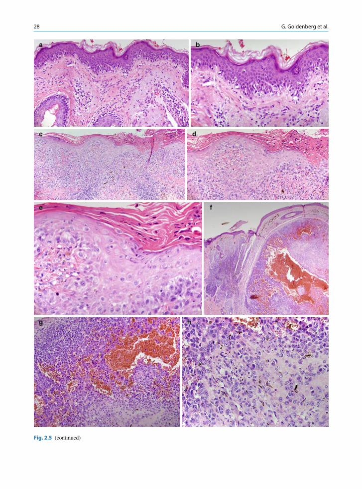
Malignant melanoma in situ (MMIS), including lentigo maligna (LM) type, presents with atypical mel-anocytes confi ned to the epidermis (Fig. 2.5a, b). The epidermis is typically atrophic in LM type of MMIS. The presence of single cell melanocytes, junctional nests of atypical melanocytes, extension of melano-cytes above the basal layer of the epidermis, confl uent spread of atypical cells, and follicular extension have been used as criteria for diagnosis of MMIS [15, 16]. Solar elastosis and melanophages are often found in the dermis. Lentigo maligna melanoma (LMM) arises from LM, and is characterized by the same fi ndings within the epidermis as LM, with dermal invasion by atypical melanocytes.
Superfi cial spreading malignant melanoma (SSMM) presents with atypical melanocytes at all levels of the epidermis, with signifi cant pagetoid spread (Figs. 2.5c–e). Atypical melanocytes found in the dermis may be present singly and in nests. Atypical dermal
melanocytes show failure of maturation with descent. While the majority of atypical melanocytes are epithe-lioid, spindle cell melanocytes may also be seen.
Nodular malignant melanoma (NMM) presents with atypical melanocytes within the epidermis, with page-toid spread, and in the underlying dermis (Fig. 2.5f–h). This MM subtype is characterized by the lack of atypi-cal melanocytes within the epidermis adjacent to the dermal component. Dermal melanocytes fail to mature and may show signifi cant mitotic activity.
Acral lentiginous malignant melanoma (ALMM) initially presents with a lentiginous radial growth phase of melanocytes within the epidermis, but as the tumor becomes thicker, nests on melanocytes with pagetoid spread may be seen (Fig. 2.5i, j) [17]. Halos surround-ing melanocytes may be seen, giving the atypical cells a lacunar appearance. Invasive dermal melanocytes may be seen singly or in nests, composed of epithe-lioid or spindle-shaped cells.
Desmoplastic malignant melanoma (DMM) is a rare type of MM that presents with dermal elongated spin-dle-shaped melanocytes, which may show nuclear hyperchromasia, bizarre nuclei, and lack melanin pig-ment (Fig. 2.5k, l). Melanocytes in DMM usually lack pigment and may be highlighted by immunochemical stains (Fig. 2.5m). Fascicles of atypical melanocytes may show an infi ltrative growth pattern, extending into subcutaneous fat. Abundant desmoplastic collagen bun-dles are usually present in the dermis. Lesions which present with less collagen and more spindle-shaped cells are referred to as spindle cell malignant melanoma (SCMM), although it has been shown that SCMM and DMM form a continuum without discrete separation [18]. An intraepidermal atypical melanocytic prolifera-tion is also observed in the majority of DMM [19].
Table 2.6 Clark’s level of invasion
Level I: Melanoma in situLevel II: Microinvasion into papillary dermisLevel III: Expansion into papillary dermisLevel IV: Invasion into reticular dermisLevel V: Invasion into subcutaneous fat
Table 2.7 Selected stains utilized in MM
S100HMB-45Melan-A/MART-1
28 G. Goldenberg et al.
a b
c d
e f
g h
Fig. 2.5 (continued)

2 Histopathology of Skin Cancer 29
Fig. 2.5 Malignant melanoma. (a, b) Lentigo maligna type showing single cell spread of melanocytes along the dermal-epidemal junction; (c, e) Superfi cial spreading malignant mela-noma showing signifi cant pagetoid spread; (f, h) Nodular malignant melanoma showing dermal invasion without signifi -cant later spread along the dermal-epidermal junction; (i, j)
Acral lentiginous malignant melanoma showing an atypical melanocytic proliferation on acral skin; (k, l) Desmoplastic malignant melanoma showing atypical melanocytes embedded in a desmoplastic stroma; and (m) S100 stain highlights atypical melanocytes in this Desmoplastic malignant melanoma
i j
k l
m

30 M. Ulrich and J. Kanitakis
2.5 Merkel cell carcinoma
Martina Ulrich, Jean Kanitakis
The diagnosis of Merkel cell carcinoma (MCC) is usu-ally made by histologic examination as the clinical features are rather nonspecifi c. Histopathologically, MCC represents a dermal tumor composed of round, small basophilic monomorphous cells with large nuclei, prominent nucleoli, and inconspicuous cyto-plasm. Mitotic fi gures are commonly seen, and necro-sis and ulceration may occur. The tumor usually spares the papillary dermis and the epidermis, and extends from the reticular dermis to the subcutaneous tissue. However, spread to the hypodermis and epidermis with pagetoid infi ltration might occur [1]. Three pathologi-cal subtypes of MCC have been described, i.e., the tra-becular, the small cell, and the intermediate type, which is the commonest one [2].
The differential diagnosis includes other tumors made of small cells, namely small cell lung cancer metastasis, lymphoma, or melanoma. Immunohistochemistry is needed for the confi rmation of diagnosis. MCC cells
express CK20 (and occasionally also neurofi laments) with a typical perinuclear dot pattern (Fig. 2.6), Neuron Specifi c Enolase, chromogranin A, and synaptophysin. Contrasting with small cell lung cancer, MCC does not express the Thyroid Transcription Factor 1 (TTF-1) (Table 2.8).
References
1. Kanitakis J, Euvrard S, Chouvet B et al (2006) Merkel cell carcinoma in organ-transplant recipients: report of two cases with unusual histological features and literature review. J Cutan Pathol 33:686–694
2. Ratner D, Nelson BR, Brown MD, et al (1993) Merkel cell carcinoma. Journal of the American Academy of Dermatology 29(2 part 1):143–156
Table 2.8 Immunohistochemical features of MCC in comparison with other small cell tumors
CK20 TTF-1 Vimentin NSE Chromogranin S100 protein LCA
Merkel cell carcinoma + − − + +/− − −Small cell lung cancer +/− + − +/− +/− − −Melanoma − − + +/− − + −Lymphoma − − + −/+ − − +
CK20 cytokeratin 20; TTF-1 thyroid transcription factor-1; NSE neuron-specifi c enolase; LCA leucocyte common antigen
Fig. 2.6 (a) Pathology of Merkel cell carcinoma: the tumour is made of uniform, large round cells with basophilic nuclei, invad-ing diffusely the dermis(haematoxylin-eosin stain). (b) Merkel
cell carcinoma: tumor cells express CK20 in a dot-like or signe-tring-like pattern (immunoperoxidase)
a b

2 Histopathology of Skin Cancer 31
2.6 Kaposi’s Sarcoma
G. Goldenberg, L.E. Golitz, J. Fitzpatrick
The histologic presentation of Kaposi’s sarcoma (KS) is similar, regardless of the clinical disease presenta-tion [20, 21]. The unifying histologic features of KS are the presence of atypical, irregular, and angulated vascular channels (Fig. 2.7a,b). The promontory sign is often described in KS and refers to irregular vascular channels that partially surround preexisting blood ves-sels. The presence of plasma cells in the surrounding stroma is also a classic fi nding.
The histologic presentation of KS evolves through patch, plaque, and tumor stages, similar to clinical dis-ease [22]. Patch stage KS present with irregular, angu-lated vascular channels in the reticular dermis. While ectatic vascular channels may be seen in this stage, the vessels may be very subtle and present with slit-like spaces and spindle-shaped cells. Plaque stage KS
presents with obvious vascular channels fi lling the entire dermis and extending into the superfi cial subcutaneous fat. The presence of a signifi cant spindle cell component is the most characteristic feature of this stage of KS. These cells intercalate between collagen bundles, form-ing slit-like, irregular vascular channels. Hemosiderin deposits and PAS-positive hyaline globules are com-monly seen in this stage of KS. Fascicles and sheets of spindle-shaped cells characterize the tumor stage of KS, along with a variable number of slit-like vascular chan-nels. Mitotic fi gures vary in number, but may be fre-quent in this stage of KS.
Evidence of human herpesvirus 8 (HHV-8) has been found by polymerase chain reaction in approximately 95% of KS lesions, and appears to be independent of the type of the disease (i.e., endemic type vs. AIDS-associated type and etc.) [23]. An immunohistochemistry stain for HHV-8 is available, and has been shown to be 99% sensi-tive and 100% specifi c for KS lesions (Fig. 2.7c) [24]. Immunohistochemical staining with CD-31, CD-34, fac-tor VIII antigen, and CD-40 has also been found useful.
a
b
c1
c2
Fig. 2.7 Kaposi’s sarcoma. (a, b) Numerous atypical, irregular, and angulated vascular channels; (c) positive staining with human herpesvirus 8 immunohistochemical stain

32 G. Goldenberg et al.
2.7 Dermatofi brosarcoma Protuberans
G. Goldenberg, L.E. Golitz, J. Fitzpatrick
Dermatofi brosarcoma protuberans (DFSP), which is associated with the rearrangement of chromosomes 17 and 22 with the fusion between the collagen type I1 gene and the platelet-derived growth factor b-chain gene, is a spindle cell neoplasm that shows an infi ltra-tive growth pattern (Fig. 2.8a, b) [25]. The main por-tion of this neoplasm shows a storiform arrangement with extension into the subcutaneous fat, with fat entrapment creating a honeycomb pattern [26]. Cytologically, there is usually little nuclear pleomor-phism and a low-to-moderate mitotic index. DFSP is a highly cellular malignancy with scant collagen. Several
histologic variants of DFSP have been reported, includ-ing myxoid, granular cell, and pigmented types.
Expression of CD-34 antigen (human progenitor cell antigen) in DFSP (Fig. 2.8c) is well described and has been used to support the view that these lesions are variants of nerve sheath tumors, which are distinct from benign fi brous histiocytomas that do not express CD-34 [27]. Immunohistochemical staining with vimentin, and more recently CD-10, has also been reported [28, 29].
Fig. 2.8 Dermatofi brosarcoma protuberans. (a, b) Atypical spindle cells show an infi ltrative growth patters and storiform arrangement, with extension into the subcutaneous fat; (c) Positive CD-34 immunohistochemical stain
a
b

2 Histopathology of Skin Cancer 33
2.8 Atypical Fibroxanthoma
G. Goldenberg, L.E. Golitz, J. Fitzpatrick
Atypical fi broxanthoma is a dermal neoplasm usually separated from the overlying epidermis by a thin Grenz. It is composed of multiple cell-types, including spindle, polyhedral, giant, clear, granular and osteoid cells (Fig. 2.9a, b) [30]. Spindle cells may predomi-nate, show pleomorphism, contain vesicular nuclei, and often form fascicles. Polyhedral cells usually show a vacuolated lipid-containing cytoplasm, and are large and haphazardly arranged. Giant cells are multinucle-ated and pleomorphic, and bizarre mitosis are com-mon in his variant. By defi nition, this is a superfi cial neoplasm, without the involvement of the deep dermis and subcutis. The overlying epidermis is usually effaced and the surrounding dermis usually shows solar elastosis.
Immunohistochemically, AFX stains positive with vimentin, and staining with CD10 and procollagen-1 (Fig. 2.9c) has also been recently described [31, 32].
a
b
c
Fig. 2.9 Atypical fi broxanthoma. (a, b) Spindle cells showing pleomorphism, nuclear hyperchromasia, numerous mitosis, and arranged in fascicles; (c) Positive Procollogen I immunohis-tochemical stain

34 G. Goldenberg et al.
2.9 Malignant Fibrous Histiocytoma
G. Goldenberg, L.E. Golitz, J. Fitzpatrick
The term “malignant fi brous histiocytoma” (MFH) has fallen out of favor, and most of these tumors have been reclassifi ed in the latest World Health Organization Classifi cation of soft tissue tumors [33]. A recent study that utilized comparative genomic hybridization dem-onstrated that most MFHs do not constitute a homoge-neous entity, but could correspond other sarcomas, particularly leiomyosarcoma and liposarcoma [34, 35]. The classic histologic presentation of MFH has been divided into fi ve types: pleomorphic, angiomatoid, myxoid, giant cell, and infl ammatory. Architecturally, MFH is a dermal neoplasm with an infi ltrative border. The pleomorphic type is most common, and presents with plump, atypical spindle cells that may be arranged in a storiform pattern, bizarre giant cells, and nodules and sheets of histiocytes. The angiomatoid variant shows large blood fi lled spaces, admixed with atypical spindle-shaped cell.
Immunohistochemical staining is usually positive with vimentin, and staining with CD74 and CD68 has also been described [36–38].
References
1. Walsh JS, Perniciaro C, Randle HW (1999) Calcifying basal cell carcinomas. Dermatol Surg 25(1):49–51
2. Crowson, AN (2006) Basal cell carcinoma: biology, mor-phology and clinical implications. Mod Pathol 19(Suppl 2):S127–S147
3. Crowson AN, Magro CM, Kadin M et al (1996) Differential expression of bcl-2 oncogene in human basal cell carcinoma. Hum Pathol 27:355–359
4. Jones CC, Ansari SJ, Tschen JA (1991) Cystic fi broepithe-lioma of Pinkus. J Cutan Pathol 18:220–222
5. Spoonemore K, Crowson AN (2004) Eccrine syringofi -broadenoma in a patient with chronic graft-versus-host dis-ease (Abstract). J Cutan Pathol 1:128–129
6. Latza U, Niedobitek G, Schwarting R, Nekarda H, Stein H (1990) Ber-EP4: new monoclonal antibody which distin-guishes epithelia from mesothelial. J Clin Pathol 43:213
7. Tellechea O, Reis JP, Domingues JC, Baptista AP (1993) Monoclonal antibody Ber EP4 distinguishes basal-cell carci-noma from squamous-cell carcinoma of the skin. Am J Dermatopathol 15:452
8. Jones MS, Helm KF, Maloney ME (1997) The immunohis-tochemical characteristics of the basosquamous cell carci-noma. Dermatol Surg 23:181
9. Beer TW, Shepherd P, Theaker JM (2000) Ber EP4 and epi-thelial membrane antigen aid distinction of basal cell, squamous cell and basosquamous carcinomas of the skin. Histopathology 37:218
10. Rossen K, Thomsen HK (2001) Ber-EP4 immunoreactivity depends on the germ layer origin and maturity of the squamous epithelium. Histopathology 39:386
11. Krahl D, Sellheyer K (2007) Monoclonal antibody Ber-EP4 reliably discriminates between microcystic adnexal carci-noma and basal cell carcinoma. J Cutan Pathol Oct;34(10): 782–787
12. Ackerman AB (1980) Malignant melanoma: a unifying con-cept. Hum Pathol 11(6):591–595
13. Elder DE (2006) Pathology of melanoma. Clin Cancer Res 12(7 Pt 2):2308s–2311s
14. Clark WH et al (1989) Model predicting survival in stage I melanoma based on tumor progression. J Natl Cancer Inst 81(24):1893–1904
15. Weyers W, Bonczkowitz M, Weyers I, Bittinger A, Schill WB (1996) Melanoma in situ versus melanocytic hyperpla-sia in sun-damaged skin. Assessment of the signifi cance of histopathologic criteria for differential diagnosis. Am J Dermatopathol Dec;18(6):560–566
16. Flotte TJ, Mihm MC Jr (1999) Lentigo maligna and malig-nant melanoma in situ, lentigo maligna type. Hum Pathol 30(5):533–536
17. Kuchelmeister C, Schaumburg-Lever G, Garbe C (2000) Acral cutaneous melanoma in caucasians: clinical features, histopathology and prognosis in 112 patients. Br J Dermatol Aug 143(2):275–280
18. Thelmo MC, Sagebiel RW, Treseler PA, Morita ET, Nguyen LH, Kashani-Sabet M, Leong SP (2001) Evaluation of sentinel lymph node status in spindle cell melanomas. J Am Acad Dermatol 44(3):451–455
19. Carlson JA, Dickersin GR, Sober AJ, Barnhill RL (1995) Desmoplastic neurotropic melanoma. A clinicopathologic analysis of 28 cases. Cancer 75(2):478–494
20. Leu HJ, Odermatt B (1985) Multicentric angiosarcoma (Kaposi’s sarcoma). Light and electron microscopic and immu-nohistological fi ndings of idiopathic cases in Europe and Africa and of cases associated with AIDS are histologically identical. Virchows Arch A Pathol Anat Histopathol 408(1):29–41
21. Chow JW, Lucas SB (1990) Endemic and atypical Kaposi’s sarcoma in Africa – histopathological aspects. Clin Exp Dermatol 15(4):253–259
22. Cottoni F, Montesu MA (1996) Kaposi’s sarcoma classifi ca-tion: a problem not yet defi ned. Int J Dermatol 35(7): 480–483
23. Olsen SJ, Moore PS (1998) Kaposi’s sarcoma-associated herpesvirus (KSHV/HHV-8) and the etiology of KS. In: Medveczky P, Friedman H, Bendinelli M (eds) Herpesviruses and immunity. Plenum, New York
24. Robin YM et al (2004) Human herpesvirus 8 immunostain-ing: a sensitive and specifi c method for diagnosing Kaposi sarcoma in paraffi n-embedded sections. Am J Clin Pathol 121:330–334
25. Shimizu A, O’Brien KP, Sjöblom T et al (1999) The der-matofi brosarcoma protuberans-associated collagen type I1/platelet-derived growth factor (PDGF) B-chain fusion gene generates a transforming protein that is processed to func-tional PDGF-BB. Cancer Res 59:3719–3723
2 Histopathology of Skin Cancer 35
26. Weiss SW, JR Goldblum (2001) Fibrohistiocytic tumors of intermediate malignancy. In: SW Weiss, JR Goldblum (eds) Enzinger and Weiss’s soft tissue tumors, 4th edn. Mosby, St Louis, pp 491–534
27. Weiss SW, Nickoloff BJ (1993) CD-34 is expressed by a distinctive cell population in peripheral nerve, nerve sheath tumors, and related lesions. Am J Surg Pathol 17: 1039–1045
28. Terrier-Lacombe MJ, Guillou L, Maire G, Terrier P, Vince DR, de Saint Aubain Somerhausen N, Collin F, Pedeutour F, Coindre JM (2003) Dermatofi brosarcoma pro-tuberans, giant cell fi broblastoma, and hybrid lesions in chil-dren: clinicopathologic comparative analysis of 28 cases with molecular data – a study from the French Federation of Cancer Centers Sarcoma Group. Am J Surg Pathol 27(1): 27–39
29. Kanitakis J, Bourchany D, Claudy A (2000) Expression of the CD10 antigen (neutral endopeptidase) by mesenchymal tumors of the skin. Anticancer Res 20(5B):3539–3544
30. Dettrick A, Geoff S (2006) Atypical fi broxanthoma with perineural or intraneural invasion: report of two cases. J Cutan Pathol 33:318–322
31. Weedon D, Williamson R, Mirza B (2005) CD10, a useful marker for atypical fi broxanthomas. Am J Dermatopathol 27(2):181
32. Jensen K, Wilkinson B, Wines N, Kossard S (2004) Procollagen 1 expression in atypical fi broxanthoma and other tumors. J Cutan Pathol 31(1):57–61
33. Fletcher CD (2006) The evolving classifi cation of soft tissue tumours: an update based on the new WHO classifi cation. Histopathology 48(1):3–12
34. Derré J, Lagace R, Nicolas A et al (2001) Leiomyosarcomas and most malignant fi brous histiocytomas share very similar comparative genomic hybridization imbalances: an analysis of a series of 27 leiomyosarcomas. Lab Invest 81:211–215
35. Chibon F, Mariani O, Mairal A et al (2003) The use of clustering software for the classifi cation of comparative genomic hybridization data. An analysis of 109 malignant fi brous histiocytomas. Cancer Genet Cytogenet 141:75–78
36. Iwasaki H, Isayama T, Johzaki H, Kikuchi M (1987) Malignant fi brous histiocytoma. Evidence of perivascular mesenchymal cell origin immunocytochemical studies with monoclonal anti-MFH antibodies. Am J Pathol 128(3):528–537
37. Lazova R, Moynes R, May D, Scott G (1997) LN-2 (CD74) A marker to distinguish atypical fi broxanthoma from malig-nant fi brous histiocytoma. Cancer 79(11):2115–2124
38. Smith ME, Costa MJ, Weiss SW (1991) Evaluation of CD68 and other histiocytic antigens in angiomatoid malignant fi brous histiocytoma. Am J Surg Pathol 15(8):757–763

http://www.springer.com/978-3-540-79346-5