Histopathology of Cutaneous Vasculitiscdn.intechweb.org/pdfs/22428.pdf · Histopathology of...
39
2 Histopathology of Cutaneous Vasculitis Ko-Ron Chen Head of Department of Dermatology, Saiseikai Central Hospital Tokyo, Japan 1. Introduction Vasculitis is an inflammatory process occurring primarily on the vessel wall. It results in vessel wall destruction and subsequent hemorrhagic and ischemic events. Skin is the most common target organ for vasculitis. 1,2,3,4 Cutaneous vasculitis includes vasculitis of the dermal small vessels 5 and subcutaneous small muscular vessels. 6 It can be a self-limited benign disease restricted to the skin or be a significant component of many systemic vasculitic syndromes such as collagen disease- associated vasculitis or anti-neutrophil cytoplasmic antibody (ANCA)-associated primary vasculitic syndromes. Cutaneous vasculitis most frequently manifests as palpable purpura or infiltrated erythema (indicating dermal small vessel vasculitis), and less frequently as nodular erythema, livedo racemosa, deep ulcers, or digital gangrene (indicating deep dermal vasculitis or subcutaneous muscular-vessel vasculitis). Cutaneous vasculitis presents as a constellation of clinical and histopathologic findings since the size of vessel involved correlates with the clinical findings. 1,2,3,4 A diagnosis of cutaneous vasculitis is best determined by the histopathologic findings of the vessel size involved and the principal inflammatory response. Histopathologic findings are also crucial for distinguishing authentic cutaneous vasculitis from cutaneous pseudovasculitis 7 (a heterogeneous collection of disorders that clinically produce hemorrhages [i.e., petechiae, purpura, and ecchymoses] or vessel occlusion [which results in livedo, cyanosis, ulcers, digital necrosis, and/or gangrene] and simulate cutaneous vasculitis but without the histopathologic evidence of vasculitis). 7 This chapter will describe the morphological approach to cutaneous vasculitis, including: 1. The diagnostic criteria for small vessel vasculitis and for subcutaneous muscular vessel vasculitis (e.g., arteritis and phlebitis); 2. The details of the inflammatory stages of muscular vessel vasculitis (based on morphological changes during the inflammatory process, which strongly affect the treatment for vasculitis); 3. The histopathologic classification of cutaneous vasculitis (based on the size of the affected vessel [e.g., small vessels or muscular vessels] and the predominant inflammatory cell types [e.g., neutrophils, eosinophils, lymphocytes, histiocytes]) and their related disorders. Distinguishing between subcutaneous muscular arteritis and phlebitis will be also discussed since the morphological features of the subcutaneous veins in the lower legs, such as a www.intechopen.com
Transcript of Histopathology of Cutaneous Vasculitiscdn.intechweb.org/pdfs/22428.pdf · Histopathology of...
2
Histopathology of Cutaneous Vasculitis
Ko-Ron Chen Head of Department of Dermatology, Saiseikai Central Hospital Tokyo,
Japan
1. Introduction
Vasculitis is an inflammatory process occurring primarily on the vessel wall. It results in vessel wall destruction and subsequent hemorrhagic and ischemic events. Skin is the most common target organ for vasculitis.1,2,3,4 Cutaneous vasculitis includes vasculitis of the dermal small vessels5 and subcutaneous
small muscular vessels.6 It can be a self-limited benign disease restricted to the skin or be a
significant component of many systemic vasculitic syndromes such as collagen disease-
associated vasculitis or anti-neutrophil cytoplasmic antibody (ANCA)-associated primary
vasculitic syndromes.
Cutaneous vasculitis most frequently manifests as palpable purpura or infiltrated erythema
(indicating dermal small vessel vasculitis), and less frequently as nodular erythema, livedo
racemosa, deep ulcers, or digital gangrene (indicating deep dermal vasculitis or
subcutaneous muscular-vessel vasculitis). Cutaneous vasculitis presents as a constellation of
clinical and histopathologic findings since the size of vessel involved correlates with the
clinical findings.1,2,3,4
A diagnosis of cutaneous vasculitis is best determined by the histopathologic findings of the vessel size involved and the principal inflammatory response. Histopathologic findings are also crucial for distinguishing authentic cutaneous vasculitis from cutaneous pseudovasculitis7 (a heterogeneous collection of disorders that clinically produce hemorrhages [i.e., petechiae, purpura, and ecchymoses] or vessel occlusion [which results in livedo, cyanosis, ulcers, digital necrosis, and/or gangrene] and simulate cutaneous vasculitis but without the histopathologic evidence of vasculitis).7 This chapter will describe the morphological approach to cutaneous vasculitis, including: 1. The diagnostic criteria for small vessel vasculitis and for subcutaneous muscular vessel
vasculitis (e.g., arteritis and phlebitis); 2. The details of the inflammatory stages of muscular vessel vasculitis (based on
morphological changes during the inflammatory process, which strongly affect the treatment for vasculitis);
3. The histopathologic classification of cutaneous vasculitis (based on the size of the affected vessel [e.g., small vessels or muscular vessels] and the predominant inflammatory cell types [e.g., neutrophils, eosinophils, lymphocytes, histiocytes]) and their related disorders.
Distinguishing between subcutaneous muscular arteritis and phlebitis will be also discussed since the morphological features of the subcutaneous veins in the lower legs, such as a
www.intechopen.com
Advances in the Diagnosis and Treatment of Vasculitis
20
compact concentric smooth muscle pattern with a round lumen and no obvious bundled formation, mimic features that are characteristic of arteries. This may lead to phlebitis or thrombophlebitis being misdiagnosed as arteritis and result in unnecessary overtreatment.
2. Morphology and distribution of cutaneous vessels
Types of vessels
Small artery Small vein Arteriole Venule and capillary
Small arteries are defined as arteries having a caliber of 100 m to 1000 m
Where a skin biopsy reaches into the subcutis, the arterial caliber level is less than 1000m. Therefore, histopathologically proven arteritis identified from skin lesions should involve small-sized arteries, rather than medium-sized arteries (which have calibers larger than 1000
m). The distribution of small arteries is from the lower dermis (where the arterial size
ranges from 100 m to 200 m) to the subcutis (where the arterial size ranges from 100 m to
1000 m). Small arteries usually have a tunica intima (containing an endothelial cell layer with an underlying layer of a sharp internal elastic lamina) and a tunica media (containing two to 10 concentric compact continuous smooth muscular layers with very few elastic fibers in the muscular layer). These are characteristic of small arteries. The tunica adventitia – the outer layer of small arteries – is generally unidentifiable, having few perivascular collagen fibers and identifiable elastic fibers (Figures 1 and 2).
Small veins are defined as veins having a caliber between 100 μm and 1000 μm.
Small veins are distributed from the lower dermis to the subcutis. The lack of an internal elastic lamina and discontinuous bundles of muscle in the elastic fiber-rich muscular medial layer are characteristic of small veins (Figures1 and 2).
Arterioles are defined as arteries having a caliber of less than 100 μm.
The tunica intima in arterioles is characterized by a layer consisting only of endothelial cells
with an underlying internal elastic lamina, as seen in small arteries. The tunica media in
arterioles is characterized by approximately one to two concentric smooth muscular layers.
The tunica adventitia in arterioles is usually unidentifiable (Figure 3).
Venules are defined as veins having a caliber of less than 100 μm.
Venules contain one to two muscular layers in the tunica media and may have a
perivascular fibrotic layer in the tunica adventitia. The lack of the internal elastic lamina is a
feature that distinguishes venules from arterioles (Figures 3 and 4).
All small vessels in the superficial to mid-dermis are arterioles, venules, and capillaries
since the vessels are smaller than 100 μm (Figure 3). All of the small vessels in the dermis
are less than 200 μm, including capillaries, venules, arterioles, small veins and small
arteries. (The small arteries and veins in the dermis range from 100 μm to 200 μm.). The
size of the small arteries at the dermo-subcutaneous junction ranges from 200 μm to 400
μm (Figures 2, 22D, and 22F). These arteries are commonly involved in cutaneous
polyarteritis nodosa.
www.intechopen.com
Histopathology of Cutaneous Vasculitis
21
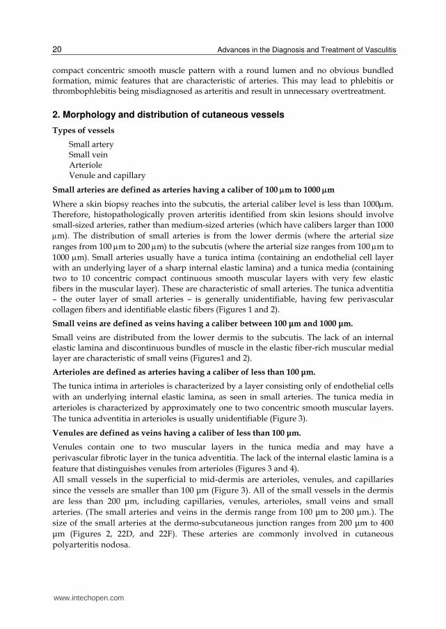
Fig. 1. (elastic tissue stain) The distinction between a deep dermal small artery and vein.
Note the characteristic sharp internal elastic fiber encircling the lumen but the absence of
elastic fiber in the muscular medial layer of the artery. By contrast, there is an absence of an
internal elastic lamina and there are multiple layers of elastic fibers in the medial muscular
layer of the vein.
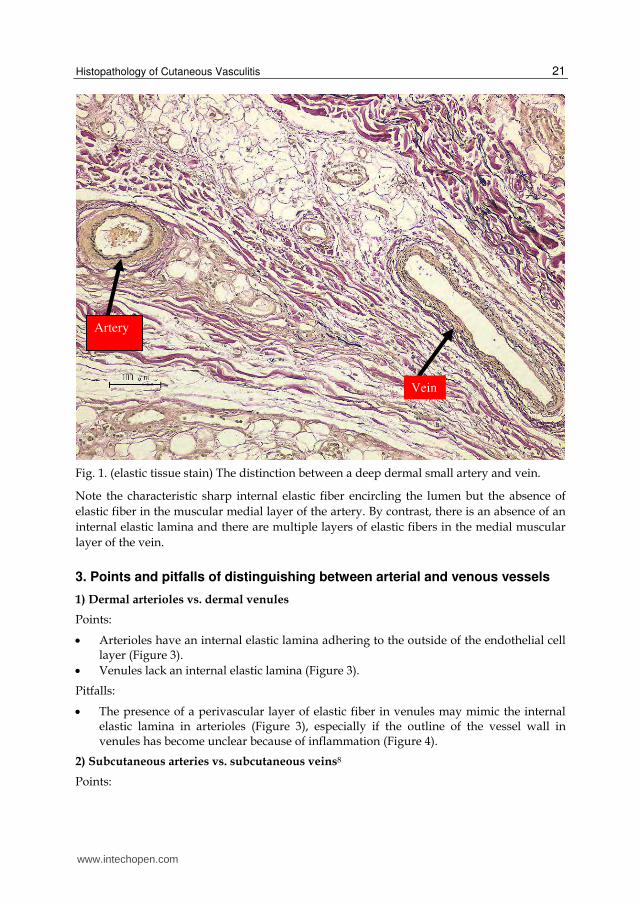
3. Points and pitfalls of distinguishing between arterial and venous vessels
1) Dermal arterioles vs. dermal venules
Points:
Arterioles have an internal elastic lamina adhering to the outside of the endothelial cell layer (Figure 3).
Venules lack an internal elastic lamina (Figure 3).
Pitfalls:
The presence of a perivascular layer of elastic fiber in venules may mimic the internal elastic lamina in arterioles (Figure 3), especially if the outline of the vessel wall in venules has become unclear because of inflammation (Figure 4).
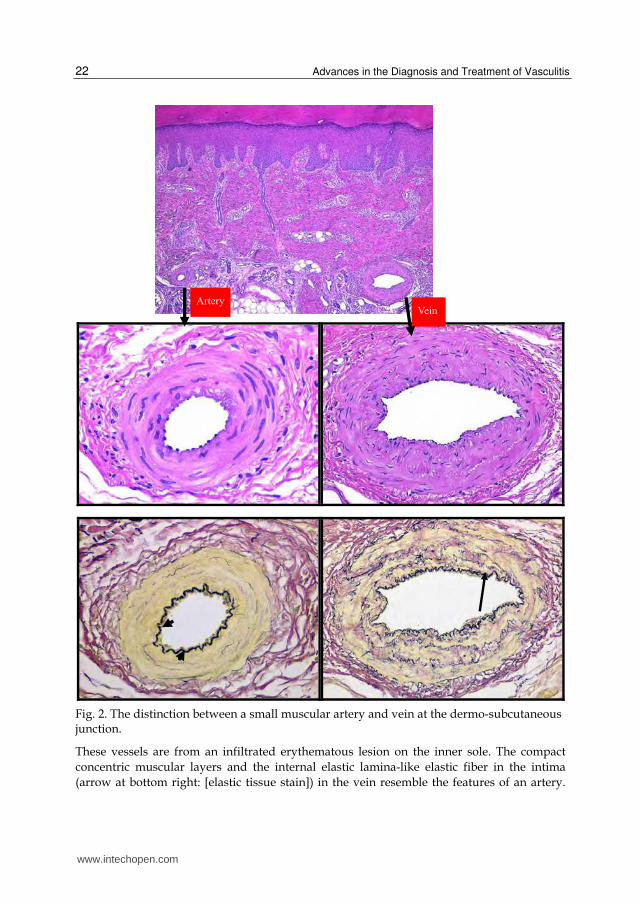
2) Subcutaneous arteries vs. subcutaneous veins8
Points:
Vein
Artery
www.intechopen.com
Advances in the Diagnosis and Treatment of Vasculitis
22
Fig. 2. The distinction between a small muscular artery and vein at the dermo-subcutaneous junction.
These vessels are from an infiltrated erythematous lesion on the inner sole. The compact
concentric muscular layers and the internal elastic lamina-like elastic fiber in the intima
(arrow at bottom right: [elastic tissue stain]) in the vein resemble the features of an artery.
www.intechopen.com
Histopathology of Cutaneous Vasculitis
23
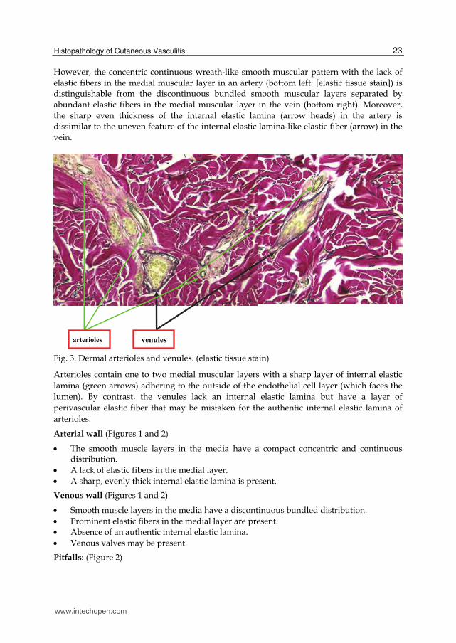
However, the concentric continuous wreath-like smooth muscular pattern with the lack of
elastic fibers in the medial muscular layer in an artery (bottom left: [elastic tissue stain]) is
distinguishable from the discontinuous bundled smooth muscular layers separated by
abundant elastic fibers in the medial muscular layer in the vein (bottom right). Moreover,
the sharp even thickness of the internal elastic lamina (arrow heads) in the artery is
dissimilar to the uneven feature of the internal elastic lamina-like elastic fiber (arrow) in the
vein.
Fig. 3. Dermal arterioles and venules. (elastic tissue stain)
Arterioles contain one to two medial muscular layers with a sharp layer of internal elastic
lamina (green arrows) adhering to the outside of the endothelial cell layer (which faces the
lumen). By contrast, the venules lack an internal elastic lamina but have a layer of
perivascular elastic fiber that may be mistaken for the authentic internal elastic lamina of
arterioles.
Arterial wall (Figures 1 and 2)
The smooth muscle layers in the media have a compact concentric and continuous distribution.
A lack of elastic fibers in the medial layer.
A sharp, evenly thick internal elastic lamina is present.
Venous wall (Figures 1 and 2)
Smooth muscle layers in the media have a discontinuous bundled distribution.
Prominent elastic fibers in the medial layer are present.
Absence of an authentic internal elastic lamina.
Venous valves may be present.
Pitfalls: (Figure 2)
www.intechopen.com
Advances in the Diagnosis and Treatment of Vasculitis
24
The presence of the intimal elastic fibers in veins (arrow at the bottom right of Figure 2えmay mimic the internal elastic lamina in arteries (arrow heads at bottom left of
Figure 2), while the uneven thickness and partial multilayer of the intimal elastic fibers in veins are distinguishable.
Morphological features of the veins obtained from skin lesions of the dorsal or plantar feet may show a thick compact and continuous smooth muscle layer distribution similar to arteries.8 The bundled smooth muscular layers, separated by abundant elastic fibers in the media of veins (bottom right of Figure 2), is distinguishable from the concentric and continuous smooth muscular layers with a lack of elastic fibers in the arterial media (bottom left of Figure 2).
4. Concept of cutaneous vasculitis
Vasculitis is an inflammatory process occurring primarily on the vessel wall, leading to
vessel wall destruction and subsequent hemorrhagic and ischemic events. Cutaneous
vasculitis comprises vasculitis of the dermal small vessels and the subcutaneous small
muscular vessels. Basic principle of histopathologic diagnosis for cutaneous vasculitis is the
morphological evidence of both (1) angiocentric infiltration of inflammatory cells and (2)
damage of vessel wall. Therefore the following vasculitis-like conditions, which are different
from the process of vessel damage in vasculitis, should not be interpreted as cutaneous
vasculitis:
1. Vessel damage that is not caused by inflammatory cells, but is caused by a direct invasion of tumor cells or organisms, including fungi, bacteria, and viruses.
2. Vessel damage as a secondary event (i.e., innocent bystanders) caused by primary
necrosis or by destruction of the connective tissue. The adjacent innocent vessels are
secondarily involved in the resultant vessel destruction. This phenomenon of innocent
bystanders-like secondary vessel damage is most often found in lesions of pyoderma
gangrenosum, overlying ulcerative lesions, dermal/subcutaneous abscesses, and
dermal/subcutaneous caseous necrosis.
5. Histopathologic diagnosis of cutaneous vasculitis
1) Biopsy: Timing, technique, and choice of lesions
An appropriate skin biopsy is the key to obtaining a significant result for cutaneous vasculitis. Timing of biopsy:
The pathologic features of vasculitis may be absent if the biopsy is poorly timed. A skin biopsy should not be performed for purpuric or infiltrated lesions after 48 hours, and a skin biopsy should not be performed for deep nodular erythematous lesions after 72 hours.
Choice of lesions and biopsy technique:
A biopsy extending to the subcutis should be taken from the most palpable purpuric lesions or the most tender, reddish nodular or infiltrated lesions.
For purpuric lesions or infiltrated erythematous lesions smaller than 5 mm, a 3 to 4 mm large punch biopsy extending to the subcutis will enable histopathologic confirmation..
www.intechopen.com
Histopathology of Cutaneous Vasculitis
25
For nodular erythema lesions or infiltrated erythema larger than 5 mm, a spindle-shaped incisional biopsy extending to the subcutis with a 5 to 6 mm large tissue sample should be performed on the central lesions.
It is better to perform two biopsies. The first, a biopsy of the most palpable and tender lesions for light microscopy examination; the second, a punch biopsy of an early erythematous or purpuric lesion for a direct immunofluorescence (DIF) examination.
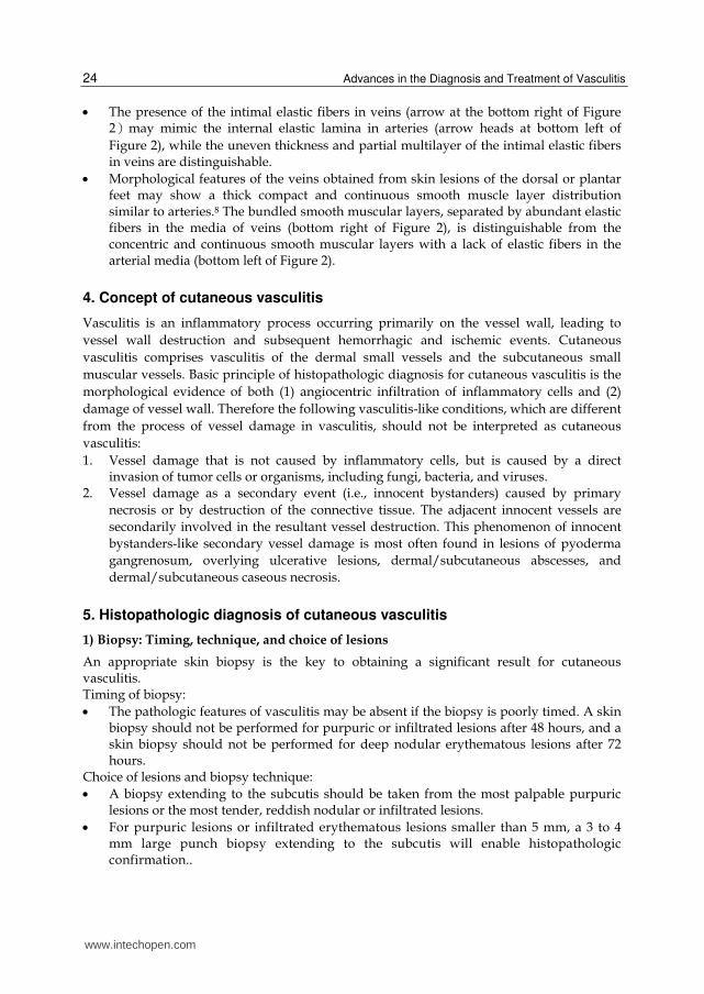
Fig. 4. Diagnostic pitfall of the internal elastic lamina-like perivascular elastic fiber (arrows) in a venule with necrotizing vasculitis. (Right): elastic tissue stain
2) Significance of direct immunofluorescence examination
A DIF examination is a crucial component of diagnosis.1 Palpable purpura on the lower legs with a histopathologic feature of leukocytoclastic vasculitis is the most common finding in vasculitis disorders with dermal small vessel involved, which can be found in ANCA-related vasculitis (e.g., Churg-Strauss syndrome, Wegener’s granulomatosis and microscopic polyangiitis); cryoglobulinemic purpura; collagen disease-associated vasculitis (e.g., rheumatoid arthritis, systemic lupus erythematosus, Sjögren disease); and Henoch-Schönlein purpura. It is therefore difficult to make a distinction just based on the cutaneous manifestations and histopathologic findings. However, the absence of immune complexes, the so-called pauci-immune vasculitis, is an expected finding in Churg-Strauss syndrome, Wegener’s granulomatosis, and microscopic polyangiitis. By contrast, deposition of IgM, and/or C3 in or around the vessels characterizes immune complex-mediated vasculitis, as found in cryoglobulinemic vasculitis, rheumatoid vasculitis,9 and systemic lupus erythematosus. Vascular deposition of IgA, rather than IgG or IgM, is required for the diagnosis of Henoch-Schönlein purpura (Figure 5). Similar to hematoxylin and eosin evaluation, the presence of diagnostic immunofluorescence patterns is inversely related to the age of the lesion biopsied. Nearly 100% of biopsies will identify immunoglobulins within the first 48 hours, followed by 70% positive results at 48-72 hours; immunoglobulins will not be detected in the lesions after 72 hours.1
3) Diagnostic criteria for small vessel vasculitis and muscular vessel vasculitis
1. Diagnostic criteria for small vessel vasculitis9
Essential findings: i. Perivascular infiltration of inflammatory cells
www.intechopen.com
Advances in the Diagnosis and Treatment of Vasculitis
26
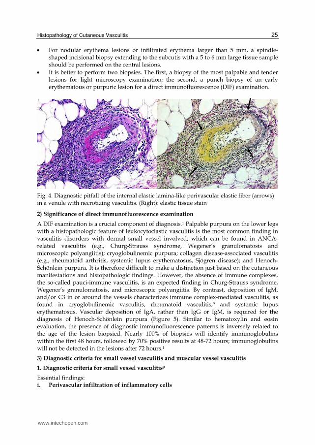
ii. Fibrinoid necrosis of the small vessel wall
Fig. 5. The presence of an IgA vascular deposit in the papillary dermis is required for a diagnosis of Henoch-Schönlein purpura.
A histopathologic diagnosis for small vessel vasculitis should fulfill both criteria I and II above.
Other minor features, including erythrocyte extravasation and nuclear dust, are not the
crucial findings for a diagnosis of small vessel vasculitis.
Q: Why is a perivascular infiltration of inflammatory cells indispensible for a diagnosis? A: Vasculitis is an inflammatory process occurring primarily at the vessel walls. A
perivascular infiltration of inflammatory cells is indispensible for a diagnosis and to avoid
the diagnostic pitfalls of:
1. Vessel damage caused by direct vessel invasion by bacteria, fungi (Figure 16), or tumor cells (Figure 20), rather than by inflammatory cells; and
2. Secondary vessel wall damage (i.e., innocent bystander damage) caused by adjacent primary necrotic connective tissue, as found in lesions of dermal or subcutaneous abscess (Figures 17 and 18).
Q: Why is fibrinoid necrosis of the vessel wall indispensible for a diagnosis of small vessel vasculitis? A: On light microscopy examination, the morphology of vessel wall fibrinoid necrosis is the sole finding of small vessel damage and is accepted as crucial in distinguishing small vessel vasculitis from vasculitis-like disorders with a perivascular infiltration of inflammatory cells (which would occur without evidence of fibrinoid necrosis of the vessel wall). Components of small vessels in the dermis mainly comprise one to two muscular layers
with an inner endothelial cell layer facing the lumen (Figure 3). The inflammatory cells can
pass from the lumen through the small vessel wall without damaging the vessel wall.
www.intechopen.com
Histopathology of Cutaneous Vasculitis
27
Therefore, the finding of perivascular inflammatory cells infiltrating into the vessel wall is
not convincing evidence of vessel wall damage. Endothelial cells, the main component of the
small vessel wall, play a key role in preventing blood from coagulation. Once inflammatory
cells such as neutrophils damage the endothelial cells, the endothelial cells lose their anti-
coagulation function (Figure 6), resulting in the formation of fibrin thrombi at the luminal
side of the damaged endothelial cell (Figures 6 and 7). A zone of a homogeneous
eosinophilic deposit, (i.e., fibrinoid necrosis) – mainly containing fibrin mixed with cellular
debris, including necrotic or apoptotic endothelial cells and, most often, degenerated
neutrophils, and serologic proteins such as immunoglobulins and complements – eventually
forms at the affected vessel wall and discharges outward to the perivascular area3,10 (Figures
6 and 8).
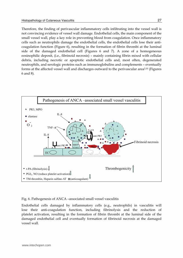
Fig. 6. Pathogenesis of ANCA –associated small vessel vasculitis
Endothelial cells damaged by inflammatory cells (e.g., neutrophils) in vasculitis will
lose their anti-coagulation function, including fibrinolysis and the reduction of
platelet activation, resulting in the formation of fibrin thrombi at the luminal side of the
damaged endothelial cell and eventually formation of fibrinoid necrosis at the damaged
vessel wall.
www.intechopen.com
Advances in the Diagnosis and Treatment of Vasculitis
28
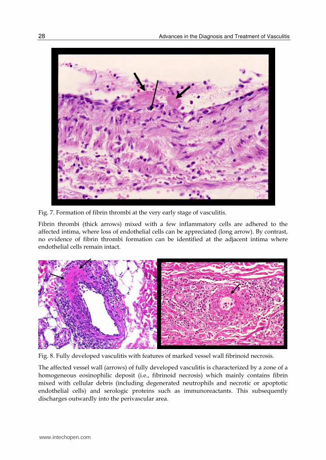
Fig. 7. Formation of fibrin thrombi at the very early stage of vasculitis.
Fibrin thrombi (thick arrows) mixed with a few inflammatory cells are adhered to the affected intima, where loss of endothelial cells can be appreciated (long arrow). By contrast, no evidence of fibrin thrombi formation can be identified at the adjacent intima where endothelial cells remain intact.
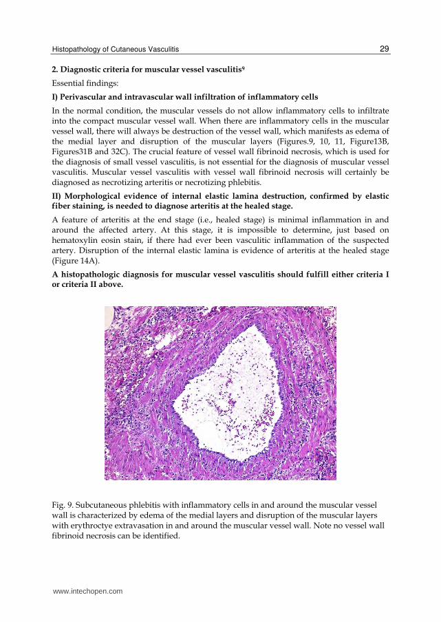
Fig. 8. Fully developed vasculitis with features of marked vessel wall fibrinoid necrosis.
The affected vessel wall (arrows) of fully developed vasculitis is characterized by a zone of a
homogeneous eosinophilic deposit (i.e., fibrinoid necrosis) which mainly contains fibrin
mixed with cellular debris (including degenerated neutrophils and necrotic or apoptotic
endothelial cells) and serologic proteins such as immunoreactants. This subsequently
discharges outwardly into the perivascular area.
www.intechopen.com
Histopathology of Cutaneous Vasculitis
29
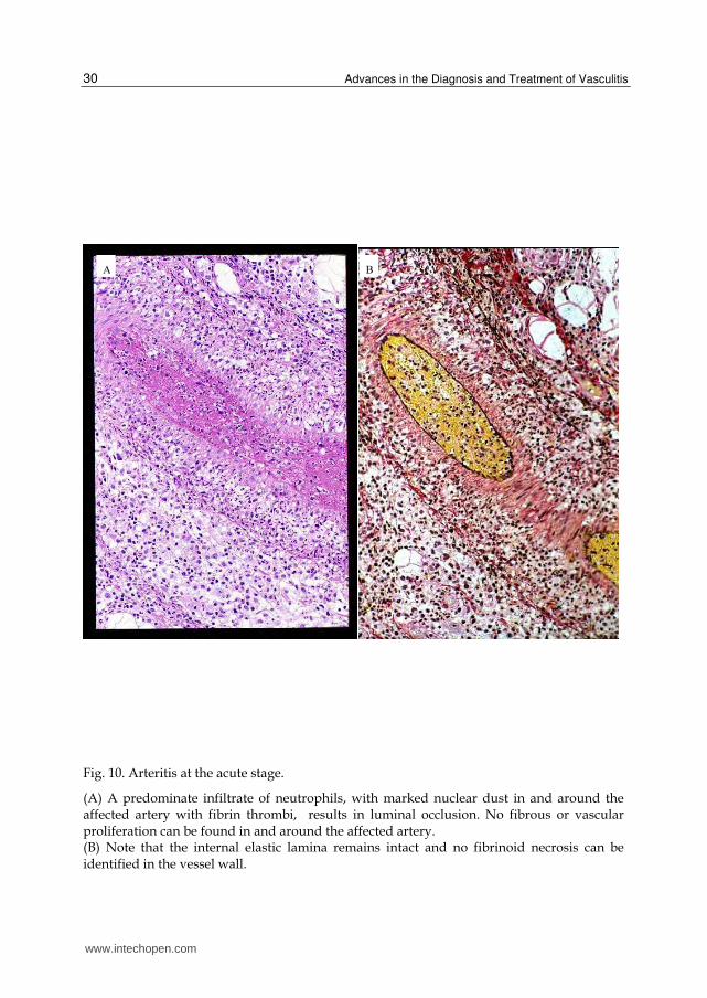
2. Diagnostic criteria for muscular vessel vasculitis9
Essential findings:
I) Perivascular and intravascular wall infiltration of inflammatory cells
In the normal condition, the muscular vessels do not allow inflammatory cells to infiltrate into the compact muscular vessel wall. When there are inflammatory cells in the muscular vessel wall, there will always be destruction of the vessel wall, which manifests as edema of the medial layer and disruption of the muscular layers (Figures.9, 10, 11, Figure13B, Figures31B and 32C). The crucial feature of vessel wall fibrinoid necrosis, which is used for the diagnosis of small vessel vasculitis, is not essential for the diagnosis of muscular vessel vasculitis. Muscular vessel vasculitis with vessel wall fibrinoid necrosis will certainly be diagnosed as necrotizing arteritis or necrotizing phlebitis.
II) Morphological evidence of internal elastic lamina destruction, confirmed by elastic fiber staining, is needed to diagnose arteritis at the healed stage.
A feature of arteritis at the end stage (i.e., healed stage) is minimal inflammation in and around the affected artery. At this stage, it is impossible to determine, just based on hematoxylin eosin stain, if there had ever been vasculitic inflammation of the suspected artery. Disruption of the internal elastic lamina is evidence of arteritis at the healed stage (Figure 14A).
A histopathologic diagnosis for muscular vessel vasculitis should fulfill either criteria I or criteria II above.
Fig. 9. Subcutaneous phlebitis with inflammatory cells in and around the muscular vessel wall is characterized by edema of the medial layers and disruption of the muscular layers with erythroctye extravasation in and around the muscular vessel wall. Note no vessel wall fibrinoid necrosis can be identified.
www.intechopen.com
Advances in the Diagnosis and Treatment of Vasculitis
30
Fig. 10. Arteritis at the acute stage.
(A) A predominate infiltrate of neutrophils, with marked nuclear dust in and around the affected artery with fibrin thrombi, results in luminal occlusion. No fibrous or vascular proliferation can be found in and around the affected artery. (B) Note that the internal elastic lamina remains intact and no fibrinoid necrosis can be identified in the vessel wall.
www.intechopen.com
Histopathology of Cutaneous Vasculitis
31
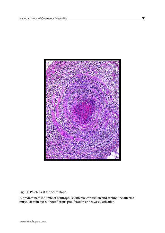
Fig. 11. Phlebitis at the acute stage.
A predominate infiltrate of neutrophils with nuclear dust in and around the affected muscular vein but without fibrous proliferation or neovascularization.
www.intechopen.com
Advances in the Diagnosis and Treatment of Vasculitis
32
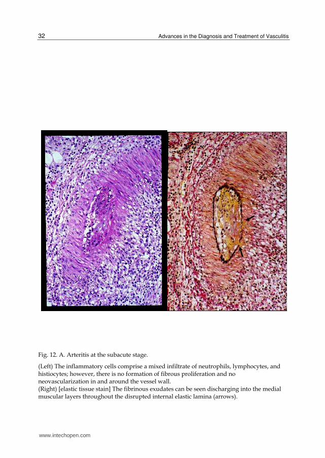
Fig. 12. A. Arteritis at the subacute stage.
(Left) The inflammatory cells comprise a mixed infiltrate of neutrophils, lymphocytes, and histiocytes; however, there is no formation of fibrous proliferation and no neovascularization in and around the vessel wall. (Right) [elastic tissue stain] The fibrinous exudates can be seen discharging into the medial muscular layers throughout the disrupted internal elastic lamina (arrows).
www.intechopen.com
Histopathology of Cutaneous Vasculitis
33
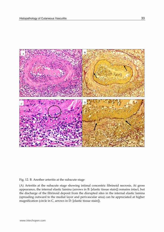
Fig. 12. B. Another arteritis at the subacute stage
(A) Arteritis at the subacute stage showing intimal concentric fibrinoid necrosis. At gross appearance, the internal elastic lamina (arrows in B: [elastic tissue stain]) remains intact, but the discharge of the fibrinoid deposit from the disrupted sites in the internal elastic lamina (spreading outward to the medial layer and perivascular area) can be appreciated at higher magnification (circle in C, arrows in D: [elastic tissue stain]).
www.intechopen.com
Advances in the Diagnosis and Treatment of Vasculitis
34
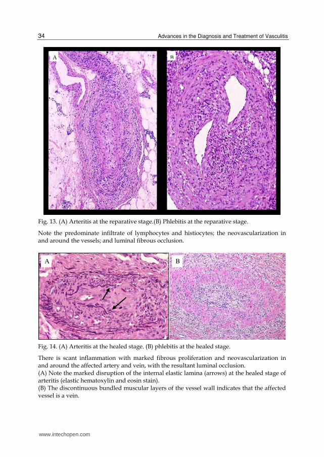
Fig. 13. (A) Arteritis at the reparative stage.(B) Phlebitis at the reparative stage.
Note the predominate infiltrate of lymphocytes and histiocytes; the neovascularization in and around the vessels; and luminal fibrous occlusion.
Fig. 14. (A) Arteritis at the healed stage. (B) phlebitis at the healed stage.
There is scant inflammation with marked fibrous proliferation and neovascularization in and around the affected artery and vein, with the resultant luminal occlusion. (A) Note the marked disruption of the internal elastic lamina (arrows) at the healed stage of arteritis (elastic hematoxylin and eosin stain). (B) The discontinuous bundled muscular layers of the vessel wall indicates that the affected vessel is a vein.
A B
www.intechopen.com
Histopathology of Cutaneous Vasculitis
35
Definition of necrotizing vasculitis and its relation to small vessel vasculitis and muscular vessel vasculitis
Necrotizing vasculitis is defined as vasculitis with vessel wall fibrinoid necrosis.
Since vessel wall fibrinoid necrosis is the crucial finding for the diagnosis of small vessel vasculitis, all small vessel vasculitis is necrotizing vasculitis.
Muscular vessel vasculitis is not necessarily identical to necrotizing vasculitis since vessel wall fibrinoid necrosis is not the crucial finding for muscular vessel vasculitis (Figures 9, 10, and 14).
4) Inflammatory stages of vasculitis (morphological evolution of vasculitis)
For muscular vessel vasculitis, morphological changes in the vessel wall and in the
luminal and perivascular areas during the dynamic inflammatory process are
diagnostically important and influence the mode of treatment. For example, patients with
ulcerative lesions (caused by arteritis at the healed stage with intimal fibrous thickening
resulting from luminal occlusion) should be administered anticoagulation agents and
vessel dilating medications, rather than systemic steroid treatment. By contrast, for
patients with lesions at either the acute or subacute stage of arteritis, initiating of systemic
steroid treatment should be considered as early as possible to suppress and cease the
ongoing inflammation occurring on the vessel wall; this prevents further destruction of
the affected vessels .
Vasculitis of the dermal small vessels (e.g., arterioles and venules) does not have these
proliferative changes as characterized by either fibrous proliferation or neovascularization
of the lumen and vessel wall. However, the formation of perivascular fibrosis may be found
in old lesions of certain small vessel vasculitis disorders such as erythema elevatum
diutinum.
Inflammatory stages of muscular vessel vasculitis start as an acute stage, followed by a subacute stage, a reparative stage, and a healed stage.11, 12
Acute stage: Characterized by a predominate infiltrate of neutrophils (or eosinophils in
cases of Churg-Strauss syndrome12) in and around the vessel wall and the lumen, but
without evidence of new capillary formation (i.e., neovascularization) and fibrous
proliferation in and around the vessel (Figures 10 and 11). Another feature is that the
internal elastic lamina remains intact (Figure 10).
Subacute stage: Characterized by a mixed infiltrate of neutrophils, lymphocytes and
histiocytes with the formation of a fibrinoid deposit (i.e., fibrinoid necrosis) in the intima of
the affected arteries. This is subsequently accompanied by its discharge into the muscular
medial layer and outward spread to the perivascular area throughout the sites of disrupted
internal elastic lamina (Figures 12A and 12B).
Reparative stage (i.e., granulation tissue stage): Characterized by a predominate infiltrate
of lymphocytes and histiocytes and fibrous and vascular proliferation in and around the
affected muscular vessels. Intimal and luminal fibrous thickening results in luminal
occlusion. (Figures 13 and 15b).
Healed stage (i.e., scar stage): Characterized by scant inflammation with marked fibrous proliferation and neovascularization in and around the affected muscular vessels (Figures 14A and 14B) and marked disruption of internal elastic lamina in the affected arteries (Figure 14A).
www.intechopen.com
Advances in the Diagnosis and Treatment of Vasculitis
36
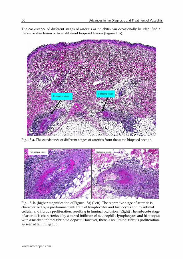
The coexistence of different stages of arteritis or phlebitis can occasionally be identified at the same skin lesion or from different biopsied lesions (Figure 15a).
Fig. 15.a. The coexistence of different stages of arteritis from the same biopsied section.
Fig. 15. b. (higher magnification of Figure 15a) (Left) The reparative stage of arteritis is characterized by a predominate infiltrate of lymphocytes and histiocytes and by intimal cellular and fibrous proliferation, resulting in luminal occlusion. (Right) The subacute stage of arteritis is characterized by a mixed infiltrate of neutrophils, lymphocytes and histiocytes with a marked intimal fibrinoid deposit. However, there is no luminal fibrous proliferation, as seen at left in Fig 15b.
www.intechopen.com
Histopathology of Cutaneous Vasculitis
37
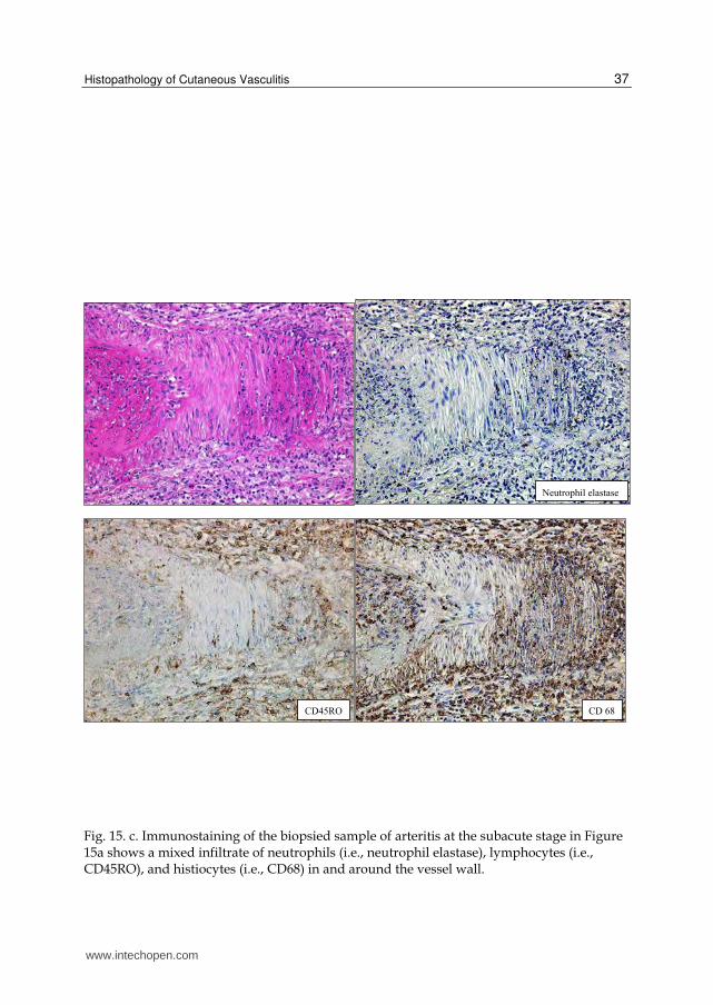
Fig. 15. c. Immunostaining of the biopsied sample of arteritis at the subacute stage in Figure 15a shows a mixed infiltrate of neutrophils (i.e., neutrophil elastase), lymphocytes (i.e., CD45RO), and histiocytes (i.e., CD68) in and around the vessel wall.
www.intechopen.com
Advances in the Diagnosis and Treatment of Vasculitis
38
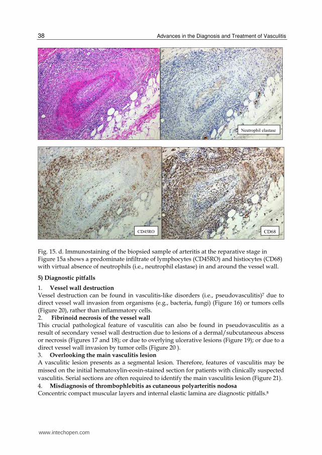
Fig. 15. d. Immunostaining of the biopsied sample of arteritis at the reparative stage in Figure 15a shows a predominate infiltrate of lymphocytes (CD45RO) and histiocytes (CD68) with virtual absence of neutrophils (i.e., neutrophil elastase) in and around the vessel wall.
5) Diagnostic pitfalls
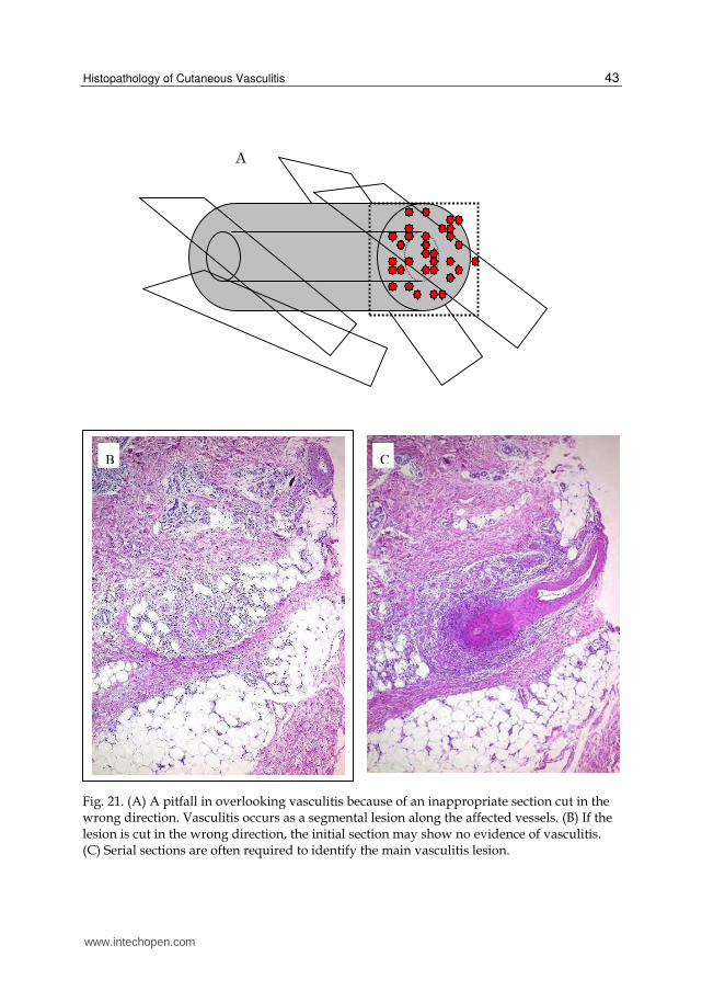
1. Vessel wall destruction Vessel destruction can be found in vasculitis-like disorders (i.e., pseudovasculitis)7 due to direct vessel wall invasion from organisms (e.g., bacteria, fungi) (Figure 16) or tumors cells (Figure 20), rather than inflammatory cells. 2. Fibrinoid necrosis of the vessel wall This crucial pathological feature of vasculitis can also be found in pseudovasculitis as a result of secondary vessel wall destruction due to lesions of a dermal/subcutaneous abscess or necrosis (Figures 17 and 18); or due to overlying ulcerative lesions (Figure 19); or due to a direct vessel wall invasion by tumor cells (Figure 20 ). 3. Overlooking the main vasculitis lesion A vasculitic lesion presents as a segmental lesion. Therefore, features of vasculitis may be
missed on the initial hematoxylin-eosin-stained section for patients with clinically suspected
vasculitis. Serial sections are often required to identify the main vasculitis lesion (Figure 21).
4. Misdiagnosis of thrombophlebitis as cutaneous polyarteritis nodosa
Concentric compact muscular layers and internal elastic lamina are diagnostic pitfalls.8
www.intechopen.com
Histopathology of Cutaneous Vasculitis
39
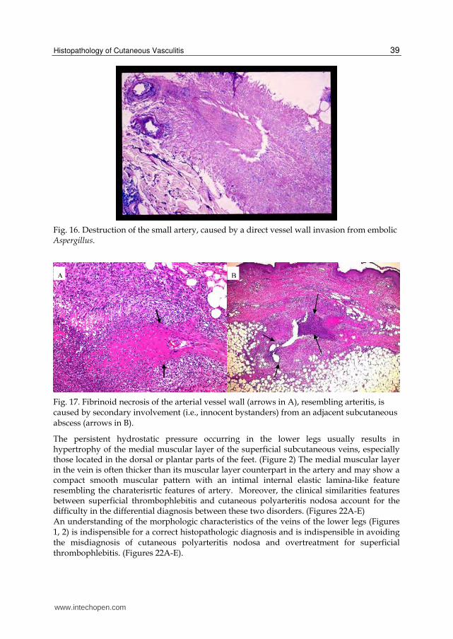
Fig. 16. Destruction of the small artery, caused by a direct vessel wall invasion from embolic Aspergillus.
Fig. 17. Fibrinoid necrosis of the arterial vessel wall (arrows in A), resembling arteritis, is caused by secondary involvement (i.e., innocent bystanders) from an adjacent subcutaneous abscess (arrows in B).
The persistent hydrostatic pressure occurring in the lower legs usually results in hypertrophy of the medial muscular layer of the superficial subcutaneous veins, especially those located in the dorsal or plantar parts of the feet. (Figure 2) The medial muscular layer in the vein is often thicker than its muscular layer counterpart in the artery and may show a compact smooth muscular pattern with an intimal internal elastic lamina-like feature resembling the charaterisrtic features of artery. Moreover, the clinical similarities features between superficial thrombophlebitis and cutaneous polyarteritis nodosa account for the difficulty in the differential diagnosis between these two disorders. (Figures 22A-E) An understanding of the morphologic characteristics of the veins of the lower legs (Figures 1, 2) is indispensible for a correct histopathologic diagnosis and is indispensible in avoiding the misdiagnosis of cutaneous polyarteritis nodosa and overtreatment for superficial thrombophlebitis. (Figures 22A-E).
www.intechopen.com
Advances in the Diagnosis and Treatment of Vasculitis
40
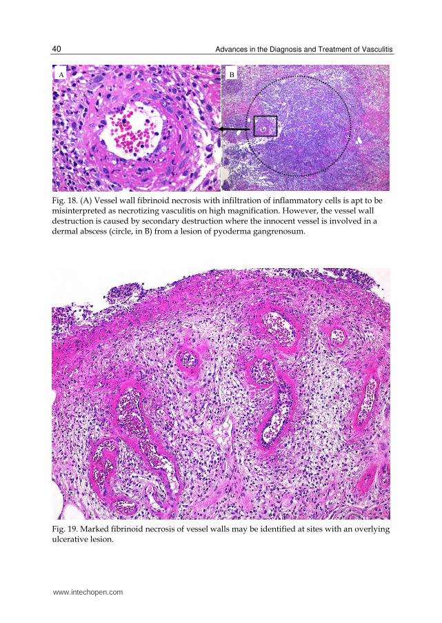
Fig. 18. (A) Vessel wall fibrinoid necrosis with infiltration of inflammatory cells is apt to be misinterpreted as necrotizing vasculitis on high magnification. However, the vessel wall destruction is caused by secondary destruction where the innocent vessel is involved in a dermal abscess (circle, in B) from a lesion of pyoderma gangrenosum.
Fig. 19. Marked fibrinoid necrosis of vessel walls may be identified at sites with an overlying ulcerative lesion.
www.intechopen.com
Histopathology of Cutaneous Vasculitis
41
Small vessel vasculitis
Neutrophilic
Immune-complex mediated (DIF +)
Cutaneous (idiopathic) leukocytoclastic angiitis (CLA) (IgM/IgG)
(also known as: hypersensitivity vasculitis, cutaneous allergic vasculitis)
Henoch-Schonlein purpura (IgA)
Acute infantile hemorrhagic edema (IgA)
Urticarial vasculitis (IgM/IgG)
Infective endocarditis (IgM/IgG/IgA)
Chronic localized fibrosing vasculitis: Erythema elevatum diutinum (IgA)
Eosinophilic
Idiopathic cutaneous eosinophilic vasculitis
Connective tissue disease (e.g. lupus, rheumatoid arthritis, Sjögren’s syndrome)
Some cases of Churg Strauss Syndrome
Granulomatous Sarcoidosis
Lymphocytic
Rickettsial & viral infections
Lichenoid dermatitides, some cases, e.g. pityriasis lichenoides, graft vs. host disease,
perniosis
Rare drug reactions and arthropod assaults
Mixed, predominately small and medium vessel vasculitis
Neutrophilic
Immune-complex mediated (DIF+)
Cryoglobulinemic vasculitis (IgM/IgG)
Hepatisis B and C related vasculitis (IgM/IgG)
Connective tissue disease vasculitis (e.g. lupus, rheumatoid arthritis, Sjögren’s syndrome)
(IgM/IgG)
ANCA associated/pauci-immune (DIF-)
Wegener's granulomatosis
Microscopic polyangiitis
Churg-Strauss syndrome
Drug-induced ANCA vasculitis
Miscellaneous/other
Behçet's disease
Inflammatory bowel disease( Crohn’s disease, ulcerative colitis)
Malignancy associated vasculitis
Eosinophilic Churg-Strauss syndrome
Lymphocytic
Degos’ disease
Rickettsial & viral infections
Collagen tissue disease vasculitis (e.g. Sjögren's syndrome, lupus vasculitis)
www.intechopen.com
Advances in the Diagnosis and Treatment of Vasculitis
42
Behçet's disease Granulomatous
Churg-Strauss syndrome
Connective tissue disease vasculitis (e.g. lupus, rheumatoid arthritis, Sjögren’s syndrome)
Inflammatory bowel disease ( Crohn’s disease, ulcerative colitis)
Post-herpetic eruptions
Muscular (Medium) vessel vasculitis
Neutrophilic
Polyarteritis nodosa (classic and cutaneous )
Idiopathic superficial thrombophlebitis
Eosinophilic Juvenile temporal arteritis
Granulomatous
Giant cell (temporal) arteritis
Nodular vasculitis (erythema induratum) Lymphocytic
Sneddon's syndrome
Beurger’s disease (thromboangiitis obliterans)
ANCA: antineutrophil cytoplasmic antibody. DIF +: direct immunfluorescence examination of skin lesions shows vessel wall immune-complexes and/or complement deposition. Modified from 1) Chen and Carlson. Am J Clin Dermatol 9:71-92.2008.
Table 1. Classification of cutaneous vasculitis, based on the size of the affected vessels, the predominate inflammatory cells, and the DIF studies
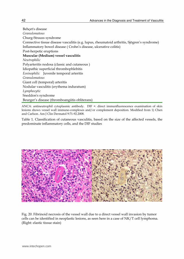
Fig. 20. Fibrinoid necrosis of the vessel wall due to a direct vessel wall invasion by tumor cells can be identified in neoplastic lesions, as seen here in a case of NK/T cell lymphoma. (Right: elastic tissue stain)
www.intechopen.com
Histopathology of Cutaneous Vasculitis
43
Fig. 21. (A) A pitfall in overlooking vasculitis because of an inappropriate section cut in the wrong direction. Vasculitis occurs as a segmental lesion along the affected vessels. (B) If the lesion is cut in the wrong direction, the initial section may show no evidence of vasculitis. (C) Serial sections are often required to identify the main vasculitis lesion.
www.intechopen.com
Advances in the Diagnosis and Treatment of Vasculitis
44
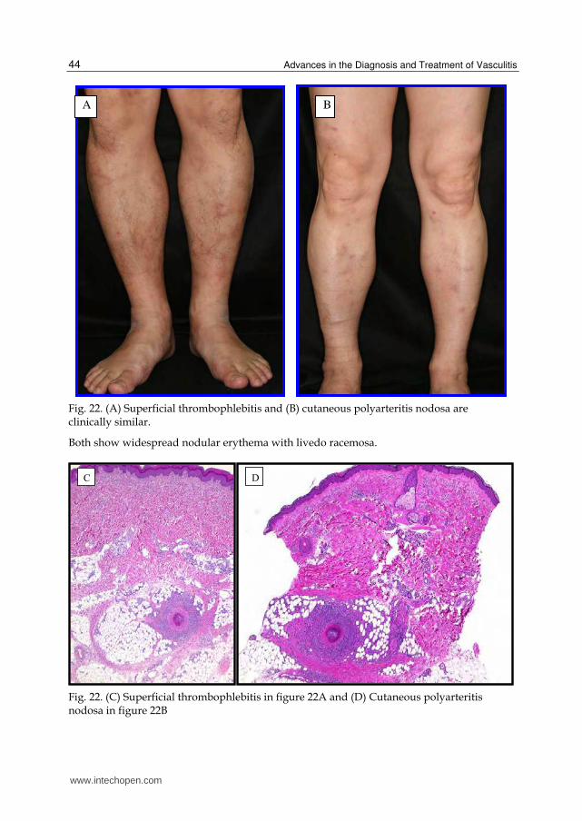
Fig. 22. (A) Superficial thrombophlebitis and (B) cutaneous polyarteritis nodosa are clinically similar.
Both show widespread nodular erythema with livedo racemosa.
Fig. 22. (C) Superficial thrombophlebitis in figure 22A and (D) Cutaneous polyarteritis nodosa in figure 22B
A B
www.intechopen.com
Histopathology of Cutaneous Vasculitis
45
They are also histopathologically similar. Both show a subcutaneous muscular vessel lesion with a compact concentric vessel wall and perivascular panniculitis.
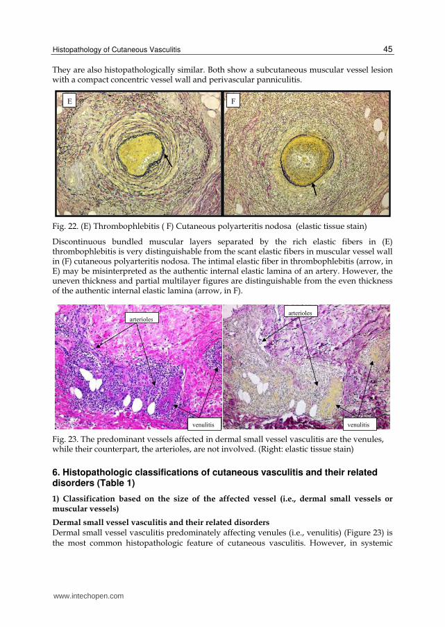
Fig. 22. (E) Thrombophlebitis ( F) Cutaneous polyarteritis nodosa (elastic tissue stain)
Discontinuous bundled muscular layers separated by the rich elastic fibers in (E) thrombophlebitis is very distinguishable from the scant elastic fibers in muscular vessel wall in (F) cutaneous polyarteritis nodosa. The intimal elastic fiber in thrombophlebitis (arrow, in E) may be misinterpreted as the authentic internal elastic lamina of an artery. However, the uneven thickness and partial multilayer figures are distinguishable from the even thickness of the authentic internal elastic lamina (arrow, in F).
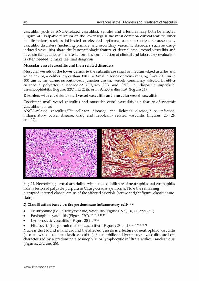
Fig. 23. The predominant vessels affected in dermal small vessel vasculitis are the venules, while their counterpart, the arterioles, are not involved. (Right: elastic tissue stain)
6. Histopathologic classifications of cutaneous vasculitis and their related disorders (Table 1)
1) Classification based on the size of the affected vessel (i.e., dermal small vessels or muscular vessels)
Dermal small vessel vasculitis and their related disorders Dermal small vessel vasculitis predominately affecting venules (i.e., venulitis) (Figure 23) is the most common histopathologic feature of cutaneous vasculitis. However, in systemic
www.intechopen.com
Advances in the Diagnosis and Treatment of Vasculitis
46
vasculitis (such as ANCA-related vasculitis), venules and arterioles may both be affected (Figure 24). Palpable purpura on the lower legs is the most common clinical feature; other manifestations, such as infiltrated or elevated erythema, occur less often. Because many vasculitic disorders (including primary and secondary vasculitic disorders such as drug-induced vasculitis) share the histopathologic feature of dermal small vessel vasculitis and have similar cutaneous manifestations, the combination of clinical and laboratory evaluation is often needed to make the final diagnosis.
Muscular vessel vasculitis and their related disorders
Muscular vessels of the lower dermis to the subcutis are small or medium-sized arteries and veins having a caliber larger than 100 um. Small arteries or veins ranging from 200 um to 400 um at the dermo-subcutaneous junction are the vessels commonly affected in either cutaneous polyarteritis nodosa1,6,8 (Figures 22D and 22F), in idiopathic superficial thrombophlebitis (Figures 22C and 22E), or in Behçet’s disease13 (Figure 26).
Disorders with coexistent small vessel vasculitis and muscular vessel vasculitis
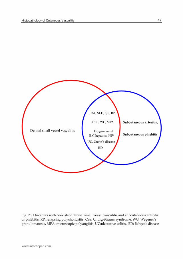
Coexistent small vessel vasculitis and muscular vessel vasculitis is a feature of systemic vasculitis such as ANCA-related vasculitis,17,18 collagen disease,9 and Behçet’s disease,13 or infection, inflammatory bowel disease, drug and neoplasm- related vasculitis (Figures. 25, 26, and 27).
Fig. 24. Necrotizing dermal arteriolitis with a mixed infiltrate of neutrophils and eosinophils from a lesion of palpable purpura in Churg-Strauss syndrome. Note the remaining disrupted internal elastic lamina of the affected arteriole (arrow at right figure: elastic tissue stain).
2) Classification based on the predominate inflammatory cell1,2,3,5,6
Neutrophilic (i.e., leukocytoclastic) vasculitis (Figures. 8, 9, 10, 11, and 26C).
Eosinophilic vasculitis (Figure 27C). 15,16,17,18,19
Lymphocytic vasculitisうFigure 28え. 13,14
Histiocytic (i.e., granulomatous vasculitis)うFigures 29 and 30). 12,18,20,21
Nuclear dust found in and around the affected vessels is a feature of neutrophilic vasculitis (also known as leukocytoclastic vasculitis). Eosinophilic and lymphocytic vascuiltis are both characterized by a predominate eosinophilic or lymphocytic infiltrate without nuclear dust
(Figures. 27C and 28).
www.intechopen.com
Histopathology of Cutaneous Vasculitis
47
Fig. 25. Disorders with coexistent dermal small vessel vasculitis and subcutaneous arteritis or phlebitis. RP: relapsing polychondritis, CSS: Churg-Strauss syndrome, WG: Wegener’s granulomatosis, MPA: microscopic polyangiitis, UC:ulcerative colitis, BD: Behçet’s disease
www.intechopen.com
Advances in the Diagnosis and Treatment of Vasculitis
48
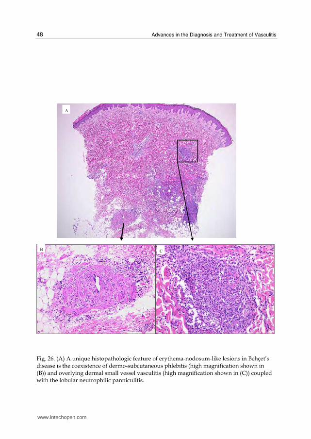
Fig. 26. (A) A unique histopathologic feature of erythema-nodosum-like lesions in Behçet’s disease is the coexistence of dermo-subcutaneous phlebitis (high magnification shown in (B)) and overlying dermal small vessel vasculitis (high magnification shown in (C)) coupled with the lobular neutrophilic panniculitis.
www.intechopen.com
Histopathology of Cutaneous Vasculitis
49
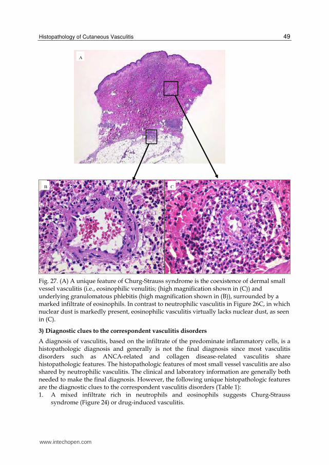
Fig. 27. (A) A unique feature of Churg-Strauss syndrome is the coexistence of dermal small vessel vasculitis (i.e., eosinophilic venulitis; (high magnification shown in (C)) and underlying granulomatous phlebitis (high magnification shown in (B)), surrounded by a marked infiltrate of eosinophils. In contrast to neutrophilic vasculitis in Figure 26C, in which nuclear dust is markedly present, eosinophilic vasculitis virtually lacks nuclear dust, as seen in (C).
3) Diagnostic clues to the correspondent vasculitis disorders
A diagnosis of vasculitis, based on the infiltrate of the predominate inflammatory cells, is a histopathologic diagnosis and generally is not the final diagnosis since most vasculitis disorders such as ANCA-related and collagen disease-related vasculitis share histopathologic features. The histopathologic features of most small vessel vasculitis are also shared by neutrophilic vasculitis. The clinical and laboratory information are generally both needed to make the final diagnosis. However, the following unique histopathologic features are the diagnostic clues to the correspondent vasculitis disorders (Table 1): 1. A mixed infiltrate rich in neutrophils and eosinophils suggests Churg-Strauss
syndrome (Figure 24) or drug-induced vasculitis.
www.intechopen.com
Advances in the Diagnosis and Treatment of Vasculitis
50
2. Subcutaneous granulomatous arteritis or phlebitis surrounded by a marked infiltrate of eosinophils is diagnostic of Churg-Strauss syndrome (Figures 27B and 29). 12,18
3. Subcutaneous eosinophilic arteritis is diagnostic of juvenile temporal arteritis19 and Churg-Strauss syndrome.17
4. Phlebitis and/or neutrophilic panniculitis with overlying dermal venulitis is a unique feature of Behçet’s disease (Figure 26). 13
5. Dermal eosinophilic vasculitis is a very rare finding. It is usually found in idiopathic cutaneous eosinophilic vasculitis,15 collagen disease,16 and Churg-Strauss syndrome (Figure27C). 18
6. Dermal granulomatous vasculitis is a unique finding. It is usually found in sarcoidosis (Figure 30), postherpetic eruption, and collagen disease.
7. Subcutaneous granulomatous muscular vessel vasculitis is a unique finding. It is usually found in classic temporal arteritis (Figure 31) and nodular vasculitis also known as erythema induratum characterized by indurated erythematous lesions in the lower legs with a subcutaneous vein predominately affected (i.e., granulomatous phlebitis) in conjunction with lobular granulomatous panniculitis. It may be occasionally be found
in Churg-Strauss syndrome,12,18 collagen disease (e.g., rheumatoid arthritis); inflammatory bowel disease (e.g., ulcerative colitis, Crohn’s disease); and
postherpetic eruption with nodular lesions. 8. The coexistence of dermal neutrophilic vasculitis and palisaded granuloma with central
basophilic degenerated collagen fibers suggests Churg-Strauss syndrome (Figure 32), Wegener’s granulomatosis, and collagen disease (e.g., rheumatoid arthritis or systemic lupus erythematosus [SLE]).
9. The coexistence of dermal small vasculitis and subcutaneous arteritis should suggest a systemic vasculitis disorder such as ANCA-related vasculitis (Figure 33), collagen disease,9 or infection-related vasculitis (e.g., hepatitis C virus-induced cryoglobulinemic purpura, inflammatory bowel disease).
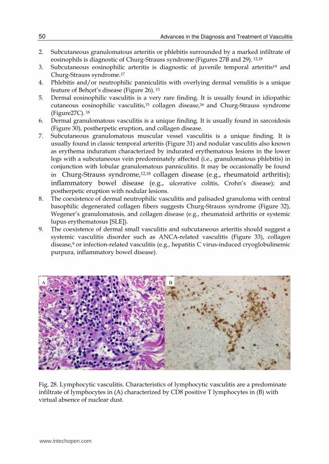
Fig. 28. Lymphocytic vasculitis. Characteristics of lymphocytic vasculitis are a predominate infiltrate of lymphocytes in (A) characterized by CD8 positive T lymphocytes in (B) with virtual absence of nuclear dust.
www.intechopen.com
Histopathology of Cutaneous Vasculitis
51
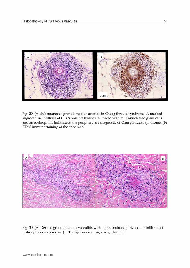
Fig. 29. (A) Subcutaneous granulomatous arteritis in Churg-Strauss syndrome. A marked angiocentric infiltrate of CD68 positive histiocytes mixed with multi-nucleated giant cells and an eosinophilic infiltrate at the periphery are diagnostic of Churg-Strauss syndrome. (B) CD68 immunostaining of the specimen.
Fig. 30. (A) Dermal granulomatous vasculitis with a predominate perivascular infiltrate of histiocytes in sarcoidosis. (B) The spccimen at high magnification.
www.intechopen.com
Advances in the Diagnosis and Treatment of Vasculitis
52
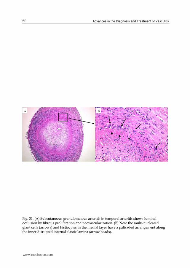
Fig. 31. (A) Subcutaneous granulomatous arteritis in temporal arteritis shows luminal occlusion by fibrous proliferation and neovascularization. (B) Note the multi-nucleated giant cells (arrows) and histiocytes in the medial layer have a palisaded arrangement along the inner disrupted internal elastic lamina (arrow heads).
www.intechopen.com
Histopathology of Cutaneous Vasculitis
53
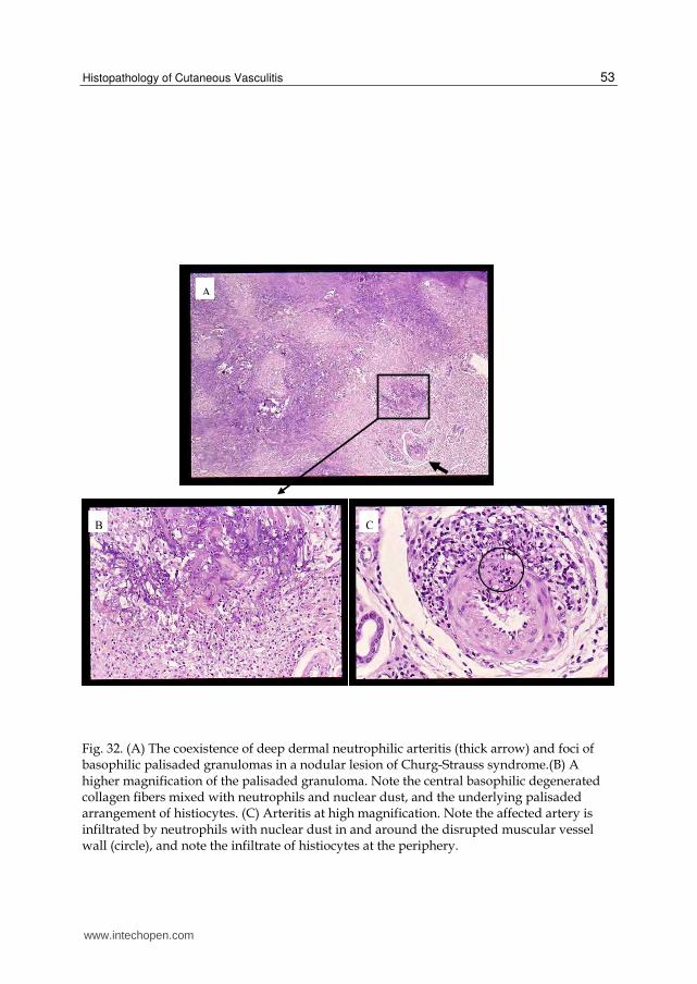
Fig. 32. (A) The coexistence of deep dermal neutrophilic arteritis (thick arrow) and foci of basophilic palisaded granulomas in a nodular lesion of Churg-Strauss syndrome.(B) A higher magnification of the palisaded granuloma. Note the central basophilic degenerated collagen fibers mixed with neutrophils and nuclear dust, and the underlying palisaded arrangement of histiocytes. (C) Arteritis at high magnification. Note the affected artery is infiltrated by neutrophils with nuclear dust in and around the disrupted muscular vessel wall (circle), and note the infiltrate of histiocytes at the periphery.
www.intechopen.com
Advances in the Diagnosis and Treatment of Vasculitis
54
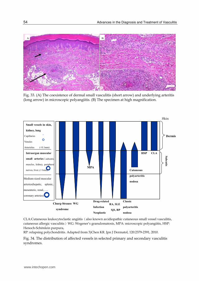
Fig. 33. (A) The coexistence of dermal small vasculitis (short arrow) and underlying arteritis (long arrow) in microscopic polyangiitis. (B) The specimen at high magnification.
CLA:Cutaneous leukocytoclastic angiitisうalso known as:idiopathic cutaneous small vessel vasculitis,
cutaneous allergic vasculitisえWG: Wegener’s granulomatosis, MPA: microscopic polyangiitis, HSP:
Henoch-Schönlein purpura,
RP: relapsing polychondritis. Adapted from 3)Chen KR. Jpn J Dermatol, 120:2379-2391, 2010.
Fig. 34. The distribution of affected vessels in selected primary and secondary vasculitis syndromes.
www.intechopen.com
Histopathology of Cutaneous Vasculitis
55
7. Summary
Skin is the most common target organ for vasculitis (Figure 34). A diagnosis of cutaneous vasculitis is best determined by the histopathologic findings. However, the diagnosis may be difficult because of the constellation of clinical and histopathologic findings. The above described contents are the best approach to the complicated histopathology of cutaneous vasculitis, including the morphology of cutaneous vessels; the diagnostic criteria; the diagnostic pitfalls; and the classification, based on the size of the involved vessels and the principal inflammatory response.
8. References
[1] Chen KR, Carlson JA. Clinical approach to cutaneous vasculitis. Am J Clin Dermatol, 9:71-92, 2008.
[2] Carlson JA, Eng BT, Chen KR. Cutaneous Vasculitis Update: Diagnostic Criteria, Classification, Epidemiology, Etiology, Pathogenesis, Evaluation and Prognosis. Am J Dermatopathol, 27:504-528, 2005.
[3] Chen KR. Pathogenesis and histopathologic diagnosis of cutaneous vasculitis.Jpn J
Dermatol, 120:2379-2391, 2010. (in Japanese) [4] Sunderkotter C, Sindrilaru A. Clinical classification of vasculitis. Eur J Dermatol. 16:114-
24, 2006. [5] Carlson JA, Chen KR: Cutaneous Vasculitis Update: Small Vessel Neutrophilic Vasculitis
Syndromes. Am J Dermatopathol. 28:486-506, 2006. [6] Carlson JA, Chen KR: Cutaneous Vasculitis Update: Neutrophilic Muscular Vessel and
Eosinophilic, Granulomatous, and Lymphocytic Vasculitis Syndromes. Am J Dermatopathol. 29:32-43, 2007.
[7] Carlson JA, Chen KR: Cutaneous pseudovasculitis. Am J Dermatopathol. 29:44-55, 2007. [8] Chen KR. The misdiagnosis of superficial thrombophlebitis as cutaneous polyarteritis
nodosa: Features of the internal elastic lamina and the compact concentric muscular layer as diagnostic pitfalls. Am J Dermatopathol 32:688-693, 2010.
[9] Chen KR, Toyohara A, Suzuki A, Miyakawa S: Clinical and histopathological spectrum of cutaneous vasculitis in rheumatoid arthritis. Br J Dermatol, 147:905-913, 2002.
[10] Bajema IM, Bruijn JA.. What stuff is this! A historical perspective on fibrinoid necrosis. J Pathol, 191:235-238, 2000.
[11] Ishibashi M, Chen KR: A morphological study of evolution of cutaneous polyarteritis nodosa. Am J Dermatopathol 30(4):319-26, 2008.
[12] Chen KR, Sakamoto M, Ikemoto K, et al: Granulomatous arteritis in cutaneous lesions of Churg-Strauss syndrome. J Cutan Pathol 34(4):330-7, 2007.
[13] Chen KR, Kawahara Y, Miyakawa S, Nishikawa T: Cutaneous vasculitis in Behçet's disease: a clinical and histopathologic study of 20 patients. J Am Acad Dermatol
36:689-96, 1997.
[14] Carlson JA, Mihm MC, Jr., LeBoit PE. Cutaneous lymphocytic vasculitis: a definition, a review, and a proposed classification. Semin Diagn Pathol. 13(1):72-90, 1996.
[15] Chen KR, Pittelkow MR, Su WPD, Gleich GJ, Newman W, Leiferman KM: Recurrent cutaneous eosinophilic necrotizing vasculitis: A novel eosinophil-mediated syndrome. Arch Dermatol 130:1159-1166, 1994.
www.intechopen.com
Advances in the Diagnosis and Treatment of Vasculitis
56
[16] Chen KR, Su WPD, Pittelkow MR, Conn DL, George T, Leiferman KM: Eosinophilic vasculitis in connective tissue disease. J Am Acad Dermatol 35:173-182, 1996.
[17] Chen KR, Ohata Y, Sakurai M, Nakayama H: Churg-Strauss syndrome: Report of a case without pre-existing asthma. J Dermatol 19:40-47, 1992.
[18] Ishibashi M, Kudo S, Yamamoto K, Shimai N, Chen KR:Churg-Strauss syndrome with coexistence of eosinophilic vasculitis, granulomatous phlebitis and granulomatous dermatitis in bullous pemphigoid-like blisters. J Cutan Pathol. 38:290-294. 2011.
[19] Lie JT, Gordon LP, Titus JL. Jevenile temporal arteritis. Biopsy study of four cases. JAMA, 234;496-499, 1975.
[20] Nordborg E, Nordberg C. The inflammatory reaction in giant cell arteritis: an immunohistochemical investigation. Clin Exp Rheumatol, 16:165-8, 1998.
[21] Mambo NC. Temporal (granulomatous) arteritis: A histopathological study of 32 cases. Histopathology. 3:209-221, 1979.[
www.intechopen.com
Advances in the Diagnosis and Treatment of VasculitisEdited by Dr. Luis M Amezcua-Guerra
ISBN 978-953-307-786-4Hard cover, 366 pagesPublisher InTechPublished online 09, November, 2011Published in print edition November, 2011
InTech EuropeUniversity Campus STeP Ri Slavka Krautzeka 83/A 51000 Rijeka, Croatia Phone: +385 (51) 770 447 Fax: +385 (51) 686 166www.intechopen.com
InTech ChinaUnit 405, Office Block, Hotel Equatorial Shanghai No.65, Yan An Road (West), Shanghai, 200040, China
Phone: +86-21-62489820 Fax: +86-21-62489821
This book represents the culmination of the efforts of a group of outstanding experts in vasculitis from all overthe world, who have endeavored to draw themselves into this volume by keeping both the text and theaccompanying figures and tables lucid and memorable. The book provides practical information about thescreening approach to vasculitis by laboratory analysis, histopathology and advanced image techniques,current standard treatment along with new and more specific interventions including biologic agents, reparativesurgery and experimental therapies, as well as miscellaneous issues such as the extra temporalmanifestations of "temporal arteritis" or the diffuse alveolar hemorrhage syndrome. The editor and each of theauthors invite you to share this journey by one of the most exciting fields of the medicine, the world ofVasculitis.
How to referenceIn order to correctly reference this scholarly work, feel free to copy and paste the following:
Ko-Ron Chen (2011). Histopathology of Cutaneous Vasculitis, Advances in the Diagnosis and Treatment ofVasculitis, Dr. Luis M Amezcua-Guerra (Ed.), ISBN: 978-953-307-786-4, InTech, Available from:http://www.intechopen.com/books/advances-in-the-diagnosis-and-treatment-of-vasculitis/histopathology-of-cutaneous-vasculitis