HISTOPATHOLOGICAL STUDY OF CHRONIC GASTRITIS · HISTOPATHOLOGICAL STUDY OF CHRONIC GASTRITIS Thesis...
20
HISTOPATHOLOGICAL STUDY OF CHRONIC GASTRITIS Thesis submitted in partial fulfillment of M.Sc. Degree in Pathology By Somaia AbduLatif Mahmoud Soliman M.B.B.Ch Faculty of Medicine, Cairo University Supervised by Prof. Dr. Dalal Anwar Elwi Professor of pathology Faculty of Medicine Cairo University Ass. Prof. Dr. Mostafa Mohamed Salem Assistant Professor of pathology Faculty of medicine Cairo University Dr. Ahmed Abd- El Monem Soliman Lecturer of Pathology Faculty of Medicine Cairo University Faculty of Medicine Cairo University 2012
Transcript of HISTOPATHOLOGICAL STUDY OF CHRONIC GASTRITIS · HISTOPATHOLOGICAL STUDY OF CHRONIC GASTRITIS Thesis...

HISTOPATHOLOGICAL STUDY OF CHRONIC GASTRITIS
Thesis submitted in partial fulfillment ofM.Sc. Degree in Pathology
By
Somaia AbduLatif Mahmoud Soliman M.B.B.Ch
Faculty of Medicine, Cairo University
Supervised by
Prof. Dr. Dalal Anwar ElwiProfessor of pathology
Faculty of MedicineCairo University
Ass. Prof. Dr. Mostafa Mohamed Salem
Assistant Professor of pathologyFaculty of medicine
Cairo University
Dr. Ahmed Abd- El Monem SolimanLecturer of PathologyFaculty of Medicine
Cairo University
Faculty of Medicine Cairo University
2012

الرحمن الرحیمهللابسم
أخرجك ھاتكم ـوهللا م من بطون أممع ـعل لك ـشیئا وج ال تعلمون م الس
.واألبصار واألفئدة لعلكم تشكرون
العظیم هللاصدق
)٧٨:النحل(

Acknowledgement
First and foremost "Thanks to God", the most merciful and kind.
I am honored to have Prof. Dr. Dalal Anwar Elwi, Professor of Pathology, Faculty of Medicine, Cairo University, as a supervisor of this work. I am deeply grateful and most appreciative to her great efforts, kind guidance and valuable advice that encouraged and helped me to finish this work.
My profound gratitude goes to Prof. Dr. MostafaMohammed Samy Salem assistant Professor of Pathology, Faculty of Medicine, Cairo University for his close supervision and precious remarks throughout the course of this study.
Special thanks are owed to Dr. Ahmed Abd-Elmonem Soliman, lecturer of Pathology, Faculty of medicine, Cairo University, who saved no time and effort in helping me, his constant support, continuous encouragement and constructive comments allowed me to accomplish this work.
Finally, I would like to thank my family for their patience, love, motivation and support throughout this work.
Somia AbduLatif

Contents
Introduction & Aim of the work 1Review of literature:
Structure of the stomach 5 Classification 9 Chronic gastritis associated with Helicobacter pylori infection 13 Autoimmune gastritis 26 Reactive gastropathy 32 Radiation gastritis 35 Lymphocytic gastritis 36 Granulomtous gastritis 38 Esinophilic gastritis 43 Other causes of infectious gastritis 46 Vascular gastropathy 51 Collagenous gastritis 54 Ureamic gastropathy 56 Graft Versus Host Disease 57
Materials & Methods 58Results 60Discussion 86Summary 93Conclusion & Recommendation 95References 96Arabic summary

List of Abbreviation
Acquired immuno deficiency syndromeAIDS1
Autoimmune gastritisAG2
Campylobacter like organism testCLO test3
Chronic antral gastritisCAG4
CytomegalovirusCMV5
Diffuse antral gastritisDAG6
Enterochromaffin-like cellsECL cells7
Enzyme linked immunosorbent assaysELISA8
Gastric antral vascular ectasiaGAVE9
Gastrin cellsG cells10
Gastrointestinal tractGIT11
Graft versus host diseaseGVHD12
Helicobacter heilmanniiH. heilmannii 13
Helicobacter pyloriH.pylori14
Hematoxylin & eosinH & E15
Human immuno deficiency virusHIV16
Intraepithelial lymphocytesIELs17
Mucosa associated lymphoid tissueMALT18
Multifocal atrophic gastritisMAG 19
Non steroidal anti-inflammatory drugsNSAIDs20
Polymerase chain reactionPCR21
ProstaglandingsPG122
Proton pump inhibitorsPPIs23
Ptoral hypertensive gastropathyPHG24
Vacuolating cytotoxin AVacA25

List of Tables
page Table
11 Sydney System Classification of Chronic
Gastritis
Table (1)
60Sydney classification systemTable (2)
61 Frequency of various histological subtypesTable (3)
62 Age frequency in chronic gastritis casesTable (4)
63 Sex distribution in the studied casesTable (5)
64 Clinical presenting symptomsTable (6)
65 Frquency of various endoscopic picturesTable (7)
66 Categorization of H.pylori subtypesTable (8)
67 Classification of intensity of inflammation in
H.pylori associated cases
Table (9)
68 Classification according to activity in
H.pylori cases
Table (10)
69 Complications in H.pylori associated casesTable (11)
70 Correlation between histological type & ageTable (12)
70 Correlation between Sex & histological typeTable (13)
71 Correlation between clinical presentation &
histological type
Table (14)
71 Correlation between clinical presentation &
histological type
Table (15)

72 Correlation between clinical presentation &
histological type
Table (15)
72 Correlation between endoscopic picture &
histological type
Table (16)
73 Correlation between endoscopic picture &
histological type
Table (17)
73 Correlation between endoscopic picture &
histological type
Table (18)
74 Corrlation between endoscopic picture &
histological type
Table (19)
74 Correlation between endoscopic picture &
subtypes of H.pylori positive group
Table (20 )
75 Correlation between endoscopic picture &
subtypes of H.pylori positive group
Table (21)
75 Correlation between endoscopic picture &
subtypes of H.pylori positive group
Table (22 )
76 Correlation between endoscopic picture &
subtypes of H.pylori positive group
Table (23)
76 Correlation between endoscopic picture &
subtypes of H.pylori positive group
Table (24)

List of Figures
PageFigure
8Diagram of 4 anatomical & 3 histological
areas of the stomach
Figure(1)
14Diagram of H.pylori structure.Figure(2)
76Chronic superficial gastritis (gastric mucosal
glands showing colonization by H.pylori
organisms). (H&E x400)
Figure (3)
76Chronic superficial gastritis (gastric mucosal
glands showing colonization by H.pylori
organisms). (H&E x400)
Figure (4)
77Chronic superficial gastritis (gastric mucosal
glands showing colonization by H.pylori
organisms). (Giemsa stain x400)
Figure (5)
77Chronic superficial gastritis (gastric mucosal
glands showing colonization by H.pylori
organisms) (Giemsa stain x400)
Figure (6)
78Chronic superficial gastritis (gastric mucosal
glands showing colonization by H.pylori
organisms) (Giemsa stain x400)
Figure (7)
78Follicullar gastritis, H.pylori associated, showing
lymphoid follicle situated deep in the antral
mucosa. (H&E x100)
Figure (8)
79Chronic atrophic gastritis, showing marked
decrease in number of gastric mucosal glands.
(H&E x100)
Figure (9)
79chronic gastritis, showing intestinal metaplasia.
(H&E x100)
Figure (10)
80Lymphocytic gastritis, showing lymphocytic
infiltration of the lamina propria & intraepithelial
lymphocytosis. (H&E x250)
Figure (11)
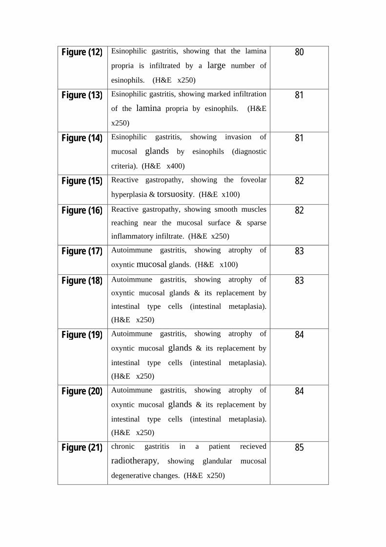
80Esinophilic gastritis, showing that the lamina
propria is infiltrated by a large number of
esinophils. (H&E x250)
Figure (12)
81Esinophilic gastritis, showing marked infiltration
of the lamina propria by esinophils. (H&E
x250)
Figure (13)
81Esinophilic gastritis, showing invasion of
mucosal glands by esinophils (diagnostic
criteria). (H&E x400)
Figure (14)
82Reactive gastropathy, showing the foveolar
hyperplasia & torsuosity. (H&E x100)
Figure (15)
82Reactive gastropathy, showing smooth muscles
reaching near the mucosal surface & sparse
inflammatory infiltrate. (H&E x250)
Figure (16)
83Autoimmune gastritis, showing atrophy of
oxyntic mucosal glands. (H&E x100)
Figure (17)
83Autoimmune gastritis, showing atrophy of
oxyntic mucosal glands & its replacement by
intestinal type cells (intestinal metaplasia).
(H&E x250)
Figure (18)
84Autoimmune gastritis, showing atrophy of
oxyntic mucosal glands & its replacement by
intestinal type cells (intestinal metaplasia).
(H&E x250)
Figure (19)
84Autoimmune gastritis, showing atrophy of
oxyntic mucosal glands & its replacement by
intestinal type cells (intestinal metaplasia).
(H&E x250)
Figure (20)
85chronic gastritis in a patient recieved
radiotherapy, showing glandular mucosal
degenerative changes. (H&E x250)
Figure (21)

List of Graphs
pageGraph
60Frequency of various histological subtypesGraph (1)
61Age frequency in chronic gastritis casesGraph (2)
62Sex distribution in the studied casesGraph (3)
63Clinical presenting symptomsGraph (4)
64Frquency of various endoscopic picturesGraph (5)
65Categorization of H.pylori subtypesGraph (6)
66Classification of intensity of inflammation in
H.pylori associated cases
Graph (7)
67Classification according to activity in H.pylori
cases
Graph (8)
68Complications in H.pylori associated casesGraph (9)

ABSTRACT
Aim of Work: Analysis of chronic gastritis cases in 2 years duration, to detect
rate of occurrence of different histological types, to reclassify cases according to
the latest grading and staging systems and to compare the clinico-pathological
features among Egyptian patients included in the study with registries of other
countries.
Methods: This was a retrospective study conducted at the Department of
Pathology, Kasr Al Aini Hospital, Cairo University between January 2009 and
December 2010. Analysis of 224 gastric biopsy specimens was done. We
routinely make Hematoxylin/Eosin stained slides and perform Giemsa stain to
check for H. pylori on all endoscopic gastric biopsies.
Results: Out of two hundred & twenty four cases, H.pylori was found in 68.7%
of the cases. Increased frequency of chronic gastritis was seen between 51 & 60
years. The most common clinical presentation was epigastric pain. The most
common endoscopic finding was gastritis. The mean age of patients was 42
years and the age of patients ranged from 2.5 to 80 years. Females constituted
52% of cases while males were 48%. The mean age of female patients 41 years
was less than that of male patients 43.4 years.
Recommendations: For proper assessment of distribution of gastritis, multiple
biopsies (at least five) from antrum, corpus & incisura angularis are important.
Key Words: Chronic gastritis, Helicobacter pylori, lymphoid follicles.
Baaed Study of Helicobacter Pylori in Egypt

Introduction & aim of the workـــــــــــــــــــــــــــــــــــــــــــــــــــــــــــــــــــــــــــــــــــــــــــــــــــــــــــــــــــــــــــــــــــــــــــــــــــــــــــــــــــــــــــــ
-1-
Introduction
The incidence and natural history of chronic gastritis has been
greatly clarified by the systematic use of endoscopic gastric biopsy (Wyatt et
al., 2001).
Chronic gastritis is a histopathologic entity characterized by chronic
inflammation of the stomach mucosa. Gastritis can be classified based on the
underlying etiologic agent (e.g, Helicobacter pylori, bile reflux, nonsteroidal
anti-inflammatory drugs [NSAIDs], autoimmunity, allergic response) and the
histopathologic pattern, which may suggest the etiologic agent and clinical
course (e.g, H. pylori –associated multifocal atrophic gastritis) (Merck, 2007.
Retrieved 2009).
Although minimal inflammation is observed in some gastropathies, such
as those associated with NSAID intake, these entities are frequently included in
the differential diagnosis of chronic gastritis. Chemical or reactive gastritis is
caused by injury of the gastric mucosa by reflux of bile and pancreatic
secretions into the stomach, but it can also be caused by exogenous substances,
including NSAIDs, acetylsalicylic acid, chemotherapeutic agents, and alcohol.
These chemicals cause epithelial damage, erosions, and ulcers that are followed
by regenerative hyperplasia detectable as foveolar hyperplasia, and damage to
capillaries, with mucosal edema, hemorrhage, and increased smooth muscle in
the lamina propria. Inflammation in these lesions caused by chemicals is
minimal or lacking; therefore, the term gastropathy or chemical gastropathy is
more appropriate to describe these lesions than is the term chemical or
reactive gastritis as proposed by the updated Sydney classification of gastritis
(Gao et al., 2009).

Introduction & aim of the workـــــــــــــــــــــــــــــــــــــــــــــــــــــــــــــــــــــــــــــــــــــــــــــــــــــــــــــــــــــــــــــــــــــــــــــــــــــــــــــــــــــــــــــ
-2-
No single classification of gastritis provides an entirely satisfactory
description of all types of gastritis. However, an etiologic classification provides
a direct target toward which therapy can be directed (Sepulveda et al., 2008).
Infectious gastritis: Chronic gastritis caused by H. pylori infection.
This is the most common cause of chronic gastritis. The most important advance
in the field of chronic gastritis & other gastric diseases (peptic ulcer, carcinoma
& malignant lymphoma) has been the awareness of the crucial role played by
H.pylori (Wu et al., 2001). Infection by Helicobacter heilmannii, granulomatous
gastritis associated with gastric infections in mycobacteriosis, syphilis,
histoplasmosis, mucormycosis, South American blastomycosis, anisakiasis, or
anisakidosis. Chronic gastritis associated with parasitic infections such as
Strongyloides species, schistosomiasis, Diphyllobothrium latum. Viral infections
such as CMV and herpes virus infection (Hasegawa et al., 2009).
Noninfectious gastritis: autoimmune gastritis, chemical gastropathy,
usually related to chronic bile reflux or NSAID and aspirin intake, Uremic
gastropathy (Siegelbaum et al, 2006). chronic noninfectious granulomatous
gastritis, associated with the following: Crohn disease, Sarcoidosis, Wegener
granulomatosis, Foreign bodies, Cocaine use, Isolated granulomatous gastritis,
Chronic granulomatous disease of childhood, eosinophilic granuloma, allergic
granulomatosis and vasculitis, plasma cell granulomas, rheumatoid nodules,
tumoral amyloidosis and granulomas associated with gastric carcinoma, gastric
lymphoma, Langerhans cell histiocytosis (Maeng et al., 2004). Eosinophilic
gastritis, radiation injury to the stomach, GVHD, ischemic gastritis, gastritis
secondary to drug therapy (Quentin et al., 2006). Lymphocytic gastritis,
including gastritis associated with celiac disease (also called collagenous
gastritis) (Leung et al., 2009).
Introduction & aim of the workـــــــــــــــــــــــــــــــــــــــــــــــــــــــــــــــــــــــــــــــــــــــــــــــــــــــــــــــــــــــــــــــــــــــــــــــــــــــــــــــــــــــــــــ
-3-
Cases of histologically documented chronic gastritis are diagnosed as
chronic gastritis of undetermined etiology or gastritis of undetermined type
when none of the findings reflect any of the described patterns of gastritis and a
specific cause cannot be identified (Galiatsatos et al., 2009).

Introduction & aim of the workـــــــــــــــــــــــــــــــــــــــــــــــــــــــــــــــــــــــــــــــــــــــــــــــــــــــــــــــــــــــــــــــــــــــــــــــــــــــــــــــــــــــــــــ
-4-
AIM OF THE WORK
Histopathological revision of all available archival material of chronic
gastritis in the last 2 years (2009-2010), collected from the pathology
department, Faculty of medicine, Cairo University Hospital, then statistical
evaluation and correlation between age, clinical data, endoscopy or any relevant
data available in the request sheets and the histopathological findings.
Assessment of histopathological types, their incidence, & complications
in order to have better treatment chances.

Review of literatureـــــــــــــــــــــــــــــــــــــــــــــــــــــــــــــــــــــــــــــــــــــــــــــــــــــــــــــــــــــــــــــــــــــــــــــــــــــــــــــــــــــــــــــــــــــــــ
-5-
Structure of the stomach
*Anatomy:
Anatomically, the stomach is divided into four regions:
Cardia: It is a narrow portion immediately distal to gastro-esophageal junction.
Fundus: It is gastric portion that extends above the level of gastro-esophageal
junction.
Body (corpus): It is the part that extends proximal to incisura angularis (angle
along the lesser curvature).
Antrum: It is the part that extends distal to incisura angularis.
(Owen, 1986)
* Histology:
I- The Mucosa:
The normal gastric mucosa consists of epithelium, delicate stroma of connective
tissue (lamina propria) and muscularis mucosa.
The epithelium:
It has as distinct glandular compartments, superficial zone, neck zone and
deep zone.
The superficial glandular zone: Composed of straight, narrow, tubular pits
(foveolae) lined by surface mucous cells and neck mucous cells. They remain the
same through out the stomach.

Review of literatureـــــــــــــــــــــــــــــــــــــــــــــــــــــــــــــــــــــــــــــــــــــــــــــــــــــــــــــــــــــــــــــــــــــــــــــــــــــــــــــــــــــــــــــــــــــــــ
-6-
The surface mucous cells: tall columnar with basal nuclei and clear luminal
cytoplasm due to accumulation of large mucin vacuoles towards the luminal
surface.
The neck mucous cells: smaller because they are compressed and distorted
by adjacent cells. They have basal nucleus and finely granular cytoplasm due to
presence of small mucin vacuoles smaller than those in the surface mucous cells
distributed throughout the cytoplasm.
The deep zone: is composed of glands which their bases lies close to or in
the muscularis mucosa and their upper ends open into the bases of the superficial
zone pits. It has variable composition throughout the different gastric histological
patterns.
The neck zone or the isthmus: it lies between superficial and deep zone. It
is lined by immature stem cells mixed with some neck mucous cells. The stem cells
proliferate and migrate upward after differentiation to replace the mucous cells of
the superficial zone and downward to replace the various cell types in the deep
zone glands (Stevens and Lowe, 1997).
There are three main histological patterns which delineate the main areas of
the stomach: the cardia, the fundus or the pylorus (Lewin et al., 1992).
The cardiac mucosa: The superficial and deep zones are of about equal
thickness. The deep zone is composed of tubular and branched glands; some of
them are complex and coiled. They are lined by mucous cells, few acid producing
cells and scattered endocrine cells. The cardiac glands are quite similar to pyloric
glands but more coiled and dilated (Helander et al., 1986).

Review of literatureـــــــــــــــــــــــــــــــــــــــــــــــــــــــــــــــــــــــــــــــــــــــــــــــــــــــــــــــــــــــــــــــــــــــــــــــــــــــــــــــــــــــــــــــــــــــــ
-7-
The body (fundic) glands: The superficial zone accounts 25% or less of the
mucosal thickness. The deep zone is composed of tightly packed long straight
tubular glands, 5-15mm long, and end blindly at the muscularis mucosa. They are
lined by acid producing cells (parietal or oxyntic cells), enzyme producing cells
(chief or peptic or zymogen cells), neck mucous cells and scattered endocrine cells
(Owen, 1997).
The pyloric (antral) glands: The superficial zone occupies slightly more
than 50% of the mucosal thickness and the crypts are often branched. The deep
zone is composed of tortuous or branched glands. They are lined by mucous cells,
scattered acid producing cells and numerous endocrine cells. The acid producing
cells become more numerous close to the pyloric spincter (Helander et al., 1986).
The lamina propria:
It is fibroreticular connective tissue that is situated between the surface
epithelium and muscularis mucosa, forming the stroma in between the glands. It
contains fine capillary plexus, nonmyelinated nerves, some fibroblasts, occasional
smooth muscle cells passing from the muscularis mucosa and rare plasma cells
(Owen, 1997).
The muscularis mucosa:
It consists of poorly defined layer of smooth muscle fibers (Owen, 1997).
II- The Submucosa:
Consists of loose connective tissue that supports the large blood vessels, lymphatics
and the Miessner’s plexus of nerves. It also contains small collection of fat cells
and some esinophils (Lawson, 1988).

Review of literatureـــــــــــــــــــــــــــــــــــــــــــــــــــــــــــــــــــــــــــــــــــــــــــــــــــــــــــــــــــــــــــــــــــــــــــــــــــــــــــــــــــــــــــــــــــــــــ
-8-
III- The Muscularis Propria:
The muscular wall of the stomach differes from the standard digestive
pattern by the presence of third layer of oblique muscle fibers (inner oblique) which
is absent in the pyloric region (Owen, 1986).
IV- The Serosa:
It consists of thin layer of connective tissue covered by a layer of flat
mesothelial cells (Owen, 1986).
Figure (1): Diagram of 4 anatomical & 3 histological areas of the stomach.

Review of literatureــــــــــــــــــــــــــــــــــــــــــــــــــــــــــــــــــــــــــــــــــــــــــــــــــــــــــــــــــــــــــــــــــــــــــــــــــــــــــــــــــــــــــــــــــــــ
-9-
Classification of chronic gastritis
Chronic gastritis represents the non-specific histopathological sequelae to
diffuse long-standing and multifactorial injury to the gastric mucosa. In a long-
standing condition, histological appearances change with time and similar
histological appearances may result from differing etiologies, Whitehead et al.,
advocate classification of chronic gastritis according to mucosal type, the grade
of gastritis, whether it is superficial or atrophic, and the type of associated
metaplasia (Whitehead et al., 1972).
Strickland, & Mackay, 1973 identified type A and type B chronic
gastritis based on topography. Type A refers to chronic atrophic gastritis
involving the corpus and often associated with parietal cell autoantibodies and
minimal antral involvement. Type B gastritis involves the distal stomach
predominantly with only patchy involvement of the corpus. This is not
associated with parietal cell autoantibodies and is considerably more prevalent
than type A.
This classification has been widely adopted and continues to be used. The
discovery of H. pylori and its association with chronic gastritis has cast new
light on the condition, Wyatt, & Dixon, 1988 proposed a classification based on
histological features and pathogenesis. This consisted of type A (autoimmune),
type B (bacterial), and reflux or chemical gastritis.
The most widely used classification of chronic gastritis is the Sydney
system which was devised by an international Working Party of experts and
attempts to draw many previous classifications together (Price. 1991).
This system combines etiology, topography and morphology. The system
relies on at least two mucosal biopsies from both the antrum and the corpus, in