HISTOPATHOLOGICAL AND ULTRAMICROSCOPIC CHANGES INDUCED … · histopathological and...
11
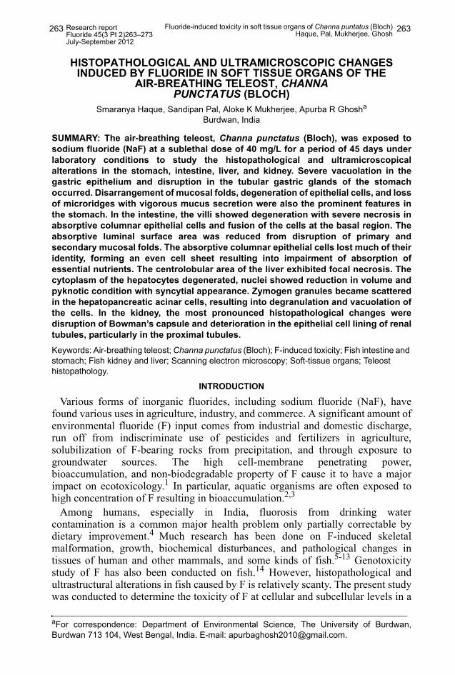
Research report Fluoride 45(3 Pt 2)263–273 July-September 2012 Fluoride-induced toxicity in soft tissue organs of Channa puntatus (Bloch) Haque, Pal, Mukherjee, Ghosh 263 263 HISTOPATHOLOGICAL AND ULTRAMICROSCOPIC CHANGES INDUCED BY FLUORIDE IN SOFT TISSUE ORGANS OF THE AIR-BREATHING TELEOST, CHANNA PUNCTATUS (BLOCH) Smaranya Haque, Sandipan Pal, Aloke K Mukherjee, Apurba R Ghosh a Burdwan, India SUMMARY: The air-breathing teleost, Channa punctatus (Bloch), was exposed to sodium fluoride (NaF) at a sublethal dose of 40 mg/L for a period of 45 days under laboratory conditions to study the histopathological and ultramicroscopical alterations in the stomach, intestine, liver, and kidney. Severe vacuolation in the gastric epithelium and disruption in the tubular gastric glands of the stomach occurred. Disarrangement of mucosal folds, degeneration of epithelial cells, and loss of microridges with vigorous mucus secretion were also the prominent features in the stomach. In the intestine, the villi showed degeneration with severe necrosis in absorptive columnar epithelial cells and fusion of the cells at the basal region. The absorptive luminal surface area was reduced from disruption of primary and secondary mucosal folds. The absorptive columnar epithelial cells lost much of their identity, forming an even cell sheet resulting into impairment of absorption of essential nutrients. The centrolobular area of the liver exhibited focal necrosis. The cytoplasm of the hepatocytes degenerated, nuclei showed reduction in volume and pyknotic condition with syncytial appearance. Zymogen granules became scattered in the hepatopancreatic acinar cells, resulting into degranulation and vacuolation of the cells. In the kidney, the most pronounced histopathological changes were disruption of Bowman’s capsule and deterioration in the epithelial cell lining of renal tubules, particularly in the proximal tubules. Keywords: Air-breathing teleost; Channa punctatus (Bloch); F-induced toxicity; Fish intestine and stomach; Fish kidney and liver; Scanning electron microscopy; Soft-tissue organs; Teleost histopathology. INTRODUCTION Various forms of inorganic fluorides, including sodium fluoride (NaF), have found various uses in agriculture, industry, and commerce. A significant amount of environmental fluoride (F) input comes from industrial and domestic discharge, run off from indiscriminate use of pesticides and fertilizers in agriculture, solubilization of F-bearing rocks from precipitation, and through exposure to groundwater sources. The high cell-membrane penetrating power, bioaccumulation, and non-biodegradable property of F cause it to have a major impact on ecotoxicology. 1 In particular, aquatic organisms are often exposed to high concentration of F resulting in bioaccumulation. 2,3 Among humans, especially in India, fluorosis from drinking water contamination is a common major health problem only partially correctable by dietary improvement. 4 Much research has been done on F-induced skeletal malformation, growth, biochemical disturbances, and pathological changes in tissues of human and other mammals, and some kinds of fish. 5-13 Genotoxicity study of F has also been conducted on fish. 14 However, histopathological and ultrastructural alterations in fish caused by F is relatively scanty. The present study was conducted to determine the toxicity of F at cellular and subcellular levels in a a For correspondence: Department of Environmental Science, The University of Burdwan, Burdwan 713 104, West Bengal, India. E-mail: [email protected].
Transcript of HISTOPATHOLOGICAL AND ULTRAMICROSCOPIC CHANGES INDUCED … · histopathological and...
Research reportFluoride 45(3 Pt 2)263–273July-September 2012
Fluoride-induced toxicity in soft tissue organs of Channa puntatus (Bloch)Haque, Pal, Mukherjee, Ghosh 263263
HISTOPATHOLOGICAL AND ULTRAMICROSCOPIC CHANGES INDUCED BY FLUORIDE IN SOFT TISSUE ORGANS OF THE
AIR-BREATHING TELEOST, CHANNA PUNCTATUS (BLOCH)
Smaranya Haque, Sandipan Pal, Aloke K Mukherjee, Apurba R Ghosha
Burdwan, India
SUMMARY: The air-breathing teleost, Channa punctatus (Bloch), was exposed tosodium fluoride (NaF) at a sublethal dose of 40 mg/L for a period of 45 days underlaboratory conditions to study the histopathological and ultramicroscopicalalterations in the stomach, intestine, liver, and kidney. Severe vacuolation in thegastric epithelium and disruption in the tubular gastric glands of the stomachoccurred. Disarrangement of mucosal folds, degeneration of epithelial cells, and lossof microridges with vigorous mucus secretion were also the prominent features inthe stomach. In the intestine, the villi showed degeneration with severe necrosis inabsorptive columnar epithelial cells and fusion of the cells at the basal region. Theabsorptive luminal surface area was reduced from disruption of primary andsecondary mucosal folds. The absorptive columnar epithelial cells lost much of theiridentity, forming an even cell sheet resulting into impairment of absorption ofessential nutrients. The centrolobular area of the liver exhibited focal necrosis. Thecytoplasm of the hepatocytes degenerated, nuclei showed reduction in volume andpyknotic condition with syncytial appearance. Zymogen granules became scatteredin the hepatopancreatic acinar cells, resulting into degranulation and vacuolation ofthe cells. In the kidney, the most pronounced histopathological changes weredisruption of Bowman’s capsule and deterioration in the epithelial cell lining of renaltubules, particularly in the proximal tubules. Keywords: Air-breathing teleost; Channa punctatus (Bloch); F-induced toxicity; Fish intestine and stomach; Fish kidney and liver; Scanning electron microscopy; Soft-tissue organs; Teleost histopathology.
INTRODUCTION
Various forms of inorganic fluorides, including sodium fluoride (NaF), havefound various uses in agriculture, industry, and commerce. A significant amount ofenvironmental fluoride (F) input comes from industrial and domestic discharge,run off from indiscriminate use of pesticides and fertilizers in agriculture,solubilization of F-bearing rocks from precipitation, and through exposure togroundwater sources. The high cell-membrane penetrating power,bioaccumulation, and non-biodegradable property of F cause it to have a majorimpact on ecotoxicology.1 In particular, aquatic organisms are often exposed tohigh concentration of F resulting in bioaccumulation.2,3
Among humans, especially in India, fluorosis from drinking watercontamination is a common major health problem only partially correctable bydietary improvement.4 Much research has been done on F-induced skeletalmalformation, growth, biochemical disturbances, and pathological changes intissues of human and other mammals, and some kinds of fish.5-13 Genotoxicitystudy of F has also been conducted on fish.14 However, histopathological andultrastructural alterations in fish caused by F is relatively scanty. The present studywas conducted to determine the toxicity of F at cellular and subcellular levels in a
aFor correspondence: Department of Environmental Science, The University of Burdwan,Burdwan 713 104, West Bengal, India. E-mail: [email protected].
Research reportFluoride 45(3 Pt 2)263–273July-September 2012
Fluoride-induced toxicity in soft tissue organs of Channa puntatus (Bloch)Haque, Pal, Mukherjee, Ghosh 264264
benthic teleostean fish, Channa punctatus (Bloch), particularly in the soft tissueorgans of the stomach, intestine, liver, and kidney.
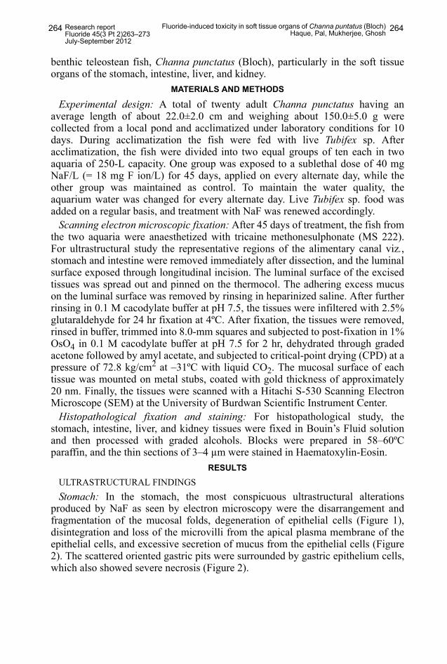
MATERIALS AND METHODS
Experimental design: A total of twenty adult Channa punctatus having anaverage length of about 22.0±2.0 cm and weighing about 150.0±5.0 g werecollected from a local pond and acclimatized under laboratory conditions for 10days. During acclimatization the fish were fed with live Tubifex sp. Afteracclimatization, the fish were divided into two equal groups of ten each in twoaquaria of 250-L capacity. One group was exposed to a sublethal dose of 40 mgNaF/L (= 18 mg F ion/L) for 45 days, applied on every alternate day, while theother group was maintained as control. To maintain the water quality, theaquarium water was changed for every alternate day. Live Tubifex sp. food wasadded on a regular basis, and treatment with NaF was renewed accordingly.
Scanning electron microscopic fixation: After 45 days of treatment, the fish fromthe two aquaria were anaesthetized with tricaine methonesulphonate (MS 222).For ultrastructural study the representative regions of the alimentary canal viz.,stomach and intestine were removed immediately after dissection, and the luminalsurface exposed through longitudinal incision. The luminal surface of the excisedtissues was spread out and pinned on the thermocol. The adhering excess mucuson the luminal surface was removed by rinsing in heparinized saline. After furtherrinsing in 0.1 M cacodylate buffer at pH 7.5, the tissues were infiltered with 2.5%glutaraldehyde for 24 hr fixation at 4ºC. After fixation, the tissues were removed,rinsed in buffer, trimmed into 8.0-mm squares and subjected to post-fixation in 1%OsO4 in 0.1 M cacodylate buffer at pH 7.5 for 2 hr, dehydrated through gradedacetone followed by amyl acetate, and subjected to critical-point drying (CPD) at apressure of 72.8 kg/cm2 at –31ºC with liquid CO2. The mucosal surface of eachtissue was mounted on metal stubs, coated with gold thickness of approximately20 nm. Finally, the tissues were scanned with a Hitachi S-530 Scanning ElectronMicroscope (SEM) at the University of Burdwan Scientific Instrument Center.
Histopathological fixation and staining: For histopathological study, thestomach, intestine, liver, and kidney tissues were fixed in Bouin’s Fluid solutionand then processed with graded alcohols. Blocks were prepared in 58–60ºCparaffin, and the thin sections of 3–4 µm were stained in Haematoxylin-Eosin.
RESULTS
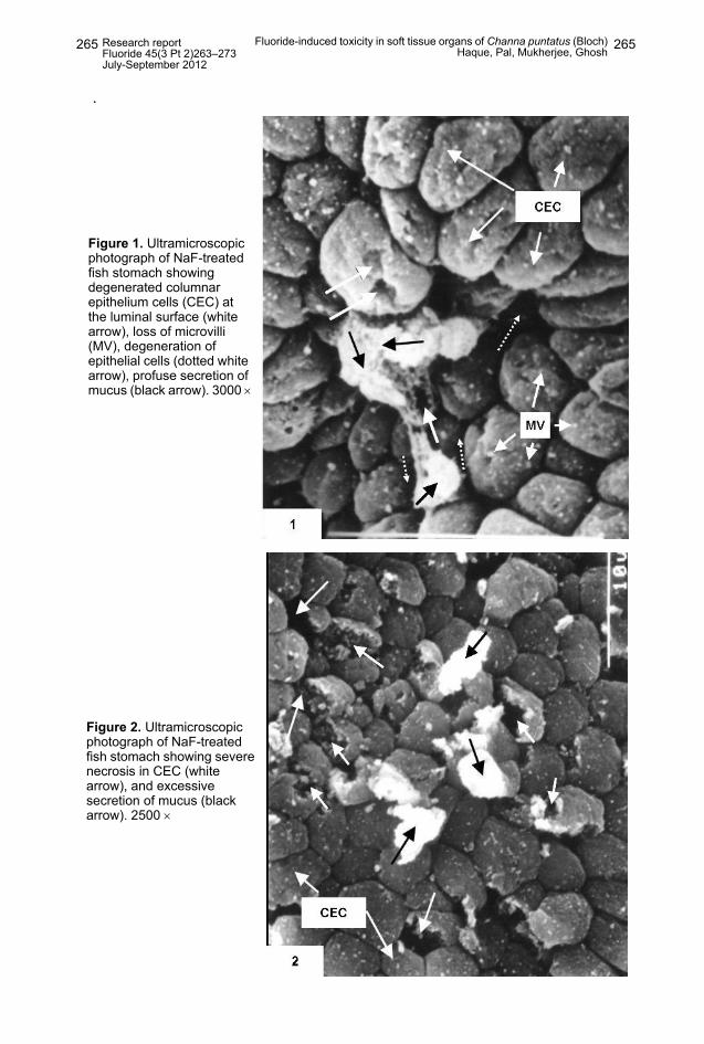
ULTRASTRUCTURAL FINDINGSStomach: In the stomach, the most conspicuous ultrastructural alterations
produced by NaF as seen by electron microscopy were the disarrangement andfragmentation of the mucosal folds, degeneration of epithelial cells (Figure 1),disintegration and loss of the microvilli from the apical plasma membrane of theepithelial cells, and excessive secretion of mucus from the epithelial cells (Figure2). The scattered oriented gastric pits were surrounded by gastric epithelium cells,which also showed severe necrosis (Figure 2).
Research reportFluoride 45(3 Pt 2)263–273July-September 2012
Fluoride-induced toxicity in soft tissue organs of Channa puntatus (Bloch)Haque, Pal, Mukherjee, Ghosh 265265
.
Figure 1. Ultramicroscopic photograph of NaF-treated fish stomach showing degenerated columnar epithelium cells (CEC) at the luminal surface (white arrow), loss of microvilli (MV), degeneration of epithelial cells (dotted white arrow), profuse secretion of mucus (black arrow). 3000 ×
Figure 2. Ultramicroscopic photograph of NaF-treated fish stomach showing severe necrosis in CEC (white arrow), and excessive secretion of mucus (black arrow). 2500 ×
Research reportFluoride 45(3 Pt 2)263–273July-September 2012
Fluoride-induced toxicity in soft tissue organs of Channa puntatus (Bloch)Haque, Pal, Mukherjee, Ghosh 266266
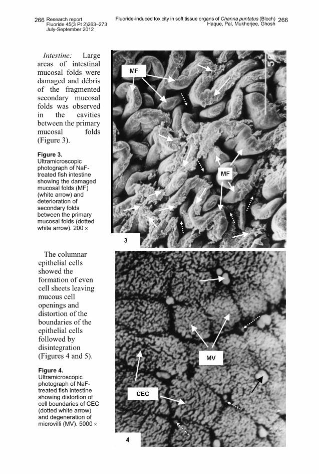
Intestine: Largeareas of intestinalmucosal folds weredamaged and débrisof the fragmentedsecondary mucosalfolds was observedin the cavitiesbetween the primarymucosal folds(Figure 3).
Figure 3. Ultramicroscopic photograph of NaF-treated fish intestine showing the damaged mucosal folds (MF) (white arrow) and deterioration of secondary folds between the primary mucosal folds (dotted white arrow). 200 ×
The columnar epithelial cells showed the formation of even cell sheets leaving mucous cell openings and distortion of the boundaries of the epithelial cells followed by disintegration (Figures 4 and 5).
Figure 4. Ultramicroscopic photograph of NaF-treated fish intestine showing distortion of cell boundaries of CEC (dotted white arrow) and degeneration of microvilli (MV). 5000 ×

Research reportFluoride 45(3 Pt 2)263–273July-September 2012
Fluoride-induced toxicity in soft tissue organs of Channa puntatus (Bloch)Haque, Pal, Mukherjee, Ghosh 267267
HISTOPATHOLOGICAL OBSERVATIONSStomach: Lesions detected by histopathology in the mucosal layer from NaF
were very pronounced as compared to the control section (Figure 6).
Figure 5. Ultramicroscopic photograph of NaF-treated intestine showing distorted mucous cell (MC) opening surrounded by degenerated CEC and MV. 5000 ×
Figure 6. Photomicrograph of stomach of control fish showing mucosal layer with prominent top plate (TP) and mucosal fold (MF). The CEC are lined beneath the mucosal layer and the tubular gastric gland (GG) with prominent nucleus (N). H & E staining. 10 × 40
Research reportFluoride 45(3 Pt 2)263–273July-September 2012
Fluoride-induced toxicity in soft tissue organs of Channa puntatus (Bloch)Haque, Pal, Mukherjee, Ghosh 268268
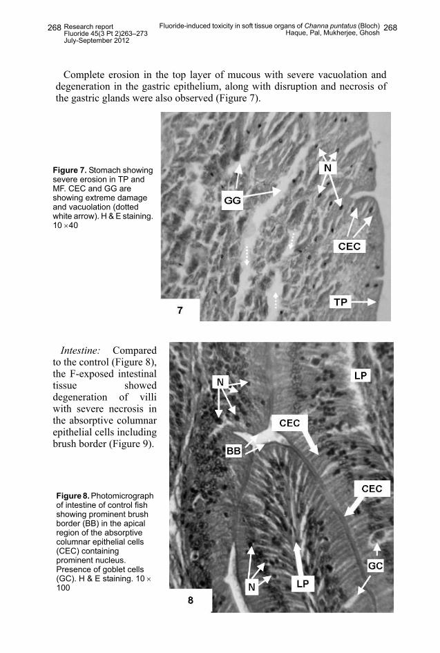
Figure 7. Stomach showing severe erosion in TP and MF. CEC and GG are showing extreme damage and vacuolation (dotted white arrow). H & E staining. 10 ×40
Complete erosion in the top layer of mucous with severe vacuolation anddegeneration in the gastric epithelium, along with disruption and necrosis ofthe gastric glands were also observed (Figure 7).
Intestine: Comparedto the control (Figure 8),the F-exposed intestinaltissue showeddegeneration of villiwith severe necrosis inthe absorptive columnarepithelial cells includingbrush border (Figure 9).
Figure 8. Photomicrograph of intestine of control fish showing prominent brush border (BB) in the apical region of the absorptive columnar epithelial cells (CEC) containing prominent nucleus. Presence of goblet cells (GC). H & E staining. 10 × 100
Research reportFluoride 45(3 Pt 2)263–273July-September 2012
Fluoride-induced toxicity in soft tissue organs of Channa puntatus (Bloch)Haque, Pal, Mukherjee, Ghosh 269269
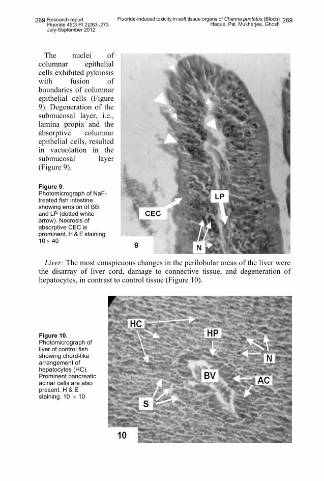
The nuclei ofcolumnar epithelialcells exhibited pyknosiswith fusion ofboundaries of columnarepithelial cells (Figure9). Degeneration of thesubmucosal layer, i.e.,lamina propia and theabsorptive columnarepithelial cells, resultedin vacuolation in thesubmucosal layer(Figure 9).
Figure 9. Photomicrograph of NaF-treated fish intestine showing erosion of BB and LP (dotted white arrow). Necrosis of absorptive CEC is prominent. H & E staining. 10 × 40
Liver: The most conspicuous changes in the perilobular areas of the liver werethe disarray of liver cord, damage to connective tissue, and degeneration ofhepatocytes, in contrast to control tissue (Figure 10).
Figure 10. Photomicrograph of liver of control fish showing chord-like arrangement of hepatocytes (HC). Prominent pancreatic acinar cells are also present. H & E staining. 10 × 10
Research reportFluoride 45(3 Pt 2)263–273July-September 2012
Fluoride-induced toxicity in soft tissue organs of Channa puntatus (Bloch)Haque, Pal, Mukherjee, Ghosh 270270
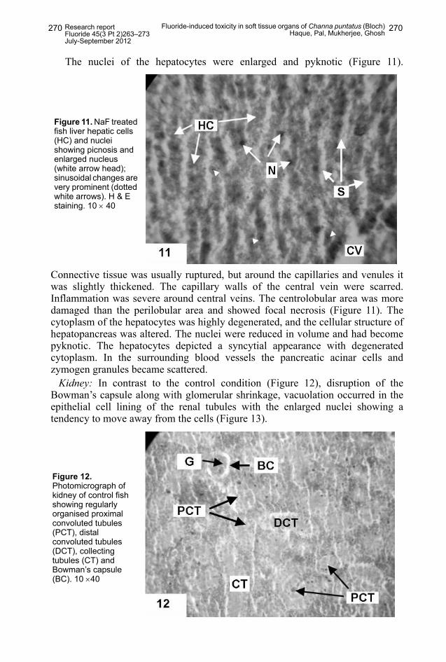
The nuclei of the hepatocytes were enlarged and pyknotic (Figure 11).
Connective tissue was usually ruptured, but around the capillaries and venules itwas slightly thickened. The capillary walls of the central vein were scarred.Inflammation was severe around central veins. The centrolobular area was moredamaged than the perilobular area and showed focal necrosis (Figure 11). Thecytoplasm of the hepatocytes was highly degenerated, and the cellular structure ofhepatopancreas was altered. The nuclei were reduced in volume and had becomepyknotic. The hepatocytes depicted a syncytial appearance with degeneratedcytoplasm. In the surrounding blood vessels the pancreatic acinar cells andzymogen granules became scattered.
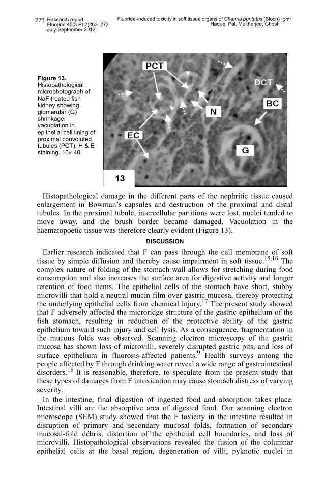
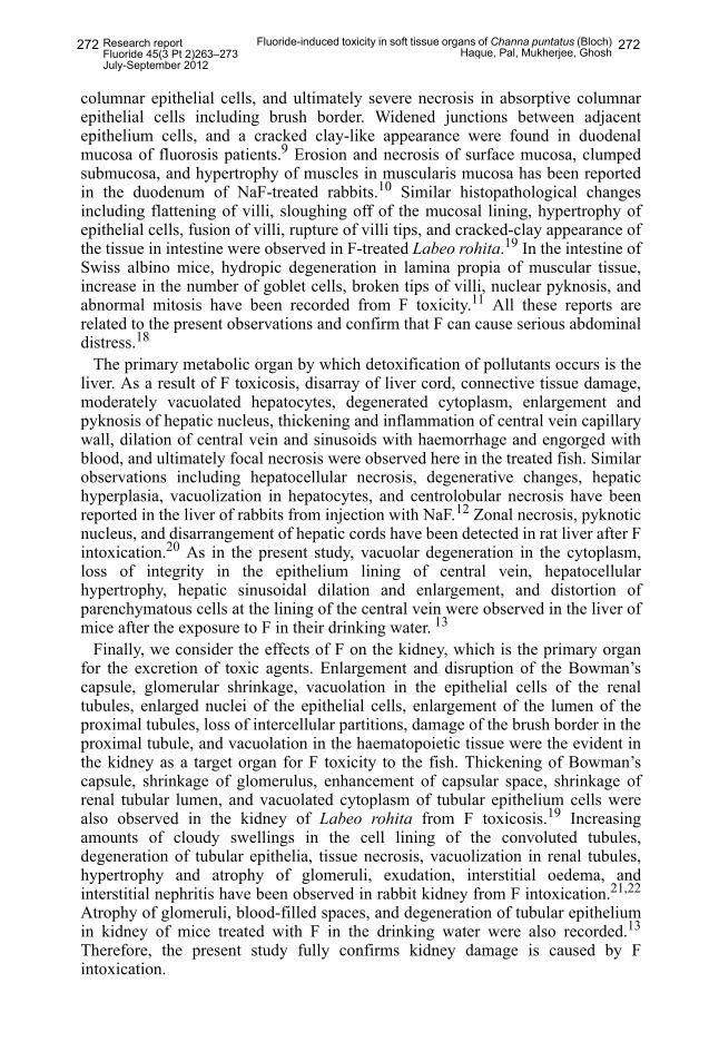
Kidney: In contrast to the control condition (Figure 12), disruption of theBowman’s capsule along with glomerular shrinkage, vacuolation occurred in theepithelial cell lining of the renal tubules with the enlarged nuclei showing atendency to move away from the cells (Figure 13).
Figure 11. NaF treated fish liver hepatic cells (HC) and nuclei showing picnosis and enlarged nucleus (white arrow head); sinusoidal changes are very prominent (dotted white arrows). H & E staining. 10 × 40
Figure 12. Photomicrograph of kidney of control fish showing regularly organised proximal convoluted tubules (PCT), distal convoluted tubules (DCT), collecting tubules (CT) and Bowman’s capsule (BC). 10 ×40
Research reportFluoride 45(3 Pt 2)263–273July-September 2012
Fluoride-induced toxicity in soft tissue organs of Channa puntatus (Bloch)Haque, Pal, Mukherjee, Ghosh 271271
Histopathological damage in the different parts of the nephritic tissue causedenlargement in Bowman’s capsules and destruction of the proximal and distaltubules. In the proximal tubule, intercellular partitions were lost, nuclei tended tomove away, and the brush border became damaged. Vacuolation in thehaematopoetic tissue was therefore clearly evident (Figure 13).
DISCUSSION
Earlier research indicated that F can pass through the cell membrane of softtissue by simple diffusion and thereby cause impairment in soft tissue.15,16 Thecomplex nature of folding of the stomach wall allows for stretching during foodconsumption and also increases the surface area for digestive activity and longerretention of food items. The epithelial cells of the stomach have short, stubbymicrovilli that hold a neutral mucin film over gastric mucosa, thereby protectingthe underlying epithelial cells from chemical injury.17 The present study showedthat F adversely affected the microridge structure of the gastric epithelium of thefish stomach, resulting in reduction of the protective ability of the gastricepithelium toward such injury and cell lysis. As a consequence, fragmentation inthe mucous folds was observed. Scanning electron microscopy of the gastricmucosa has shown loss of microvilli, severely disrupted gastric pits, and loss ofsurface epithelium in fluorosis-affected patients.9 Health surveys among thepeople affected by F through drinking water reveal a wide range of gastrointestinaldisorders.18 It is reasonable, therefore, to speculate from the present study thatthese types of damages from F intoxication may cause stomach distress of varyingseverity.
In the intestine, final digestion of ingested food and absorption takes place.Intestinal villi are the absorptive area of digested food. Our scanning electronmicroscope (SEM) study showed that the F toxicity in the intestine resulted indisruption of primary and secondary mucosal folds, formation of secondarymucosal-fold débris, distortion of the epithelial cell boundaries, and loss ofmicrovilli. Histopathological observations revealed the fusion of the columnarepithelial cells at the basal region, degeneration of villi, pyknotic nuclei in
Figure 13. Histopathological microphotograph of NaF treated fish kidney showing glomerular (G) shrinkage, vacuolation in epithelial cell lining of proximal convoluted tubules (PCT). H & E staining. 10× 40
Research reportFluoride 45(3 Pt 2)263–273July-September 2012
Fluoride-induced toxicity in soft tissue organs of Channa puntatus (Bloch)Haque, Pal, Mukherjee, Ghosh 272272
columnar epithelial cells, and ultimately severe necrosis in absorptive columnarepithelial cells including brush border. Widened junctions between adjacentepithelium cells, and a cracked clay-like appearance were found in duodenalmucosa of fluorosis patients.9 Erosion and necrosis of surface mucosa, clumpedsubmucosa, and hypertrophy of muscles in muscularis mucosa has been reportedin the duodenum of NaF-treated rabbits.10 Similar histopathological changesincluding flattening of villi, sloughing off of the mucosal lining, hypertrophy ofepithelial cells, fusion of villi, rupture of villi tips, and cracked-clay appearance ofthe tissue in intestine were observed in F-treated Labeo rohita.19 In the intestine ofSwiss albino mice, hydropic degeneration in lamina propia of muscular tissue,increase in the number of goblet cells, broken tips of villi, nuclear pyknosis, andabnormal mitosis have been recorded from F toxicity.11 All these reports arerelated to the present observations and confirm that F can cause serious abdominaldistress.18
The primary metabolic organ by which detoxification of pollutants occurs is theliver. As a result of F toxicosis, disarray of liver cord, connective tissue damage,moderately vacuolated hepatocytes, degenerated cytoplasm, enlargement andpyknosis of hepatic nucleus, thickening and inflammation of central vein capillarywall, dilation of central vein and sinusoids with haemorrhage and engorged withblood, and ultimately focal necrosis were observed here in the treated fish. Similarobservations including hepatocellular necrosis, degenerative changes, hepatichyperplasia, vacuolization in hepatocytes, and centrolobular necrosis have beenreported in the liver of rabbits from injection with NaF.12 Zonal necrosis, pyknoticnucleus, and disarrangement of hepatic cords have been detected in rat liver after Fintoxication.20 As in the present study, vacuolar degeneration in the cytoplasm,loss of integrity in the epithelium lining of central vein, hepatocellularhypertrophy, hepatic sinusoidal dilation and enlargement, and distortion ofparenchymatous cells at the lining of the central vein were observed in the liver ofmice after the exposure to F in their drinking water. 13
Finally, we consider the effects of F on the kidney, which is the primary organfor the excretion of toxic agents. Enlargement and disruption of the Bowman’scapsule, glomerular shrinkage, vacuolation in the epithelial cells of the renaltubules, enlarged nuclei of the epithelial cells, enlargement of the lumen of theproximal tubules, loss of intercellular partitions, damage of the brush border in theproximal tubule, and vacuolation in the haematopoietic tissue were the evident inthe kidney as a target organ for F toxicity to the fish. Thickening of Bowman’scapsule, shrinkage of glomerulus, enhancement of capsular space, shrinkage ofrenal tubular lumen, and vacuolated cytoplasm of tubular epithelium cells werealso observed in the kidney of Labeo rohita from F toxicosis.19 Increasingamounts of cloudy swellings in the cell lining of the convoluted tubules,degeneration of tubular epithelia, tissue necrosis, vacuolization in renal tubules,hypertrophy and atrophy of glomeruli, exudation, interstitial oedema, andinterstitial nephritis have been observed in rabbit kidney from F intoxication.21,22
Atrophy of glomeruli, blood-filled spaces, and degeneration of tubular epitheliumin kidney of mice treated with F in the drinking water were also recorded.13
Therefore, the present study fully confirms kidney damage is caused by Fintoxication.
Research reportFluoride 45(3 Pt 2)263–273July-September 2012
Fluoride-induced toxicity in soft tissue organs of Channa puntatus (Bloch)Haque, Pal, Mukherjee, Ghosh 273273
In conclusion, we have shown that sub-lethal exposure of the teleost fish Channapunctatus to excess F causes significant disturbance and impairment of soft tissueorgans involving the important life processes of digestion, metabolism, andexcretion.
REFERENCES1 Sharma PD. Environment Pollution In: Ecology and environment. 7th ed. Meerut, India:
Rastogi Publications; 2002. 2 Neuhold JM, Sigler WF. Effects of sodium fluoride on carp and rainbow trout. Trans Am Fish
Sco 1960;89:358-70.3 Lee CF, Nilson HW. Study of the metabolism of naturally occurring fluorine in canned
salmon and mackerel. U.S. Bureau of Fish Invest 1939. Report 44.4 Susheela AK, Bhatnagar M. Reversal of fluoride induced cell injury through elimination of
fluoride and consumption of diet rich in essential nutrients and antioxidants. Mol CellBiochem 2002;234-235(1-2):335-40.
5 Wang Y, Yin Y, Gilula LA, Wilson AJ. Endemic fluorosis of the skeleton: radiological featuresin 127 patients. AJR Am J Roentgenol 1994;162:93–8.
6 Shupe JL, Bruner RH, Seymour J, Alden CL. The pathology of chronic bovine fluorosis: areview. Toxicol Pathol 1992;20(2):274-85; discussion 285-8.
7 Bajpai S, Tripathi M. Effect of fluoride on growth bioindicators in stinging catfish,Heteropneustes fossilis (Bloch). Fluoride 2010;43(4)232-36.
8 Kumar A, Tripathi N, Tripathi M. Fluoride-induced biochemical changes in fresh watercatfish (Clarias batrachus, Linn). Fluoride 2007;40(1):37-41.
9 Dasarathy S, Das TK, Gupta IP, Susheela AK, Tandon RK. Gastroduodenal manifestationsin patients with skeletal fluorosis. J Gastroenterol 1996; 31:333-7.
10 Shashi A. Histopathological effects of sodium fluoride on the duodenum of rabbits Fluoride2002;35(1):28-37.
11 Sondhi H, Gupta ML, Gupta GL. Intestinal effects of sodium fluoride in Swiss albino mice.Fluoride 1995;28(1):21-4.
12 Shashi A, Thapar SP. Histopathology of fluoride-induced hepatotoxicity in rabbits. Fluoride2001;34(1):34-42.
13 Chattopadhyay A, Podder S, Agarwal S, Bhattacharya S. Fluoride-induced histopathologyand synthesis of stress protein in liver and kidney of mice. Arch Toxicol 2010;85(4):327-35.
14 Tripathi N, Bajpai S, Tripathi M. Genotoxic alterations induced by fluoride in Asian catfish,Clarias batrachus (Linn). Fluoride 2009;42(4):292-6.
15 Carlson CH, Armstrong WD, Singer L. Distribution and excretion of radiofluoride in thehuman. Proc Soc Exp Biol Med 1960;104:235-9.
16 Jacyszyn K, Marut A. Fluoride in blood and urine in humans administered fluoride andexposed to fluoride-polluted air. Fluoride 1986;19(1):26-32.
17 Chakrabarti P, Sinha GM. Mucosal surface of the alimentary canal in Mystus vittatus(Bloch): a scanning electron microscopic study. Proceedings of Indian National ScienceAcademy 1987;53B:317-22.
18 Sharma JD, Jain P, Sohu D. Gastric discomforts from fluoride in drinking water in SanganerTehsil, Rajasthan, India. Fluoride 2009;42(4)286-91.
19 Bhatnagar C, Bhatnagar M, Regarc BC. Fluoride-induced histopathological changes in gill,kidney, and intestine of fresh water Teleost, Labeo rohita. Fluoride 2007;40(1):55-61.
20 Chinoy NJ, Joseph R, Sequeira E, Narayana MV. Effects of sodium fluoride on the muscleand liver of albino rats. Ind J Environ Toxicol 1991;1:129-34.
21 Shashi A, Singh JP, Thapar SP. Toxic effects of fluoride on rabbit kidney. Fluoride2002;35(1):38-50.
22 Kawahara H. Experimental studies on the changes of the kidney due to fluorosis. Part III.Morphological studies on the changes of the kidney of rabbits and growing albino rats dueto sodium fluoride. Shikoku Acta Med 1956;8:283-8.
Copyright © 2012 The International Society for Fluoride Research Inc. www.fluorideresearch.org www.fluorideresearch.com www.fluorideresearch.net
Editorial Office: 727 Brighton Road, Ocean View, Dunedin 9035, New Zealand.