Histology I - Mt. San Antonio Collegeinstruction2.mtsac.edu/crexach/anatomy 35/pdf...
46
Histology I Histology I Histology I Dr. Carmen E. Rexach Anatomy 35 Mt San Antonio College
Transcript of Histology I - Mt. San Antonio Collegeinstruction2.mtsac.edu/crexach/anatomy 35/pdf...

Histology IHistology IHistology I
Dr. Carmen E. RexachAnatomy 35
Mt San Antonio College
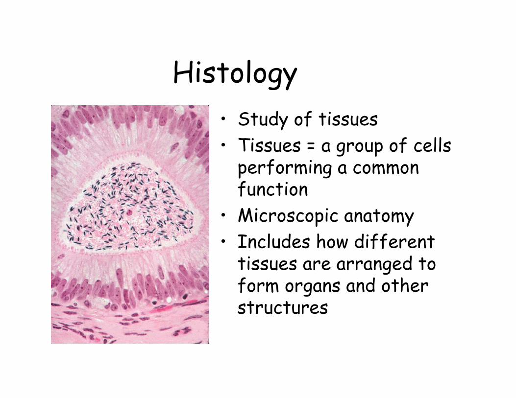
Histology• Study of tissues • Tissues = a group of cells
performing a common function
• Microscopic anatomy• Includes how different
tissues are arranged to form organs and other structures

Extracellular matrix: cell surface
• Composition– Fibrous structural and adhesion proteins
embedded in gelatinous polysaccharide ground substance
• Function– Binds cells together to form tissues

Extracellular matrix: cell surface
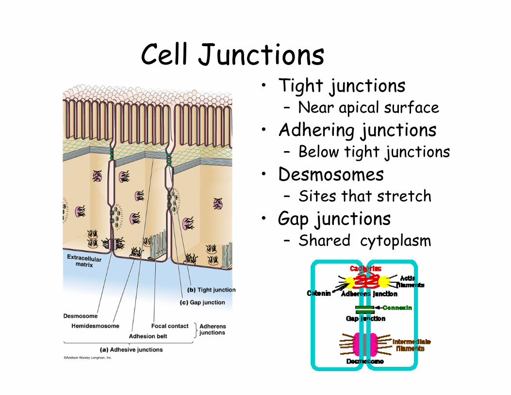
Cell Junctions• Tight junctions
– Near apical surface• Adhering junctions
– Below tight junctions• Desmosomes
– Sites that stretch• Gap junctions
– Shared cytoplasm
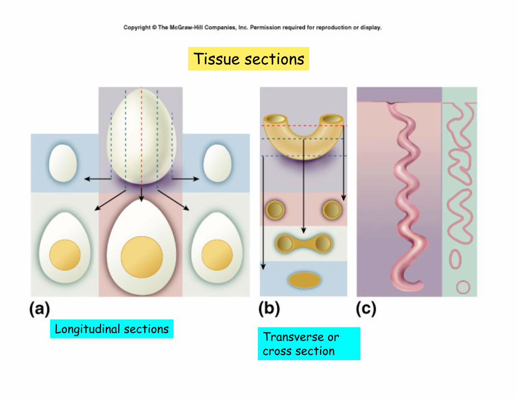
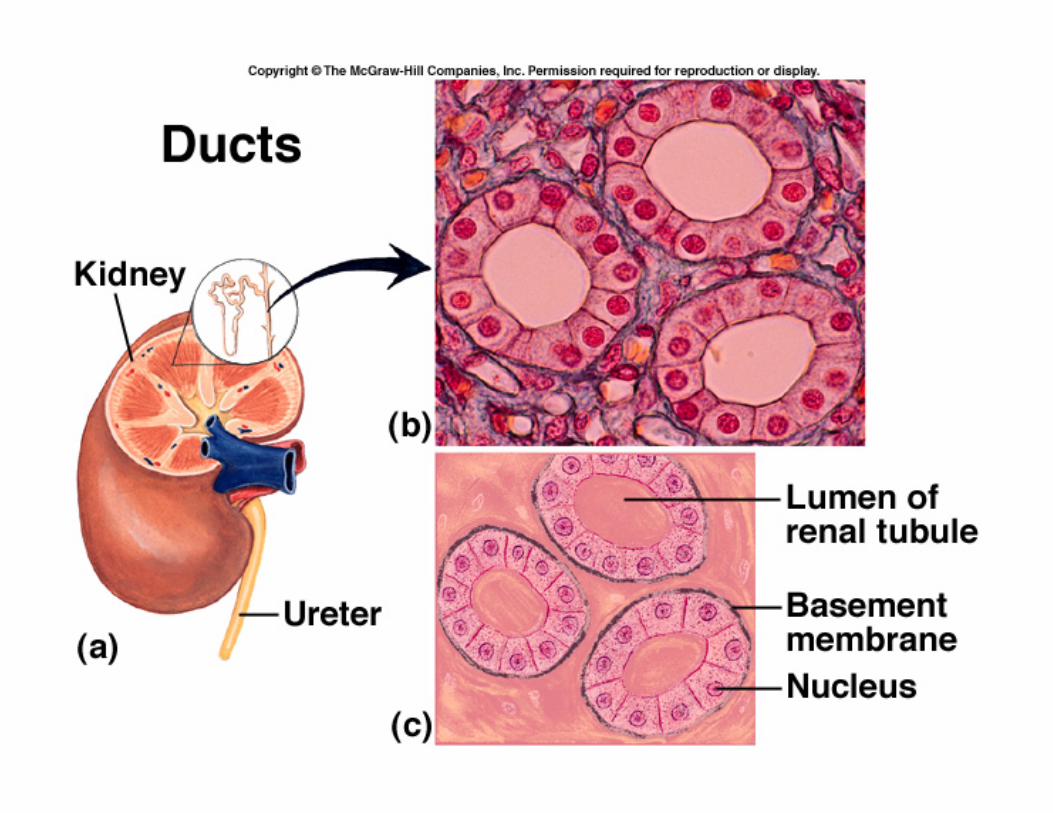
Tissue sections
Longitudinal sections Transverse or cross section
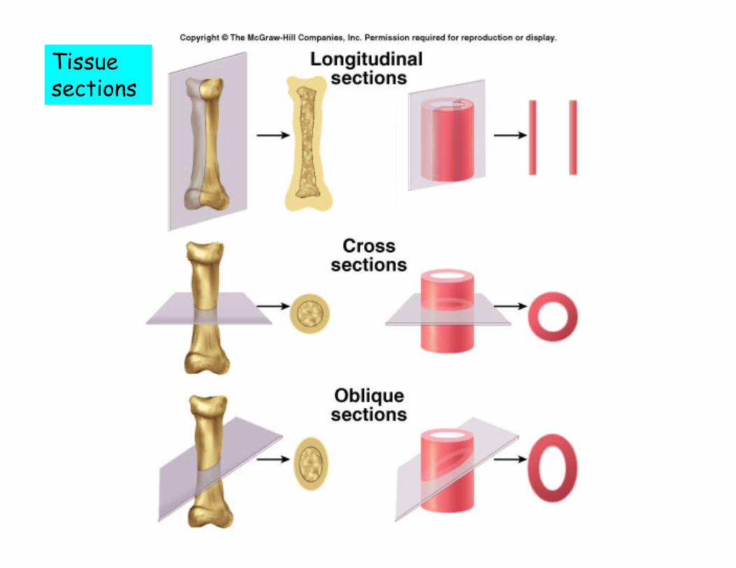
Tissue sections

Primary tissue classes
• Epithelial• Connective
• Differences– Matrix
• variation• Primary constituents
– Fibrous proteins– Ground substance
– Amount of cells vs matrix
• Nervous• Muscular

Epithelial tissue• Composition
– One or more layers of closely adhering cells– Apical surface and basal surface– Rest on a basement membrane– Avascular– Supported by connective tissues
• Function– Cover organ surfaces– Form secretory tissue– Form ducts of glands

Classification
• Criteria– Number of cell layers– Morphology of the
surface cells• Two major categories
– Simple (one layer)– Stratified (two or more
layers)

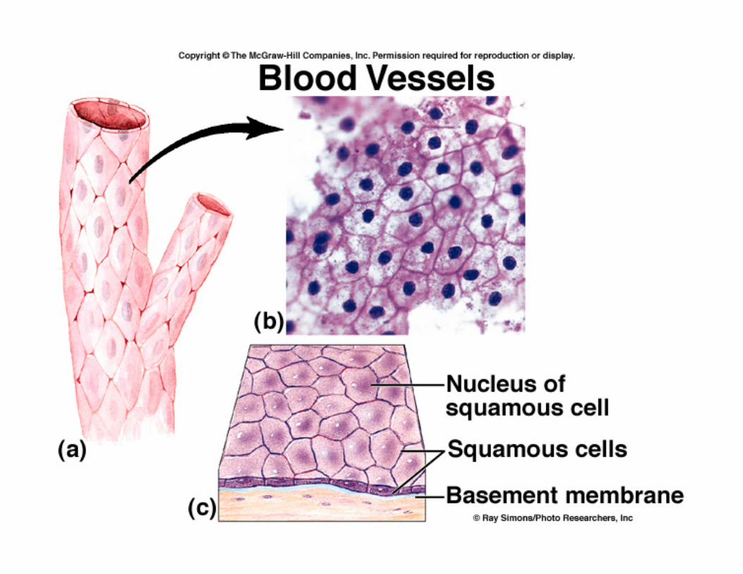
Cell shape• Squamous
• Cuboidal
• Columnar
• Acinar

Simple Epithelia
• General– All cells touch basement membrane
• Exception: Pseudostratified columnar
• Categories– Simple squamous– Simple cuboidal– Simple columnar– Pseudostratified columnar

Simple Epithelia

Microvilli vs. Cilia
Microvilli are folds in the apical surface of the plasma membrane. They increase cell surface area.
Cilia are composed of microtubules and project from the apical surface. Function: to move things across the cell surface
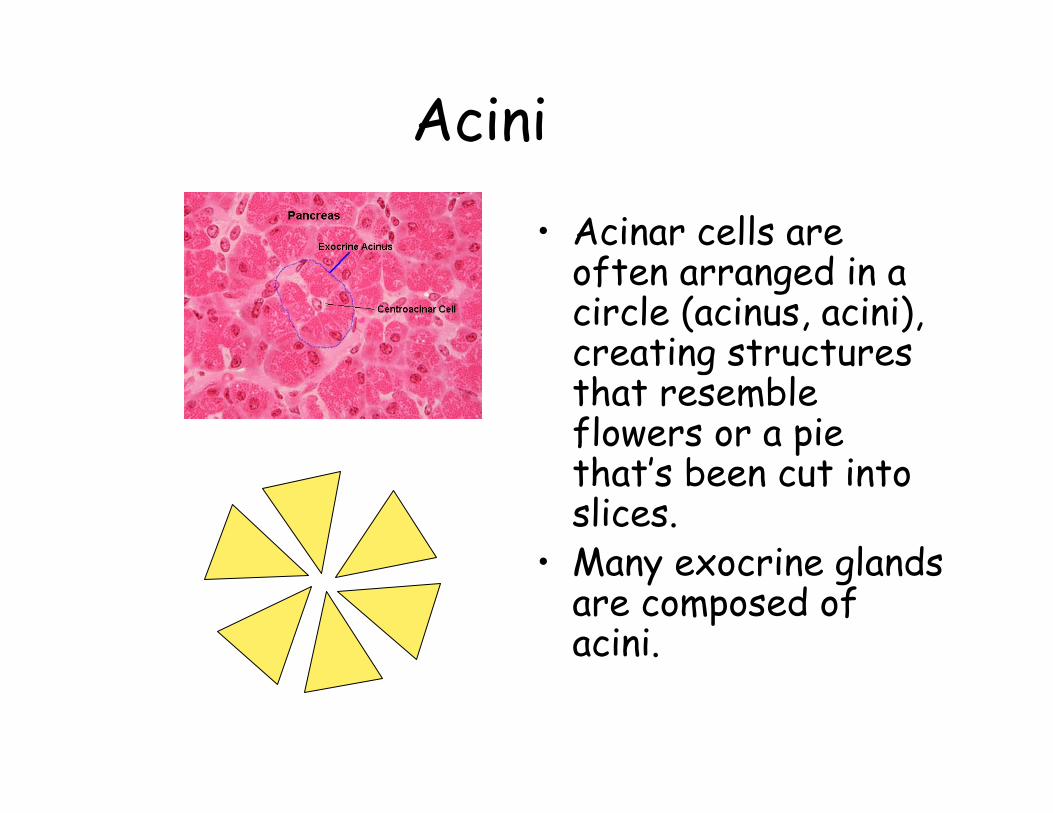
Acini
• Acinar cells are often arranged in a circle (acinus, acini), creating structures that resemble flowers or a pie that’s been cut into slices.
• Many exocrine glands are composed of acini.

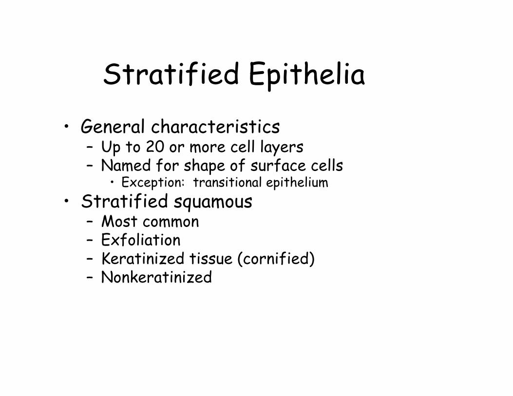
Stratified Epithelia• General characteristics
– Up to 20 or more cell layers– Named for shape of surface cells
• Exception: transitional epithelium• Stratified squamous
– Most common– Exfoliation– Keratinized tissue (cornified)– Nonkeratinized
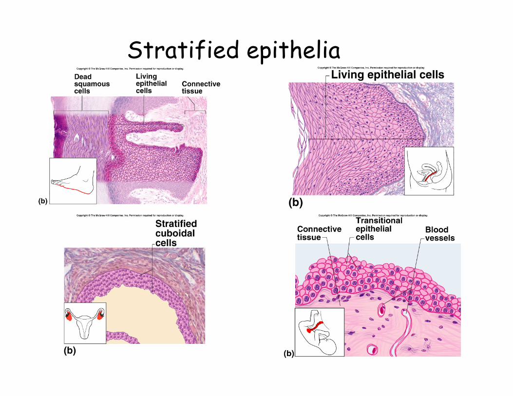
Stratified epithelia

Transitional epithelium
Note the “dome cells” at the apical surface

Connective Tissue
• Less cells, more ECF (ground substance)• Variable, widely distributed, most
abundant• Three categories of mature tissue
– Fibrous CT– Supportive CT (bone, cartilage)– Fluid CT (blood)

Functions
• Binds organs• Support• Protection
– Physical– Immune
• Movement• Storage• Heat production• Transportation

Fibrous Connective Tissue: Components
• Cells– Fibroblasts– Macrophage– Leukocytes– Plasma Cells– Mast Cells– Adipocytes
• Fibers – Collagenous– Reticular– Elastic
• Ground substance

Types of Fibrous CT• Loose
– Lots of ground substance– dissolves in vitro– Types
• Areolar, reticular, adipose• Dense
– Fiber is predominant component– Types
• Dense regular, dense irregular

Loose CT
Areolar connective tissue
Reticular connective tissue
Adipose connective tissue

Areolar Connective Tissue• Cells
– All six types• Matrix
– Predominately collagenous– Some elastic/reticular
• General– Very vascular– Loosely organized– Varied– Underlies almost all
epithelial tissue

Reticular• Reticular fibers and fibroblasts• Stroma of organs and tissues such as
lymph nodes, spleen, thymus, bone marrow
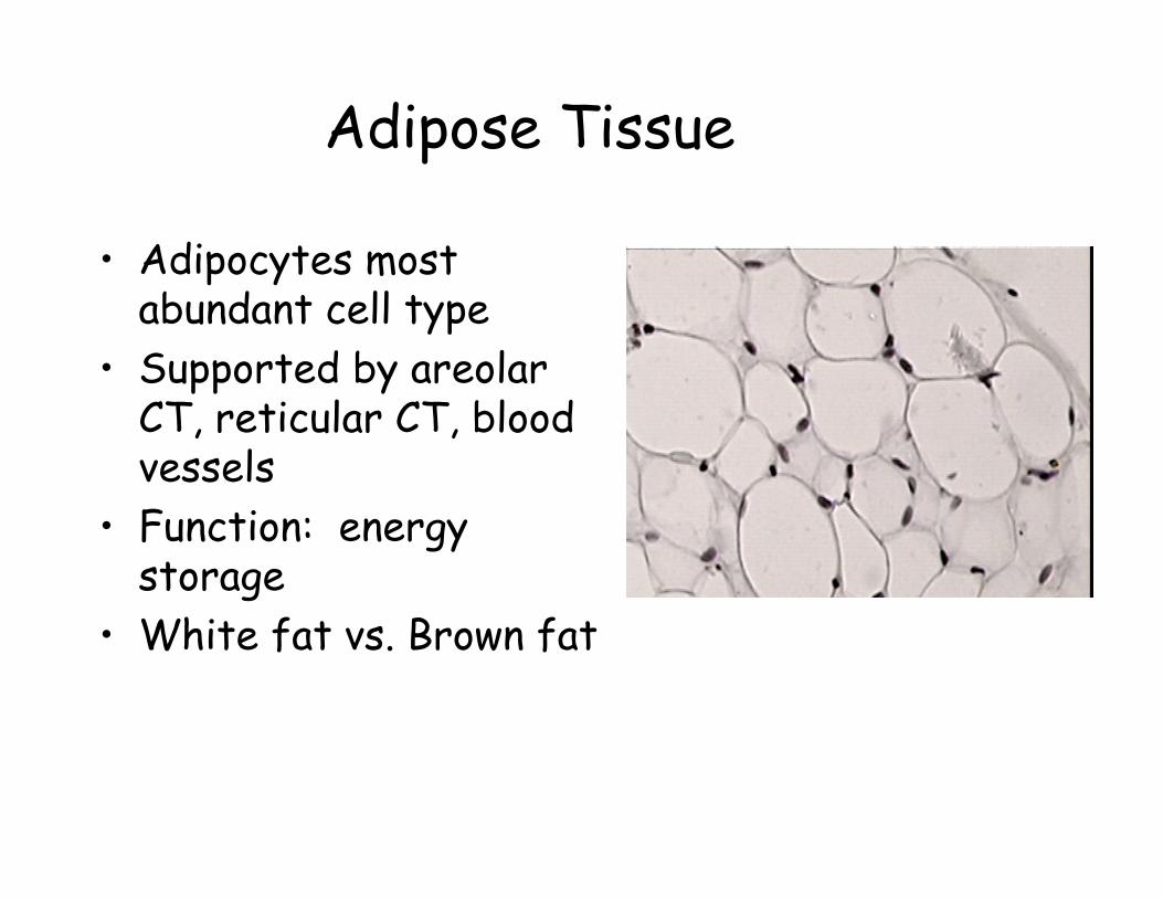
Adipose Tissue
• Adipocytes most abundant cell type
• Supported by areolarCT, reticular CT, blood vessels
• Function: energy storage
• White fat vs. Brown fat

Dense Connective Tissue• Dense Regular
– Closely packed parallel collagen fibers + fibroblasts
– Tendons and ligaments
– Yellow elastic tissue• Elastic fibers• Fibroblasts with
larger nuclei– Wavy elastic sheets
in walls of medium/large arteries
• Dense Irregular– Collagen fibers in
random arrangement + fibroblasts
– Dermis, protective capsule around organs, fibrous sheet around bones, etc.
– Similar to areolar, but less “open”space

Dense Connective Tissue
Dense Regular Dense Irregular
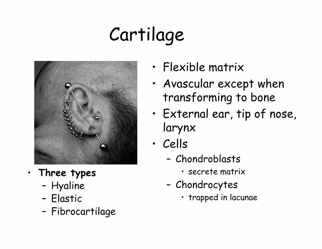
Cartilage• Flexible matrix• Avascular except when
transforming to bone• External ear, tip of nose,
larynx• Cells
– Chondroblasts• secrete matrix
– Chondrocytes• trapped in lacunae
• Three types– Hyaline– Elastic– Fibrocartilage

Hyaline Cartilage• Matrix
– smooth, frictionless – mix of collagen, elastin,
chondroitin (a glucosaminoglycan)
• Cells– Cell nests
• Usually covered by perichondrium– Dense irregular CT covering– inner layer produces new
chondrocytes• Examples:
– Costal cartilage– Nasal septum– C-ring cartilage of trachea– Fetal skeleton

Elastic Cartilage
• Matrix– Web-like mesh of
elastic fibers– Appearance of fur
• Perichondriumalways present
• Examples:– Pinna– Epiglottis

Fibrocartilage• Characteristics
– Parallel collagen fibers
– Rows of chondrocytes in lacunae
– No perichondrium• Examples
– Pubic symphysis– Intervertebral
discs– Menisci of the
synovial joints

Bone• Two forms
– Spongy• Heads of long
bone• Named for
appearance– Compact
• No visible spaces• Dense calcified
tissue• External surface
of all bone
• Structure of compact bone– Haversian canal– Osteon– Lamellae– Lacunae– Canaliculi
• Periosteum

Compact Bone

Blood• Plasma + formed
elements• Erythrocytes• Leukocytes
– Neutrophils– Basophils– Eosinophils– Monocytes– Lymphocytes

Hemopoiesisformation of formed elements
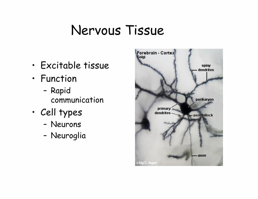
Nervous Tissue
• Excitable tissue• Function
– Rapid communication
• Cell types– Neurons– Neuroglia

Neurons
• impulse: an all or none electrical event
• neurotransmitters: chemical messengers released by neurons

Neuron

Neuroglia (nerve glue)
• 6 subtypes• highly branched
like neurons• not capable of
impulses• perform supportive
(connective tissue type) functions
astrocyte

Nerves: Cross section

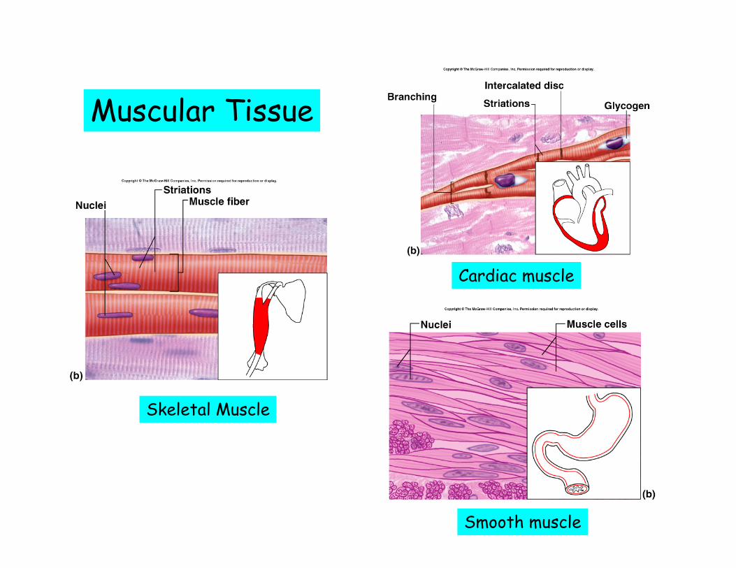
Muscular Tissue• Contracts when stimulated• Three types
– Skeletal• Striated, multinucleated, voluntary• Muscle fibers
– Cardiac• Striated, uninucleate, involuntary• Short branching cells• Intercalated discs
– gap junctions– desmosomes
– Smooth• No striations, uninucleate, involuntary• Short fusiform cells• Visceral muscle
Muscular Tissue
Skeletal Muscle
Cardiac muscle
Smooth muscle