Hirudotherapy: A Case Report Non-Occlusive Mesenteric ...
7
Received 07/15/2020 Review began 07/16/2020 Review ended 07/20/2020 Published 07/30/2020 © Copyright 2020 Akalın et al. This is an open access article distributed under the terms of the Creative Commons Attribution License CC-BY 4.0., which permits unrestricted use, distribution, and reproduction in any medium, provided the original author and source are credited. Non-Occlusive Mesenteric Ischemia Due to Hirudotherapy: A Case Report Çağrı Akalın , Nergis Ekmen 1. Department of General Surgery, Ordu University Training and Research Hospital, Ordu, TUR 2. Department of Gastroenterology, Gazi University Hospital, Ankara, TUR Corresponding author: Çağrı Akalın, [email protected] Abstract Leech therapy, which can lead to complications such as anemia and bleeding, has been used to treat many diseases since ancient times. Furthermore, some substances in leech saliva are known to have anticoagulant effects. Acute mesenteric ischemia, which develops due to mesenteric vascular obstruction, can be treated medically or surgically. Non-occlusive mesenteric ischemia (NOMI) occurs as a result of decreased blood flow in mesenteric vessels due to hypovolemia, hypotension, etc. In this report, we mentioned a 57-year-old male patient who was admitted to the emergency department with syncope and weakness. In his medical history, the patient was stated to have used leech therapy to treat diabetic wounds on his feet, and prolonged and unstoppable bleeding was seen after leech bites. On his physical examination, there was tenderness in all quadrants of the abdomen. Abdominal computed tomography without contrast agent showed hepatic portal venous gas and pneumatosis cystoides intestinalis (PSI). The patient underwent laparotomy owing to the development of acute abdomen during the follow-up. Necrosis was seen in the terminal ileum and entire colon. Low flow in mesenteric vascular vessels of these necrotic segments was indicated with intraoperative Doppler ultrasonography. All necrotic segments were resected and open end- ileostomy was performed. The patient was discharged on the 17th day of follow-up. In conclusion, excessive bleeding caused by leech therapy can cause NOMI. Categories: Emergency Medicine, General Surgery, Other Keywords: leech therapy, acute mesenteric ischemia, diabetic foot Introduction Leech therapy has been used in the treatment of some diseases since ancient times in the medical field. This treatment is known as hirudotherapy and is used for many diseases such as cardiovascular disease, diabetes mellitus and its complications, after replantation treatment, revascularization, and soft tissue injury [1]. Bleeding and anemia are among complications of hirudotherapy [2]. Vascular obstruction of the intestine causes acute mesenteric ischemia (AMI). AMI has a high prevalence and rates of mortality (50%-69%) in patients with cardiovascular disease and over 50 years old [3]. In this paper, the aim was to report a case of AMI which developed after acute bleeding and anemia leech therapy of a diabetic foot ulcer. Case Presentation A 57-year-old man was admitted to the ED with complaints of syncope and weakness. We noticed that in his medical history, leeches were applied to treat the diabetic wounds on his feet 1 2 Open Access Case Report DOI: 10.7759/cureus.9467 How to cite this article Akalın Ç, Ekmen N (July 30, 2020) Non-Occlusive Mesenteric Ischemia Due to Hirudotherapy: A Case Report. Cureus 12(7): e9467. DOI 10.7759/cureus.9467
Transcript of Hirudotherapy: A Case Report Non-Occlusive Mesenteric ...

Received 07/15/2020 Review began 07/16/2020 Review ended 07/20/2020 Published 07/30/2020
© Copyright 2020Akalın et al. This is an open accessarticle distributed under the terms ofthe Creative Commons AttributionLicense CC-BY 4.0., which permitsunrestricted use, distribution, andreproduction in any medium, providedthe original author and source arecredited.
Non-Occlusive Mesenteric Ischemia Due toHirudotherapy: A Case ReportÇağrı Akalın , Nergis Ekmen
1. Department of General Surgery, Ordu University Training and Research Hospital, Ordu, TUR 2.Department of Gastroenterology, Gazi University Hospital, Ankara, TUR
Corresponding author: Çağrı Akalın, [email protected]
AbstractLeech therapy, which can lead to complications such as anemia and bleeding, has been used totreat many diseases since ancient times. Furthermore, some substances in leech saliva areknown to have anticoagulant effects. Acute mesenteric ischemia, which develops due tomesenteric vascular obstruction, can be treated medically or surgically. Non-occlusivemesenteric ischemia (NOMI) occurs as a result of decreased blood flow in mesenteric vesselsdue to hypovolemia, hypotension, etc. In this report, we mentioned a 57-year-old male patientwho was admitted to the emergency department with syncope and weakness. In his medicalhistory, the patient was stated to have used leech therapy to treat diabetic wounds on his feet,and prolonged and unstoppable bleeding was seen after leech bites. On his physicalexamination, there was tenderness in all quadrants of the abdomen. Abdominal computedtomography without contrast agent showed hepatic portal venous gas and pneumatosiscystoides intestinalis (PSI). The patient underwent laparotomy owing to the development ofacute abdomen during the follow-up. Necrosis was seen in the terminal ileum and entire colon.Low flow in mesenteric vascular vessels of these necrotic segments was indicated withintraoperative Doppler ultrasonography. All necrotic segments were resected and open end-ileostomy was performed. The patient was discharged on the 17th day of follow-up. Inconclusion, excessive bleeding caused by leech therapy can cause NOMI.
Categories: Emergency Medicine, General Surgery, OtherKeywords: leech therapy, acute mesenteric ischemia, diabetic foot
IntroductionLeech therapy has been used in the treatment of some diseases since ancient times in themedical field. This treatment is known as hirudotherapy and is used for many diseases such ascardiovascular disease, diabetes mellitus and its complications, after replantation treatment,revascularization, and soft tissue injury [1]. Bleeding and anemia are among complications ofhirudotherapy [2]. Vascular obstruction of the intestine causes acute mesenteric ischemia(AMI). AMI has a high prevalence and rates of mortality (50%-69%) in patients withcardiovascular disease and over 50 years old [3].
In this paper, the aim was to report a case of AMI which developed after acute bleeding andanemia leech therapy of a diabetic foot ulcer.
Case PresentationA 57-year-old man was admitted to the ED with complaints of syncope and weakness. Wenoticed that in his medical history, leeches were applied to treat the diabetic wounds on his feet
1 2
Open Access CaseReport DOI: 10.7759/cureus.9467
How to cite this articleAkalın Ç, Ekmen N (July 30, 2020) Non-Occlusive Mesenteric Ischemia Due to Hirudotherapy: A CaseReport. Cureus 12(7): e9467. DOI 10.7759/cureus.9467

two hours before he attended our clinic. After this, the leeches spontaneously detached but heobserved bleeding from his bites and bleeding did not stop in spite of compression with tightbandages. A review of the patient’s past medical history revealed hypertension, diabetesmellitus, diabetic foot, ischemic heart disease and coronary artery bypass surgery.
Vital signs were as follows; fever: 36.6°C, blood pressure: 85/50 mmHg, heart rate: 112/min andrespiratory rate: 28/min. Physical examination revealed tenderness in the whole abdomen.Ampulla was empty with rectal examination. There was an ulcerative lesion with size 5x3 cm onthe site of the amputated first toe. Additionally, there were multiple bites due to leech therapyon both feet (Figure 1).
FIGURE 1: Diabetic foot ulcer under the first metatarsal head of
2020 Akalın et al. Cureus 12(7): e9467. DOI 10.7759/cureus.9467 2 of 7
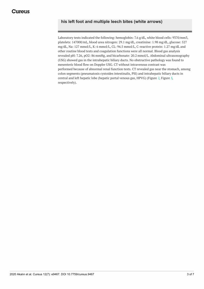
his left foot and multiple leech bites (white arrows)
Laboratory tests indicated the following: hemoglobin: 7.6 g/dL, white blood cells: 9370/mm3,platelets: 147000/mL, blood urea nitrogen: 29.1 mg/dL, creatinine: 1.98 mg/dL, glucose: 527mg/dL, Na: 127 mmol/L, K: 6 mmol/L, CL: 94.5 mmol/L, C-reactive protein: 1.27 mg/dL andother routine blood tests and coagulation functions were all normal. Blood gas analysisrevealed pH: 7.26, pO2: 86 mmHg, and bicarbonate: 20.2 mmol/L. Abdominal ultrasonography(USG) showed gas in the intrahepatic biliary ducts. No obstructive pathology was found tomesenteric blood flow on Doppler USG. CT without intravenous contrast wasperformed because of abnormal renal function tests. CT revealed gas near the stomach, amongcolon segments (pneumatosis cystoides intestinalis, PSI) and intrahepatic biliary ducts incentral and left hepatic lobe (hepatic portal venous gas, HPVG) (Figure 2, Figure 3,respectively).
2020 Akalın et al. Cureus 12(7): e9467. DOI 10.7759/cureus.9467 3 of 7

FIGURE 2: CT scan, coronal planeRed arrows are showing the pneumatosis cystoides intestinalis in the bowel wall
2020 Akalın et al. Cureus 12(7): e9467. DOI 10.7759/cureus.9467 4 of 7

FIGURE 3: CT scan, axial planeRed arrows are showing the hepatic portal venous gas in the center and left lobe of liver
The patient was referred to another hospital that had an intensive care unit and startedconservative treatment with antibiotics and intravenous fluid. Twelve hours after treatment,his general condition did not improve and signs of diffused peritonitis were observed onabdominal examination. The patient underwent exploratory laparotomy. Necrosis was found inthe terminal ileum and the entire colon during laparotomy. Doppler USG detected low flow inthe vessels supplying these necrotic segments. However, resection was decided due to necrosisof these segments. These necrotic intestinal segments were resected and an end-ileostomy wasperformed. He had an uneventful recovery and was discharged on the 17th postoperative day.
2020 Akalın et al. Cureus 12(7): e9467. DOI 10.7759/cureus.9467 5 of 7

DiscussionHirudotherapy is known as a complementary treatment method applied with blood-suckingleeches. Leeches are attached to the skin of the diseased area and the aim is to acquire potentialutility from leech saliva that is secreted while the leeches are feeding. It has been used forcenturies and is still used in Asian and European countries [4,5]. The saliva of the leechcontains hirudin, histamine-like substances, hyaluronidase and collagenase. Hirudin has only atransient antithrombin effect in humans and is a powerful anticoagulant. While leech bites,hyaluronidase and collagenase allow it to reach the tissues and blood vessels; histamine-likemolecules cause vasodilatation, and platelet functions, kinin activity, and the coagulationcascade are inhibited [6,7]. We think that hypovolemia due to prolonged bleeding causedsyncope in this case.
AMI occurs as a result of decreased blood flow to the intestines. AMI is classified into fourgroups according to its etiology: arterial embolism of the superior mesenteric artery (SMA),arterial thrombosis of SMA in a pre-existing atherosclerotic lesion, mesenteric venousthrombosis, and non-occlusive mesenteric ischemia (NOMI). NOMI is responsible for 20%-30%of AMI episodes. Especially in the presence of atherosclerotic disease, mesenteric ischemiaoccurs due to mesenteric vasospasm without arterial or venous obstruction [8]. We think thistheory formed the etiology in this case. Besides, mesenteric vasospasm without arterial orvenous obstruction is known. This knowledge helps us to understand the development of NOMIin a hemodynamically-stabilized patient after treatment [9]. Doppler USG is insufficient toshow distal embolism of main vessels and for the diagnosis of NOMI. Angiography is the goldstandard for diagnosis of mesenteric ischemia. Angiography is the most reliable method toshow the presence and localization of occlusion [10]. In this case, we could not performmesenteric angiography because of abnormal renal function tests. We did not find the exactreason for AMI. However, the detection of blood flow in necrotic segments supports thediagnosis of NOMI.
HPVG is thought to be caused by bacterial gas formation from an abscess in the intestinallumen or liver and bacteria that cause gas formation in the portal vein [11,12]. The mortalityrate in HPVG is between 75% and 90% and is higher in patients associated with necrotizingenterocolitis and mesenteric vascular occlusions [13]. PSI is characterized by intramural gas-filled cystic formations involving any part of the gastrointestinal system. Most of the time thedisease involves the jejunum and ileum; moreover, the colon is affected in 6% [14]. The findingsof AMI on CT are gas in the portal vein, edema of the intestinal wall, mesenteric edema andPSI [15]. Also, in our case, PSI and HPVG were detected on CT. Peritoneal irritation signs werefound during the physical examination and after this diagnosis was established with emergencylaparotomy.
ConclusionsIn summary, hirudotherapy may induce NOMI due to prolonged bleeding. Leech therapy shouldbe performed by certified experts in appropriate conditions. Patients should be closelymonitored for its complications.
Additional InformationDisclosuresHuman subjects: Consent was obtained by all participants in this study. Conflicts of interest:In compliance with the ICMJE uniform disclosure form, all authors declare the following:Payment/services info: All authors have declared that no financial support was received fromany organization for the submitted work. Financial relationships: All authors have declaredthat they have no financial relationships at present or within the previous three years with any
2020 Akalın et al. Cureus 12(7): e9467. DOI 10.7759/cureus.9467 6 of 7

organizations that might have an interest in the submitted work. Other relationships: Allauthors have declared that there are no other relationships or activities that could appear tohave influenced the submitted work.
AcknowledgementsThe authors would like to thank Dr.Cagatay Sisman for sharing information about the patient'soperation.
References1. Duruhan S, Bicer B, Tuncay MS, et al.: Hirudotherapy following trauma and plastic surgery
procedures. Turk J Integr Med. 2014, 2:32-37.2. Blumberg D, Wald A: Other diseases of the colon and rectum . In Sleisenger and Fordtran's
Gastrointestinal and Liver Disease. Feldman M, Friedman LS, Sleisenger MH (ed): Saunders,China; 2002. 7th edition:2307-2309.
3. Roussel A, Castier Y, Nuzzo A, et al.: Revascularization of acute mesenteric ischemia aftercreation of a dedicated multidisciplinary center. J Vasc Surg. 2015, 62:1251-1256.10.1016/j.jvs.2015.06.204
4. Whitaker IS, Rao J, Izadi D, et al.: Historical article: Hirudo medicinalis: ancient origins of,and trends in the use of medicinal leeches throughout history. Br J Oral Maxillofac Surg. 2004,42:133-137. 10.1016/S0266-4356(03)00242-0
5. Mory RN, Mindell D, Bloom DA: The leech and the physician: biology, etymology, and medicalpractice with Hirudinea medicinalis. World J Surg. 2000, 24:878-883. 10.1007/s002680010141
6. Gileva OS, Mumcuoglu KY: Hirudotherapy. In Biotherapy-history, principles and practice: apractical guide to the diagnosis and treatment of disease using living organisms. GrassbergerM, Sherman RA, Gileva OS, Kim CMH, Mumcuoglu KY (ed): Springer Science, Business Media,London; 2013. 31:76. 10.1007/978-94-007-6585-6_3
7. Abdullah S, Dar LM, Rashid A, et al.: Hirudotherapy/Leech therapy: applications andindications in surgery. Arch Clin Exp Surg. 2012, 1:172-180. 10.5455/aces.20120402072447
8. Klar E, Rahmanian PB, Bucker A, et al.: Acute mesenteric ischemia: a vascular emergency .Dtsch Arztebl Int. 2012, 109:249-256. 10.3238/arztebl.2012.0249
9. Trompeter M, Brazda T, Remy CT, et al.: Non-occlusive mesenteric ischemia: etiology,diagnosis, and interventional therapy. Eur Radiol. 2002, 12:1179-1187. 10.1007/s00330-001-1220-2
10. Bowersox JC, Zwolak RM, Walsh DB, et al.: Duplex ultrasonography in the diagnosis of celiacand mesenteric artery occlusive disease. J Vasc Surg. 1991, 14:780-788.10.1067/mva.1991.33215
11. Thompson AM, Bizzarro MJ: Necrotizing enterocolitis in newborns: pathogenesis, preventionand management. Drugs. 2008, 68:1227-1238. 10.2165/00003495-200868090-00004
12. Merritt CR, Goldsmith JP, Sharp MJ: Sonographic detection of portal venous gas in infantswith necrotizing enterocolitis. Am J Roentgenol. 1984, 143:1059-1062. 10.2214/ajr.143.5.1059
13. Wiesner W, Mortele KJ, Glickman JN, et al.: Pneumatosis intestinalis and portomesentericvenous gas in intestinal ischemia: correlation of CT findings with severity of ischemia andclinical outcome. Am J Roentgenol. 2001, 177:1319-1323. 10.2214/ajr.177.6.1771319
14. Morris MS, Gee AC, Cho SD: Management and outcome of pneumatosis intestinalis . Am JSurg. 2008, 195:679-683. 10.1016/j.amjsurg.2008.01.011
15. Barmase M, Kang M, Wig J, et al.: Role of multidetector CT angiography in the evaluation ofsuspected mesenteric ischemia. Eur J Radiol. 2011, 80:e582-e587. 10.1016/j.ejrad.2011.09.015
2020 Akalın et al. Cureus 12(7): e9467. DOI 10.7759/cureus.9467 7 of 7