HIP MRI SEQUENCES folder/MSK MR... · 2018-01-04 · synovial herniation pit) --PINCER=acet...
17
HIP MRI SEQUENCES: Bones: scout, cor T1, cor PD FS FAI: axial T1 (angled) Capsule/cartilage: cor PD FS (angled), sag PD FS Labrum: cor PD FS (angled), axial T1 FS and PD FS radial sag PD (parallel to neck)+ axial scout (place crosshair) sag PD FS (not as useful) Muscles/Tendons: axial T1 FS, cor PD FS, and any T1 without FS OSSEOUS STRUCTURES (for hip imaging, axial and coronal are the most important) -Acetabulum (bony rim and labrum absent inferiorly at “acet notch” covered by transverse lig; “acet fossa” is devoid of cartilage and filled w/ fibrofatty tissue & synovium) -acetabular dysplasia; protusio (when femoral head projects medial to ilioischial line); retroversion (figure of 8; anterior acet is more lateral than post acet) -post disloc (avulsion of post-acet; labral and chondral injuries +/- entrapment; disruption of iliofemoral lig (ant); hemarthrosis; sciatic nerve injury; risk=AVN) -Femur (lot of red marrow in femur/pelvis which has int signal, higher than muscle; fatty marrow epiphysis & trochanters; marrow conversion: epidiametaphysis) -small amount of red marrow in subchondral femur head epi is normal; diffuse red marrow seen with anemia or chronic illness; marrow packing or infiltrative d/o -femoral neck 115-140deg with respect to femoral shaft -head/neck (fovea centralis does not have articular cartilage but contains low T1/T2 lig teres & pulvinar aka fibrofatty tissuelig teres inserts into transverse lig) -trochanters (enthesophytes) -Iliac -Sacrum (insuff fx parallels SI joint & may cross midline if bilateral aka “Honda sign”), Ilium (supracetabular “raised eyebrow” insuff fx) -Ischium (esp ischial tuberosity) -Pubic rami and symphysis -SI joints (true synovial joint ant-inf half w/ thinner cartilage on iliac side where DJD begins first): bilat symm=ank spond & IBD -Red marrow (usually symmetrically involves metaphysis; conversion to yellow from appendicular axial and epidiametaphysis; small amount of red marrow in subchondral region OK but can mimic AVN; signal > than muscle on T1 o/w think infiltrative marrow ddx=tumor, OM, edema/trauma, marrow-packing d/o) -Fracture (T1 coronal +/- STIR, axial T2)—stress fx=sclerotic T1/T2 w/ T2 edema compressive>tensile neck; fatigue fx= may lack sclerosis) -Stress injury of tibia (gradeI=periosteal edema, gradeII=also endosteal/BM edema on T2, gradeIII=also abnl BM signal on T1, gradeIV=fx line visible) -AVN early=diffuse then focal edema (can mimic TOH)serpentine low T1 signal surrounding fatty center (ant-sup head) sharp inner line and ill-defined outer margin due to gray edemadouble line sign T2 (hi signal granulation tissue paralleling along inside margin of dark sclerotic band)late=sclerosis/collapse (subchondral fx and epiphyseal collapse); mimic=gray signal subchondral normal red marrow; “ASEPTIC” anemia (sickle-cell), steroids, ethanol, pancreatitis, trauma, idiopathic, CVD, XRT, LCP, Gaucher’s -TOH=preg women 3 rd tri < middle-aged men 40-50yo M>F 3:1 no trauma, spont res in 6-8mos, hi T2 from head to intertrochanteric region (unilat), no subchondral low T2 signal (<4mm thick and <1.25cm long c/w irreversible AVN), acetabulum uninvolved, may have effusion, MR +ve 48hrs after pain onset; penia on xray within 4wks (ddx=early AVN or insuff fx or osteoid osteoma) -Lig teres tear (normally homogen low T1/T2; post-traumatic partial/complete tear vs degeneration=hypertrophied lig can be assoc w/ CPPD--osseous erosion/irreg of fovea) -Inflamm arthritis (septic and aspectic inflamm arthritis appear similar)—thickened enhancing synovium, bone marrow edema, effusion, erosions -Septic hip (effusion + synovitis; synovial thickening and enhancement; BM edema both sides of joint; mono-articular; aspiration fluid PMN>80k is diagnostic criteria) -Sickle cell (OM vs Infarct; cannot differentiate on MR; consider In-WBC + Tc-SC) FAI (pain w/ flexion/internal rot; assoc w/ ant-sup labral tear; bony frag or os acetabulae at sup-lat acet rim; fibrocystic lesion or
Transcript of HIP MRI SEQUENCES folder/MSK MR... · 2018-01-04 · synovial herniation pit) --PINCER=acet...

HIP MRI
SEQUENCES:
Bones: scout, cor T1, cor PD FS
FAI: axial T1 (angled)
Capsule/cartilage: cor PD FS (angled), sag PD FS
Labrum: cor PD FS (angled), axial T1 FS and PD FS
radial sag PD (parallel to neck)+ axial scout (place crosshair)
sag PD FS (not as useful)
Muscles/Tendons: axial T1 FS, cor PD FS, and any T1 without FS
OSSEOUS STRUCTURES (for hip imaging, axial and coronal are the most important)
-Acetabulum (bony rim and labrum absent inferiorly at “acet notch” covered by transverse lig; “acet fossa” is devoid of cartilage and
filled w/ fibrofatty tissue & synovium)
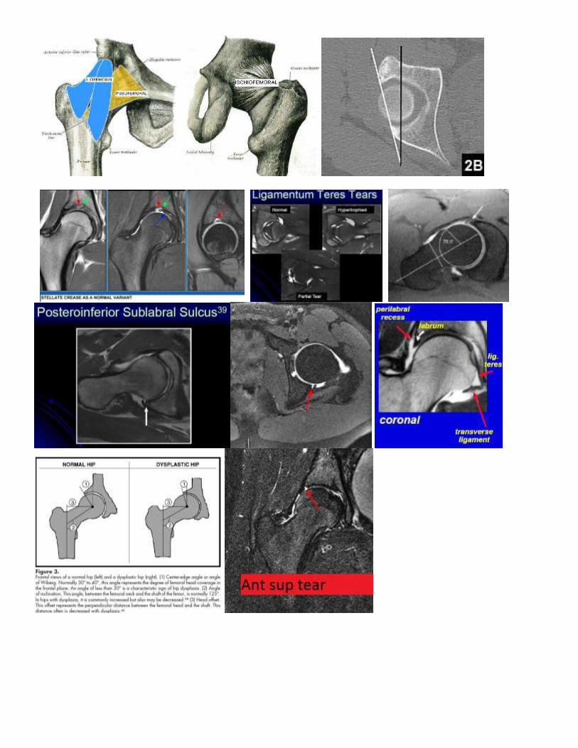
-acetabular dysplasia; protusio (when femoral head projects medial to ilioischial line); retroversion (figure of 8; anterior acet is
more lateral than post acet)
-post disloc (avulsion of post-acet; labral and chondral injuries +/- entrapment; disruption of iliofemoral lig (ant); hemarthrosis;
sciatic nerve injury; risk=AVN)
-Femur (lot of red marrow in femur/pelvis which has int signal, higher than muscle; fatty marrow epiphysis & trochanters; marrow
conversion: epidiametaphysis)
-small amount of red marrow in subchondral femur head epi is normal; diffuse red marrow seen with anemia or chronic illness;
marrow
packing or infiltrative d/o
-femoral neck 115-140deg with respect to femoral shaft
-head/neck (fovea centralis does not have articular cartilage but contains low T1/T2 lig teres & pulvinar aka fibrofatty tissuelig
teres
inserts into transverse lig)
-trochanters (enthesophytes)
-Iliac
-Sacrum (insuff fx parallels SI joint & may cross midline if bilateral aka “Honda sign”), Ilium (supracetabular “raised eyebrow”
insuff fx)
-Ischium (esp ischial tuberosity)
-Pubic rami and symphysis
-SI joints (true synovial joint ant-inf half w/ thinner cartilage on iliac side where DJD begins first): bilat symm=ank spond & IBD
-Red marrow (usually symmetrically involves metaphysis; conversion to yellow from appendicularaxial and epidiametaphysis;
small amount of red marrow in subchondral region OK but can mimic AVN; signal > than muscle on T1 o/w think infiltrative marrow
ddx=tumor, OM, edema/trauma, marrow-packing d/o)
-Fracture (T1 coronal +/- STIR, axial T2)—stress fx=sclerotic T1/T2 w/ T2 edema compressive>tensile neck; fatigue fx= may lack
sclerosis)
-Stress injury of tibia (gradeI=periosteal edema, gradeII=also endosteal/BM edema on T2, gradeIII=also abnl BM signal on T1,
gradeIV=fx line visible)
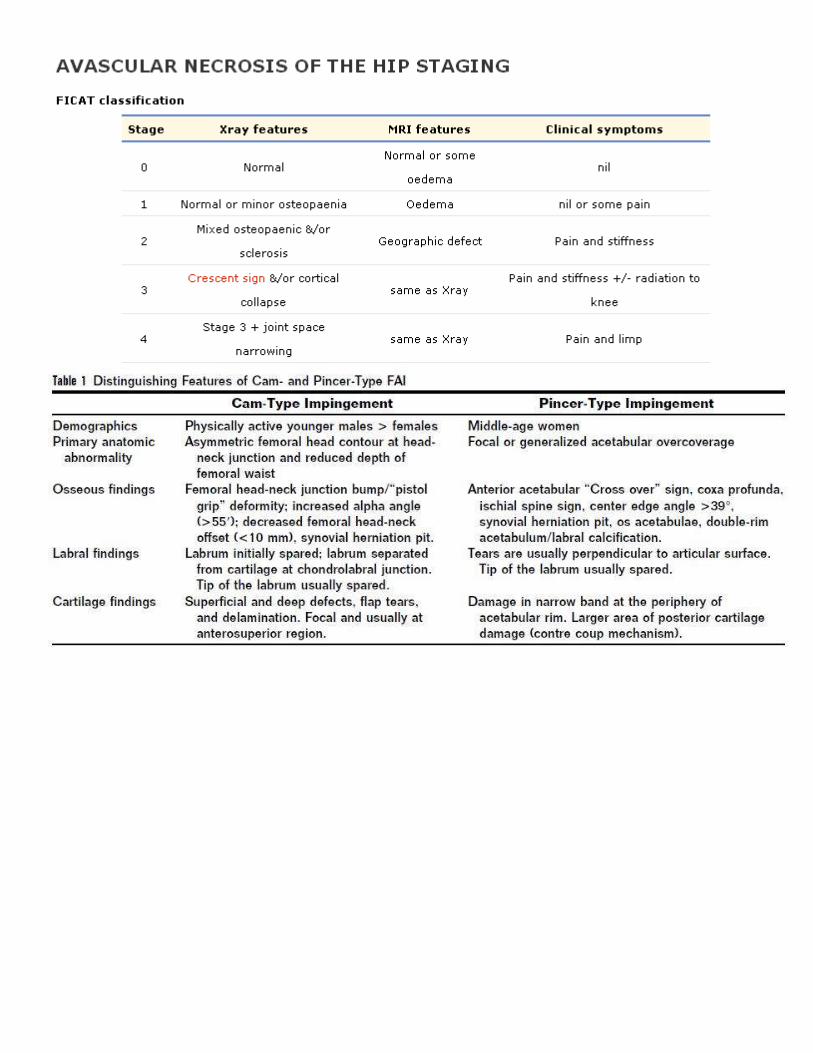
-AVN early=diffuse then focal edema (can mimic TOH)serpentine low T1 signal surrounding fatty center (ant-sup head) sharp inner
line and ill-defined outer margin due to gray edemadouble line sign T2 (hi signal granulation tissue paralleling along inside margin
of dark sclerotic band)late=sclerosis/collapse (subchondral fx and epiphyseal collapse); mimic=gray signal subchondral normal red
marrow; “ASEPTIC” anemia (sickle-cell), steroids, ethanol, pancreatitis, trauma, idiopathic, CVD, XRT, LCP, Gaucher’s
-TOH=preg women 3rd tri < middle-aged men 40-50yo M>F 3:1 no trauma, spont res in 6-8mos, hi T2 from head to intertrochanteric
region (unilat), no subchondral low T2 signal (<4mm thick and <1.25cm long c/w irreversible AVN), acetabulum uninvolved, may
have effusion, MR +ve 48hrs after pain onset; penia on xray within 4wks (ddx=early AVN or insuff fx or osteoid osteoma)
-Lig teres tear (normally homogen low T1/T2; post-traumatic partial/complete tear vs degeneration=hypertrophied lig can be assoc w/
CPPD--osseous erosion/irreg of fovea)
-Inflamm arthritis (septic and aspectic inflamm arthritis appear similar)—thickened enhancing synovium, bone marrow edema,
effusion, erosions
-Septic hip (effusion + synovitis; synovial thickening and enhancement; BM edema both sides of joint; mono-articular; aspiration fluid
PMN>80k is diagnostic criteria)
-Sickle cell (OM vs Infarct; cannot differentiate on MR; consider In-WBC + Tc-SC)
FAI (pain w/ flexion/internal rot; assoc w/ ant-sup labral tear; bony frag or os acetabulae at sup-lat acet rim; fibrocystic lesion or

synovial herniation pit)
--PINCER=acet overcoverage due to coxa profunda or acetabular protusio resulting in retroversion “crossover” sign of ant acet c/w
pincer-type morphology assoc with FAI + anterior-superior labral tear (perpendicular to articular surface) + post-inf acet countercoup
chondral injury
--CAM=femoral head-neck offset with anterior-superior (seen best on axial-oblique) vs lateral (seen best on AP or radial sequences)
dysplastic/aspherical bump (“pistol grip” deformity); alpha angle>55 on axial-oblique images (prescribed parallel to long axis of
femoral neck); head-neck offset <10mm; labral initially sparedthen ALAD=acetabulolabral articular disruption (labrum separated
from cartilage at chondralabral jct); may have associated anterior-superior cartilage tear (deep / superficial / flap tear) vs delamination
(inverted oreo cookie sign with contrast or fluid undermining articular cartilage)
--Herniation pit (always abnl; in-growth of fibrocartilaginous tissue thru cortical defect; aka fibrocystic lesion; ant-sup-lat at head/neck
jc; can be uni/bilateral)
--also look for BM edema, subchondral cystic changes, intraosseous ganglion, synovitis (obscuration of normal sulcus btwn sup
labrum and capsule may be an early sign), and capsular thickening (hypertrophy of iliofemoral lig)
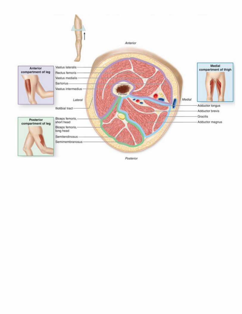
MUSCLES/TENDONS
Tendonitis/Tendinopthy; partial/full-thickness tear; avulsion; HADD (calcific tendonitis); myotendinous strain; stress injury
-Anterior: flex (quadriceps=rectus femoris + vastus lateralis/intermedius/medalis; sartorius)
-Medial: adduct (GPA=gracilis, pectineus, adductor longus/brevis/magnus) GROIN PULL
-Lateral: abduct (tensor fascia lata, gluteus max/med/min)GR TROCHANTERIC PAIN SYNDROME (glut med/min
tendinopathy/tear > avulsion of gr torchanter)
-Posterior: extend (hamstrings= biceps femoris + semimemb + semitend) HAMSTRING INJURY tear (usually SM) vs ischial
tuberosity avulsion (usually of conjoint tendon of BF/ST)
-External rotators: (piriformis, quadratus femoris, GOGO’s=gemellus sup/inf and obturator int/ext) PIRIFORMIS SYNDROME
(sciatic nerve neuropathy; mimics radiculopathy)—sciatic nerve located ant or may split thru piriformis and exits via greater sciatic
notch
ORIGIN/INSERTION:
ASISsartorius
AIISrectus femoris
Iliac alaGluteal muscle origin
Superior pubic ramuspectineus muscle
Inferior pubic ramusadductor longus
Pubic symphysisadductor brevis and gracilis
Ischial ramusorigin of other adductors
Ischial tuberosityhamstring origin (superolateral=semimembranosis; inferomedial=conjoint tendon of semitendinosis and long head
biceps femoris; BF muscle located lateral, SM inbetween, ST medially; BF and ST come together as conjoint tendon); also insertion of
sacrotuberous lig; note: adductor magnus inserts slong inferior aspect of ischial tuberosity
Gr trochanter”GOP” Glut med (lateral and superoposterior), Glut min (anterior), Obturator internus, Piriformis insertion, Bursa
(post)
Ls trochanterIliopsoas insertion
Gluteal tuberosity of prox femurGlut maximus insertion
Linea aspera (femoral diaphysis)insertion on many muscles
-Muscle strain:
-strain (edema at myotendinous junction vs less commonly peripheral edema in epimysial strain' commonly involves hamstring, rectus
femoris, medial gastroc, and biceps brachii); laceration (knife); contusion (localized edema with associated ST and BM edema); tear
(discontinuity); hemorrhage (can mimic mass so give gad on f/u exam); compartment syndrome (edematous muscle compartment with
fascial edema)
-GradeI (pain)=microscopic at musculotendinous jct w/ T2 feathery edema w/ preserved morphology; GradeII (pain/weakness)=partial
thickness tear w/ discrete T1 bright intramuscular hematoma or fluid collection (tearing of up to <50% of muscle fibers); GradeIII
(pain/weakness/loss of fxn)=complete tear “bull’s eye” appearance +/- retraction
-Longer the length of muscle involvement, longer it would take to resolve; more edema/fluid means higher grade; complete tendon or
myotendinous tear is Grade III; chronic tear associated with thickened tendon and peritendinous or diffuse muscle atrophy.
-Muscle edema ddx=muscle injury (strain/tear); myositis (increased T2 signal; may be subtle if autoimmune; more obvious and more
enhancement if infectious); inflammatory myopathy; DOMS; acute/subacute denervation
-Muscle denervation= acute (increased muscle volume, T2 bright, no fatty infiltration, +muscle enhancement), subacute (normal
muscle volume, T2 bright, early fatty infiltration, +muscle enhancement), chronic (decreased muscle volume, no longer T2 bright,
increased muscle infiltration, no muscle enhancement)
-Fascitis (inflammation/fluid/edema tracks along deep fascial planes; +/- assoc myositis; +/-abscess/microabacesses)
-Nec Fascitis (clinical diagnosis; destruction & necrosis of subcutaneous and deep fascial tissues with dull gray appearance of fascial
edema; +/-myonecrosis; +/-abscess/microabscesses; look for vascular thrombosis; late=gas gangrene; spreads rapidly; Gad not

necessary but useful to identify abscess but can underestimate extent of necrosis; typeI=polymicrobial 90% vs typeII=flesh-eating
10%)
-Snapping hip (multiple causes including slipping of iliopsoas tendon over iliopectineal eminence w/ hip flexion/extension)
-Athletic pubalgia (rectus abdominus, pectineus, adductor longus, and levator ani insert at pubic symphysis; tear of rectus abdominis
leads to compartment syndrome in region of adductor muscle near sup margin of inf pubis; marrow edema in pubic symphysis and
nearby portion of pubic rami and adj muscle edema esp adductors along with tendon tears and even hematoma)
-Osteitis pubis (multiparous women; marrow edema and irreg of pubic symphysis w/ fluid within cartilaginous joint ; may have AP or
sup-inf joint incongruity)
-Gluteal tear (aka rotator cuff of hip) from greater trochanter which has 4 facets=“anterior” (GlutMin), “lateral” (GlutMed—lateral
tendons), “posterior” (trochanteric bursa), “superoposterior” (GlutMed—main tendon); partial/complete tear, avulsion of gr trochanter,
glut muscle atrophy
-Abnl muscle signal DDX: bursitis, strain/tear (usually at myotendinous jct), DOMS, contusion/hematoma, myositis, acute
denervation (<2wks-1year)
-Intramuscular tumors DDX: hemangioma (contains fat, calc, serpiginous vessels), lipoma, myxoma (mimics cyst but enhances),
sarcoma (no surrounding edema), mets (has surrounding edema), lymphoma, neurofibroma (“target”), desmoid & MFH(=low T1 and
low/int T2), hematoma, myositis ossificans
LABRUM/CARTILAGE (dedicated hip arthrogram)
Labrum
-post-sup labrum is thicker; labrum usually triangular but may be non-triangular (rounded/flat) in older pts
->90% are anterior or antsup tears (there are no normal variants here!); clockface on sag plane (ant is 3’oclock)
-base vs periphery of labrum
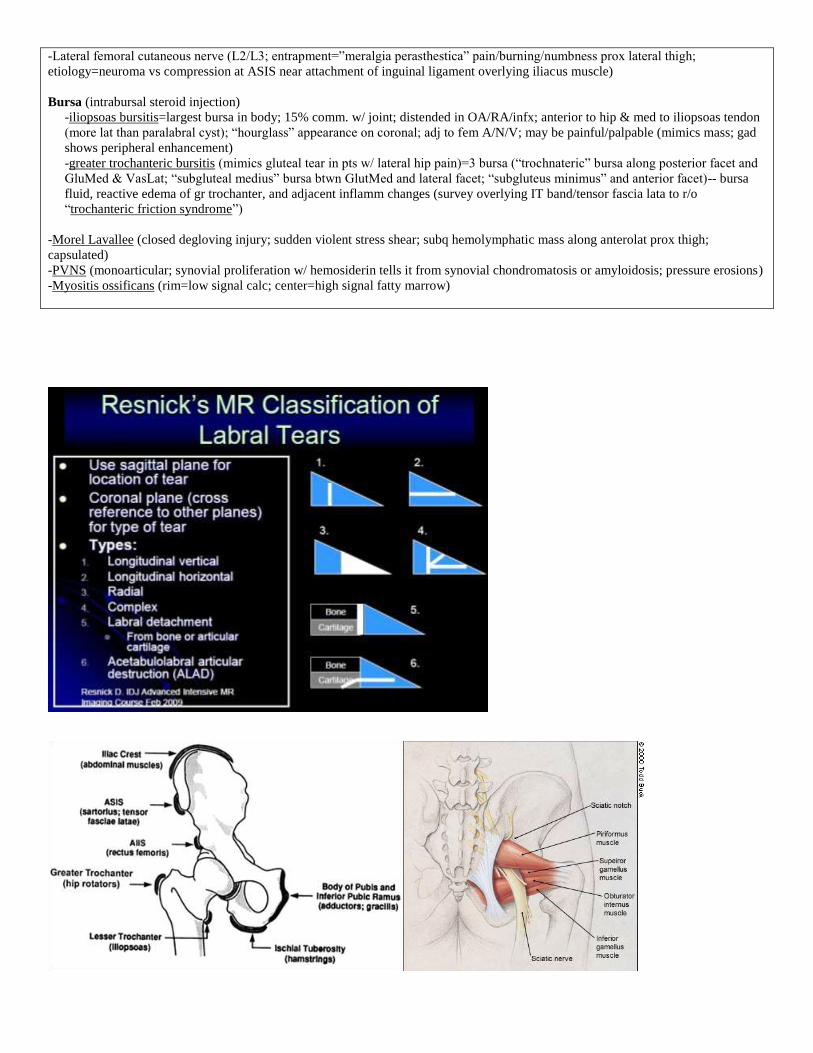
-Resnick classification: longitudinal vertical, longitudinal horiz, radial, complex, labral detachment, acetabulolabral articular
destruction (ALAD)
-Others use: degeneration (common>50yo) vs tear (linear/heterogenous signal; deformed/distorted contour) vs detached
(partial/complete +/-displacement; contrast interposed at acetabular-labral jct or interface)
pitfalls:
-normal labral variants more common in lower quadrant; no normal variant within ant to sup labrum
-post-inf sublabral sulcus on axial
-perilabral recess btwn labrum and capsule (small ant and post; larger superiorly)
-?ant-inf sublabral sulcus (linear-cleft, only partial separation, no perilabral abnl)
-labro-ligamentous sulcus/recess btwn labrum and transverse lig anteriorly and posteriorly on cor/sag; anterior labrum pitfall is adj
iliopsoas tendon on axial
-cartilage undercutting
-Paralabral/para-articular cyst—may be intra-osseous subchondral (within acetabulum)
Capsule
-Stellate crease or supra-acetabular fossa (focal notch in superior acet 12’oclock devoid of cartilage w/o BM edema in young pts; may
contain plica or fibrous cord)
-capsule inserts directly at base of labrum anteriorly and posteriorly with a small “perilabral recess”; larger “perilabral recess” seen
superiorly
-capsule inserts anteriorly along intertrochanteric line and posteriorly halfway down femoral neck
-capsule reinforced by 3 longitudinal capsular ligs: ANT: Iliofemoral=strongest (Y-shaped=medial and lateral bands) + Pubofemoral
(hiatus btwn iliofem and pubofem allow comm. w/ iliopsoas bursa in 15%), POST: Ischiofemoral (sup and inf bands)
-circularly oriented fibers known Zona Orbicularis encircles the capsule at base of femoral neck (forms a collar)
Cartilage (“acet fossa” not covered with cartilage): thinning, fissuring, delamination, partial/full thickness defect, chondral flap seen
w/ full-thickness tears; PINCER=assoc w/ countercoup post-inf cartilage degen; CAM=assoc w/ ant-sup cartilage degen
MISCELLANEOUS
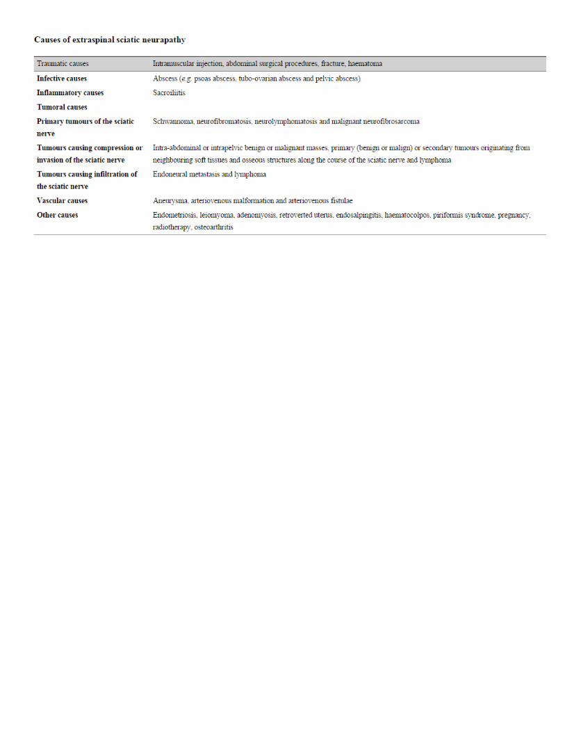
Nerve
-Sciatic nerve=tibial nerve+common peroneal nerve (immed post to post column of acet and lat to ischial tuberosity hamstring
insertion; ant to piriformis thru greater sciatic foramen; located btwn quadratus femoris muscle ant and glut max post; normal
intermediate stippled signal surrounded by fat; compressive neuropathy or fibrolipomatous hamartoma)
-Piriformis syndrome (piriformis muscle hypertrophy vs inflammation vs anatomic variation leads to compression on sciatic nerve;
seen as abnl signal within piriformis muscle; 85% sciatic nerve anterior to piriformis muscle; 12% TN and CPN are split by piriformis
with CPN thru muscle; 3% CPN posterior to piriformis with TN thru muscle; 1% sciatic nerve thru muscle)
-Femoral nerve (inguinal canal; etiology include inguinal hernia, iliopsoas bursitis, psoas abcess, aneurysm, etc)
-Obturator nerve (obturator canal; etiology include obturator hernia, osteitis pubis, paralabral cyst)
-Lateral femoral cutaneous nerve (L2/L3; entrapment=”meralgia perasthestica” pain/burning/numbness prox lateral thigh;
etiology=neuroma vs compression at ASIS near attachment of inguinal ligament overlying iliacus muscle)
Bursa (intrabursal steroid injection)
-iliopsoas bursitis=largest bursa in body; 15% comm. w/ joint; distended in OA/RA/infx; anterior to hip & med to iliopsoas tendon
(more lat than paralabral cyst); “hourglass” appearance on coronal; adj to fem A/N/V; may be painful/palpable (mimics mass; gad
shows peripheral enhancement)
-greater trochanteric bursitis (mimics gluteal tear in pts w/ lateral hip pain)=3 bursa (“trochnateric” bursa along posterior facet and
GluMed & VasLat; “subgluteal medius” bursa btwn GlutMed and lateral facet; “subgluteus minimus” and anterior facet)-- bursa
fluid, reactive edema of gr trochanter, and adjacent inflamm changes (survey overlying IT band/tensor fascia lata to r/o
“trochanteric friction syndrome”)
-Morel Lavallee (closed degloving injury; sudden violent stress shear; subq hemolymphatic mass along anterolat prox thigh;
capsulated)
-PVNS (monoarticular; synovial proliferation w/ hemosiderin tells it from synovial chondromatosis or amyloidosis; pressure erosions)
-Myositis ossificans (rim=low signal calc; center=high signal fatty marrow)