High%Class%1%essay%% Discussthenewinsightsintheund ...
13
Medical Microbiology Coursework Essay High Class 1 essay 1 Discuss the new insights in the understanding of Haemolytic Uraemic Syndrome and its worldwide implications following the large scale outbreak of E.Coli O104:H4 diarrhea in Germany 2011 In May 2011 there was a large scale outbreak of Haemolytic Uraemic Syndrome (HUS) in North Germany. This outbreak caused the highest frequency of HUS cases caused by Escherichia coli (E.coli) on record, with the number of cases and deaths being 2.4 and 1.4 times higher than the number reported for 350 different outbreaks between 1982 and 2001 (Safadi et al. 2012). On the 24 th of May 2011 the Oantigen, a lipopolysaccharide antigen on the cell wall, was determined to be O104 and further genetic testing by The Robert Koch Institute concluded it possessed the H4 antigen, an antigen present on the flagella of E.coli (Robert Koch Institute. 2011). Up until 2011 there had only been a limited number of cases of this serotype in humans; for example in Korea 2006 a woman presented with ‘bloody diarrhea, abdominal pain, haemolytic anaemia, thrombocytopenia and acute renal failure.’ She was diagnosed with Escherichia Coli O104:H4 associated haemolytic uraemic syndrome (Bae et al. 2006). Once the serotype was known, the treatment of plasmapheresis and haemodialysis could start. During the 2011 outbreak in Germany, E.coli caused a total of 3842 people to be hospitalized. Within this figure were 855 cases of HUS and 2,987 cases of gastroenteritis without development of HUS, which shows that 22% of cases contracted HUS. In the previous five years (20062010) a median of 13 HUS cases and 218 gastroenteritis cases were reported (Robert Koch Institute. 2011). This is a 67 fold increase of HUS cases and a 17 fold increase of gastroenteritis. Of the 3842 cases, 1.4% died as a result of the infection Prabha Parthasarathy 30/1/14 12:09 Comment [1]: FEEDBACK ON SUSSEX DIRECT An outstanding essay which shows not only a complete understanding , but an ability to think around the topic. Especially with regards to the pathogenic features. Very good evidence and an indepth discussion which highlights the role of the unique features of the German outbreak. Also, good use of evidence to highlight the unusual epidemiology. The essay is logical, moves step by step in the sequence of events chronologically . Excellent presentation. Very good use of diagrams, especially the one on the plasmids. Good referencing. Very minor errors highlighted on script. Prabha Parthasarathy 30/1/14 11:53 Comment [2]: Very good use of data to highlight the magnitude of the problem, this one of the key points as well, as the main issue with the German outbreak was a very unsusual occurence
Transcript of High%Class%1%essay%% Discussthenewinsightsintheund ...

Medical Microbiology Coursework Essay
High Class 1 essay
1
Discuss the new insights in the understanding of Haemolytic
Uraemic Syndrome and its worldwide implications following
the large scale outbreak of E.Coli O104:H4 diarrhea in
Germany 2011
In May 2011 there was a large scale outbreak of Haemolytic Uraemic Syndrome (HUS) in
North Germany. This outbreak caused the highest frequency of HUS cases caused by
Escherichia coli (E.coli) on record, with the number of cases and deaths being 2.4 and 1.4
times higher than the number reported for 350 different outbreaks between 1982 and 2001
(Safadi et al. 2012). On the 24th of May 2011 the O-‐antigen, a lipopolysaccharide antigen on
the cell wall, was determined to be O104 and further genetic testing by The Robert Koch
Institute concluded it possessed the H4 antigen, an antigen present on the flagella of E.coli
(Robert Koch Institute. 2011). Up until 2011 there had only been a limited number of cases
of this serotype in humans; for example in Korea 2006 a woman presented with ‘bloody
diarrhea, abdominal pain, haemolytic anaemia, thrombocytopenia and acute renal failure.’
She was diagnosed with Escherichia Coli O104:H4 associated haemolytic uraemic syndrome
(Bae et al. 2006). Once the serotype was known, the treatment of plasmapheresis and
haemodialysis could start.
During the 2011 outbreak in Germany, E.coli caused a total of 3842 people to be
hospitalized. Within this figure were 855 cases of HUS and 2,987 cases of gastroenteritis
without development of HUS, which shows that 22% of cases contracted HUS. In the
previous five years (2006-‐2010) a median of 13 HUS cases and 218 gastroenteritis cases
were reported (Robert Koch Institute. 2011). This is a 67 fold increase of HUS cases and a 17
fold increase of gastroenteritis. Of the 3842 cases, 1.4% died as a result of the infection
Prabha Parthasarathy� 30/1/14 12:09Comment [1]: FEEDBACK ON SUSSEX DIRECT An outstanding essay which shows not only a complete understanding , but an ability to think around the topic. Especially with regards to the pathogenic features. Very good evidence and an indepth discussion which highlights the role of the unique features of the German outbreak. Also, good use of evidence to highlight the unusual epidemiology. The essay is logical, moves step by step in the sequence of events chronologically . Excellent presentation. Very good use of diagrams, especially the one on the plasmids. Good referencing. Very minor errors highlighted on script.
Prabha Parthasarathy� 30/1/14 11:53Comment [2]: Very good use of data to highlight the magnitude of the problem, this one of the key points as well, as the main issue with the German outbreak was a very unsusual occurence

Medical Microbiology Coursework Essay
High Class 1 essay
2
which is an increase of 0.9% when compared with death from the most common type of
E.coli that causes HUS, E.coli O157:H7 (Denis Piérard et al. 2012).
Figure 1 : showing the epidemiology curve from 1st May to 4th July 2011. With thep Peak of
cases being on May 22nd 2011
(Robert Koch Institute. 2011, Figure 2)
Prabha Parthasarathy� 30/1/14 11:53Comment [3]: Very good figure, but could be discussed or referred in the text

Medical Microbiology Coursework Essay
High Class 1 essay
3
Escherichia coli as the Cause of the May 2011 Outbreak
Haemolytic Uraemia Syndrome is a disease that is characterised by 3 factors,
thrombocytopenia (low platelet count), thrombic microangiopathy (blood clots in small
blood vessels) and haemolytic anaemia (low haemoglobin count due to increased
destruction of red blood cells) (Mayer et al. 2012). The infections of this due to E.coli
typically originate from uncooked, or undercooked, vegetables or meat that is contaminated
with faecal matter. The most common type of E.coli that causes this is E.coli O157:H7, which
is an enterohaemorrhagic strain of E.coli. This serotype was responsible for an outbreak in
1993 due to undercooked hamburgers (Mayer et al. 2012).
Therodore Escherich was the first to note that E.coli had a high prevalence in the normal
intestinal flora of healthy individuals. However, he also noted that it had the ability to cause
disease in humans when it was directly introduced in to ‘extra-‐intestinal’ sites (R.M.R.
Browne et EL. Hartland. 2002). Enterohaemorrhagic E.Coli (EHEC) is primarily a human
pathogen but their main reservoir hosts are cattle, sheep and goats (ruminants) which are
all asymptomatic carriers. This was why, in the first days of the outbreak, ruminants were
suspected but later were found not to be the source (Denis Piéard et al. 2012). The actual
source of the Germany outbreak was due to contaminated bean sprouts, and once these
were identified their distribution was halted. However, this didn’t stop the infection
spreading by secondary transmission. Secondary Transmission is transmission of the
pathogen by person to person contact and mainly occurs between people in the same
house-‐hold (Robert Koch Institute. 2011).
Detection of EHEC O104:H4 became quicker and easier when F.Scheutz et al. published a
paper containing a quick screening method that could be used in ‘primary and secondary
Prabha Parthasarathy� 30/1/14 11:54Comment [4]: Very clear description of how the epidemiology was different in the German outbreak

Medical Microbiology Coursework Essay
High Class 1 essay
4
testing laboratories’. Scheutz et al. discovered that the O104 antigen present on EHEC
O104:H4 was identical to the K9 capsular antigen also present on other E.coli serotypes
(Orskov F et Orskov I. 1992). A simple agglutination reaction was then used using the K9
antiserum. (F Scheutz. 2011)
On the 31st of May the strain was sent to the European Union Reference Laboratory for
E.coli, who used real time PCR to detect the serotype O104:H4. Once this serotype had been
detected, many members of the scientific community shared their knowledge of E.Coli to
describe the specific characteristics of this stain. They found that the isolates were positive
for genes aggR, attA, aap, aggA and aggC which are typical of an Enteroaggregative
Escherichia coli (EAEC) (Christina Frank et al. 2011). Further analysis showed that O104:H4
was an EAHC, but has acquired a gene coding for a Shiga Toxin (ST) via a bacteriophage (F
Scheutz et al. 2011). This classed E.coli O104:H4 as an Enterohaemorrhagic E.coli.
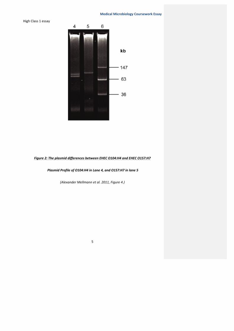
Figure 2 shows the plasmid differences between the outbreak strain and the most common
EHEC outbreak serotype, O157:H7. We can see from this plasmid profile that EHEC O104:H4
has 2 plasmids approximately 83 and 90kb in size, while EHEC O157:H7 has only 1 plasmid of
approximately 76kb (Alexander Mellmann et al. 2011).
These plasmids may have been acquired from Horizontal Gene Transfer (HGT) between
different E.coli, which allowed them to colonize in a completely new environment.
A theory posed by Matthew A. Croxen and B.Brett Finlay is that many HGT transfer events
can cause bacteria to be under new selective pressures, which select for the most virulent
organisms (Matthew A. Croxen and B.Brett Finlay. 2009).
Prabha Parthasarathy� 30/1/14 11:57Comment [5]: This is an outstanding discussion and hits at the heart of the topic. The use of figure has enhanced discussion as it clearly shows how the outbreak strain was different. The key finding form the figure is discussed in the text
Medical Microbiology Coursework Essay
High Class 1 essay
5
Figure 2: The plasmid differences between EHEC O104:H4 and EHEC O157:H7
Plasmid Profile of O104:H4 in Lane 4, and O157:H7 in lane 5
(Alexander Mellmann et al. 2011, Figure 4.)

Medical Microbiology Coursework Essay
High Class 1 essay
6
Pathogenesis of Enterohaemorrhagic E.Coli
Due to the 2011 outbreak, our knowledge of the pathogenicity mechanisms E.coli uses has
increased. The pathogenesis for O157:H7 EHEC is the most well-‐known as it is the most
common cause of EHEC infection.
The main virulence factor that all Enterohaemorrhagic E.coli have present is the gene coding
for a Shiga Toxin (Stx). This has 2 distinct forms that have equally distinct antigens, Stx1 and
Stx2. Both these have ‘shared AB5 domain structure, but only 56% amino acid homology’
(Chad.L.Mayer et al). These 2 types can be distinguished from each other by using an
Antibody-‐Antigen reaction, where antibodies for Stx1 neutralize the toxin and antibodies
against Stx2 have no effect on the toxin (David Greenwood, Richard C.B.Slack et John
F.Peutherer 2002). EHEC O157:H7 has both Stx1 and Stx2 located adjacent to each other on
an Open Reading Frame (Nurmohammad Shaikh and Phillip I. Tarr. 2003).
Both types of Shiga Toxin have A and B subunits, with A having the biological/enzymatic
activity and B aiding in receptor binding and specificity. Stx1 binds to Gb3 receptors on the
surface of intestinal epithelial cells, while stx2 binds to Gb4 receptors. These receptors then
use clathrin coated pits to endocytose the toxin (David Greenwood, Richard C.B.Slack et
John F.Peutherer 2002). The toxin-‐receptor complex now undergoes retrograde transport to
the Endoplasmic Reticulum (ER) via the Golgi body (Helge Karch 2001) (Chad L Mayer et al.
2012). Once processed by the ER, the A1 subunit has an N-‐glycosidase function and
hydrolyses an adenine residue on the 28S subunit of ribosomes in the cell, which inhibits
protein synthesis (David Greenwood, Richard C.B.Slack et John F.Peutherer 2002) (R.M.R
Prabha Parthasarathy� 30/1/14 12:02Comment [6]: The discussion here highlights what is the usual pathogenic mechanisms and then relates it to the mechanisms that were unique to the German outbreak. Additionally, at some places the student has discussed why this is important as some of these differences explain the unusual epidemic pattern.
Medical Microbiology Coursework Essay
High Class 1 essay
7
Browne et EL. Hartland 2002). The overall effect of this mechanism is cellular apoptosis,
necrosis and thrombotic microangiopathy all which contribute to the HUS seen in patients
(Chad L Mayer et al. 2012). Nevertheless the mechanism for E.coli O104:H4 is similar;
however it has specific differences that contribute to the difference in epidemiology
observed.
EHEC O104:H4 only has the stx2 gene while O157:H7 has both (Robert Koch Institute. 2011).
This toxin was different in 1 nucleotide position, making it identical to toxin found in
Germany in 2002 and 2005 (F Scheutz et al. 2012). In addition to this, O104:H4 possesses a
gene that codes for a Pic serine protease. This protease allows the bacterium to utilize
mucus as a growth substrate, and so aids in intestinal colonisation. Both stx2 and the Pic
serine protease are chromosomally encoded EAHC virulence factors that EHEC O104:H4 had
acquired (Safadi et al. 2012).
Due to further research conducted by the Robert Koch Institute a median incubation period
of 8 days was given as the median for the outbreak. Previously, EHEC O157:H7 had an
incubation period of 3-‐4days. (Robert Koch Institute. 2011) (Christina Frank et al. 2011).
Safadi et al conducted a study and showed that this increased incubation period and
enhanced virulence gene expression was due to the formation of a biofilm in vivo, which
protected the bacterium from the host’s defences. This bio-‐film produced by O104:H4 was
7.3 fold larger than the biofilm produced by O157:H7 and had an abundance of complex
structures e.g. microcolonies and water channels that allowed nutrient and oxygen intake
into the biolfilm. This Bio-‐film contributed to the increased incubation time observed
(Safadi et al. 2012). Alexander Mellmann et al. also demonstrated that O104:H4 had a
higher acid resistance when compared with O157:H7. This may aid to the increased severity

Medical Microbiology Coursework Essay
High Class 1 essay
8
and incubation time as more Colony Forming Units can pass through the stomach and
colonise in the intestine (Alexander Mellmann et al. 2011).
However, the main difference during this outbreak was the age of cases. ‘Only 1% of the
case patients with HUS are younger than 5 years of age, which was the median age in
Germany from 2001-‐2010’ (Christina Frank et al. 2011). F. Scheutz et al hypothesised that
this difference in age may have been due to differences in adherence and/or colonisation
factors present on EHEC O104:H4. These differences could add to the effect of susceptibility
that age incurred (F.Scheutz et all 2011).
O104:H4 also showed weakness to some antibiotics that O157:H7 did not e.g. meropenem
and chloramphenicol (Martina Bielaszewska et al. 2012). If antibiotics are given to a patient
with EHEC O157:H7 it can exacerbate the condition. This is due to the fact that release of
Shiga Toxin occurs through lambdoid phage mediated lysis in response to any DNA damage,
and therefore antibiotics cause the release of the toxin. (Chad L. Mayer 2012, Matthew A.
Croxen et B. Brett Finlay 2010).
Prabha Parthasarathy� 30/1/14 12:03Comment [7]: This was one of the concepts which many students got wrong. Although student have said that the germ n outbreak was highly resistant to anitbiotics, in effect we do not give antibiotics for treating HUS.

Medical Microbiology Coursework Essay
High Class 1 essay
9
Implications of the German Outbreak
The implications of this outbreak were made clear in the Final report made by the Robert
Koch institute 2011. A repeated survey was given to an online panel between 24th May and
24th June to analyse what had changed in consumption patterns and personal hygiene. The
analysis showed that 43% of the population of northern Germany stopped eating raw
vegetables.
Furthermore, once sprouts were the main suspicion with regards to the cause of the
outbreak, 84% of the population quit eating raw sprouts in week 3 of the survey. During the
next week this figure fell to 80%. (Robert Koch Institute 2011)
With regards to hygiene ‘63% of the population paid more attention to personal hygiene, for
example more frequent hand washing.’ This cautiousness was also applied to meal
preparation where 62% of the population surveyed paid more attention to cleanliness when
preparing meals. (Robert Koch Institute 2011)
Prabha Parthasarathy� 30/1/14 12:06Comment [8]: The essay did not have any clear conclusions at the end, however, this could have been easily achieved by summarises some of the main points earlier. Hence the essay seems to end abruptly. Considered a minor point here as it does not reveal a lack of understanding.

Medical Microbiology Coursework Essay
High Class 1 essay
10

Medical Microbiology Coursework Essay
High Class 1 essay
11
References
• Rim Al Safadi et al. (2012) ‘ Correlation between In Vivo Biofilm Formation and Virulence Gene Expression in Escherichia Coli O104:H4’. PLoS ONE, Volume 7(7), e41628, pp 1-‐7. (Accessed 26th February) Accessed online at: http://www.plosone.org/article/info:doi/10.1371/journal.pone.0041628
• Robert Koch Institute. (2011), ‘Report: Final presentation and evaluation of epidemiological finding in the EHEC O104:H4 outbreak’. Germany 2011’, Berlin 2011 pp 1-‐43. (Accessed 26th February) Accessed online : (http://www.rki.de/EN/Home/EHEC_final_report.pdf;jsessionid=AE23F8632C0DFE488FB2EF63BCDE3EA5.2_cid290?__blob=publicationFile) (Accessed 26th February)
• Woo Kyun Bae et al. (2006) ‘ A case of Haemolytic Uremic syndrome Caused by Escherichia coli O104:H4’. Yonsei Medical Journal, Volume 47 (3), pp. 437-‐439 (Accessed 26th February)
• Denis Piérard et al. (2012) ‘O157:H7 and O104:H4 Vero/Shiga toxin producing Escherichia coli outbreaks: respective role of cattle and humans’. Veterinary Research, Volume 43 (13), pp 1-‐12. (Accessed 26th February) Accessed online at: http://www.veterinaryresearch.org/content/43/1/13
• Orskov, F et Orskov I. (1992) ‘Escherichia coli Serotyping and Disease in Man and Animals’. Canadian Journal of Microbiology, Volume 38 (7), Abstract. (Accessed 12th March) Accessed online at: http://www.ncbi.nlm.nih.gov/pubmed/1382824?dopt=Abstract

Medical Microbiology Coursework Essay
High Class 1 essay
12
• Chad L.Mayer et al. (2012) ’Shiga Toxin and the Pathophysiology of Hemolytic Uremic Syndrome in Humans and Animals’. Toxins, Volume 4, pp 1261 – 1287. (Accessed 3rd March)
Accessed online at : http://www.mdpi.com/2072-‐6651/4/11/1261
• F Scheutz et al. (2011) ‘Characteristics of the enteroaggregative Shiga toxin/Verotoxin producing Escherichia Coli O104:H4 strain causing the outbreak of haemolytic uraemic syndrome in German, May to June 2011’. Eurosurveillance, Volume 16 (24), Article 2. (Accessed 3rd March) Accessed online: http://www.eurosurveillance.org/ViewArticle.aspx?ArticleId=19889
• Christina Frank et al. (2011) ‘Epidemic Profile of Shiga Toxin-‐Producing Escherichia coli O104:H4 Outbreak in Germany – Preliminary Report’. New England Journal of Medicine, Volume 365 (19), pages 1771-‐1780. (accessed 3rd March) Accessed online at: http://www.nejm.org/doi/full/10.1056/NEJMoa1106483
• Alexander Mellmann et al. (2011) ‘Prospective Genomic Characterisation of the German Enterohaemorrhagic Escherichia coli O104:H4 Outbreak by Rapid Next Generation Sequencing Technology’. PLoS ONE , Volume 6 (7),e22751. (Accessed 3rd March) Accessed online at : http://www.plosone.org/article/info%3Adoi%2F10.1371%2Fjournal.pone.0022751
• Matthew A. Croxen et B. Brett Finlay.( 2009) ‘ Molecular Mechanisms of Escherichia coli Pathogenicity’. Nature Review Microbiology, Volume 8 (1) , pg 26-‐28. (Accessed 10th March)
• R.M.R.Browne et EL. Hartland. (2002) ‘Escherichia coli as a cause of diarrhea’. Journal of Gastroeterology and Hepatology, Volume 17(4), pp 467-‐475. (Accessed 26th February)

Medical Microbiology Coursework Essay
High Class 1 essay
13
• David Greenwood, Richard C.B.Slack et John F.Peutherer. (2003) ‘ Medical Microbiology: A guide to microbial infections: Pathogenesis, Immunity, Laboratory diagnosis and Control’. Sixteenth Edition, Philadelphia: Churchhill Livingstone, pp 271-‐273. (Accessed 3rd March)
• Nurmohammad Sharkh et Phillip I Tarr. (2003), ‘ Escherichia coli O157:H7 Shiga Toxin-‐Encoding Bacteriophages : Integrations, Excisions, Truncations, and evolutionary Implications’. Journal of Bacteriology, volume 185 (12), pp 3596-‐3605. (Accessed 10th March)
Accessed online at: http://www.ncbi.nlm.nih.gov/pubmed/12775697
• Helge Karch. (2001) ‘The Role of Virulence Factors in Enterohemorrhagic Escherichia
coli (EHEC) -‐ Associated Hemolytic-‐Uremic Syndrome’. Semin Thromb Hemost, Volume 27 (3), pp 207-‐214. (Accessed 10th March) Accessed online at: http://www.ncbi.nlm.nih.gov/pubmed/11446654
• Martina Bielaszawsak et al. (2012) ‘ Effects of Antibiotics on Shiga Toxin 2 Production and Bacteriophage Induction by Epidemic Escherichia coli O104:H4 Strain’. Antimicrobial Agents Chemotherapy, Volume 56 (6), pp 3277-‐3282. (Accessed 20th March) Accessed online at: http://www.ncbi.nlm.nih.gov/pmc/articles/PMC3370775/