High-speed myocardial perfusion imaging using a novel detector
26
High-speed myocardial perfusion imaging using a novel detector technique: validation of CT attenuation correction Bernhard A. Herzog, Ronny R. Buechel, Lars Husmann, Aju P. Pazhenkottil, Irene A. Burger, Mathias Wolfrum, Rene N.Nkoulou, Ines Valenta, Jelena R. Ghadri, Valery Treyer Philipp A. Kaufmann Cardiac Imaging University Hospital Zurich, Switzerland
Transcript of High-speed myocardial perfusion imaging using a novel detector

High-speed myocardial perfusion imaging using a novel
detector technique:
validation of CT attenuation correction
Bernhard A. Herzog, Ronny R. Buechel, Lars Husmann, Aju P.
Pazhenkottil, Irene A. Burger, Mathias Wolfrum, Rene N.Nkoulou, Ines
Valenta, Jelena R. Ghadri, Valery Treyer
Philipp A. Kaufmann
Cardiac Imaging
University Hospital Zurich, Switzerland

Disclosures
None

GE Alcyone Technology
Cadmium Zinc Telluride Detectors
Concept
• Reduced size
• Multiple detectors n=19
• Focus on the heart
• NO detector motion
Cadmium Zinc Telluride detectors
• higher spatial resolution
• higher energy resolution

Scan time evaluation study
“To establish the optimal scan time for nuclear myocardial
perfusion imaging (MPI) on an ultrafast cardiac gamma
camera using a novel cadmium-zinc-telluride (CZT) solid-
state detector technology.“
Herzog et al. JNM 2010;51:46-51

Methods
• n = 20 (BMI: 21.7-35.5kg/m2)
• 1-day 99mTc-Tetrofosmin Adenosine-stress / rest MPI
• 15 min scan time - standard dual-detector SPECT camera
(Ventri, GE Healthcare)
• 10 min scan time - CZT camera (Discovery NM 530c, GE)
• 19 stationary detectors
• spatial resolution: 4.3 mm (2x), sensitivity 21.0 counts/s/mCi (4x)

Listfile Modus
1min
______2min
___________ 3min
_________________4min
______________________5min
___________________________6min

1 2 3 4 5 6
AlcyoneStandardtime
15 min
Stress
300MBq Tc
15 min
Rest
900MBq Tc
Alcyone CZT: Optimation of scan time

Herzog et al. JNM 2010;51:46-51
Clinical Agreement
LowLowHighHigh
Alcyone CZT: Optimation of scan time

Ventri (Standard) vs. Alcyone
- normal perfusion -
Alcyone

Ventri (Standard) vs. Alcyone
- perfusion defect -
Alcyone

Attenuation correction (high BMI)


Attenuation correction (high BMI)

Aims
to validate attenuation correction
• using low-dose standard CT for MPI
• on a novel ultra fast gamma camera
with CZT detector technology

Study Design
● 66 consecutive patients
● referred to MPI for the assessment of ischemic CAD
● Each patient underwent MPI on a
● standard dual-detector SPECT camera (Ventri)
● novel ultrafast CZT camera (Discovery NM 530c)
● with and without CT attenuation correction (AC)
● Attenuation correction
● unenhanced low dose CT

Study Protocoll

Images acquisition
● Standard dual-head SPECT camera
● low-energy, high-resolution collimator
● 20% symmetric window at 140 keV, 64x64 matrix
● elliptic orbit with step-and-shoot acquisition

Images acquisition
● CZT camera
● semiconductor detectors
● pinhole collimation
● stationary array of 19 small gamma cameras
- focusing the heart; no detector rotation
Compared to Ventri
● Improved energy resolution / spatial resolution by a factor of 2
● Improved sensitivity 21.0 counts/sec/mCi by a factor of 4

Images reconstruction
● standard short as well as vertical, and horizontal long axis
● polar maps of perfusion encompassing the entire left ventricle
● Ventri
● standard iterative reconstruction algorithm
● Ordered Subset Expectation Maximization 2 iterations, 10 subsets
● CZT
● new dedicated iterative algorithm
● integrated collimator geometry modeling
● Maximum Penalized Likelihood Iterative Reconstruction,
low counts 40, high counts 50 iterations

Attenuation correction
● unenhanced 64-slices CT examination
on a standalone Light Speed VCT Scanner (GE Healthcare)
● prospectively ECG-triggering (75% of the R-R interval)
● 2.5-mm section thickness, 0.35 s gantry rotation times
● 120 kV / 200 – 250 mA, depending on the patient’s size
● reconstruction 5.0-mm thickness, 512 x 512 matrix
● full-chest-size-adapted FOV of 50 cm x 50 cm

Data analysis
Two experienced nuclear cardiologists made the clinical analysis
● non-corrected and AC-corrected images
● low and high dose scans
● Quantitative
● on MPI polar maps, using a 20-segment left ventricular model
● relative percentage counts uptake of gamma ray emissions was
assessed for each segment
● Visual
● Presence or absence of perfusion defects in the three main
coronary territories
AlcyoneStandard
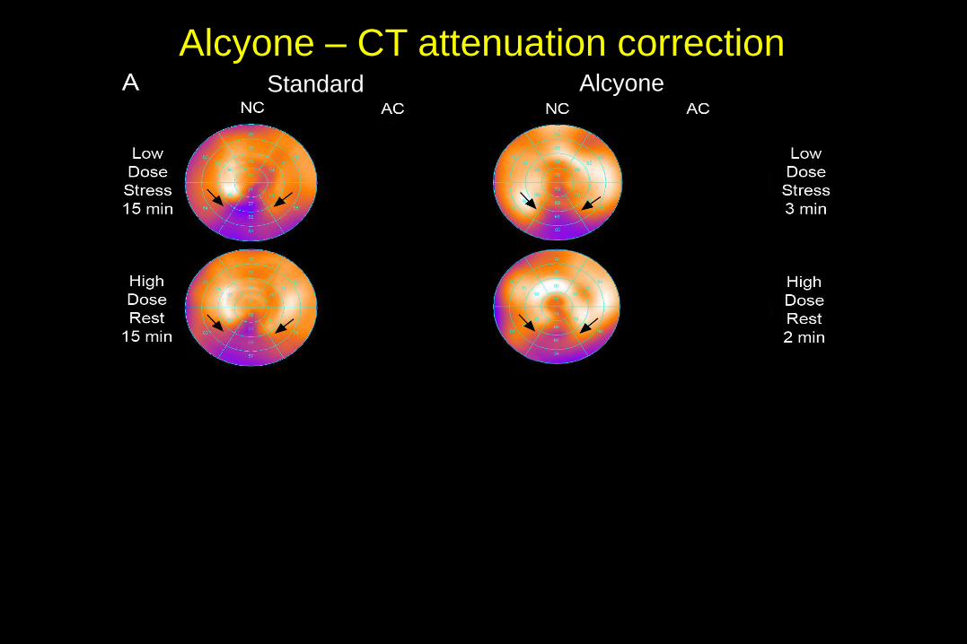
Alcyone – CT attenuation correction

Quantitative Analysis

Qualitative Analysis

Conclusion
● Our results support that AC of MPI on the novel CZT camera is
feasible as it provides high correlation of segmental tracer uptake
and an excellent clinical agreement compared to AC MPI on a
conventional SPECT camera.