High-resolution ultrasound visualization of the …...ULTRASOUND High-resolution ultrasound...
9
ULTRASOUND High-resolution ultrasound visualization of the recurrent motor branch of the median nerve: normal and first pathological findings Georg Riegler 1 & Christopher Pivec 1 & Hannes Platzgummer 1 & Doris Lieba-Samal 2 & Peter Brugger 3 & Suren Jengojan 1 & Martin Vierhapper 4 & Gerd Bodner 1 Received: 24 May 2016 /Revised: 11 September 2016 /Accepted: 21 November 2016 /Published online: 12 December 2016 # The Author(s) 2016. This article is published with open access at Springerlink.com Abstract Purpose To evaluate in a prospective study the possibility of visualization and diagnostic assessment of the recurrent motor branch (RMB) of the median nerve with high-resolution ultra- sound (HRUS). Materials and methods HRUS with high-frequency probes (18–22 MhZ) was used to locate the RMB in eight fresh ca- daveric hands. To verify correct identification, ink-marking and consecutive dissection were performed. Measurement of the RMB maximum transverse-diameter, an evaluation of the origin from the median nerve and its course in relation to the transverse carpal ligament, was performed in both hands of ten healthy volunteers (n = 20). Cases referred for HRUS ex- aminations for suspected RMB lesions were also assessed. Results The RMB was clearly visible in all anatomical spec- imens and all volunteers. Dissection confirmed HRUS find- ings in all anatomical specimens. Mean RMB diameter in volunteers was 0.7 mm ± 0.1 (range, 0.6–1). The RMB orig- inated from the radial aspect in 11 (55%), central aspect in eight (40%) and ulnar aspect in one (5%) hand. Nineteen (95%) extraligamentous courses and one (5%) subliga mentous course were detected. Three patients with visible RMB abnormalities on HRUS were identified. Conclusion HRUS is able to reliably visualize the RMB, its variations and pathologies. Key Points • Ultrasound allows visualization of the recurrent motor branch of the median nerve. • Ultrasound may help clinicians to assess patients with re- current motor branch pathologies. • Patient management may become more appropriate and targeted therapy could be improved. Keywords Median nerve . Carpal tunnel syndrome . Ultrasound . Iatrogenic disease . Anatomical variation Introduction The recurrent motor branch (RMB), sometimes also referred to as the muscular thenar branch of the median nerve, classi- cally supplies innervation to the thenar musculature, including the abductor pollicis brevis, the opponens pollicis and the superficial head of the flexor pollicis brevis (Fig. 1)[1, 2]. These contribute to the most important movements of the hand: opposition and abduction of the thumb. Damage to the RMB may lead to severely impaired func- tion in patients, with loss of dexterity, pinch and grasp func- tion. The main clinical relevance of the RMB is its suscepti- bility to iatrogenic injury, due to its variants, during decom- pression surgery for carpal tunnel syndrome (CTS) [3]. This is because anatomical studies have shown that there is high Electronic supplementary material The online version of this article (doi:10.1007/s00330-016-4671-1) contains supplementary material, which is available to authorized users. * Georg Riegler [email protected] 1 Department of Biomedical Imaging and Image-guided Therapy, Medical University of Vienna, Währingergürtel 18-20, 1090 Vienna, Austria 2 Department of Neurology, Medical University of Vienna, Währingergürtel 18-20, 1090 Vienna, Austria 3 Department of Anatomy, Center for Anatomy and Cell Biology, Medical University of Vienna, Währingerstrasse 13, 1090 Vienna, Austria 4 Department of Surgery, Division of Plastic and Reconstructive Surgery, Medical University of Vienna, Währingergürtel 18-20, 1090 Vienna, Austria Eur Radiol (2017) 27:2941–2949 DOI 10.1007/s00330-016-4671-1
Transcript of High-resolution ultrasound visualization of the …...ULTRASOUND High-resolution ultrasound...
ULTRASOUND
High-resolution ultrasound visualization of the recurrent motorbranch of the median nerve: normal and first pathologicalfindings
Georg Riegler1 & Christopher Pivec1 & Hannes Platzgummer1 & Doris Lieba-Samal2 &
Peter Brugger3 & Suren Jengojan1& Martin Vierhapper4 & Gerd Bodner1
Received: 24 May 2016 /Revised: 11 September 2016 /Accepted: 21 November 2016 /Published online: 12 December 2016# The Author(s) 2016. This article is published with open access at Springerlink.com
AbstractPurpose To evaluate in a prospective study the possibility ofvisualization and diagnostic assessment of the recurrent motorbranch (RMB) of the median nerve with high-resolution ultra-sound (HRUS).Materials and methods HRUS with high-frequency probes(18–22 MhZ) was used to locate the RMB in eight fresh ca-daveric hands. To verify correct identification, ink-markingand consecutive dissection were performed. Measurement ofthe RMB maximum transverse-diameter, an evaluation of theorigin from the median nerve and its course in relation to thetransverse carpal ligament, was performed in both hands often healthy volunteers (n = 20). Cases referred for HRUS ex-aminations for suspected RMB lesions were also assessed.Results The RMB was clearly visible in all anatomical spec-imens and all volunteers. Dissection confirmed HRUS find-ings in all anatomical specimens. Mean RMB diameter in
volunteers was 0.7 mm ± 0.1 (range, 0.6–1). The RMB orig-inated from the radial aspect in 11 (55%), central aspect ineight (40%) and ulnar aspect in one (5%) hand. Nineteen(95%) extraligamentous courses and one (5%) subligamentous course were detected. Three patients with visibleRMB abnormalities on HRUS were identified.Conclusion HRUS is able to reliably visualize the RMB, itsvariations and pathologies.Key Points• Ultrasound allows visualization of the recurrent motorbranch of the median nerve.
• Ultrasound may help clinicians to assess patients with re-current motor branch pathologies.
• Patient management may become more appropriate andtargeted therapy could be improved.
Keywords Median nerve . Carpal tunnel syndrome .
Ultrasound . Iatrogenic disease . Anatomical variation
Introduction
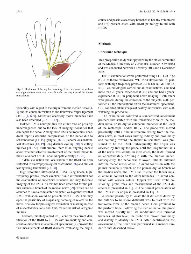
The recurrent motor branch (RMB), sometimes also referredto as the muscular thenar branch of the median nerve, classi-cally supplies innervation to the thenar musculature, includingthe abductor pollicis brevis, the opponens pollicis and thesuperficial head of the flexor pollicis brevis (Fig. 1) [1, 2].These contribute to the most important movements of thehand: opposition and abduction of the thumb.
Damage to the RMB may lead to severely impaired func-tion in patients, with loss of dexterity, pinch and grasp func-tion. The main clinical relevance of the RMB is its suscepti-bility to iatrogenic injury, due to its variants, during decom-pression surgery for carpal tunnel syndrome (CTS) [3]. This isbecause anatomical studies have shown that there is high
Electronic supplementary material The online version of this article(doi:10.1007/s00330-016-4671-1) contains supplementary material,which is available to authorized users.
* Georg [email protected]
1 Department of Biomedical Imaging and Image-guided Therapy,Medical University of Vienna, Währingergürtel 18-20,1090 Vienna, Austria
2 Department of Neurology, Medical University of Vienna,Währingergürtel 18-20, 1090 Vienna, Austria
3 Department of Anatomy, Center for Anatomy and Cell Biology,Medical University of Vienna, Währingerstrasse 13,1090 Vienna, Austria
4 Department of Surgery, Division of Plastic and ReconstructiveSurgery, Medical University of Vienna, Währingergürtel 18-20,1090 Vienna, Austria
Eur Radiol (2017) 27:2941–2949DOI 10.1007/s00330-016-4671-1
variability with regard to the origin from the median nerve [4,5] and its course in relation to the transverse carpal ligament(TCL) [4, 6–9]. Moreover, accessory motor branches havealso been described [2, 4, 10–12].
Isolated RMB neuropathies are either rare or possiblyunderdiagnosed due to the lack of imaging modalities thatcan depict the nerve. Among these RMB neuropathies, anec-dotal reports describe compression of the nerve due toschwannomas [13–15], ganglia [16, 17], anomalous anatom-ical structures [18, 19], long distance cycling [20] or cuttinginjuries [21, 22]. Furthermore, there is an ongoing debateabout whether selective involvement of the thenar motor fi-bres is a variant of CTS or an idiopathic entity [23–26].
To date, evaluation and localization of the RMB has beenrestricted to electrophysiological assessment [26] and clinicaltesting using landmarks [27, 28].
High-resolution ultrasound (HRUS), using linear, high-frequency probes, offers excellent tissue differentiation forthe examination of superficial structures and may facilitateimaging of the RMB. As this has been described for the pal-mar cutaneous branch of the median nerve [29], which can beassumed to have a comparable diameter, we hypothesized thatRMB evaluation would be possible with HRUS. This mayopen the possibility of diagnosing pathologies related to thenerve, or allow for pre-surgical evaluation or marking in caseof suspected variations, and, thus, reduce the risk of iatrogenicinjuries.
Therefore, this study aimed to: (i) confirm the correct iden-tification of the RMB by HRUS with ink-marking and con-secutive dissection in anatomical specimens; (ii) provide thefirst measurements of RMB diameter, evaluating the origin,
course and possible accessory branches in healthy volunteers;and (iii) present cases with RMB pathology found withHRUS.
Methods
Ultrasound technique
This prospective study was approved by the ethics committeeof the Medical University of Vienna (EC-number 1529/2015)and was conducted between 1 February 2015 and 1 December2015.
HRUS examinations were performed using a GE LOGIQ e(GE Healthcare, Wauwatosa, WI, USA) ultrasound (US) plat-form with high-frequency probes (GE L8-18i-D, GE L10-22-RS). Two radiologists carried out all examinations. One hadmore than 20 years’ experience (G.B.) and one had 4 years’experience (G.R.) in peripheral nerve imaging. Both raterswere present during the collection of the subjects. G.B. per-formed all the interventions on all the anatomical specimens.G.R. collected all the images of healthy individuals, with G.R.watching the procedure.
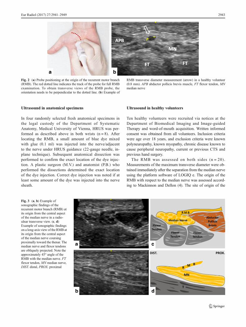
The examination followed a standardized assessmentprotocol that started with the transverse view of the me-dian nerve or its digital cutaneous branches at the levelof the metacarpal bodies III-IV. The probe was movedproximally until a tubular structure arising from the me-dian nerve, in most cases curving radially and proximallyand coursing toward the thenar musculature, was pre-sumed to be the RMB. Subsequently, the origin wasassessed by turning the probe until the longitudinal axisof the nerve was visible. In most cases, the RMB formedan approximately 45° angle with the median nerve.Subsequently, the nerve was followed until its entranceinto the thenar musculature. To avoid confusion with thepalmar cutaneous branch or the palmar digital branch ofthe median nerve, the RMB had to enter the thenar mus-culature in contrast to the other branches. To avoid con-fusion with vessels, colour Doppler was used. Probe po-sitioning, probe track and measurement of the RMB di-ameter is presented in Fig. 2. The normal presentation ofthe RMB at its origin is presented in Fig. 3.
A second possibility to locate the RMB (considered bythe authors to be more difficult) was to start with thetransverse view of the median nerve 3 cm proximal tothe pisiform bone. Following the median nerve, the probewas moved distally until its subdivision into terminalbranches. At this level, the probe was moved proximallyand distally to identify the RMB. After identification, theassessment of the nerve was performed in a manner sim-ilar to that described above.
Fig. 1 Illustration of the regular branching of the median nerve with anextraligamentous recurrent motor branch coursing toward the thenarmusculature
2942 Eur Radiol (2017) 27:2941–2949
Ultrasound in anatomical specimens
In four randomly selected fresh anatomical specimens inthe legal custody of the Department of SystematicAnatomy, Medical University of Vienna, HRUS was per-formed as described above in both wrists (n = 8). Afterlocating the RMB, a small amount of blue dye mixedwith glue (0.1 ml) was injected into the nerve/adjacentto the nerve under HRUS guidance (22-gauge needle, in-plane technique). Subsequent anatomical dissection wasperformed to confirm the exact location of the dye injec-tion. A plastic surgeon (M.V.) and anatomist (P.B.) whoperformed the dissections determined the exact locationof the dye injection. Correct dye injection was noted if atleast some amount of the dye was injected into the nervesheath.
Ultrasound in healthy volunteers
Ten healthy volunteers were recruited via notices at theDepartment of Biomedical Imaging and Image-guidedTherapy and word-of-mouth acquisition. Written informedconsent was obtained from all volunteers. Inclusion criteriawere age over 18 years, and exclusion criteria were knownpolyneuropathy, known myopathy, chronic disease known tocause peripheral neuropathy, current or previous CTS andprevious hand surgery.
The RMB was assessed on both sides (n = 20).Measurements of the maximum transverse diameter were ob-tained immediately after the separation from the median nerveusing the platform software of LOGIQ e. The origin of theRMB with respect to the median nerve was assessed accord-ing to Mackinnon and Dellon (4). The site of origin of the
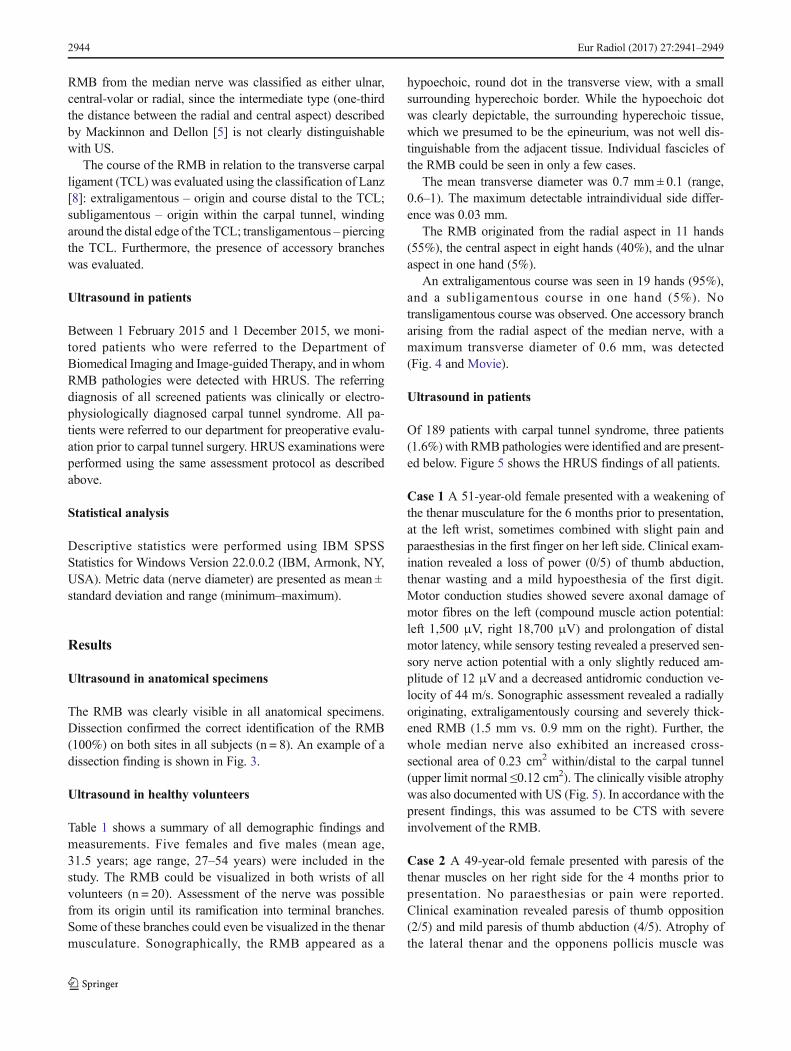
Fig. 3 (a, b) Example ofsonographic findings of therecurrent motor branch (RMB) atits origin from the central aspectof the median nerve in a radio-ulnar transverse view. (c, d)Example of sonographic findingson a long-axis view of the RMB atits origin from the central aspectof the median nerve coursingproximally toward the thenar. Themedian nerve and flexor tendonsare obliquely projected. Note theapproximately 45° angle of theRMB with the median nerve. FTflexor tendon, MN median nerve,DIST. distal, PROX. proximal
Fig. 2 (a) Probe positioning at the origin of the recurrent motor branch(RMB). The red dotted line indicates the track of the probe for full RMBexamination. To obtain transverse views of the RMB probe, theorientation needs to be perpendicular to the dotted line. (b) Example of
RMB transverse diameter measurement (arrow) in a healthy volunteer(0.8 mm). APB abductor pollicis brevis muscle, FT flexor tendon, MNmedian nerve
Eur Radiol (2017) 27:2941–2949 2943
RMB from the median nerve was classified as either ulnar,central-volar or radial, since the intermediate type (one-thirdthe distance between the radial and central aspect) describedby Mackinnon and Dellon [5] is not clearly distinguishablewith US.
The course of the RMB in relation to the transverse carpalligament (TCL) was evaluated using the classification of Lanz[8]: extraligamentous – origin and course distal to the TCL;subligamentous – origin within the carpal tunnel, windingaround the distal edge of the TCL; transligamentous – piercingthe TCL. Furthermore, the presence of accessory brancheswas evaluated.
Ultrasound in patients
Between 1 February 2015 and 1 December 2015, we moni-tored patients who were referred to the Department ofBiomedical Imaging and Image-guided Therapy, and in whomRMB pathologies were detected with HRUS. The referringdiagnosis of all screened patients was clinically or electro-physiologically diagnosed carpal tunnel syndrome. All pa-tients were referred to our department for preoperative evalu-ation prior to carpal tunnel surgery. HRUS examinations wereperformed using the same assessment protocol as describedabove.
Statistical analysis
Descriptive statistics were performed using IBM SPSSStatistics for Windows Version 22.0.0.2 (IBM, Armonk, NY,USA). Metric data (nerve diameter) are presented as mean ±standard deviation and range (minimum–maximum).
Results
Ultrasound in anatomical specimens
The RMB was clearly visible in all anatomical specimens.Dissection confirmed the correct identification of the RMB(100%) on both sites in all subjects (n = 8). An example of adissection finding is shown in Fig. 3.
Ultrasound in healthy volunteers
Table 1 shows a summary of all demographic findings andmeasurements. Five females and five males (mean age,31.5 years; age range, 27–54 years) were included in thestudy. The RMB could be visualized in both wrists of allvolunteers (n = 20). Assessment of the nerve was possiblefrom its origin until its ramification into terminal branches.Some of these branches could even be visualized in the thenarmusculature. Sonographically, the RMB appeared as a
hypoechoic, round dot in the transverse view, with a smallsurrounding hyperechoic border. While the hypoechoic dotwas clearly depictable, the surrounding hyperechoic tissue,which we presumed to be the epineurium, was not well dis-tinguishable from the adjacent tissue. Individual fascicles ofthe RMB could be seen in only a few cases.
The mean transverse diameter was 0.7 mm ± 0.1 (range,0.6–1). The maximum detectable intraindividual side differ-ence was 0.03 mm.
The RMB originated from the radial aspect in 11 hands(55%), the central aspect in eight hands (40%), and the ulnaraspect in one hand (5%).
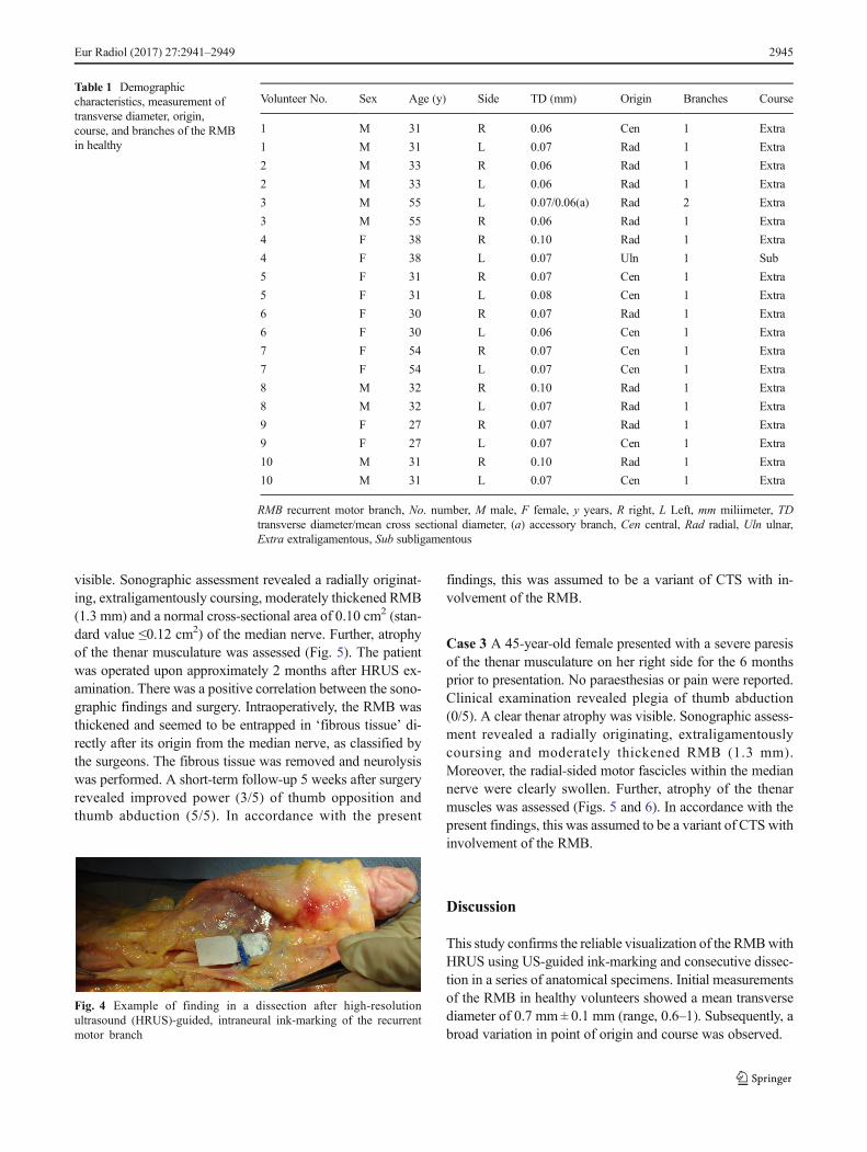
An extraligamentous course was seen in 19 hands (95%),and a subligamentous course in one hand (5%). Notransligamentous course was observed. One accessory brancharising from the radial aspect of the median nerve, with amaximum transverse diameter of 0.6 mm, was detected(Fig. 4 and Movie).
Ultrasound in patients
Of 189 patients with carpal tunnel syndrome, three patients(1.6%) with RMB pathologies were identified and are present-ed below. Figure 5 shows the HRUS findings of all patients.
Case 1 A 51-year-old female presented with a weakening ofthe thenar musculature for the 6 months prior to presentation,at the left wrist, sometimes combined with slight pain andparaesthesias in the first finger on her left side. Clinical exam-ination revealed a loss of power (0/5) of thumb abduction,thenar wasting and a mild hypoesthesia of the first digit.Motor conduction studies showed severe axonal damage ofmotor fibres on the left (compound muscle action potential:left 1,500 μV, right 18,700 μV) and prolongation of distalmotor latency, while sensory testing revealed a preserved sen-sory nerve action potential with a only slightly reduced am-plitude of 12 μV and a decreased antidromic conduction ve-locity of 44 m/s. Sonographic assessment revealed a radiallyoriginating, extraligamentously coursing and severely thick-ened RMB (1.5 mm vs. 0.9 mm on the right). Further, thewhole median nerve also exhibited an increased cross-sectional area of 0.23 cm2 within/distal to the carpal tunnel(upper limit normal ≤0.12 cm2). The clinically visible atrophywas also documented with US (Fig. 5). In accordance with thepresent findings, this was assumed to be CTS with severeinvolvement of the RMB.
Case 2 A 49-year-old female presented with paresis of thethenar muscles on her right side for the 4 months prior topresentation. No paraesthesias or pain were reported.Clinical examination revealed paresis of thumb opposition(2/5) and mild paresis of thumb abduction (4/5). Atrophy ofthe lateral thenar and the opponens pollicis muscle was
2944 Eur Radiol (2017) 27:2941–2949
visible. Sonographic assessment revealed a radially originat-ing, extraligamentously coursing, moderately thickened RMB(1.3 mm) and a normal cross-sectional area of 0.10 cm2 (stan-dard value ≤0.12 cm2) of the median nerve. Further, atrophyof the thenar musculature was assessed (Fig. 5). The patientwas operated upon approximately 2 months after HRUS ex-amination. There was a positive correlation between the sono-graphic findings and surgery. Intraoperatively, the RMB wasthickened and seemed to be entrapped in ‘fibrous tissue’ di-rectly after its origin from the median nerve, as classified bythe surgeons. The fibrous tissue was removed and neurolysiswas performed. A short-term follow-up 5 weeks after surgeryrevealed improved power (3/5) of thumb opposition andthumb abduction (5/5). In accordance with the present
findings, this was assumed to be a variant of CTS with in-volvement of the RMB.
Case 3 A 45-year-old female presented with a severe paresisof the thenar musculature on her right side for the 6 monthsprior to presentation. No paraesthesias or pain were reported.Clinical examination revealed plegia of thumb abduction(0/5). A clear thenar atrophy was visible. Sonographic assess-ment revealed a radially originating, extraligamentouslycoursing and moderately thickened RMB (1.3 mm).Moreover, the radial-sided motor fascicles within the mediannerve were clearly swollen. Further, atrophy of the thenarmuscles was assessed (Figs. 5 and 6). In accordance with thepresent findings, this was assumed to be a variant of CTS withinvolvement of the RMB.
Discussion
This study confirms the reliable visualization of the RMBwithHRUS using US-guided ink-marking and consecutive dissec-tion in a series of anatomical specimens. Initial measurementsof the RMB in healthy volunteers showed a mean transversediameter of 0.7 mm ± 0.1 mm (range, 0.6–1). Subsequently, abroad variation in point of origin and course was observed.
Table 1 Demographiccharacteristics, measurement oftransverse diameter, origin,course, and branches of the RMBin healthy
Volunteer No. Sex Age (y) Side TD (mm) Origin Branches Course
1 M 31 R 0.06 Cen 1 Extra
1 M 31 L 0.07 Rad 1 Extra
2 M 33 R 0.06 Rad 1 Extra
2 M 33 L 0.06 Rad 1 Extra
3 M 55 L 0.07/0.06(a) Rad 2 Extra
3 M 55 R 0.06 Rad 1 Extra
4 F 38 R 0.10 Rad 1 Extra
4 F 38 L 0.07 Uln 1 Sub
5 F 31 R 0.07 Cen 1 Extra
5 F 31 L 0.08 Cen 1 Extra
6 F 30 R 0.07 Rad 1 Extra
6 F 30 L 0.06 Cen 1 Extra
7 F 54 R 0.07 Cen 1 Extra
7 F 54 L 0.07 Cen 1 Extra
8 M 32 R 0.10 Rad 1 Extra
8 M 32 L 0.07 Rad 1 Extra
9 F 27 R 0.07 Rad 1 Extra
9 F 27 L 0.07 Cen 1 Extra
10 M 31 R 0.10 Rad 1 Extra
10 M 31 L 0.07 Cen 1 Extra
RMB recurrent motor branch, No. number, M male, F female, y years, R right, L Left, mm miliimeter, TDtransverse diameter/mean cross sectional diameter, (a) accessory branch, Cen central, Rad radial, Uln ulnar,Extra extraligamentous, Sub subligamentous
Fig. 4 Example of finding in a dissection after high-resolutionultrasound (HRUS)-guided, intraneural ink-marking of the recurrentmotor branch
Eur Radiol (2017) 27:2941–2949 2945

Ultrasound measurements of the transverse diameter of theRMB in healthy volunteers revealed clearly lower values than
previously described in anatomical studies. Wang and col-leagues [30] reported a mean diameter of 1.7 ± 0.3 mm in
Fig. 6 (a, b) Findings in patient 1, showing an enlarged recurrent motorbranch (RMB) (encircled) atop the enlarged median nerve just distal tothe carpal tunnel and a clear thenar atrophy on the patient’s left side. (c, d)Findings in patient 2, showing a thickened RMB (encircled). The median
nerve is obliquely projected. (d) Intraoperative findings of the RMB afterremoval of fibrous tissue surrounding the branch. (e, f) Findings in patient3, with swollen radial-sidedmotor fascicles within themedian nerve and aclear thenar atrophy on the patient’s right side
Fig. 5 (a, b) Example ofsonographic findings of therecurrent motor branch (RMB)originating from the ulnar aspectof the median nerve. (c, d)Example of sonographic findingsin the same volunteer. The RMBcrosses the anterior aspect of themedian and courses beneath thetransverse carpal ligament (TCL;subligamentous) toward the the-nar musculature. FPB flexorpollicis brevis muscle, FT flexortendon, MN median nerve, SUBLsubligamentous
2946 Eur Radiol (2017) 27:2941–2949

seven fresh forearm amputation specimens, and Üstün et al.[31] reported a mean diameter of 1.4 mm ± 0.12 mm in tenfresh cadaver arms. This discrepancy in nerve diameter maybe due to the different visualization methods employed. In ourstudy, we could measure only the hypoechoic pattern of thenerve without the surrounding neural tissue. Measurements ofthe transverse diameter of the RMBwith US have not yet beenreported. As presented, the transverse diameter in patientsclearly exceeded the upper limit observed in healthy volun-teers. Therefore, HRUS may help to evaluate RMB patholo-gies, and our data may serve as a reference for further, moredetailed US characterizations of the RMB.
In addition to the depiction of the RMB, HRUS providesfurther information about the course, origin and branching ofthis nerve. The course of the RMB in relation to the TCL hasgained extensive attention in various studies, due to the factthat the ‘anomalous’ trans- and subligamentous variants, inparticular, are at risk during both open and endoscopic surgery[3, 7, 8, 32]. Our results are comparable with previous studies[4, 6–9] (extraligamentous, 46–97%; subligamentous, 2–34%; transligamentous, 1–23%), although we did not detecta transligamentous course, which is mainly attributable to thesmall study sample.
The origin from the radial aspect of the median nerve wasdescribed as a possible site of entrapment because of separateobliquely arranged fibres from the TCL encircling the RMB[7]. As the radial origin is most common and the results ofprevious studies are in accordance with our data (55% vs. 60–80%) [4, 5], we think this condition can be easily detected byHRUS. The less frequent origin from the ulnar aspect of themedian nerve (5% in our study vs. 1.1% in a study of 821hands that had undergone carpal tunnel (CT) release surgery)[9] represents a major risk for iatrogenic injury with the ulnarside approach, as well as with the median approach, since thenerve crosses the anterior aspect of the median nerve duringCT release [33].
Iatrogenic RMB injury during CT release seems to be a rarecomplication, with approximately 0.5% in the reported litera-ture [3, 32]. Nevertheless, it represents a severe complicationalso called the ‘million dollar injury’ due to the compensationawarded in lawsuits because of the loss of thenar function [34,35]. To date, no consensus exists about whether the branchshould be examined intra- or preoperatively to avoid damage.Of 153 surgeons responding to a questionnaire, the majority(>70%) did not explore the nerve routinely and did not rec-ommend doing so [36]. Other authors suggest exploring theRMB intraoperatively, at least for some special conditions[37]. Indeed, whenever the surgeon encounters muscle fibreslying superficial to or interposed within the TCL, there is agreater than 90% likelihood that the motor branch would beanomalous [37]. The preoperative RMB localization is limitedto the use of surface landmarks, such as the Kaplan’s cardinaland middle finger radial-side-line, or a physical examination
manoeuvre, such as the middle finger flexion test [27, 28].Nevertheless, these tests are inaccurate when the RMB has avarying course. As an example, in middle finger flexion tests,the transligamentous course showed a deviation of 10–25 mmfrom where it was expected [27, 28]. RMB evaluation withHRUS overcomes all these limitations. It allows visualizationof the nerve along its entire course, which may help surgeonsto plan their approach for CT release. In the case of a ‘danger-ous’ variation, preoperative skin-marking could be providedto facilitate exploration of the nerve. Although we did notobserve an iatrogenic injury prior to re-operation during ourshort observation period, we saw one patient postoperativelywho was treated for a complete transection of the RMB duringcarpal tunnel release. Therefore, the results of this study sug-gest that HRUS evaluation of the RMB should be included aspart of the conventional sonographic examination for CTS tominimize iatrogenic injury during CT release.
In 1982, Bennet and Crouch [23] reported two cases ofisolated compression of the RMB, characterized by selec-tive involvement of thenar motor fibres. In these cases,the surgical observation showed compression of thebranch due to a transligamentous course or an excessiveangle of the thenar branch at the distal edge of the trans-verse ligament with neuroma formation proximal to theentrapment sites. Four subsequent electrophysiologicaland clinical studies underlined the theory that motor fas-cicles alone may be involved in CTS, or represent a sep-arate entity without the classic CTS [24–26, 38]. Our case2 and case 3 may provide a hint that these conditions maybe detectable by HRUS in the future. Nevertheless, furthercomparative HRUS studies between normal and patholog-ical RMB conditions are needed to reliably answer thisquestion.
This study has several strengths and limitations. Itsstrengths include the first-time use of HRUS for specificassessment of the RMB and confirmation of the find-ings by the gold standard of anatomical dissection. Itslimitations include the fact that findings in vivo wereuncontrolled. However, accuracy in anatomical speci-mens was 100% and RMB in volunteers could befollowed into the abductor muscle. A further limitationis the fact that pathological findings in this manuscriptare case reports and do not provide reliable informationabout the future role of HRUS in RMB pathology de-tection. For this reason, further comparative studies be-tween normal and pathological conditions are needed.
In conclusion, this study confirms the reliable ability tovisualize the RMB and its variations with HRUS, in anatom-ical specimens and in healthy volunteers. We therefore en-courage the use of HRUS, especially for preoperative evalua-tion for carpal tunnel release or if thenar muscle weakening ispresent. Further studies are needed to assess the value ofHRUS in diagnosing RMB pathologies.
Eur Radiol (2017) 27:2941–2949 2947
Acknowledgements Open access funding provided by MedicalUniversity of Vienna. The authors thank Mary McAllister for her com-ments on themanuscript. This workwas orally presented at the ECR 2016in Vienna. The scientific guarantor of this publication is Gerd Bodner.The authors of this manuscript declare no relationships with any compa-nies whose products or services may be related to the subject matter of thearticle. The authors state that this work has not received any funding. Nocomplex statistical methods were necessary for this paper. InstitutionalReview Board approval was obtained. Written informed consent wasobtained from all subjects (patients) in this study. Methodology: prospec-tive, experimental, performed at one institution.
Open Access This article is distributed under the terms of the CreativeCommons At t r ibut ion 4 .0 In te rna t ional License (h t tp : / /creativecommons.org/licenses/by/4.0/), which permits unrestricted use,distribution, and reproduction in any medium, provided you giveappropriate credit to the original author(s) and the source, provide a linkto the Creative Commons license, and indicate if changes were made.
References
1. Mazurek MT, Shin AY (2001) Upper extremity peripheral nerveanatomy: current concepts and applications. Clin Orthop RelatRes 7–20
2. Mumford J, Morecraft R, Blair WF (1987) Anatomy of the thenarbranch of the median nerve. J Hand Surg 12:361–365
3. Benson LS, Bare AA, Nagle DJ et al (2006) Complications ofendoscopic and open carpal tunnel release. Arthrosc: J ArthroscRelated Surg: Off Publ Arthrosc Assoc North Am Int ArthroscAssoc 22:919–924, 924 e911–912
4. Hurwitz PJ (1996) Variations in the course of the thenar motorbranch of the median nerve. J Hand Surg 21:344–346
5. Mackinnon SE, Dellon AL (1988) Anatomic Investigations ofNerves at the Wrist.1. Orientation of the Motor Fascicle of theMedian Nerve in the Carpal-Tunnel. Ann Plast Surg 21:32–35
6. Al-Qattan MM (2010) Variations in the course of the thenar motorbranch of the median nerve and their relationship to the hypertro-phic muscle overlying the transverse carpal ligament. J Hand Surg35:1820–1824
7. Kozin SH (1998) The anatomy of the recurrent branch of the me-dian nerve. J Hand Surg 23:852–858
8. Lanz U (1977) Anatomical variations of the median nerve in thecarpal tunnel. J Hand Surg 2:44–53
9. Tountas CP, Bihrle DM, MacDonald CJ et al (1987) Variations ofthe median nerve in the carpal canal. J Hand Surg 12:708–712
10. Alp M, Marur T, Akkin SM et al (2005) Ramification patternof the thenar branch of the median nerve entering the thenarfascia and the distribution of the terminal branches in the the-nar musculature: Anatomic cadaver study in 144 hands. ClinAnat 18:195–199
11. Olave E, Prates JC, Gabrielli C et al (1996) Morphometricstudies of the muscular branch of the median nerve. J Anat189(Pt 2):445–449
12. Sacks JM, Kuo YR, McLean K et al (2007) Anatomical relation-ships among the median nerve thenar branch, superficial palmararch, and transverse carpal ligament. Plast Reconstr Surg 120:713–718
13. Fiaschi P, Pacetti M, Secci F et al (2015) A rare case of first motorbranch ofmedian nerve schwannoma. Neur Sci: Off J Italian NeurolSoc Italian Soc Clin Neurophysiol 36:659–661
14. Josty IC, Sykes PJ (2001) An unusual schwannoma of the mediannerve: effects on the motor branch. Br J Plast Surg 54:71–73
15. Squarzina PB, Adani R, Cerofolini E et al (1993) Ancientschwannoma of the motor branch of the median nerve: aclinical case. La Chirurgia Degli Organi Di Movimento 78:19–23
16. Jensen TT (1990) Isolated compression of the motor branch of themedian nerve by a ganglion. Case report. Scan J Plastic ReconstrucSurg Hand Surg/Nordisk Plastikkirurgisk Forening [and] NordiskKlubb Handkirurgi 24:171
17. Kato H, Ogino T, Nanbu T et al (1991) Compression neuropathy ofthe motor branch of the median nerve caused by palmar ganglion. JHand Surg 16:751–752
18. Widder S, Shons AR (1988) Carpal tunnel syndrome associatedwith extra tunnel vascular compression of the median nerve motorbranch. J Hand Surg 13:926–927
19. Yamanaka K, Horiuchi Y, Yabe Y (1994) Compression neuropathyof themotor branch of the median nerve due to an anomalous thenarmuscle. J Hand Surg 19:711–712
20. Ali E, Delamont RS, Jenkins D et al (2013) Bilateral recurrentmotor branch of median nerve neuropathy following long-distance cycling. Clin Neurophysiol: Of J Int Fed ClinNeurophysiol 124:1258–1260
21. Haussmann P (1980) Isolated traumatic lesions of the motor nervebranches in the hand. Handchirurgie 12:23–25
22. Nara Y, Hasegawa O,MatsumotoM et al (1995) Electrophysiologicevaluation of entrapment neuropathy caused by scar formation inrecurrent motor branch of the median nerve. No to Shinkei BrainNerve 47:779
23. Bennett JB, Crouch CC (1982) Compression syndrome of therecurrent motor branch of the median nerve. J Hand Surg 7:407–409
24. Cosgrove JL, Chase PM, Mast NJ (2002) Thenar motor syndrome:median mononeuropathy of the hand. Am J Phys Med Rehab/Assoc Acad Phys 81:421–423
25. Mondelli M, Aretini A, Ginanneschi F et al (2010) Thenar motorneuropathy electrophysiological study of 28 cases. J ClinNeurophysiol: Off Publ Am Electroencephalographic Soc 27:344–349
26. Repaci M, Torrieri F, Di Blasio F et al (1999) Exclusiveelectrophysiological motor involvement in carpal tunnel syn-drome. Clin Neurophysiol: Off J Int Fed Clin Neurophysiol110:1471–1474
27. Eskandari MM, Yilmaz C, Oztuna V et al (2005) Topographic lo-calization of the motor branch of the median nerve. J Hand Surg 30:803–807
28. Rodriguez R, Strauch RJ (2013) The middle finger flexion test tolocate the thenar motor branch of the median nerve. J Hand Surg 38:1547–1550
29. Tagliafico A, Pugliese F, Bianchi S et al (2008) High-resolutionsonography of the palmar cutaneous branch of the median nerve.AJR Am J Roentgenol 191:107–114
30. Wang Y, Zhu S (1997) Transfer of a branch of the anteriorinterosseus nerve to the motor branch of themedian nerve and ulnarnerve. Chin Med J 110:216–219
31. Ustun ME, Ogun TC, Karabulut AK et al (2001) An alternativemethod for restoring opposition after median nerve injury: an ana-tomical feasibility study for the use of neurotisation. J Anat 198:635–638
32. Palmer AK, Toivonen DA (1999) Complications of endoscopic andopen carpal tunnel release. J Hand Surg 24:561–565
33. Lilly CJ, Magnell TD (1985) Severance of the thenar branch of themedian nerve as a complication of carpal tunnel release. J HandSurg 10:399–402
2948 Eur Radiol (2017) 27:2941–2949
34. Krishnan P, Mishra R, JenaM et al (2013) Transligamentous thenarbranch of the median nerve: the million dollar nerve. Neurol India61:311–312
35. Virgilio C (2014) Surgery. Springer Science + Business Media,New York
36. Candal-Couto JJ, Sher JL (2007) The thenar motor branch duringcarpal tunnel decompression: "the expert opinion". Arch OrthopTrauma Surg 127:431–434
37. Green DP, Morgan JP (2008) Correlation between muscle mor-phology of the transverse carpal ligament and branching pat-tern of the motor branch of median nerve. J Hand Surg 33:1505–1511
38. Mondelli M, Baldasseroni A, Aretini A et al (2010) Prevalent in-volvement of thenar motor fibres in vineyard workers with carpaltunnel syndrome. Clin Neurophysiol:Off J Int Fed ClinNeurophysiol 121:1251–1255
Eur Radiol (2017) 27:2941–2949 2949