
High resolution ultrasound in equine ophthalmology to display the
20
High resolution ultrasound in equine ophthalmology to display the anterior segment of the eye M. Cronau, H. Gerhards
Transcript of High resolution ultrasound in equine ophthalmology to display the
High resolution ultrasound in equine ophthalmology to display the anterior segment of the eye
M. Cronau, H. Gerhards
High resolution ultrasound
• Difference HRUS ⇔ normal ultrasound• Indications for ophthalmic ultrasound• Method• Normal anterior segment anatomy• Some case examples
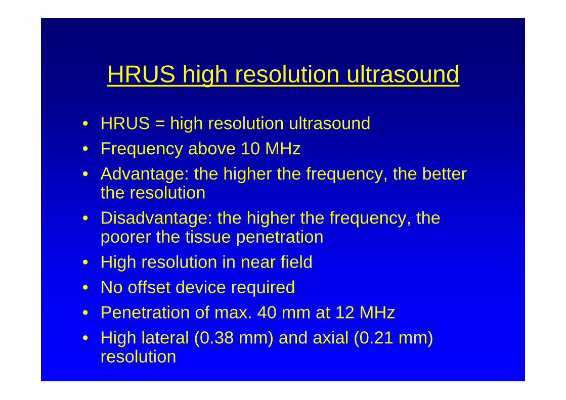
HRUS high resolution ultrasound
• HRUS = high resolution ultrasound• Frequency above 10 MHz• Advantage: the higher the frequency, the better
the resolution • Disadvantage: the higher the frequency, the
poorer the tissue penetration • High resolution in near field• No offset device required• Penetration of max. 40 mm at 12 MHz • High lateral (0.38 mm) and axial (0.21 mm)
resolution

Indications for ophthalmic ultrasound
• Opacities of the ocular media (corneal opacities, hypopyon, hyphema, cataract, cloudy vitreous)
• Differentiate between e.g. iridal cysts ⇔melanoma
• Glaucoma: determine diameter of globe, corneal thickness

Method
• Requires no sedation
• Dark room
• Ultrasound contact gel
• No clipping required
• Comparable quality of both transpalpebraland direct coppling images
• No offset device
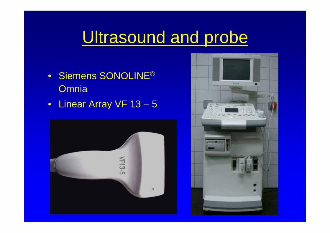
Ultrasound and probe
• Siemens SONOLINE®
Omnia• Linear Array VF 13 – 5

Normal anterior segment anatomy
• C = cornea
• AC = anterior chamber
• I = iris
• PC = posterior chamber
• L = lens

Normal anterior segment anatomy
• C = cornea
• NB = nigroid bodies
• AC = anterior chamber
• I = iris
• L = lens

Ulcer
• Corneal appearance after placement of conjunctival flap
• Further ocular irritation• HRUS- examination to
determine eventual internal ocular structure damage

Anterior synechia
• C: cornea
• I: iris
• AC: anterior chamber
• Anterior synechiapersistent irritation ⇒synechiolysis
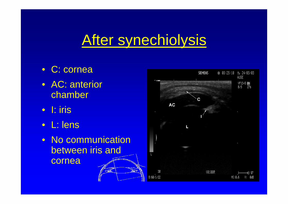
After synechiolysis
• C: cornea• AC: anterior
chamber• I: iris• L: lens• No communication
between iris and cornea

Hyphema
• Post traumatic tear in the sclera
• C: cornea
• AC: anterior chamber
• L: lens

Posterior synechia (Iris Bombé)
• C: cornea
• I: iris
• AC: anterior chamber
• ALS: anterior lens surface
• L: lens

Iris cyst
• C: cornea• CY: cyst• AC: anterior chamber• L: lens• Cyst: anechogenic• Melanoma: echogenic
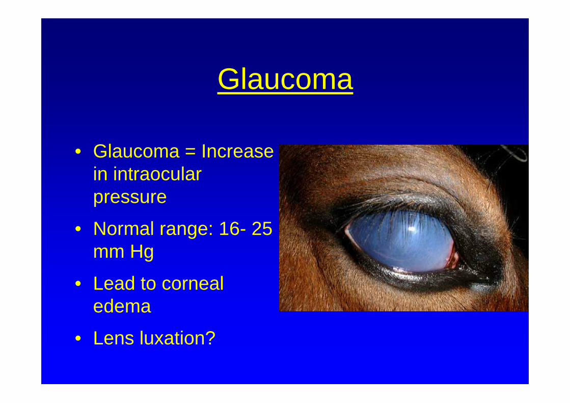
Glaucoma
• Glaucoma = Increase in intraocular pressure
• Normal range: 16- 25 mm Hg
• Lead to corneal edema
• Lens luxation?

Luxatio lentis anterior
• Luxated lens in iridocorneal angle ⇒reduction in aqueous outflow ⇒ Increase in intraocular pressure
• C: cornea
• L: lens
• AC: anterior chamber
• VB: vitreous body
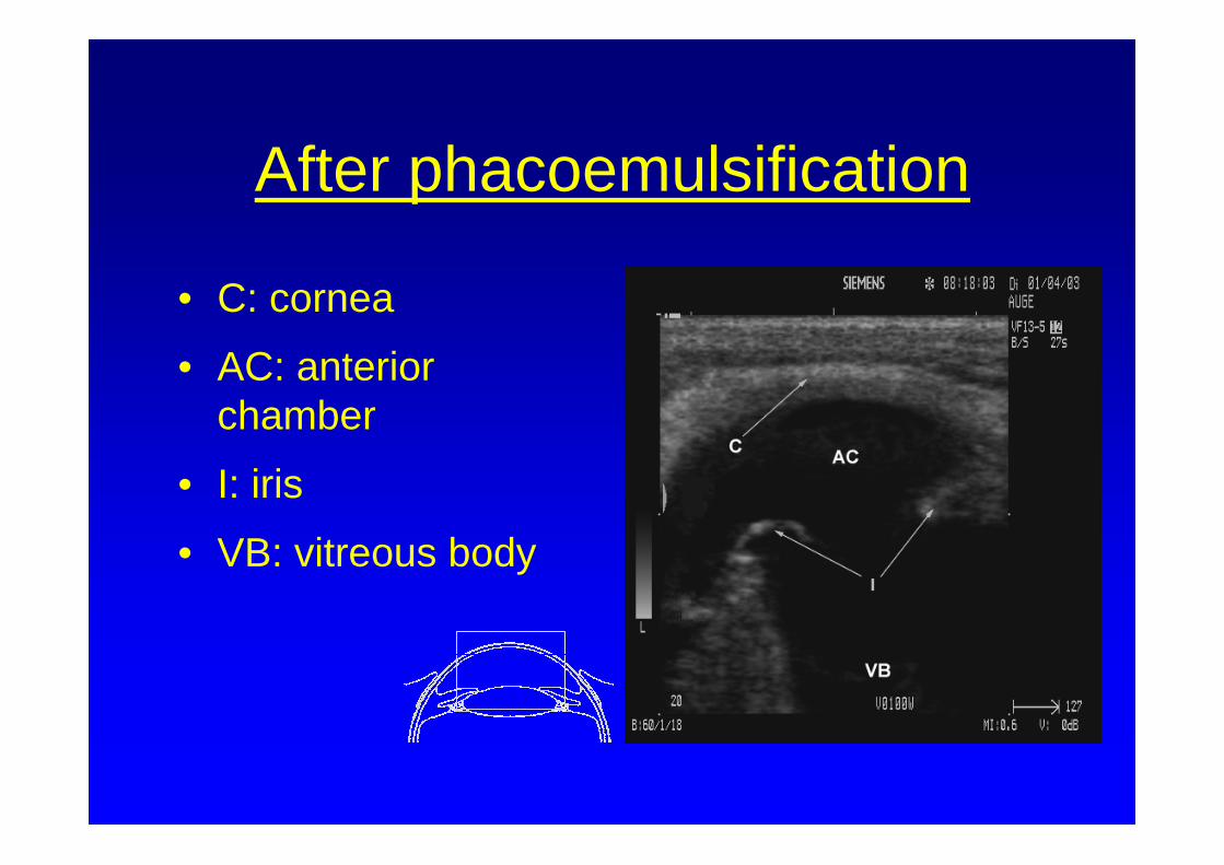
After phacoemulsification
• C: cornea
• AC: anterior chamber
• I: iris
• VB: vitreous body

Absolute glaucoma
• Distinct corneal edema
• Buphthalmos

Zonules
• C: cornea
• AC: anterior chamber
• I: iris
• Z: zonules
• L: lens

Conclusion
• Ultrasonic examination is a simple and efficient means of evaluating eyes with opaque optic media
• Improved presentation of individual ocular structures when compared to conventional ultrasound, especially those located within the anterior segment