High-resolution karyotyping by oligonucleotide microarrays ...
15
High-resolution karyotyping by oligonucleotide microarrays : the next revolution in cytogenetics Gijsbers, A.C.J. Citation Gijsbers, A. C. J. (2010, November 30). High-resolution karyotyping by oligonucleotide microarrays : the next revolution in cytogenetics. Retrieved from https://hdl.handle.net/1887/16187 Version: Corrected Publisher’s Version License: Licence agreement concerning inclusion of doctoral thesis in the Institutional Repository of the University of Leiden Downloaded from: https://hdl.handle.net/1887/16187 Note: To cite this publication please use the final published version (if applicable).
Transcript of High-resolution karyotyping by oligonucleotide microarrays ...
High-resolution karyotyping by oligonucleotide microarrays : the nextrevolution in cytogeneticsGijsbers, A.C.J.
CitationGijsbers, A. C. J. (2010, November 30). High-resolution karyotyping by oligonucleotidemicroarrays : the next revolution in cytogenetics. Retrieved fromhttps://hdl.handle.net/1887/16187 Version: Corrected Publisher’s Version
License: Licence agreement concerning inclusion of doctoral thesis in theInstitutional Repository of the University of Leiden
Downloaded from: https://hdl.handle.net/1887/16187 Note: To cite this publication please use the final published version (if applicable).
Chapter 3.3
X-chromosome duplications in males with mental retardation: pathogenic or
benign variants?
Antoinet CJ Gijsbers, Nicolette S den Hollander, Apollonia TJM Helderman-van de Enden, Janneke HM Schuurs-Hoeijmakers, Linda Vijfhuizen, Emilia K Bijlsma, Arie van Haeringen, Kerstin BM Hansson, Egbert Bakker, Martijn H Breuning and Claudia AL Ruivenkamp
Center for Human and Clinical Genetics; Leiden University Medical Center (LUMC), Leiden, The Netherlands
Clin Genet, in press

89
Chapter 3
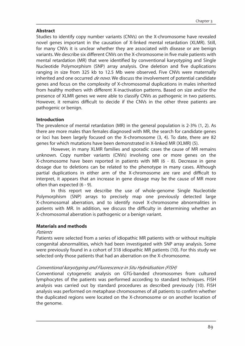
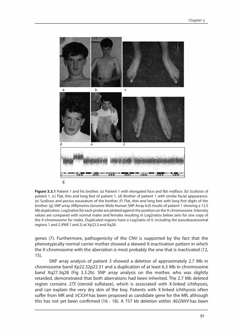
AbstractStudies to identify copy number variants (CNVs) on the X-chromosome have revealed novel genes important in the causation of X-linked mental retardation (XLMR). Still, for many CNVs it is unclear whether they are associated with disease or are benign variants. We describe six different CNVs on the X-chromosome in five male patients with mental retardation (MR) that were identified by conventional karyotyping and Single Nucleotide Polymorphism (SNP) array analysis. One deletion and five duplications ranging in size from 325 kb to 12.5 Mb were observed. Five CNVs were maternally inherited and one occurred de novo. We discuss the involvement of potential candidate genes and focus on the complexity of X-chromosomal duplications in males inherited from healthy mothers with different X-inactivation patterns. Based on size and/or the presence of XLMR genes we were able to classify CNVs as pathogenic in two patients. However, it remains difficult to decide if the CNVs in the other three patients are pathogenic or benign.
IntroductionThe prevalence of mental retardation (MR) in the general population is 2-3% (1, 2). As there are more males than females diagnosed with MR, the search for candidate genes or loci has been largely focused on the X-chromosome (3, 4). To date, there are 82 genes for which mutations have been demonstrated in X-linked MR (XLMR) (5). However, in many XLMR families and sporadic cases the cause of MR remains unknown. Copy number variants (CNVs) involving one or more genes on the X-chromosome have been reported in patients with MR (6 - 8). Decrease in gene dosage due to deletions can be related to the phenotype in many cases. Although partial duplications in either arm of the X-chromosome are rare and difficult to interpret, it appears that an increase in gene dosage may be the cause of MR more often than expected (6 - 9). In this report we describe the use of whole-genome Single Nucleotide Polymorphism (SNP) arrays to precisely map one previously detected large X-chromosomal aberration, and to identify novel X-chromosome abnormalities in patients with MR. In addition, we discuss the difficulty in determining whether an X-chromosomal aberration is pathogenic or a benign variant.
Materials and methodsPatientsPatients were selected from a series of idiopathic MR patients with or without multiple congenital abnormalities, which had been investigated with SNP array analysis. Some were previously found in a cohort of 318 idiopathic MR patients (10). For this study we selected only those patients that had an aberration on the X-chromosome.
Conventional karyotyping and Fluorescence in Situ Hybridisation (FISH) Conventional cytogenetic analysis on GTG-banded chromosomes from cultured lymphocytes of the patients was performed according to standard techniques. FISH analysis was carried out by standard procedures as described previously (10). FISH analysis was performed on metaphase chromosomes of all patients to confirm whether the duplicated regions were located on the X-chromosome or on another location of the genome.
90
X-chromosome duplications
SNP ArraysThe Affymetrix GeneChip Human Mapping 262K NspI (used for patients 2, 3 and 5), 238K StyI (used for patient 4) and Affymetrix Genome-Wide Human SNP Array 6.0 arrays (used for patient 1) (Affymetrix, Santa Clara, CA, USA) were performed following the manufacturer’s instructions. The 262K and 238K arrays have an average probe spacing of 12 kb and were analyzed as described previously (10). The average probe spacing of the 6.0 array is 0.7 kb. SNP copy number was assessed in the patient using Affymetrix Genotyping Console 2.1 software. Aberrations of at least 100 kb were analyzed. All CNVs identified in this study were evaluated as described earlier (10). All patients with a (potentially) pathogenic CNV were added to the DECIPHER database. Multiplex Ligation-dependent Probe Amplification (MLPA) analysisMLPA experiments were performed as described previously to narrow down the breakpoints of patient 2 (10).
X-inactivation X-inactivation was investigated with Southern blot analysis with a probe for the M27B (DXS255) locus for the mothers of patients 1 and 4. The intensity of the unmethylated (2.8 kb) and the methylated (5.2 kb) bands were compared to determine random or non-random X-inactivation. The ratio of active to inactive X could not be calculated by this method. X-inactivation was investigated with a methylation-specific PCR for family members of patients 2 and 3 (11). Inactivation was considered to be random if the ratio was less than 75:25 (12). Results and discussionWe present six different CNVs, five interstitial duplications and one interstitial deletion, on the X-chromosome in five male MR patients (Table 3.3.1). Five CNVs were inherited from the mother (patients 1-4) and one had occurred de novo (patient 5). Clinical features of these patients are shown in Table 3.3.2 and an extended clinical description is provided as supplementary material. The array-based technology for the detection of CNVs in MR patients has proven to be a powerful tool. However, the main challenge is ranking these CNVs as pathogenic, potentially pathogenic or polymorphic variants. Here, CNVs were classified into these groups based on various factors, including size, gene content, and whether they were inherited or occurred de novo (13).
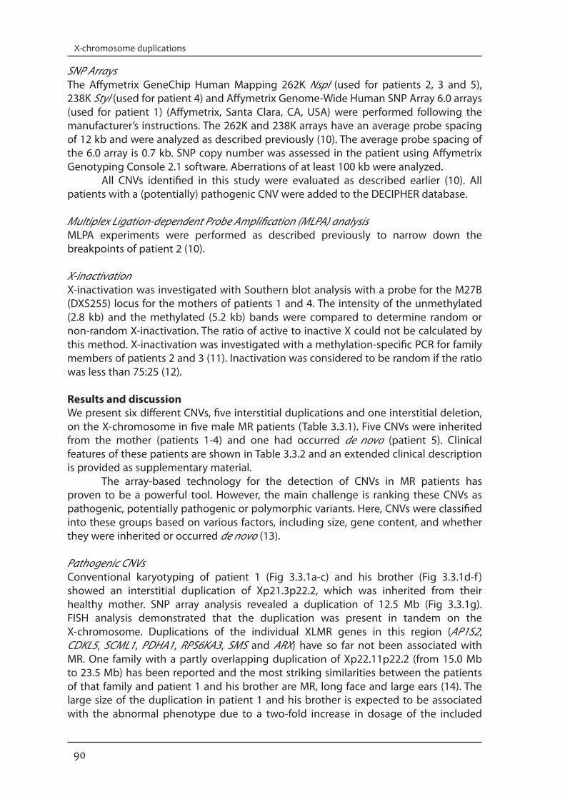
Pathogenic CNVsConventional karyotyping of patient 1 (Fig 3.3.1a-c) and his brother (Fig 3.3.1d-f ) showed an interstitial duplication of Xp21.3p22.2, which was inherited from their healthy mother. SNP array analysis revealed a duplication of 12.5 Mb (Fig 3.3.1g). FISH analysis demonstrated that the duplication was present in tandem on the X-chromosome. Duplications of the individual XLMR genes in this region (AP1S2, CDKL5, SCML1, PDHA1, RPS6KA3, SMS and ARX) have so far not been associated with MR. One family with a partly overlapping duplication of Xp22.11p22.2 (from 15.0 Mb to 23.5 Mb) has been reported and the most striking similarities between the patients of that family and patient 1 and his brother are MR, long face and large ears (14). The large size of the duplication in patient 1 and his brother is expected to be associated with the abnormal phenotype due to a two-fold increase in dosage of the included
91
Chapter 3
Figure 3.3.1 Patient 1 and his brother. (a) Patient 1 with elongated face and flat midface. (b) Scoliosis of patient 1. (c) Flat, thin and long feet of patient 1. (d) Brother of patient 1 with similar facial appearance. (e) Scoliosis and pectus excavatum of the brother. (f ) Flat, thin and long feet with long first digits of the brother. (g) SNP array (Affymetrix Genome-Wide Human SNP Array 6.0) results of patient 1 showing a 12.5 Mb duplication. Log2ratios for each probe are plotted against the position on the X-chromosome. Intensity values are compared with normal males and females resulting in Log2ratios below zero for one copy of the X-chromosome for males. Duplicated regions have a Log2ratio of 0, including the pseudoautosomal regions 1 and 2 (PAR 1 and 2) at Xp22.3 and Xq28.
genes (7). Furthermore, pathogenicity of the CNV is supported by the fact that the phenotypically normal carrier mother showed a skewed X-inactivation pattern in which the X-chromosome with the aberration is most probably the one that is inactivated (12, 15). SNP array analysis of patient 3 showed a deletion of approximately 2.7 Mb in chromosome band Xp22.32p22.31 and a duplication of at least 6.3 Mb in chromosome band Xq27.3q28 (Fig 3.3.2b). SNP array analysis on the mother, who was slightly retarded, demonstrated that both aberrations had been inherited. The 2.7 Mb deleted region contains STS (steroid sulfatase), which is associated with X-linked ichthyosis, and can explain the very dry skin of the boy. Patients with X-linked ichthyosis often suffer from MR and VCX3A has been proposed as candidate gene for the MR, although this has not yet been confirmed (16 - 18). A 757 kb deletion within NLGNX4 has been

92
X-chromosome duplications
Tabl
e 3.
3.1.
Ove
rvie
w o
f the
X-c
hrom
osom
al C
NVS
det
ecte
d in
mal
es
3
93
Chapter 3
Tabl
e 3.
3.2
Clin
ical
des
crip
tion
of th
e pa
tient
s w
ith a
n X-
chro
mos
omal
abe
rrat
ion

94
X-chromosome duplications
reported in a boy with autism and mental retardation (19). The 6.3 Mb duplicated region contains one known XLMR gene, FMR1 which is associated with the fragile X-syndrome. Recently, a familial interstitial Xq27.3q28 duplication has been reported partly overlapping the duplication of patient 3 (20). The phenotype of patient 3 shows no striking similarities with affected members of this family. The combination of the loss of these genes and the relative large size of the duplication in our patient most probably accounts for his cognitive phenotype as well as his other clinical features. Additionally, his slightly retarded mother, who carries the same X-chromosomal aberrations, showed a random X-inactivation (70:30) which might support the causative role of the aberrations.
Figure 3.3.2 Patient 3. (a) Facial appearance showing oval eyes, short nose and a relative small chin. (b) SNP array (Affymetrix GeneChip Human Mapping 262K NspI and 238K StyI) results showing the 2.71 Mb deletion and 6.22 Mb duplication. Intensity values are compared with normal males resulting in Log2ratios of zero for one copy of the X-chromosome. Log2ratios for each probe are plotted against the position on the X-chromosome.
Potentially pathogenic CNVsHigh-resolution whole-genome screening arrays have enabled the detection of small abnormalities. Yet, the discovery of small CNVs in healthy individuals has revealed that many CNVs are not pathogenic (21-23). We found three CNVs of relatively small sizes (325 kb – 606 kb) in patients 2, 4 and 5. As these CNVs are neither described in the DGV nor do they contain known XLMR genes they could be potentially pathogenic. SNP array analysis of patient 2 presented an interstitial duplication of at least 349 kb in chromosome band Xp22.12. The healthy mother and grandmother carried the same Xp22.12 duplication. The small duplication was located in the same duplicated region as observed in patient 1. However, the aberration contains only two genes, SH3KBP1 and MAP3K15. The SH3-domain kinase-binding protein, a multifunctional protein which is expressed in the nervous system (24), plays key roles in endocytic down-regulation of receptor tyrosine kinases, in apoptosis, in cell adhesion and in the rearrangement of the cytoskeleton. The MAP3K15 gene is a member of the Mitogen activated protein (MAP) kinase family. Although a mutation has been recently described in this gene in a patient with MR, the clinical significance of the mutation is not known (25). So far no MR patients with a similar duplication of these genes have been described. The unaffected carrier mother and grandmother of the patient showed skewed X-inactivation patterns (both ratios were 90:10), which could be an indicator for the functional effect of the aberration.
95
Chapter 3
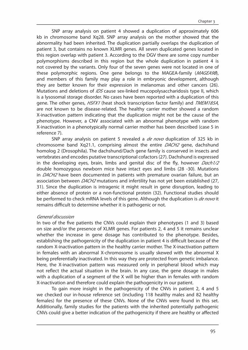
SNP array analysis on patient 4 showed a duplication of approximately 606 kb in chromosome band Xq28. SNP array analysis on the mother showed that the abnormality had been inherited. The duplication partially overlaps the duplication of patient 3, but contains no known XLMR genes. All seven duplicated genes located in this region overlap with patient 3. According to the DGV there are some copy number polymorphisms described in this region but the whole duplication in patient 4 is not covered by the variants. Only four of the seven genes were not located in one of these polymorphic regions. One gene belongs to the MAGEA-family (MAGEA9B), and members of this family may play a role in embryonic development, although they are better known for their expression in melanomas and other cancers (26). Mutations and deletions of IDS cause sex-linked mucopolysaccharidosis type II, which is a lysosomal storage disorder. No cases have been reported with a duplication of this gene. The other genes, HSFX1 (heat shock transcription factor family) and TMEM185A, are not known to be disease-related. The healthy carrier mother showed a random X-inactivation pattern indicating that the duplication might not be the cause of the phenotype. However, a CNV associated with an abnormal phenotype with random X-inactivation in a phenotypically normal carrier mother has been described (case 5 in reference 7). SNP array analysis on patient 5 revealed a de novo duplication of 325 kb in chromosome band Xq21.1, comprising almost the entire DACH2 gene, dachshund homolog 2 (Drosophila). The dachshund/Dach gene family is conserved in insects and vertebrates and encodes putative transcriptional cofactors (27). Dachshund is expressed in the developing eyes, brain, limbs and genital disc of the fly, however Dach1/2 double homozygous newborn mice have intact eyes and limbs (28 -30). Mutations in DACH2 have been documented in patients with premature ovarian failure, but an association between DACH2 mutations and infertility has not yet been established (27, 31). Since the duplication is intragenic it might result in gene disruption, leading to either absence of protein or a non-functional protein (32). Functional studies should be performed to check mRNA levels of this gene. Although the duplication is de novo it remains difficult to determine whether it is pathogenic or not.
General discussionIn two of the five patients the CNVs could explain their phenotypes (1 and 3) based on size and/or the presence of XLMR genes. For patients 2, 4 and 5 it remains unclear whether the increase in gene dosage has contributed to the phenotype. Besides, establishing the pathogenicity of the duplication in patient 4 is difficult because of the random X-inactivation pattern in the healthy carrier mother. The X-inactivation pattern in females with an abnormal X-chromosome is usually skewed with the abnormal X being preferentially inactivated. In this way they are protected from genetic imbalance. Here, the X-inactivation pattern was measured only in peripheral blood which may not reflect the actual situation in the brain. In any case, the gene dosage in males with a duplication of a segment of the X will be higher than in females with random X-inactivation and therefore could explain the pathogenicity in our patient. To gain more insight in the pathogenicity of the CNVs in patient 2, 4 and 5 we checked our in-house reference set (including 118 healthy males and 82 healthy females) for the presence of these CNVs. None of the CNVs were found in this set. Additionally, family studies for the patients with the inherited potentially pathogenic CNVs could give a better indication of the pathogenicity if there are healthy or affected
96
X-chromosome duplications
male carriers. Furthermore, the DGV should be regularly checked for these CNVs, since the data in this database is increasing rapidly. More patients or normal individuals with overlapping X chromosomal duplications will be needed to elucidate whether the CNVs that we have reported are pathogenic or benign variants In conclusion, this study highlights the difficulty in pinpointing the underlying causes of disease. Nevertheless knowing whether a CNV is pathogenic or benign is of major importance for counselling and diagnosis.
AcknowledgementsWe thank the patients and their parents for their cooperation and Patrick van Bunderen and Martine van Belzen for X-inactivation studies. We thank Dr. K. Madan for critical comments on the manuscript.
Supplementary materialPatient descriptionsPatient 1 was a 33-year old male. He was born by caesarian section at 43 weeks of gestation following a normal pregnancy. The parents were healthy and non-consanguineous. Birth weight was 2460 g. He had bradycardia and hypotonia and assisted ventilation had been necessary for a short period. A submucous cleft of the palate was closed before the age of 1 year. As an infant he developed myoclonic jerks which stopped spontaneously after a few years. His psychomotor development was delayed, he was able to sit without support at 15 months. His dental development was abnormal: at the age of 11 he had only 5 teeth, several incisors and molars had never appeared. Examination at the age of 20 showed: length 182 cm, span 170 cm and head circumference 56 cm. He had a friendly deposition and was able to speak a few single words. He had a long and slender habitus. The face was elongated with a relatively small mouth and a flat midface (Fig. 3.3.1a). The uvula was bifid. The maxillary lateral incisors were absent. His hands showed bilateral simian creases and long fingers. He had scoliosis (Fig. 3.3.1b). His feet were long and thin (Fig. 3.3.1c). The 31-year old younger brother of patient 1 had a similar phenotype. He was born by caesarian section at 34 weeks. Birth weight was 1610 g. Assisted ventilation was necessary in the neonatal period because of respiratory distress syndrome. An open ductus Botalli closed after medication. Like his brother he developed myoclonic jerks, which ceased spontaneously before the age of four. Also as in his brother the psychomotor development was delayed, he walked independently at the age of four. Neurological examination at the age of 16 years showed a spastic gait. On examination at the age of 18 years his length was 190 cm, span 187 cm and head circumference 54 cm. He spoke a few words and communicated with signing. The face was somewhat asymmetric (Fig. 3.3.1d). The maxillary lateral incisors were absent. He had pectus excavatum and scoliosis (Fig. 3.3.1e). The hand creases were normal. The feet were flat, thin and long with long first digits (Fig 3.3.1f ).
Patient 2 was a boy, born at term after an uncomplicated pregnancy. He had a normal birth weight. His development has been delayed form infancy, he was able to walk at the age of 4 years and he has no speech. He has a high threshold for pain. He has no sleeping problems. When first seen at the age of 4 years, he had a happy disposition and made stereotypic movements with his head and hands. Height, weight and head
97
Chapter 3
References1. Roeleveld N, Zielhuis GA, Gabreëls F. 1997. The prevalence of mental retardation: a critical review of
recent literature. Dev Med Child Neurol 39:125-132.2. Leonard H, Wen X. 2002. The epidemiology of mental retardation: challenges and opportunities in
the new millennium. Ment Retard Dev Disabil Res Rev 8:117-134.3. Mandel JL, Chelly J. 2005. Monogenic X-linked mental retardation: is it as frequent as currently
estimated? The paradox of the ARX (Aristaless X) mutations. Eur J Hum Genet 12:689-693. 4. Ropers HH, Hamel BC. 2005. X-linked mental retardation. Nat Rev Genet 6:46-57. 5. Chiurazzi P, Schwartz CE, Gecz J, Neri G. 2008. XLMR genes: update 2007. Eur J Hum Genet 16:422-
434.6. Madrigal I, Rodríguez-Revenga L, Armengol L, González E, Rodriguez B, Badenas C, Sánchez A,
Martínez F, Guitart M, Fernández I, Arranz JA, Tejada M, Pérez-Jurado LA, Estivill X, Milà M. 2007. X-chromosome tiling path array detection of copy number variants in patients with chromosome X-linked mental retardation. BMC Genomics 8:443.
7. Froyen G, Van Esch H, Bauters M, Hollanders K, Frints SG, Vermeesch JR, Devriendt K, Fryns JP, Marynen P. 2007. Detection of genomic copy number changes in patients with idiopathic mental retardation by high-resolution X-array-CGH: important role for increased gene dosage of XLMR genes. Hum Mutat 28:1034-1042.
8. Bauters M, Weuts A, Vandewalle J, Nevelsteen J, Marynen P, Van Esch H, Froyen G. 2008. Detection and validation of copy number variation in X-linked mental retardation. Cytogenet Genome Res 123:44-53.
10. Gijsbers AC, Lew JY, Bosch CA, Schuurs-Hoeijmakers JH, van Haeringen A, den Hollander NS, Kant SG, Bijlsma EK, Breuning MH, Bakker E, Ruivenkamp CA. 2009. A new diagnostic workflow for patients with mental retardation and/or multiple congenital abnormalities: test arrays first. Eur J Hum Genet 17:1394-1402
circumference were within normal limits. He had several dysmorphic features, including bitemporal narrowing, narrow palpebral fissures, deep set eyes, large mouth, and widely spaced teeth. He had a broad-based gait.
Patient 3 was born at 41 weeks of gestation with a normal birth weight (3350 g). The pregnancy had been complicated by pre-eclampsia. He was admitted to the department of pediatrics for observation because of hypotonia. His skin was very dry. There were no feeding problems during infancy. He did not speak any words at the age of three. At 13 years of age he was found to be severely retarded. He displayed palilalia and was not toilet-trained. His height was normal. His facial features were slightly dysmorphic with oval eyes, a short nose and a relatively small chin (Fig. 3.3.2a). He had inverted nipples, gynaecomastia, and an unusual subcutaneous fat distribution with large pads of fat above the buttocks.
Patient 4 was born at 41 weeks of gestation with a normal birth weigth (3930 g). He had an early closure of the fontanelles and some facial dysmorphic features, including trigonocephaly with narrow and sloping forehead and metopic ridge, flat face, upslant and short palpebral fissures with telecanthus and epicanthic folds, hypotelorism, infra-orbital creases, wide nasal bridge. He had proximal placement of thumbs, single palmar creases, fetal finger pads, and widely-spaced nipples.
Patient 5 was born at 40 weeks of gestation with a normal birth weight (3050 g). He had hypotonia and moderate MR. His facial features were very similar to those of his father with deep set small eyes, hypotelorism, narrow nose and small mouth.
98
X-chromosome duplications
11. Kubota T, Nonoyama S, Tonoki H, Masuno M, Imaizumi K, Kojima M, Wakui K, Shimadzu M, Fukushima Y. 1999. A new assay for the analysis of X-chromosome inactivation based on methylation-specific PCR. Hum Genet 104:49–55.
12. Orstavik KH. 2009. X chromosome inactivation in clinical practice. Hum Genet 126: 363-373.13. Lee C, Iafrate AJ, Brothman AR. 2007. Copy number variations and clinical cytogenetic diagnosis of
constitutional disorders. Nat Genet 39: S48-54.14. Tzschach A, Chen W, Erdogan F, Hoeller A, Ropers HH, Castellan C, Ullmann R, Schinzel A. 2008.
Characterization of interstitial Xp duplications in two families by tiling path array CGH. Am J Med Genet A 146A:197-203.
15. Plenge RM, Stevenson RA, Lubs HA, Schwartz CE, Willard HF. 2002. Skewed X-chromosome inactivation is a common feature of X-linked mental retardation disorders. Am J Hum Genet 71: 168-173.
16. Fukami M, Kirsch S, Schiller S, Richter A, Benes V, Franco B, Muroya K, Rao E, Merker S, Niesler B, Ballabio A, Ansorge W, Ogata T, Rappold GA. 2000. A member of a gene family on Xp22.3, VCX-A, is deleted in patients with X-linked nonspecific mental retardation. Am J Hum Genet 67:563-573.
17. Van Esch H, Hollanders K, Badisco L, Melotte C, Van Hummelen P, Vermeesch JR, Devriendt K, Fryns JP, Marynen P, Froyen G. 2005. Deletion of VCX-A due to NAHR plays a major role in the occurrence of mental retardation in patients with X-linked ichthyosis. Hum Mol Genet 14:1795-1803.
18. Cuevas-Covarrubias SA, González-Huerta LM. 2008. Analysis of the VCX3A, VCX2 and VCX3B genes shows that VCX3A gene deletion is not sufficient to result in mental retardation in X-linked ichthyosis. Br J Dermatol 158:483-486.
19. Lawson-Yuen A, Saldivar JS, Sommer S, Picker J. 2008. Familial deletion within NLGN4 associated with autism and Tourette syndrome. Eur J Hum Genet 16:614-618.
20. Rio M, Malan V, Boissel S, Toutain A, Royer G, Gobin S, Morichon-Delvallez N, Turleau C, Bonnefont JP, Munnich A, Vekemans M, Colleaux L. 2010. Familial interstitial Xq27.3q28 duplication encompassing the FMR1 gene but not the MECP2 gene causes a new syndromic mental retardation condition. Eur J Hum Genet 18:285-290.
21. Iafrate AJ, Feuk L, Rivera MN, Listewnik ML, Donahoe PK, Qi Y, Scherer SW, Lee C. 2004. Detection of large-scale variation in the human genome. Nat Genet 36:949-951.
22. Sebat J, Lakshmi B, Troge J, Alexander J, Young J, Lundin P, Månér S, Massa H, Walker M, Chi M, Navin N, Lucito R, Healy J, Hicks J, Ye K, Reiner A, Gilliam TC, Trask B, Patterson N, Zetterberg A, Wigler M. 2004. Large-scale copy number polymorphism in the human genome. Science 305:525-528.
23. Redon R, Ishikawa S, Fitch KR, Feuk L, Perry GH, Andrews TD, Fiegler H, Shapero MH, Carson AR, Chen W, Cho EK, Dallaire S, Freeman JL, González JR, Gratacòs M, Huang J, Kalaitzopoulos D, Komura D, MacDonald JR, Marshall CR, Mei R, Montgomery L, Nishimura K, Okamura K, Shen F, Somerville MJ, Tchinda J, Valsesia A, Woodwark C, Yang F, Zhang J, Zerjal T, Zhang J, Armengol L, Conrad DF, Estivill X, Tyler-Smith C, Carter NP, Aburatani H, Lee C, Jones KW, Scherer SW, Hurles ME. 2006. Global variation in copy number in the human genome. Nature 444:444-454.
24. Ababou A, Pfuhl M, Ladbury JE. 2009. Novel insights into the mechanisms of CIN85 SH3 domains binding to Cbl proteins: solution-based investigations and in vivo implications. J Mol Biol 387:1120-1136.
25. Tarpey PS, Smith R, Pleasance E, Whibley A, Edkins S, Hardy C, O’Meara S, Latimer C, Dicks E, Menzies A, Stephens P, Blow M, Greenman C, Xue Y, Tyler-Smith C, Thompson D, Gray K, Andrews J, Barthorpe S, Buck G, Cole J, Dunmore R, Jones D, Maddison M, Mironenko T, Turner R, Turrell K, Varian J, West S, Widaa S, Wray P, Teague J, Butler A, Jenkinson A, Jia M, Richardson D, Shepherd R, Wooster R, Tejada MI, Martinez F, Carvill G, Goliath R, de Brouwer AP, van Bokhoven H, Van Esch H, Chelly J, Raynaud M, Ropers HH, Abidi FE, Srivastava AK, Cox J, Luo Y, Mallya U, Moon J, Parnau J, Mohammed S, Tolmie JL, Shoubridge C, Corbett M, Gardner A, Haan E, Rujirabanjerd S, Shaw M, Vandeleur L, Fullston T, Easton DF, Boyle J, Partington M, Hackett A, Field M, Skinner C, Stevenson RE, Bobrow M, Turner G, Schwartz CE, Gecz J, Raymond FL, Futreal PA, Stratton MR. 2009. A systematic, large-scale resequencing screen of X-chromosome coding exons in mental retardation. Nat Genet 41:535-543.
26. De Plaen E, Arden K, Traversari C, Gaforio J J, Szikora JP, De Smet C, Brasseur F, van der Bruggen P, Lethe B, Lurquin C, Brasseur R, Chomez P, De Backer O, Cavenee W, Boon T. 1994. Structure, chromosomal localization, and expression of 12 genes of the MAGE family. Immunogenetics 40:360-369.
27. Davis RJ, Harding M, Moayedi Y, Mardon G. 2008. Mouse Dach1 and Dach2 are redundantly required for Müllerian duct development. Genesis 46:205-213.
28. Mardon G, Solomon NM, Rubin GM. 1994. Dachshund encodes a nuclear protein required for normal eye and leg development in Drosophila. Development 120:3473-3486.
99
Chapter 3
29. Keisman EL, Baker BS. 2001. The Drosophila sex determination hierarchy modulates wingless and decapentaplegic signaling to deploy dachshund sex-specifically in the genital imaginal disc. Development 128:1643-1656.
30. Davis RJ, Pesah YI, Harding M, Paylor R, Mardon G. 2006. Mouse Dach2 mutants do not exhibit gross defects in eye development or brain function. Genesis 44:84-92.
31. Bione S, Rizzolio F, Sala C, Ricotti R, Goegan M, Manzini MC, Battaglia R, Marozzi A, Vegetti W, Dalprà L, Crosignani PG, Ginelli E, Nappi R, Bernabini S, Bruni V, Torricelli F, Zuffardi O, Toniolo D. 2004. Mutation analysis of two candidate genes for premature ovarian failure, DACH2 and POF1B. Hum Reprod 19(12):2759-2766.
32. Thienpont B, de Ravel T, Van Esch H, Van Schoubroeck D, Moerman P, Vermeesch JR, Fryns JP, Froyen G, Lacoste C, Badens C, Devriendt K. 2007. Partial duplications of the ATRX gene cause the ATR-X syndrome. Eur J Hum Genet 15:1094-1097.