High-Performance Liquid Chromatography (HPLC ...
10
High-Performance Liquid Chromatography (HPLC) Quantification of Liposome- Delivered Doxorubicin in Arthritic Joints of Collagen-Induced Arthritis Rats The Harvard community has made this article openly available. Please share how this access benefits you. Your story matters Citation Niu, Hongqing, Menghua Xu, Shuangtian Li, Junwei Chen, Jing Luo, Xiangcong Zhao, Chong Gao, and Xiaofeng Li. 2017. “High- Performance Liquid Chromatography (HPLC) Quantification of Liposome-Delivered Doxorubicin in Arthritic Joints of Collagen-Induced Arthritis Rats.” Medical Science Monitor Basic Research 23 (1): 150-158. doi:10.12659/MSMBR.904103. http:// dx.doi.org/10.12659/MSMBR.904103. Published Version doi:10.12659/MSMBR.904103 Citable link http://nrs.harvard.edu/urn-3:HUL.InstRepos:32630600 Terms of Use This article was downloaded from Harvard University’s DASH repository, and is made available under the terms and conditions applicable to Other Posted Material, as set forth at http:// nrs.harvard.edu/urn-3:HUL.InstRepos:dash.current.terms-of- use#LAA
Transcript of High-Performance Liquid Chromatography (HPLC ...

High-Performance Liquid Chromatography(HPLC) Quantification of Liposome-
Delivered Doxorubicin in Arthritic Jointsof Collagen-Induced Arthritis Rats
The Harvard community has made thisarticle openly available. Please share howthis access benefits you. Your story matters
Citation Niu, Hongqing, Menghua Xu, Shuangtian Li, Junwei Chen, JingLuo, Xiangcong Zhao, Chong Gao, and Xiaofeng Li. 2017. “High-Performance Liquid Chromatography (HPLC) Quantificationof Liposome-Delivered Doxorubicin in Arthritic Joints ofCollagen-Induced Arthritis Rats.” Medical Science Monitor BasicResearch 23 (1): 150-158. doi:10.12659/MSMBR.904103. http://dx.doi.org/10.12659/MSMBR.904103.
Published Version doi:10.12659/MSMBR.904103
Citable link http://nrs.harvard.edu/urn-3:HUL.InstRepos:32630600
Terms of Use This article was downloaded from Harvard University’s DASHrepository, and is made available under the terms and conditionsapplicable to Other Posted Material, as set forth at http://nrs.harvard.edu/urn-3:HUL.InstRepos:dash.current.terms-of-use#LAA

Received: 2016.12.16Accepted: 2017.03.02
Published: 2017.04.14
High-Performance Liquid Chromatography (HPLC) Quantification of Liposome-Delivered Doxorubicin in Arthritic Joints of Collagen-Induced Arthritis Rats
ABEF 1 Hongqing Niu ABC 1 Menghua Xu BF 1 Shuangtian Li AD 1 Junwei Chen CG 1 Jing Luo BC 1 Xiangcong Zhao AF 2 Chong Gao AEG 1 Xiaofeng Li
Corresponding Author: Xiaofeng Li, e-mail: [email protected] Source of support: This work was supported by the Postgraduate Education and Innovation Program of the Education Department of Shanxi Province,
China (Number 2016BY085) and the Scientific Research Foundation of Health and Family Planning Commission of Shanxi Province, China (Number 201601136)
Background: Neoangiogenesis occurring in inflamed articular synovium in early rheumatoid arthritis (RA) is characterized by enhanced vascular permeability that allows nanoparticle agents, including liposomes, to deliver encapsulated drugs to arthritic joints and subsequently improve therapeutic efficacy and reduce adverse effects. However, the targeting distribution of liposomes in arthritic joints during RA has not been quantitatively demonstrated. We performed this study to evaluate the targeting distribution of PEGylated doxorubicin liposomes in the ar-thritic joints of collagen-induced arthritis (CIA) rats by high-performance liquid chromatography (HPLC).
Material/Methods: Two doxorubicin formulations were administered to CIA rats via tail intravenous injection at a single dose (50 mg/m2). CIA rats were sacrificed and the tissues of the inflamed ankle joints were collected. The content of doxorubicin in the arthritic joints was analyzed by a validated and reproducible HPLC method. A two-way ANOVA for 2×5 factorial design was used for statistical analysis.
Results: The developed HPLC method was sensitive, precise, and reproducible. The method was successfully applied to quantify doxorubicin content in arthritic tissues. At each time point (6, 12, 24, 48, and 72 h), doxorubicin con-tent in the arthritic joints of the doxorubicin liposome group (DOX-LIP group) was higher than in the free doxo-rubicin group (DOX group) (P<0.05). In the DOX-LIP group, doxorubicin levels in the arthritic joints increased gradually and significantly in the interval of 6–72 h post-administration.
Conclusions: PEGylated doxorubicin liposomes were targeted to, accumulated, and retained in the arthritic joints of CIA rats. The present study indicates that liposome encapsulation increases the therapeutic efficacy of antirheumatic drugs, presenting a promising therapeutic strategy for RA.
MeSH Keywords: Arthritis, Experimental • Arthritis, Rheumatoid • Chromatography, High Pressure Liquid • Doxorubicin • Drug Delivery Systems • Liposomes
Full-text PDF: http://www.basic.medscimonit.com/abstract/index/idArt/904103
Authors’ Contribution: Study Design A
Data Collection B Statistical Analysis CData Interpretation D
Manuscript Preparation E Literature Search FFunds Collection G
1 Department of Rheumatology, The Second Hospital of Shanxi Medical University, Taiyuan, Shanxi, P.R. China
2 Department of Pathology, Brigham and Women’s Hospital, Harvard Medical School, Boston, MA, U.S.A.
3842 3 3 26
eISSN 2325-4416© Med Sci Monit Basic Res, 2017; 23: 150-158
DOI: 10.12659/MSMBR.904103
150
ANIMAL STUDIES
Indexed in: [Index Medicus/MEDLINE] [EMBASE/Excerpta Medica] [Chemical Abstracts/CAS] [Index Copernicus]
This work is licensed under Creative Common Attribution-NonCommercial-NoDerivatives 4.0 International (CC BY-NC-ND 4.0)

Background
Rheumatoid arthritis (RA) is a chronic immune-mediated dis-ease characterized by inflammation of synovial tissues, even-tually resulting in joint destruction and subsequent loss of function. Neoangiogenesis occurring in the early stage of the disease is an important histopathologic change in RA, neces-sary for the development of synovium pannus [1,2] and lead-ing to the destruction of articular cartilage and bone. Current therapeutic strategies for RA include reducing disease activi-ty, minimizing joint damage, and improving physical function and prognosis [3,4]. Because of low bioavailability and limited tissue selectivity, several important drugs for RA treatment re-quire high doses and frequent administration to achieve suffi-cient therapeutic efficiency [5], increasing the risk of system-ic adverse effects.
Liposomes are nanoparticle lipid vesicles that are composed of continuous bilayers of phospholipids surrounding an aqueous phase, and have been investigated as drug delivery systems for improved targeted delivery of therapeutic agents [5]. Typical li-posomal formulations containing polyethylene glycol (PEG) in-crease the stability of liposomes in circulation and avoid rap-id elimination by the mononuclear phagocyte system (MPS) of the liver and the spleen, resulting in increased localization at the cancer/inflamed sites, caused by the enhanced perme-ability and retention (EPR) effect [5]. After systemic adminis-tration, liposomal systems deliver the encapsulated drugs to the cancer/inflamed sites by ‘passive targeting’ [6].
Neovascularization occurring at the articular synovium in ear-ly RA is characterized by enhanced permeability of the cap-illaries, allowing small, PEGylated, liposomal formulations to permeate the inflamed sites and be retained in the extravas-cular space via the EPR effect [7,8]. Subsequently, a large pro-portion of liposomes are taken up by macrophages in the in-flamed synovium [9]. Passive targeting and the EPR effect make long-circulating PEGylated liposomal drug formulations attractive for increasing the therapeutic efficiency of the an-tirheumatic drugs.
At present, only a few liposomal products, including doxorubicin liposomes, are used in clinics for cancer therapy. Leaky blood vessels in cancer and inflamed synovial tissue share common properties [10,11], indicating that liposomes could be used in clinical settings for RA. Chemotherapeutic agents, including methotrexate and azathioprine, are widely used in the treat-ment of RA. When powerful chemotherapeutic agents such as doxorubicin and cyclophosphamide are delivered selectively to inflamed synovial tissues using nanoparticles, their therapeutic efficacy is improved, whereas toxicity is reduced, as systemic exposure to the drug is minimized [12]. This study examined PEGylated doxorubicin liposome targeting of inflamed joints
after intravenous administration in collagen-induced arthritis (CIA) rats, a classical animal model of RA.
High-performance liquid chromatography (HPLC) is a sensitive and precise analytical method for quantifying the content of drugs in biological specimens. Determination of doxorubicin concentration in different biological samples utilizing HPLC has been previously reported [13,14]. However, HPLC coupled with a UV detector has the disadvantage of a high detection lim-it (µM levels or higher). As a detector, the mass spectrometer has a low detection limit (nM levels or lower); however, special equipment is required that may not be affordable for all labora-tories. In contrast, the fluorescence detector is affordable and it was shown to display sufficient sensitivity and precision in de-termining the content of doxorubicin in biological samples [14].
In this paper, we report a fully validated, reproducible HPLC method for sensitive doxorubicin quantification in arthritic joints, which employs gradient elution and is coupled with a fluorescence detector. The reversed-phase HPLC method de-scribed determines the sum total content of doxorubicin (en-capsulated and released) in tissues and was modified from previous studies [13–16]. In addition, we evaluated and com-pared the difference between PEGylated liposomal doxorubi-cin and free doxorubicin targeting the inflamed joints of CIA rats. To the best of our knowledge, we are the first group to quantitatively demonstrate with HPLC that PEGylated liposo-mal formulations accumulate in the inflamed joints.
Material and Methods
Chemicals and reagents
PEGylated doxorubicin hydrochloride liposome injections were obtained from Changzhou Kinyond Manufacturing Co., Ltd. (Jintan, Jiangsu, China). Doxorubicin hydrochloride for injection was purchased from Zhejiang Hisun Pharmaceutical Co., Ltd. (Taizhou, Zhejiang, China). Immunization-grade bovine type II collagen, complete Freund’s adjuvant containing 2 mg/mL M. tuberculosis, and incomplete Freund’s adjuvant were purchased from Chondrex, Inc (Redmond, WA, USA). Other chemicals were of analytical grade and obtained from commercial sources.
Doxorubicin hydrochloride and daunorubicin hydrochloride (in-ternal standard, IS) were purchased from the National Institutes for Food and Drug Control (Beijing, China). Acetonitrile and methanol were of HPLC grade and purchased from Tianjin Shield Fine Chemicals Co., Ltd (Tianjin, China). Ammonium ac-etate and glacial acetic acid were of analytical grade and ob-tained from Tianjin Hengxing Chemical Reagent Co., Ltd (Tianjin, China). Ultrapure water was obtained using a Millipore Milli-Q Academic system (Millipore Corporation, Bedford, MA, USA).
151
Niu H. et al.: Targeting distribution of liposomes in CIA rats© Med Sci Monit Basic Res, 2017; 23: 150-158
ANIMAL STUDIES
Indexed in: [Index Medicus/MEDLINE] [EMBASE/Excerpta Medica] [Chemical Abstracts/CAS] [Index Copernicus]
This work is licensed under Creative Common Attribution-NonCommercial-NoDerivatives 4.0 International (CC BY-NC-ND 4.0)

HPLC instrumentation and system conditions
The HPLC system consisted of a Waters e2695 separations module (Waters, USA) and a Waters 2475 multi-l fluorescence detector (Waters, USA). The chromatographic column was a Waters Symmetry C18 column (4.6×150 mm, 5 μm, Waters, USA). Chromatographic data were acquired by Empower 2 software from Waters.
The optimized mobile phases consisted of 5 mmol/L aqueous ammonium acetate containing 0.5% (v/v) glacial acetic acid (A) and acetonitrile (B) filtered through a membrane filter (0.2 μm) prior to use. The mobile-phase gradient program (A/B) was 80: 20 (t=0 min), 65: 35 (t=2 min), and 80: 20 (t=8 min) and was applied over 10 min at a flow rate of 0.4 mL/min. The column temperature was maintained at 30°C. The excitation and emission wavelengths were 470 and 590 nm, respective-ly. The injection volume was 20 µL.
Induction of CIA and treatment procedures
Wistar rats (6–8 weeks old) were purchased from Beijing Weitonglihua Laboratory Animal Co., Ltd., Beijing, China (License Number: SCXK Beijing 2012-0001) and housed under specif-ic pathogen-free (SPF) conditions with a 12 h light/dark cycle at ambient temperature (21–25°C), with water provided ad libitum. The rats were acclimatized for 1 week in cages pri-or to model induction. Afterwards, the rats were injected in-tradermally at the tail base with 200 μg bovine type II colla-gen emulsified in Freund’s complete adjuvant (1: 1, v/v). Two weeks later, the rats were given intradermal booster injections of 100 μg bovine type II collagen in incomplete Freund’s adju-vant (1: 1, v/v) [17,18].
The arthritis index (AI) was monitored and ankle joint structure was analyzed histologically to determine the development of CIA. Rats were examined at least 2 times per week for signs of arthritis onset, based on paw swelling and AI score. Lateral joint diameters were measured using a vernier caliper. Each paw was evaluated by 2 independent observers and the total grades were summed, yielding a maximum AI score of 16 for each animal [17]. Ten days after the second injection of type II collagen, CIA development was declared in rats with the AI score ³8. CIA and normal rats were sacrificed and the inflamed ankles were fixed with 10% neutral formalin for 24 h and de-calcified with 10% EDTA for 30 days. The ankles were incised longitudinally, embedded with paraffin, and stained with he-matoxylin-eosin (HE) to observe joint inflammation. Slides were analyzed with an Olympus BX51 microscope and photo-graphed with an Olympus digital camera. Pathological mani-festations are shown in Figure 1.
The CIA rats were randomly allocated into the doxorubicin lipo-some group (DOX-LIP group, n=30, male: female=1: 1) and the free doxorubicin group (DOX group, n=30, male: female=1: 1). Doxorubicin liposomes or free doxorubicin at a single dose of 50 mg/m2 were prepared as a solution containing doxorubi-cin (1 mg/mL) in 5% glucose solution and administered to the CIA rats via tail intravenous injection. The CIA model rats (n=6) were sacrificed at predetermined time points (6, 12, 24, 48, and 72 h) after intravenous administration and the tissues of the inflamed ankle joints were collected, homogenized in normal saline, and blotted with filter paper to remove residual blood. All samples were stored at –80°C for further analysis. Tissues from untreated mice (n=6) were collected for the preparation of standard curves. A 2×5 factorial design was used to deter-mine the contents of doxorubicin in the 2 groups.
All procedures involving animals were carried out in accor-dance with the guidelines and regulations approved by the Ethics Committee for Animal Experiments of Shanxi Medical University, Taiyuan, China.
Sample preparation
The tissue of the inflamed ankle joints was ground in a mor-tar with liquid nitrogen. The obtained powder was weighed precisely with a Sartorius scale (BSA224S, Sartorius Scientific Instruments (Beijing) Co., Limited) and mixed with 0.5 mL of ultrapure water. IS solution (40 µL, 10 µg/mL daunorubicin hydrochloride in methanol) was added to the mixture above and vortex-mixed for 2 min. Drug extraction was performed by adding 1 mL of methanol. After vortex-mixing for 2 min and centrifuging at 12 000 rpm (10 303× g) for 10 min, the organic layer was transferred into a clean tube and evaporat-ed to dryness at 50°C under a gentle stream of nitrogen. The residue was resuspended in methanol (100 µL), vortex-mixed for 2 min, and centrifuged at 12 000 rpm (10 303× g) for 10 min. Afterwards, the supernatant (20 µL) was injected onto the chromatographic column for analysis.
Determination of doxorubicin content
Preparation of stock and working solutions
The primary stock solutions of doxorubicin hydrochloride and IS were prepared in methanol at the concentration of 2 mg/mL. The stock solutions were stored at 4°C. The working solutions of doxorubicin were freshly prepared by appropriate dilution of the stock solutions with methanol. Working solutions con-taining 10 µg/mL IS were freshly prepared by serial dilution of stock solutions with methanol.
152
Niu H. et al.: Targeting distribution of liposomes in CIA rats
© Med Sci Monit Basic Res, 2017; 23: 150-158ANIMAL STUDIES
Indexed in: [Index Medicus/MEDLINE] [EMBASE/Excerpta Medica] [Chemical Abstracts/CAS] [Index Copernicus]
This work is licensed under Creative Common Attribution-NonCommercial-NoDerivatives 4.0 International (CC BY-NC-ND 4.0)

Preparation of calibration standards
Calibration standards and quality control (QC) samples were prepared by spiking 500 µL of the homogenized tissue samples of drug-free inflamed joints with 50 µL of doxorubicin solution or 40 µL of IS (10 µg/mL). The samples were processed follow-ing the ‘Sample preparation’ procedure described above and analyzed. The final concentrations of doxorubicin in the calibra-tion samples used to draw the standard curve were 0.8, 3, 8, 24, 50, 80, and 120 µg/mL. The peak-area ratios of doxorubicin to IS (Y) and the concentrations of doxorubicin (X) were used to plot the calibration curves. The linear correlation coefficient (r) was required to be equal to or greater than 0.98 [19–21].
Validation of the analytical method
Precision and accuracy. To evaluate the intra- and inter-day precision of the method, QC samples spiked with
doxorubicin solutions at 5 concentration levels (0.8, 3, 12, 60, and 120 µg/mL) were prepared following the steps indicated in the ‘Sample preparation’ section. The intra-day precision was determined by analyzing 6 replicates of tissue samples at each concentration level. The inter-day precision was deter-mined by analyzing the samples at each concentration level for 3 consecutive days. The precision and accuracy of the method were expressed by the relative standard deviation (RSD%) and relative error (RE%), respectively, at each concentration level. Variation for precision and accuracy was required to be < 15%.
Recovery. The extraction recovery was determined by compar-ing the peak areas of doxorubicin and IS in the QC samples spiked with standard solutions prior to extraction with peak areas from samples to which standard solutions were added after extraction. The extraction recovery of doxorubicin was performed at 3 concentration levels (0.8, 12, and 60 µg/mL), whereas the concentration of IS was 10 µg/mL. The samples
A
C
B
D
Figure 1. Histopathological manifestations of ankles in normal and CIA rats. (A, B) Synovial membrane is clear in normal rats, without hyperplasia and infiltration of inflammatory cells. (C, D) Altered synovial membrane in CIA rats, with proliferating synovial cells, infiltration of inflammatory cells, and pannus. (HE staining; A, C: ×40; B, D: ×100).
153
Niu H. et al.: Targeting distribution of liposomes in CIA rats© Med Sci Monit Basic Res, 2017; 23: 150-158
ANIMAL STUDIES
Indexed in: [Index Medicus/MEDLINE] [EMBASE/Excerpta Medica] [Chemical Abstracts/CAS] [Index Copernicus]
This work is licensed under Creative Common Attribution-NonCommercial-NoDerivatives 4.0 International (CC BY-NC-ND 4.0)
were processed as indicated in the ‘Sample preparation’ sec-tion and were analyzed 5 times for each concentration level using HPLC. The recovery was calculated according to the fol-lowing formula:
Recovery (%)=[(measured doxorubicin content)/ (added doxorubicin content)]×100%
Stability. The stability studies were performed as follows. Short-term stability was evaluated by spiking 500 µL of QC samples with 50 µL of working solutions and 40 µL of IS so-lution (10 µg/mL) to obtain final concentrations of 0.8, 12, and 60 µg/mL for doxorubicin. The samples were processed as described in the ‘Sample preparation’ section and kept at 21–25°C during routine sample handling time (at least 6 h). Freeze-thaw stability was assessed by analyzing the QC sam-ples spiked with doxorubicin solution at 3 different concen-trations (0.8, 12, and 60 µg/mL), after 3 freeze-and-thaw cy-cles, from – 80°C to room temperature. Stability is presented as a percent based on the measured concentration at the giv-en time point compared with the concentration measured at time zero (recovery) at the same concentration levels. All sta-bility studies were performed in 6 replicates. Deviations > ±5% from the initial concentration were considered unstable.
Application of the developed method to the targeting distribution study
To demonstrate that the liposomal drug formulations accumu-late in the inflamed joints due to passive targeting and EPR
effect, PEGylated doxorubicin liposomes or free doxorubicin were administered to the CIA rats via tail intravenous injec-tion. The developed HPLC method was applied to determine the content of doxorubicin in the inflamed joint tissues. The samples were stored at –80°C for further analysis. The HPLC method with fluorescence detection was used to measure the sum total doxorubicin (encapsulated and released) found in tissues. Doxorubicin content in inflamed joints was compared between the DOX-LIP and DOX groups.
Statistical analysis
Analysis was performed using SPSS 20.0 statistical software. Data are expressed as mean ± standard deviation (M ±SD). A two-way classification ANOVA for 2×5 factorial design was used for statistical analysis. P values of <0.05 were considered statistically significant.
Results
Determination of doxorubicin content
HPLC chromatograms and retention times
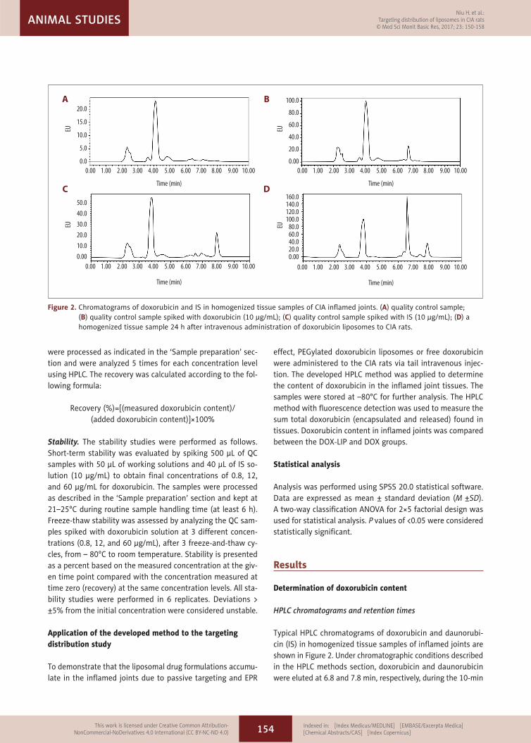
Typical HPLC chromatograms of doxorubicin and daunorubi-cin (IS) in homogenized tissue samples of inflamed joints are shown in Figure 2. Under chromatographic conditions described in the HPLC methods section, doxorubicin and daunorubicin were eluted at 6.8 and 7.8 min, respectively, during the 10-min
Figure 2. Chromatograms of doxorubicin and IS in homogenized tissue samples of CIA inflamed joints. (A) quality control sample; (B) quality control sample spiked with doxorubicin (10 µg/mL); (C) quality control sample spiked with IS (10 µg/mL); (D) a homogenized tissue sample 24 h after intravenous administration of doxorubicin liposomes to CIA rats.
Time (min)
EU
0.00
20.0
15.0
10.0
5.0
0.01.00 2.00 3.00 4.00 5.00 6.00 7.00 8.00 9.00 10.00
Time (min)
EU
0.00
50.0
40.0
30.0
20.0
10.0
0.001.00 2.00 3.00 4.00 5.00 6.00 7.00 8.00 9.00 10.00
Time (min)
EU
0.00
100.0
80.0
60.0
40.0
20.0
0.001.00 2.00 3.00 4.00 5.00 6.00 7.00 8.00 9.00 10.00
Time (min)EU
0.00
160.0140.0120.0100.0
80.060.040.020.00.00
1.00 2.00 3.00 4.00 5.00 6.00 7.00 8.00 9.00 10.00
A
C
B
D
154
Niu H. et al.: Targeting distribution of liposomes in CIA rats
© Med Sci Monit Basic Res, 2017; 23: 150-158ANIMAL STUDIES
Indexed in: [Index Medicus/MEDLINE] [EMBASE/Excerpta Medica] [Chemical Abstracts/CAS] [Index Copernicus]
This work is licensed under Creative Common Attribution-NonCommercial-NoDerivatives 4.0 International (CC BY-NC-ND 4.0)

run-time, and no significant interfering peaks of endogenous tissue components were observed (Figure 2).
Calibration curve, linearity, and the limit of quantification (LOQ)
Good linearity was observed over the concentration range of 0.8–120 µg/mL between peak-area ratios of doxorubicin to IS and the respective doxorubicin concentrations in QC sam-ples. The regression equation of the calibration curve was Y=0.4277 X–0.7058. The correlation coefficient was excellent (r2=0.9924). The LOQ of doxorubicin in inflamed joint tissues of CIA rats was 0.8 µg/mL.
Precision and accuracy
Intra- and inter-day precision of the method, expressed as relative standard deviation (RSD%), are presented in Table 1. The RSD values ranged from 3.16% to 7.01% for intra-day and 4.20% to 8.48% for inter-day analysis, indicating excellent pre-cision of the developed method. Accuracy, shown in Table 1 as a relative error (RE%), varied from 2.40% to 5.12% for in-tra-day and from – 2.07% to 5.13% for inter-day analysis. The method showed satisfactory accuracy.
Recovery
Recovery of doxorubicin was 76.6±4.9, 75.7±4.8, and 78.8±5.2%, at the concentrations of 0.8, 12, and 60 µg/mL, respectively, whereas the recovery of IS was 85.9±4.4% at 10 µg/mL. The
average recovery for doxorubicin in the tissues of inflamed joints was >75%, with RSD values of 6.39, 6.34, and 6.65%. RSD value for the recovery of IS was 5.18%. The results above indicate that extraction recoveries of doxorubicin and IS using methanol are satisfactory and highly reproducible.
Stability
Stability studies were performed to ensure the reliability of results related to handling and storing of inflamed joint tis-sue samples. As presented in Table 2, RSD of the stability re-sults were < 5%, indicating that doxorubicin was stable in the inflamed rat joint tissues for 6 h at ambient temperature and after 3 freeze-thaw cycles. No spontaneous degradation oc-curred during the routine analysis and storage of samples, un-der any of the tested conditions.
Application of the HPLC method to the targeting distribution study
The developed HPLC method was successfully applied to the determination of doxorubicin content in inflamed joint tissues of CIA rats after intravenous administration of doxorubicin li-posomes or free doxorubicin at a single dose of 50 mg/m2. The time profile of M ±SD of doxorubicin content in the arthritic joint tissues is presented in Table 3 and Figure 3.
To demonstrate whether doxorubicin content in the inflamed joint tissues after administration differed between PEGylated doxorubicin liposomes and free doxorubicin, a two-way
Concentration(µg/mL)
Intra-day (n=6) Inter-day (n=3)
Precision (RSD%) Accuracy (RE%) Precision (RSD%) Accuracy (RE%)
0.8 3.16 3.67 4.20 3.54
3 5.65 4.23 8.48 5.13
12 7.01 5.12 5.94 –2.07
60 4.34 4.07 6.58 2.25
120 4.18 2.40 5.32 2.50
Table 1. Intra- and inter-day precision and accuracy of doxorubicin quantification.
Concentration(µg/mL)
Short-term stability Freeze-thaw stability
Recovery (%) RSD (%) Recovery (%) RSD (%)
0.8 91.9±3.2 3.48 95.5±3.5 3.63
12 93.0±3.7 3.97 96.3±3.4 3.56
60 95.4±3.0 3.10 96.8±2.3 2.33
Table 2. Stability data for doxorubicin under different conditions (n=6).
155
Niu H. et al.: Targeting distribution of liposomes in CIA rats© Med Sci Monit Basic Res, 2017; 23: 150-158
ANIMAL STUDIES
Indexed in: [Index Medicus/MEDLINE] [EMBASE/Excerpta Medica] [Chemical Abstracts/CAS] [Index Copernicus]
This work is licensed under Creative Common Attribution-NonCommercial-NoDerivatives 4.0 International (CC BY-NC-ND 4.0)

classification ANOVA model for 2×5 factorial design was used to examine the interaction of drug formulations (PEGylated doxorubicin liposomes or free doxorubicin) and time (the sac-rifice time points of CIA rats). The ANOVA model showed a sig-nificant drug formulations * time interactive effect on changes in the content of doxorubicin (F=13.151, P<0.001).
The factorial analysis was restricted to the above outcome associated with significant drug formulations * time interac-tion. Mean changes in doxorubicin content were compared at each time point using t tests between the DOX-LIP and DOX group CIA rats. Mean levels of doxorubicin were compared among different time points in the same group using ANOVA and LSD t tests. The factorial analysis allowed us to establish which drug formulation accumulated better in the arthritic joints and how these differences changed over time. Analytical results indicated that in the DOX-LIP group, doxorubicin con-tent in the arthritic joints was significantly higher compared to the DOX group, at each time point (Table 3). In the DOX-LIP group, the levels of doxorubicin in the arthritic joints in-creased gradually with time. No significant differences were observed between doxorubicin levels 6 and 12 h post-admin-istration. However, doxorubicin levels in arthritic joints 24, 48, and 72 h after treatment differed significantly from levels
observed 6 h after administration. In the DOX group, doxo-rubicin content decreased gradually over time, but the differ-ences between time points were not significant (Figure 3 and Table 3). These results suggest that PEGylated liposomal drug formulations offer targeted delivery and retention of encap-sulated drugs at sites of inflammation.
Discussion
HPLC is a sensitive and precise analytical method for quanti-fying the content of drugs in biological tissues. Optimal chro-matographic conditions for determining the analyte in biological specimens should offer efficient separation of the analyte from other exogenous components, a favorable mobile phase with simple composition and appropriate pH value, a concise and efficient procedure, and excellent accuracy and precision [15].
In the present study, a fully validated and reproducible HPLC method employing gradient elution and coupled with conve-nient fluorescence detection was developed for determining the content of doxorubicin in inflamed joints of CIA rats after intravenous administration. As previously described, an opti-mal mobile phase consisted of 5 mmol/L aqueous ammonium acetate containing 0.5% (v/v) glacial acetic acid and acetoni-trile. The HPLC system conditions and procedures were easi-ly controlled. Under chromatographic conditions used in this study, doxorubicin was eluted at 6.8 min within 10-min run-time without any interfering endogenous tissue components. Peak areas and doxorubicin content displayed excellent lin-earity. Accuracy and precision were good and met the require-ments for the quantification of doxorubicin in arthritic joint tissues. The developed HPLC method displayed favorable ac-curacy and recovery and was suitable for extracting and de-termining the content of doxorubicin.
RA is associated with angiogenesis and inflammation of synovi-um that leads to tissue proliferation and joint destruction. Collagen-induced arthritis is a well-established experimental model that shows many similarities to human RA. Experimental
Time (h) N DOX-LIP group (µg/g) DOX group (µg/g) t value P value
6 6 0.976±0.127*#$ 0.545±0.126 2.2331 0.030
12 6 1.061±0.461#$ 0.465±0.062 3.0932 0.003
24 6 1.376±0.345#$ 0.356±0.060 5.2901 <0.001
48 6 2.147±0.551& 0.441±0.115 8.8393 <0.001
72 6 2.540±0.644 0.491±0.117 10.6165 <0.001
Table 3. Doxorubicin content in the arthritic joints of the 2 CIA rat groups (n=6).
*P<0.05 compared with 24 h; # P<0.001 compared with 48 h; $ P<0.001 compared with 72 h; & P<0.05 compared with 72 h.
6
3.5
3.0
2.5
2.0
1.5
1.0
0.5
0.0
12 24
Time (hours)
Cont
ent o
f dox
orub
icin
(μg/
g)
48 72
DOX groupDOX-LIP group
Figure 3. Time profile of doxorubicin content in the arthritic joints of the 2 groups (n=6).
156
Niu H. et al.: Targeting distribution of liposomes in CIA rats
© Med Sci Monit Basic Res, 2017; 23: 150-158ANIMAL STUDIES
Indexed in: [Index Medicus/MEDLINE] [EMBASE/Excerpta Medica] [Chemical Abstracts/CAS] [Index Copernicus]
This work is licensed under Creative Common Attribution-NonCommercial-NoDerivatives 4.0 International (CC BY-NC-ND 4.0)

evidence suggests that angiogenesis in the synovium is in-volved in the generation of collagen-induced arthritis [22–24]. Leaky capillaries present in the inflamed synovium and tumor tissue share some unique characteristics that allow liposomal drug formulations to extravasate into the inflammatory/tumor tissues and be retained in the extravascular space [10,11]. Targeted accumulation of liposomal drug formulations in tu-mor tissue has been previously demonstrated [13–15], where-as similar research on delivery of liposomal drug formulations to inflamed RA/CIA joints is rare. To the best of our knowledge, we quantified the amount of liposomal drug formulations in arthritic joints by using HPLC for the first time.
The significance of our study is a quantitative demonstration of the accumulation of PEGylated liposome formulations in in-flamed joints. After successful induction of arthritis, doxorubi-cin liposomes or free doxorubicin were administered intrave-nously to CIA rats. The amount of doxorubicin in the arthritic joints was determined using the developed HPLC method. The results show that the doxorubicin levels in the inflamed joints rapidly increased in the DOX-LIP group compared to the DOX group, with differences between the 2 groups significant at every time point. Doxorubicin content in the inflamed joints increased gradually over time in the DOX-LIP group, and the retention time of the PEGylated doxorubicin liposomes was longer. This phenomenon might be connected with the longer half-life, targeting, and sustained release effects of the lipo-somal drug formulation. The circulation time of the liposomal formulation in vivo was prolonged compared to the free drug and the bioavailability was improved due to the targeting and sustained release effects of the liposomal formulation in the in-flamed joints [5]. This effect might be attributed to the encap-sulation of the drug into PEGylated liposomes, which protect-ed the drug from rapid uptake by the mononuclear phagocyte system after administration. Liposomal encapsulation allowed the drug to be transported through vascular walls into inflam-matory tissues [5]. Our results suggest that liposomes can pas-sively target and accumulate in the arthritic joints due to in-creased vascular permeability of inflamed tissues.
Sandanaraj et al. [25] encapsulated indocyanine green (ICG), a clinically approved fluorescent dye, inside the liposomes and injected the fluorescent liposomes into mice with antigen-in-duced arthritis (AIA), an arthritis model also presenting leaky vasculature in the arthritic tissues. Their results showed a 2-fold increase in fluorescence from arthritic joints compared to non-arthritic joints after 24 h. The study indicated that nanopar-ticles passively target leaky vasculature and are retained in
arthritic joints for a longer time. This conclusion is in accor-dance with our results.
The vascular permeability of inflamed tissues differs between the acute and chronic stages in the RA model [26]. As a re-sult, the extravasation of liposomal formulations during the acute inflammatory phase is expected to be higher than in the chronic phase; therefore, the targeted accumulation of anti-rheumatic drugs encapsulated in liposomes should be higher in the acute phase. It should be noted that the present study has demonstrated the targeted delivery of the liposomal for-mulation to inflamed joints in the acute phase. Our study sug-gests that liposomes may be a promising drug delivery option for encapsulating antirheumatic drugs and targeting arthrit-ic joints in RA.
Conclusions
A sensitive, precise, and reproducible HPLC method with gra-dient elution and fluorescence detection was developed and used to quantify the content of doxorubicin in arthritic tis-sues. In this study, doxorubicin liposomes administered to CIA rats were decorated with PEG to enhance the stability of li-posomes and passively target the arthritic tissue. The signifi-cance of this study is that it, for the first time, quantitatively demonstrates that PEGylated liposome formulations can deliv-er drugs to inflamed arthritic joints. Our results indicate that liposomal formulations could be used as drug carriers, deliv-ering antirheumatic drugs to inflamed joints via leaky vascu-lature, increasing the therapeutic efficacy of the encapsulated drugs, and decreasing the exposure and toxicity to non-tar-geted organs and tissues. Taken together, these findings in-dicate that liposomes could present a new, promising thera-peutic strategy for RA.
Acknowledgments
We thank Mr Hua Guo and Mr Chen Zhang (Rheumatism Laboratory, The Second Hospital of Shanxi Medical University) for preparing the animals for RA model induction. We also thank Ms Jinju Duan and Mr Xuezhi Liu (Clinical Pharmacology Laboratory, The Second Hospital of Shanxi Medical University) for their support and assistance in HPLC detection.
Disclosure of interest
The authors report no conflicts of interest.
157
Niu H. et al.: Targeting distribution of liposomes in CIA rats© Med Sci Monit Basic Res, 2017; 23: 150-158
ANIMAL STUDIES
Indexed in: [Index Medicus/MEDLINE] [EMBASE/Excerpta Medica] [Chemical Abstracts/CAS] [Index Copernicus]
This work is licensed under Creative Common Attribution-NonCommercial-NoDerivatives 4.0 International (CC BY-NC-ND 4.0)

References:
1. Kelly S, Bombardieri M, Humby F et al: Angiogenic gene expression and vascular density are reflected in ultrasonographic features of synovitis in early rheumatoid arthritis: An observational study. Arthritis Res Ther, 2015; 17: 58
2. Yi JP, Wu YZ, Yu N et al: VEGF gene polymorphisms affect serum protein levels and alter disease activity and synovial lesions in rheumatoid arthri-tis. Med Sci Monit, 2016; 22: 316–24
3. Smolen JS, Breedveld FC, Burmester GR et al: Treating rheumatoid arthritis to target: 2014 update of the recommendations of an internation-al task force. Ann Rheum Dis, 2016; 75(1): 3–15
4. Monti S, Montecucco C, Bugatti S, Caporali R: Rheumatoid arthritis treat-ment: The earlier the better to prevent joint damage. RMD Open, 2015; 1(Suppl. 1): e000057
5. van den Hoven JM, Van Tomme SR, Metselaar JM et al: Liposomal drug for-mulations in the treatment of rheumatoid arthritis. Mol Pharm, 2011;8(4): 1002–15
6. Matsumoto Y, Ichihara H, Hino M et al: Therapeutic effects of hybrid li-posomes without drugs for rheumatoid arthritis. Drug Deliv, 2015; 22(5): 619–26
7. Reddy KR: Controlled-release, pegylation, liposomal formulations: new mechanisms in the delivery of injectable drugs. Ann Pharmacother, 2000; 34(7–8): 915–23
8. Vanniasinghe AS, Bender V, Manolios N: The potential of liposomal drug delivery for the treatment of inflammatory arthritis. Semin Arthritis Rheum, 2009; 39(3): 182–96
9. Hofkens W, Storm G, van den Berg WB, van Lent PL: Liposomal targeting of glucocorticoids to the inflamed synovium inhibits cartilage matrix de-struction during murine antigen-induced arthritis. Int J Pharm, 2011; 416(2): 486–92
10. Maeda H, Fang J, Inutsuka T, Kitamoto Y: Vascular permeability enhance-ment in solid tumor: various factors, mechanisms involved and its impli-cations. Int Immunopharmacol, 2003; 3(3): 319–28
11. Levick JR: Permeability of rheumatoid and normal human synovium to spe-cific plasma proteins. Arthritis Rheum, 1981; 24(12): 1550–60
12. Ji J, Liu M, Meng Y et al: Experimental study of magnetic multi-walled car-bon nanotube-doxorubicin conjugate in a lymph node metastatic model of breast cancer. Med Sci Monit, 2016; 22: 2363–73
13. Anders CK, Adamo B, Karginova O et al: Pharmacokinetics and efficacy of PEGylated liposomal doxorubicin in an intracranial model of breast can-cer. PLoS One, 2013; 8(5): e61359
14. Lucas AT, O’Neal SK, Santos CM et al: A sensitive high performance liquid chromatography assay for the quantification of doxorubicin associated with DNA in tumor and tissues. J Pharm Biomed Anal, 2016; 119: 122–29
15. Zhang C, Wu Y, Dong Y et al: Quantification of DOX bioavailability in bio-logical samples of mice by sensitive and precise HPLC assay. Pharm Biol, 2016; 54(1): 55–61
16. Ohnishi N, Tomida H, Ito Y et al: Characterization of a doxorubicin lipo-some formulation by a novel in vitro release test methodology using col-umn-switching high-performance liquid chromatography. Chem Pharm Bull (Tokyo), 2014; 62(6): 538–44
17. Brand DD, Latham KA, Rosloniec EF: Collagen-induced arthritis. Nat Protoc, 2007; 2(5): 1269–75
18. Rosloniec EF, Cremer M, Kang AH et al: Collagen-induced arthritis. Curr Protoc Immunol, 2010; Chapter 15: Unit 15.5.1-25
19. United States Food and Drug Administration. Bioanalytical method vali-dation guideline. Guidence for industry: Bioanalytical method validation. U.S. Department of Health and Human Service. Rockville: Food and Drug Administration. 2001.5
20. Chinese Pharmacopoeia Commission. Guidance for drug pharmaceu-tics: Bioavailability and bioequivalence test in human body. Chinese Pharmacopoeia, 2010 edition II. 2009.8
21. China Food and Drug Administration. Guidance of research and technique for chemico-pharmaceutics: Bioavailability and bioequivalence in human body. 2005.7
22. Andersson SE, Ekström GM: Changes in vascular porosity and joint blood flow during development of collagen induced arthritis in the rat. Modulation by indomethacin and L-NAME. J Rheumatol, 1997; 24(11): 2188–95
23. Liu C, Kong X, Li X et al: Wen Luo Yin inhibits angiogenesis in collagen-induced arthritis rat model and in vitro. J Ethnopharmacol, 2013; 149(2): 478–89
24. Wang Z, Chen Z, Yang S et al: Berberine ameliorates collagen-induced ar-thritis in rats associated with anti-inflammatory and anti-angiogenic ef-fects. Inflammation, 2014; 37(5): 1789–98
25. Sandanaraj BS, Gremlich HU, Kneuer R et al. Fluorescent nanoprobes as a biomarker for increased vascular permeability: Implications in diagnosis and treatment of cancer and inflammation. Bioconjug Chem, 2010; 21(1): 93–101
26. Hansch A, Sauner D, Hilger I et al: Noninvasive diagnosis of arthritis by au-tofluorescence. Invest Radiol, 2003; 38(9): 578–83
158
Niu H. et al.: Targeting distribution of liposomes in CIA rats
© Med Sci Monit Basic Res, 2017; 23: 150-158ANIMAL STUDIES
Indexed in: [Index Medicus/MEDLINE] [EMBASE/Excerpta Medica] [Chemical Abstracts/CAS] [Index Copernicus]
This work is licensed under Creative Common Attribution-NonCommercial-NoDerivatives 4.0 International (CC BY-NC-ND 4.0)


![What is HPLC? High Performance Liquid Chromatography High Pressure Liquid Chromatography (usually true] Hewlett Packard Liquid Chromatography (a joke)](https://static.fdocuments.net/doc/165x107/56649c855503460f9493c784/what-is-hplc-high-performance-liquid-chromatography-high-pressure-liquid-chromatography.jpg)