Biochemistry 3070 – Nucleic Acids 1 Nucleic Acids Biochemistry 3070.

1074 | Mater. Chem. Front., 2020, 4, 1074--1088 This journal is©The Royal Society of Chemistry and the Chinese Chemical Society 2020
Cite this:Mater. Chem. Front.,
2020, 4, 1074
High-order structures from nucleic acidsfor biomedical applications
Alyssa C. Hill * and Jonathan Hall *
Over the past 40 years, research in the fields of DNA nanotechnology and RNA nanotechnology has
taken nucleic acid molecules out of their biological contexts and harnessed their unique base-pairing
and self-assembly properties to generate well-defined, organized, and functional supramolecular
architectures. Capitalizing on an intrinsic biocompatibility and the ability to tailor size, shape, and
functionality from the bottom up, recent work has positioned high-order nucleic acid structures as
powerful biomedical tools. This review summarizes advances in nanotechnology that have enabled the
fabrication of synthetic nucleic acid structures. Nucleic acid-based platforms for biosensing and
therapeutic drug delivery are highlighted. Finally, an outlook that considers the limitations and future
challenges for this field is presented.
Advances in DNA nanotechnologyDNA junctions
Deoxyribonucleic acid (DNA) is well known as the macromoleculethat encodes genetic information. Taken out of its biologicalcontext, DNA is also an attractive material for bottom-up
Institute of Pharmaceutical Sciences, Department of Chemistry and Applied
Biosciences, ETH Zurich, Switzerland. E-mail: [email protected],
Alyssa C. Hill
Alyssa C. Hill graduated in 2013with a BS (Summa Cum Laude) inmicrobiology from the Universityof Oklahoma (USA). She carriedout her PhD at the sameinstitution as a National ScienceFoundation Graduate ResearchFellow under the supervision ofProf. Susan Schroeder. In 2017,Alyssa began her postdoctoralresearch in the laboratory ofJonathan Hall at ETH Zurich(CH) with the support of a grantfrom the Novartis Research
Foundation (Novartis Forschungsstiftung). Her current work isfocused on developing unusually stable viral RNA motifs asnucleic acid-based platforms for therapeutic drug delivery.
Jonathan Hall
Jonathan Hall received his PhDin organic chemistry at ImperialCollege in London. He completedpost-doctoral work with J.-M.Lehn in Strasbourg (FR) and withY. Kishi in Cambridge (USA). Hejoined the nucleic acids section atNovartis Pharmaceuticals in Baselin 1992. In the following six yearshis group established high-throughput oligonucleotide syn-thesis and genome-wide screeningusing siRNAs. Working with theneuroscience department his
group developed methods to use siRNAs in vivo which resulted intherapeutic effects of siRNAs in clinical models of neuropathic pain viaintrathecal delivery. Jonathan Hall became full professor forPharmaceutical Chemistry at the ETHZ in 2007. From 2012–2014he was chair of the Institute of Pharmaceutical Sciences. JonathanHall serves on the steering committee of the Drug Discovery NetworkZurich (DDNZ), of which he is a co-founder. The long-term objective ofhis group is to help bring RNA as a target and a drug into mainstreampharma research by making RNA-targeting ligands more drug-like andby identifying new RNA targets that drive disease mechanisms.
Received 14th October 2019,Accepted 23rd January 2020
DOI: 10.1039/c9qm00638a
rsc.li/frontiers-materials
MATERIALS CHEMISTRYFRONTIERS
REVIEW
Ope
n A
cces
s A
rtic
le. P
ublis
hed
on 0
6 Fe
brua
ry 2
020.
Dow
nloa
ded
on 1
/16/
2022
12:
00:3
1 A
M.
Thi
s ar
ticle
is li
cens
ed u
nder
a C
reat
ive
Com
mon
s A
ttrib
utio
n-N
onC
omm
erci
al 3
.0 U
npor
ted
Lic
ence
.
View Article OnlineView Journal | View Issue

This journal is©The Royal Society of Chemistry and the Chinese Chemical Society 2020 Mater. Chem. Front., 2020, 4, 1074--1088 | 1075
fabrication. First, the composition of DNA is known. DNAsequences are made up of four nucleotides: adenine, cytosine,guanine, and thymine (A, C, G, and T, respectively). Second,DNA participates in some of the most predictable interactionsof any natural or synthetic molecule.1 Indeed, DNA sequencesform hydrogen bonds according to Watson–Crick base pairingrules (i.e., C pairs with G and A pairs with T), and theseinteractions confer on DNA the capacity for precise molecularrecognition and programmable self-assembly.1,2 Finally, thestructure of DNA is defined at the nanometer (nm) scale:DNA helices adopt B-form geometry, with 10.5 base pairs (bp)per turn, a diameter of 2 nm, a helical pitch of 3.4 nm, and apersistence length of 50 nm.3 However, in nature, DNA existspredominantly as a duplex with a linear helical axis, which ispoorly suited for fabrication in three dimensions (3D).
In 1982, Nadrian Seeman conceived of using branched DNAmolecules, or junctions, to assemble DNA structures in 2- and3D.4 Seeman’s inspiration was the Holliday structure, a mobileintermediate of genetic recombination that consists of a singlestrand exchange, or crossover, between two DNA duplexes. In aseminal publication, Seeman proposed rules for constructingimmobile DNA junctions from multiple strands and suggestedfitting the junctions with ‘sticky ends’.4 Sticky ends are single-stranded overhangs, and cohesion between sticky ends in DNAgenerates a helix with standard B-form local geometry.5 Byprogramming DNA junctions to self-assemble via sticky-endedcohesion (Fig. 1A), Seeman imagined the creation of extendedDNA arrays, including 2D lattices and 3D crystals.4 Seeman’s visionwas that DNA crystals with embedded recognition motifs could beused as hosts to organize proteins and other macromolecule guestsfor structure determination by X-ray crystallography (Fig. 1B).4
In the years following Seeman’s innovative proposal, a variety ofDNA structures were created, including multi-way junctions,6,7
geometric shapes,8 knots9,10 (Fig. 1C), Borromean rings11
(Fig. 1D), and polyhedra12,13 (Fig. 1E and F), and thus the fieldof structural DNA nanotechnology was established.
DNA tiles
While early work in the field of DNA nanotechnology demon-strated that target topologies could be generated by mani-pulating flexible junctions, the creation of specific 2- and 3Dgeometries hinged on the development of more rigid motifs.15
To this end, a variety of DNA ‘tiles’ with high structural integrityhave been developed.16 One notable example is the double-crossover (DX) tile.17 In contrast to the Holliday junction, whichfeatures one crossover between two DNA duplexes, the DX tilefeatures two crossovers (Fig. 2A). Accordingly, the DX tile hasa stiffness that is twice that of linear, double-stranded DNA.18
DX tiles with sticky ends have been shown to self-assembleinto periodic19 and aperiodic20 2D lattices (Fig. 2A). Furtherresearch has produced triple crossover (TX)21 and paranemiccrossover (PX)22 tiles, multi-point stars23–25 (Fig. 2B–D), double-decker tiles26 (Fig. 2E), T-junctions27 (Fig. 2F), Wang tiles,28 andother tiles for assembling DNA lattices of varying patterns andperiodicities, 3D DNA objects with controlled sizes29 (Fig. 2G),and even DNA-based nano-mechanical devices.30,31
Additionally, in 2009, Seeman’s group used a tile known asthe tensegrity triangle to produce the first rationally designed,self-assembled DNA crystal.32,33 In the tensegrity triangle, sevenoligonucleotide strands come together to form a structure com-prising three struts and three four-way junction vertices, whiletwo-nucleotide sticky ends mediate self-assembly in 3D (Fig. 2H).32,33
Fig. 1 DNA junctions as units of assembly in extended DNA arrays and discrete DNA topologies. (A) A DNA four-way junction with sticky ends self-assembles into a 2D DNA lattice. (B) A 3D DNA crystal (black) scaffolds proteins (blue) for structure determination by X-ray crystallography. (C) A DNAtrefoil knot. (D) Borromean rings made of DNA. (E) A DNA cube. (F) A DNA octahedron. Panels (A) and (B) reprinted from ref. 1 by permission from SpringerNature: Nature Reviews Materials. Copyright 2017. Panels (C–F) reprinted from ref. 14 by permission from Elsevier: Trends in Biotechnology. Copyright1999.
Review Materials Chemistry Frontiers
Ope
n A
cces
s A
rtic
le. P
ublis
hed
on 0
6 Fe
brua
ry 2
020.
Dow
nloa
ded
on 1
/16/
2022
12:
00:3
1 A
M.
Thi
s ar
ticle
is li
cens
ed u
nder
a C
reat
ive
Com
mon
s A
ttrib
utio
n-N
onC
omm
erci
al 3
.0 U
npor
ted
Lic
ence
.View Article Online

1076 | Mater. Chem. Front., 2020, 4, 1074--1088 This journal is©The Royal Society of Chemistry and the Chinese Chemical Society 2020
The development of self-assembling DNA crystals constituted animportant step toward realizing Seeman’s aforementioned visionfor the field of DNA nanotechnology. However, the crystalsdiffracted to only 4 Å resolution.33 Similarly, self-assemblingDNA crystals designed to contain two tensegrity triangles perasymmetric unit diffracted to only 5 Å resolution.34 In recentyears, improvements to resolution and crystal stability have beenmade by incorporating 50 terminal phosphates into certaincomponent DNA strands,35 by changing the length36 andcomposition37 of the sticky ends, and by introducing additionalstabilizing DNA strands,38 crosslinks,39 and covalent bonds.40
However, further improvements to resolution will be necessaryin order to maximize the utility of DNA crystals as tools formacromolecular structure determination by X-ray crystallography.
DNA origami
In a 2003 publication, Yan et al. described the creation of DNAlattices from DX tiles self-assembled around long (B300nucleotide; nt) strands of DNA.20 The following year, Shih et al.reported a DNA octahedron self-assembled from a 1.7 kilobase(kb) strand of DNA and five 40-nt strands.42 Inspired by theseadvances, in 2006 Paul Rothemund generalized the approach intoa method called DNA origami, which consists of folding a large‘scaffold’ strand of DNA with many short ‘staple’ strands in order
to generate defined shapes of arbitrary complexity.43 Specifically,designs are created by raster-filling a shape with the scaffoldstrand and using staple strands that base pair to the scaffoldto hold it in place (Fig. 3A).43 In a now-famous publication,Rothemund folded the B7 kb M13 bacteriophage genome withover 200 32-nt DNA strands into rectangles, stars, and smileyfaces (Fig. 3B).43 Rothemund’s technique revolutionized thefield by achieving high yields of the designed structures whilesimultaneously avoiding requirements for the purificationof component strands, multiple assembly steps, and exactstoichiometries.43
Follow-on research extended Rothemund’s design principlesinto 3D with impressive results. Using DNA origami, Ke et al.created DNA cages,44 and Douglas et al. created DNA monoliths,bridges, and crosses.45 Other studies generated 3D structures thattwist and curve at the nano-scale. For example, Dietz et al.produced DNA beach balls and square-tooth gears46 (Fig. 3C),and Han et al. produced DNA spheres, shells, and flasks(Fig. 3D).47 A particularly notable example of 3D DNA origamiwas reported in a publication by Andersen et al., who usedthe M13 bacteriophage genome as a scaffold to create fullyaddressable, self-assembling DNA boxes.48 Each box measured42 � 36 � 36 nm and was folded from six interconnectedsheets of DNA with 220 staple strands bridging the edges.48
Fig. 2 DNA tiles and tile-based structures. (A) Double-crossover (DX) tile (left) and a periodic DNA lattice self-assembled from the DX tile (right). Scalebar: 150 nm. (B) Three-point star motif (left) and a periodic DNA lattice self-assembled from the three-point star motif (right). Scale bar: 50 nm. (C) Four-point star motif (left) and a periodic DNA lattice self-assembled from the four-point star motif (right). Scale bar: 50 nm. (D) Six-point star motif (left) and aperiodic DNA lattice self-assembled from the six-point star motif (right). Scale bar: 50 nm. (E) Double-decker tile (left) and a periodic DNA lattice self-assembled from the double-decker tile (right). Scale bar: 200 nm. (F) T-junction (left) and a periodic DNA lattice self-assembled from the T-junction(right). Scale bar: 25 nm. (G) Cryo-electron microscopy (cryo-EM) reconstructed models of DNA polyhedra self-assembled from the three-point starmotif. Top panel: DNA tetrahedron, middle panel: DNA dodecahedron, bottom panel: DNA buckyball. Scale bars: 5 nm, 20 nm, and 20 nm, respectively.(H) Tensegrity triangle (left) and DNA crystals self-assembled from the tensegrity triangle (right). Scale bar: 500 mm. Panel (A) tile reprinted from ref. 41 bypermission from Springer Nature: Methods in Molecular Biology. Copyright 2005. Panel (A) lattice reprinted from ref. 19 by permission from SpringerNature: Nature. Copyright 1998. Panel (B) reprinted with permission from ref. 24. Copyright 2005 American Chemical Society. Panel (C) from ref. 23.Reprinted with permission from AAAS. Panel (D) reprinted with permission from ref. 25. Copyright 2006 American Chemical Society. Panel (E) reprintedwith permission from ref. 26. Copyright 2011 American Chemical Society. Panel (F) reprinted with permission from ref. 27. Copyright 2009 John Wileyand Sons. Panel (G) reprinted from ref. 29 by permission from Springer Nature: Nature. Copyright 2008. Panel (H) reprinted from ref. 33 by permissionfrom Springer Nature: Nature. Copyright 2009.
Materials Chemistry Frontiers Review
Ope
n A
cces
s A
rtic
le. P
ublis
hed
on 0
6 Fe
brua
ry 2
020.
Dow
nloa
ded
on 1
/16/
2022
12:
00:3
1 A
M.
Thi
s ar
ticle
is li
cens
ed u
nder
a C
reat
ive
Com
mon
s A
ttrib
utio
n-N
onC
omm
erci
al 3
.0 U
npor
ted
Lic
ence
.View Article Online

This journal is©The Royal Society of Chemistry and the Chinese Chemical Society 2020 Mater. Chem. Front., 2020, 4, 1074--1088 | 1077
Furthermore, by programming the lids of these boxes with DNA‘locks’, the authors showed that they could control theiropening with externally supplied DNA ‘keys’ (Fig. 3E).48 In thiscase, the locks comprised DNA duplexes with sticky ends thatfacilitated toehold-mediated strand displacement by auxiliaryDNA strands.
Variations on DNA origami
More recent work has produced variations on the DNA origamitechnique. For example, Wei et al. developed a 42-nt single-stranded DNA tile that self-assembles into complex 2D patterns,
including alphanumeric characters, punctuation marks, andsmiley faces, without the need for a scaffold strand (Fig. 3F).49
A recent publication from the same group showed that 32-ntsingle-stranded DNA tiles, or ‘bricks’, self-assemble into pre-scribed 3D shapes with up to 10 000 unique components(Fig. 3G).50,51 Another recent study using DNA bricks generatedpolyhedral assemblies with atomic masses up to 1.2 gigadaltonsand long, thick tubes similar in size to some bacilli.52 Becausethey do not require a scaffold strand, these approaches contrastwith Rothemund’s ‘scaffolded’ DNA origami. A separate approachis single-stranded origami, which folds multi-kilobase nucleic
Fig. 3 2- and 3D DNA origami shapes. (A) Scaffolded DNA origami: the folding of a long scaffold strand of DNA (black) into a defined 2D shape isaccomplished with the help of short staple strands (colors). (B) Complex 2D shapes folded using scaffolded DNA origami. Images are 165 nm � 165 nm.(C) Schematic representation (left) and transmission electron microscopy (TEM) images (right) of a DNA square-tooth gear. Scale bars: 20 nm.(D) Schematic representations of a DNA sphere (top left) and flask (bottom left) and TEM images of the sphere (top right) and flask (bottom right).Scale bars: 50 nm. (E) Illustration of a DNA origami box with a controllable lid. (F) Complex 2D shapes self-assembled from single-stranded DNA tiles.Images are 150 nm � 150 nm. (G) 3D DNA shapes self-assembled from DNA bricks. Each shape is 25 � 25 � 27 nm. (H) Atomic force microscopy (AFM)images of single-stranded DNA origami hearts. Scale bars: left panel, 50 nm; right panel, 200 nm. Panels (A) and (B) reprinted from ref. 43 by permissionfrom Springer Nature: Nature. Copyright 2006. Panel (C) from ref. 46. Reprinted with permission from AAAS. Panel (D) from ref. 47. Reprinted withpermission from AAAS. Panel (E) reprinted from ref. 48 by permission from Springer Nature: Nature. Copyright 2009. Panel (F) reprinted from ref. 49 bypermission from Springer Nature: Nature. Copyright 2012. Panel (G) from ref. 50. Reprinted with permission from AAAS. Panel (H) from ref. 53. Reprintedwith permission from AAAS.
Review Materials Chemistry Frontiers
Ope
n A
cces
s A
rtic
le. P
ublis
hed
on 0
6 Fe
brua
ry 2
020.
Dow
nloa
ded
on 1
/16/
2022
12:
00:3
1 A
M.
Thi
s ar
ticle
is li
cens
ed u
nder
a C
reat
ive
Com
mon
s A
ttrib
utio
n-N
onC
omm
erci
al 3
.0 U
npor
ted
Lic
ence
.View Article Online

1078 | Mater. Chem. Front., 2020, 4, 1074--1088 This journal is©The Royal Society of Chemistry and the Chinese Chemical Society 2020
acid strands into complex 2D patterns without using staplestrands. In one example, Han et al. folded hearts from a singlestrand of DNA approximately 3000 nt in length (Fig. 3H).53
Another recent example described the combination of single-stranded origami with DNA tiles to fold highly knotted 2- and3D topologies.54 Since the advent of DNA origami, semi-automated55,56 and fully-automated57,58 approaches for produ-cing target 2- and 3D geometries have emerged. Today, the easeof the origami technique, combined with the commercialavailability of chemically synthesized DNA sequences,59 makesthe design and fabrication of complex DNA structures acces-sible even to non-specialists.1
Emergence of RNA nanotechnology
In parallel with developments in DNA nanotechnology, the lasttwo decades have witnessed the emergence of the field ofribonucleic acid (RNA) nanotechnology.60,61 Owing to its 20-hydroxyl group, RNA is more chemically labile than DNA,62 butit nevertheless has several desirable features for nano-scalefabrication. Like DNA, RNA comprises four nucleotides: adenine,cytosine, guanine, and uracil (A, C, G, and U, respectively). It ishighly programmable, with molecular recognition and self-assembly properties governed by canonical Watson–Crick inter-actions (i.e., C:G and A:U). However, unlike DNA, RNA alsoengages in many non-canonical interactions (e.g., G:U wobblepairs, sheared G:A pairs, reverse Hoogsteen pairs, and G:A iminopairs),63,64 which permit the formation of a breathtaking range ofcomplex 3D structures and the execution of catalytic and recogni-tion functions that rival the activities of proteins.65,66 Indeed,in nature, messenger RNA (mRNA), transfer RNA (tRNA), andribosomal RNA (rRNA) have active roles in protein synthesis.67–70
Additionally, ribozymes, riboswitches, small RNAs, and long,noncoding RNAs are central players in genome replication, intronsplicing, regulation of gene expression, epigenetic modificationand scaffolding, and more.66
Moreover, RNA structure is organized on the primary,secondary, and tertiary levels. Primary structure is simply thenucleotide sequence of an RNA molecule. Secondary structurecomprises recurrent motifs such as helices, hairpins, bulges, internalloops, and multi-way junctions, and tertiary structure comprisesnoncovalent interactions that connect these motifs together in 3D.71
Hierarchical folding confers modularity on all levels of RNAstructure.72 Therefore, RNA designers can shop the natural repertoireand mix and match different structural and functional elements(‘modules’) in order to create composite RNA structures with tailoredfunctionalities.73 These features, combined with the groundworklaid by research in the field of DNA nanotechnology and advances inchemical RNA synthesis,74 have enabled the construction of a widevariety of synthetic RNA structures. Important advances in the fieldof RNA nanotechnology are summarized below.
RNA tectonics
In 1996, Westhof et al. proposed ‘RNA tectonics’ to describethe idea that RNA can be resolved into and reassembled from
component modules, like a 3D mosaic.75 A few years later,Jaeger and Leontis put this idea into practice by generatingsynthetic ‘tectoRNA’ units using a hairpin tetraloop and atetraloop receptor extracted from the Tetrahymena thermophilagroup I intron.76 Their pioneering work showed that therational placement of interacting loops and loop-receptorscould direct the self-assembly of RNA dimers and 1D arrays.76
Further studies revealed that the self-assembly behavior oftectoRNAs could be fine-tuned by changing the length, helicaltwist, and flexibility of the linker between interacting motifs.77
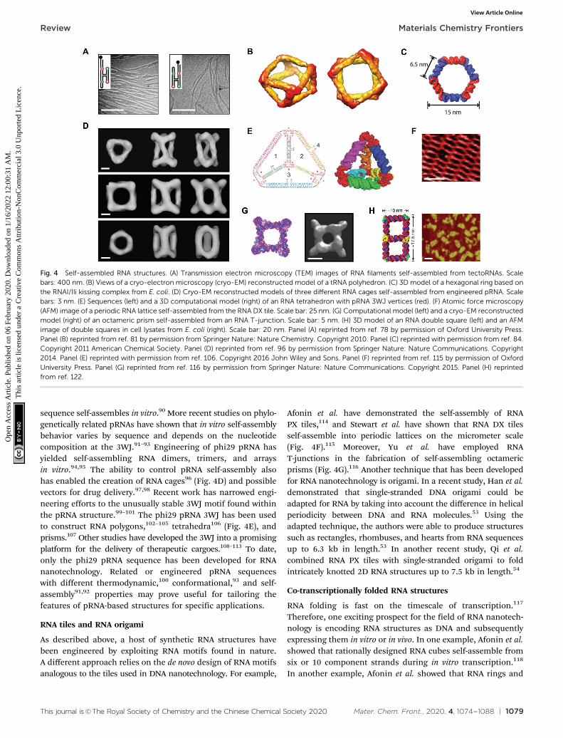
Additionally, by modifying the loop:loop-receptor system witha four-way junction derived from the hairpin ribozyme,Nasalean et al. later demonstrated the self-assembly of long,micrometer-scale RNA structures that resemble actin filamentsfrom the protein world (Fig. 4A).78
Using a different approach, Horiya et al. showed that kissingloops taken from the genome of human immunodeficiencyvirus (HIV) could mediate the formation of large RNAassemblies.79 Kissing loops are short hairpin loops that basepair, and their interactions have been shown to be 102–104 timesmore stable than loop:loop-receptor interactions.79 Chworos et al.combined HIV kissing loops with a right angle motif conserved inrRNA to generate RNA squares, square patterns, and finite grids ofdefined size and shape.80 In a separate study, a five-way junctionderived from class II tRNA was engineered with HIV kissingloops to generate self-assembling, thermostable RNA polyhedra(Fig. 4B).81 Using 50-biotinylated tectoRNAs, the authors of thisstudy further illustrated a remarkable degree of spatial controlby directing the precise positioning and encapsulation ofstreptavidins within the RNA structures.81
Yet other studies have adapted a kissing complex fromEscherichia coli (E. coli) with a 1201 bend to generate multimericRNA rings with potential drug delivery applications (Fig. 4C).82–84
Notably, these circularized RNA assemblies show increasedresistance to ribonucleases relative to their linear RNAcounterparts.84 Recent work by Geary et al. has generalizedthe RNA tectonics approach by composing a ‘syntax’ of struc-tural modules, including kissing loops, tail–tail interactions,triple helices, bulges, and three- and five-way junctions.85 Usingthis syntax, the authors demonstrated the formation of variousRNA shapes, including polygons, ladders, grids, and evenhearts85 that together provide a glimpse into the versatility ofRNA as a medium for generating complex nanostructures.
Engineering of viral pRNA
Alongside tectoRNAs, another molecule that has significantlyshaped the current RNA nanotechnology landscape is proheador packaging RNA (pRNA).60,61 pRNA is a naturally-occurringRNA molecule that derives from the phi29 bacteriophage andrelated bacteriophages in the phi29-like family.86 Full-lengthpRNA is approximately 170 nt long,87 and it has a conservedsecondary structure that features six helical regions, a three-wayjunction (3WJ), and two kissing loops that mediate pRNA self-assembly in the context of a DNA packaging motor, where pRNAperforms an essential but yet unknown function.87–89 In 1998,Guo et al. demonstrated that the prototype phi29 pRNA
Materials Chemistry Frontiers Review
Ope
n A
cces
s A
rtic
le. P
ublis
hed
on 0
6 Fe
brua
ry 2
020.
Dow
nloa
ded
on 1
/16/
2022
12:
00:3
1 A
M.
Thi
s ar
ticle
is li
cens
ed u
nder
a C
reat
ive
Com
mon
s A
ttrib
utio
n-N
onC
omm
erci
al 3
.0 U
npor
ted
Lic
ence
.View Article Online
This journal is©The Royal Society of Chemistry and the Chinese Chemical Society 2020 Mater. Chem. Front., 2020, 4, 1074--1088 | 1079
sequence self-assembles in vitro.90 More recent studies on phylo-genetically related pRNAs have shown that in vitro self-assemblybehavior varies by sequence and depends on the nucleotidecomposition at the 3WJ.91–93 Engineering of phi29 pRNA hasyielded self-assembling RNA dimers, trimers, and arraysin vitro.94,95 The ability to control pRNA self-assembly alsohas enabled the creation of RNA cages96 (Fig. 4D) and possiblevectors for drug delivery.97,98 Recent work has narrowed engi-neering efforts to the unusually stable 3WJ motif found withinthe pRNA structure.99–101 The phi29 pRNA 3WJ has been usedto construct RNA polygons,102–105 tetrahedra106 (Fig. 4E), andprisms.107 Other studies have developed the 3WJ into a promisingplatform for the delivery of therapeutic cargoes.108–113 To date,only the phi29 pRNA sequence has been developed for RNAnanotechnology. Related or engineered pRNA sequenceswith different thermodynamic,100 conformational,93 and self-assembly91,92 properties may prove useful for tailoring thefeatures of pRNA-based structures for specific applications.
RNA tiles and RNA origami
As described above, a host of synthetic RNA structures havebeen engineered by exploiting RNA motifs found in nature.A different approach relies on the de novo design of RNA motifsanalogous to the tiles used in DNA nanotechnology. For example,
Afonin et al. have demonstrated the self-assembly of RNAPX tiles,114 and Stewart et al. have shown that RNA DX tilesself-assemble into periodic lattices on the micrometer scale(Fig. 4F).115 Moreover, Yu et al. have employed RNAT-junctions in the fabrication of self-assembling octamericprisms (Fig. 4G).116 Another technique that has been developedfor RNA nanotechnology is origami. In a recent study, Han et al.demonstrated that single-stranded DNA origami could beadapted for RNA by taking into account the difference in helicalperiodicity between DNA and RNA molecules.53 Using theadapted technique, the authors were able to produce structuressuch as rectangles, rhombuses, and hearts from RNA sequencesup to 6.3 kb in length.53 In another recent study, Qi et al.combined RNA PX tiles with single-stranded origami to foldintricately knotted 2D RNA structures up to 7.5 kb in length.54
Co-transcriptionally folded RNA structures
RNA folding is fast on the timescale of transcription.117
Therefore, one exciting prospect for the field of RNA nanotech-nology is encoding RNA structures as DNA and subsequentlyexpressing them in vitro or in vivo. In one example, Afonin et al.showed that rationally designed RNA cubes self-assemble fromsix or 10 component strands during in vitro transcription.118
In another example, Afonin et al. showed that RNA rings and
Fig. 4 Self-assembled RNA structures. (A) Transmission electron microscopy (TEM) images of RNA filaments self-assembled from tectoRNAs. Scalebars: 400 nm. (B) Views of a cryo-electron microscopy (cryo-EM) reconstructed model of a tRNA polyhedron. (C) 3D model of a hexagonal ring based onthe RNAI/IIi kissing complex from E. coli. (D) Cryo-EM reconstructed models of three different RNA cages self-assembled from engineered pRNA. Scalebars: 3 nm. (E) Sequences (left) and a 3D computational model (right) of an RNA tetrahedron with pRNA 3WJ vertices (red). (F) Atomic force microscopy(AFM) image of a periodic RNA lattice self-assembled from the RNA DX tile. Scale bar: 25 nm. (G) Computational model (left) and a cryo-EM reconstructedmodel (right) of an octameric prism self-assembled from an RNA T-junction. Scale bar: 5 nm. (H) 3D model of an RNA double square (left) and an AFMimage of double squares in cell lysates from E. coli (right). Scale bar: 20 nm. Panel (A) reprinted from ref. 78 by permission of Oxford University Press.Panel (B) reprinted from ref. 81 by permission from Springer Nature: Nature Chemistry. Copyright 2010. Panel (C) reprinted with permission from ref. 84.Copyright 2011 American Chemical Society. Panel (D) reprinted from ref. 96 by permission from Springer Nature: Nature Communications. Copyright2014. Panel (E) reprinted with permission from ref. 106. Copyright 2016 John Wiley and Sons. Panel (F) reprinted from ref. 115 by permission of OxfordUniversity Press. Panel (G) reprinted from ref. 116 by permission from Springer Nature: Nature Communications. Copyright 2015. Panel (H) reprintedfrom ref. 122.
Review Materials Chemistry Frontiers
Ope
n A
cces
s A
rtic
le. P
ublis
hed
on 0
6 Fe
brua
ry 2
020.
Dow
nloa
ded
on 1
/16/
2022
12:
00:3
1 A
M.
Thi
s ar
ticle
is li
cens
ed u
nder
a C
reat
ive
Com
mon
s A
ttrib
utio
n-N
onC
omm
erci
al 3
.0 U
npor
ted
Lic
ence
.View Article Online

1080 | Mater. Chem. Front., 2020, 4, 1074--1088 This journal is©The Royal Society of Chemistry and the Chinese Chemical Society 2020
cubes co-transcriptionally assemble from up to 22 differentcomponent strands in vitro.119 An artful study by Geary et al.combined principles of origami with tiles, hairpins, and kissingloops to create hexagonal RNA lattices that co-transcriptionallyfold and self-assemble in vitro.120 Other studies have extendedthese principles into living systems. For example, Delebecqueet al. co-transcriptionally assembled RNA scaffolds in E. colithat were capable of organizing proteins in a hydrogen-producing biosynthetic pathway.121 More recently, Li et al.showed that an RNA double square design could be cloned,expressed, and folded in E. coli (Fig. 4H).122 Compared toconventional origami techniques, the gene expression of RNAassemblies avoids costly chemical synthesis as well as lengthyannealing steps120 and moves the field a step closer to one ofthe holy grails of RNA nanotechnology, i.e., rationally designingRNA objects as large and complex as natural RNA machines.123
Biomedical applications
As described above, numerous advances in nucleic acid nano-technology have contributed to the development of supra-molecular DNA and RNA assemblies with precise structuraland dynamic control. Here, we highlight examples of high-order nucleic acid structures in two promising biomedicalapplications: biosensing and therapeutic drug delivery.
Biosensing
Biosensors are tools that convert signals from biological analytes(e.g., cells, proteins, nucleic acids, and small metabolites) intorecordable signals. They consist of a recognition component(i.e., a probe), a transducer, and a signal amplification device.124
One challenge in biosensing is facilitating maximum interactionsbetween analytes and probes while preventing agglomeration dueto an irregular distribution of probes across a sensing surface.124
Rationally designed DNA tetrahedra have proven particularlyuseful for addressing this problem. DNA tetrahedra are mechani-cally robust structures that self-assemble rapidly and in nearlyquantitative yields from four component strands.125,126 In 2010,Pei et al. developed a sensitive electrochemical biosensor usingDNA tetrahedra assembled from three DNA strands bearing aterminal thiol group (–SH) and one DNA strand containing aprobe.127 The tetrahedra self-assembled on a gold (Au) electrodeby Au–S chemistry and were shown to enforce uniform probe-to-probe spacing.127 Additionally, tetrahedra with pendant DNAprobes showed a lower detection limit for target DNA of approxi-mately 1 picomolar (pM), which represented a 250-fold improve-ment over the same probe without tetrahedron scaffolding.127
Moreover, aptamers are synthetic nucleic acid sequences that areevolved to bind a user-specified target ligand with high affinityusing a process known as systematic evolution of ligandsby exponential enrichment (SELEX).128,129 Replacement of thependant DNA probe with a DNA aptamer enabled the detectionof thrombin, a potential tumor marker, with a lower detectionlimit of 100 pM, or three orders of magnitude lower than thedetection limit for the aptamer alone.127
New work has shown that DNA tetrahedra functionalizedwith antibodies can detect other disease biomarkers, includingtumor necrosis factor alpha (TNF-a),130 a well-known tumormarker, and prostate-specific antigen (PSA),131 a biomarker forprostate cancer. Yet other DNA tetrahedra have been developedfor detecting microRNAs (miRNAs).132–136 In one particularlynotable example, Wen et al. showed that a DNA tetrahedron-based biosensor could discriminate human let-7 sequenceswith single nucleotide variations.132 The same system equippedwith a different probe could detect the cancer-associatedmiRNA-21 on the attomolar range.132 Furthermore, using DNAtetrahedra, Zhou et al. were able to detect MCF-7 breast cancercells.137 Lin et al. since have developed a protocol for the fabrica-tion of DNA tetrahedron-based biosensors for small molecule,nucleic acid, protein, and whole cell detection (Fig. 5A).138 Recentstudies have further shown that DNA tetrahedron-based bio-sensors can detect RNAs,139–142 proteins,143 small molecules,144
and metal ions145 inside living cells.
Drug delivery
Owing to their intrinsic biocompatibility, nucleic acids alsohold promise as platforms for therapeutic drug delivery.In 2006, Erben et al. illustrated the potential of DNA structuresto serve as molecular containers for therapeutic cargo byloading cytochrome c into a self-assembled DNA tetrahedron.146
Follow-on research showed that DNA tetrahedra are stable toenzymatic degradation,147 and DNA tetrahedra,148 icosahedra,149
and cages150 may be reconfigured in response to external signalsfor controlled drug release. Furthermore, many studies havedemonstrated that DNA structures are taken up by culturedcells without transfection reagents.151–154 Recently, Wiraja et al.tested a variety of nano-scale DNA structures, including tetra-hedra, cylindrical rods, rectangles, and triangles, and showedthat highly-ordered structures smaller than 75 nm penetratethe skin.155 Using a mouse model of melanoma, the studyfurther showed that DNA tetrahedra loaded with the chemo-therapeutic drug doxorubicin could achieve over 2-fold higherdrug accumulation and tumor inhibition relative to topicallyapplied doxorubicin and liposome- or nanoparticle-formulateddoxorubicin.155
While some DNA structures achieve passive uptake, otherplatforms have been developed with active targeting mechanismsto promote uptake by specific populations of cells. In oneexample, Chang et al. showed that DNA icosahedra self-assembled from five- and six-point star motifs could be func-tionalized with a MUC 1 aptamer for targeted delivery ofdoxorubicin to MUC 1+ MCF-7 breast cancer cells.156 In aseparate, pioneering study, Lee et al. developed a DNA tetra-hedron with a small interfering RNA (siRNA) hybridized to eachedge (Fig. 5B).157 SiRNAs are short, double-stranded RNAmolecules that elicit potent gene silencing by co-opting anendogenous RNA interference (RNAi) pathway.158 Currently,major challenges for the delivery of oligonucleotide drugs suchas siRNAs include nuclease protection, systemic delivery,and targeted cellular uptake.159 Notably, siRNA-functionalizedtetrahedra administered in a mouse tumor xenograft model
Materials Chemistry Frontiers Review
Ope
n A
cces
s A
rtic
le. P
ublis
hed
on 0
6 Fe
brua
ry 2
020.
Dow
nloa
ded
on 1
/16/
2022
12:
00:3
1 A
M.
Thi
s ar
ticle
is li
cens
ed u
nder
a C
reat
ive
Com
mon
s A
ttrib
utio
n-N
onC
omm
erci
al 3
.0 U
npor
ted
Lic
ence
.View Article Online

This journal is©The Royal Society of Chemistry and the Chinese Chemical Society 2020 Mater. Chem. Front., 2020, 4, 1074--1088 | 1081
displayed longer half-lives in blood circulation relative tosiRNAs alone.157 Additionally, tetrahedra with siRNAs bearingfolate distributed to several tissues but accumulated in folatereceptor-overexpressing KB cells, where they efficiently silenceda luciferase reporter.157 In line with these results, other studieshave shown that DNA structures can confer stability on theirtherapeutic payloads while also maintaining or even improvingtheir therapeutic efficacies.160,161
Yet other DNA platforms have been developed with sophis-ticated mechanisms for cargo release. In a recent example,Bujold et al. designed DNA ‘nanosuitcases’ that encapsulatesiRNA and release it in the presence of specific mRNA ormiRNA triggers (Fig. 5C).162 In another example, Douglaset al. developed DNA origami ‘nanorobots’ with AND logicgates.163 These logic gates consisted of two aptamer ‘locks’ thattriggered a drastic reconfiguration of the robot and exposedmolecular payloads upon binding both antigenic ‘keys’.163
Recent work by Li et al. combined both active targeting and
controlled release mechanisms in a DNA nanorobot loadedwith thrombin and functionalized with nucleolin-bindingaptamers (Fig. 5D).164 Thrombin is a protease that inducescoagulation and may be useful for starving tumors of nutrientsand oxygen by selective occlusion of tumor blood vessels.164
Nucleolin is a protein that is expressed on tumor-associatedendothelial cells.165 Remarkably, the nanorobots were capableof depositing thrombin at tumor cells in mouse models ofbreast cancer, and they also proved safe and immunologicallyinert in both mice and miniature pigs.164
Several high-order RNA structures also have been developedfor therapeutic drug delivery. Because RNA is more chemicallylabile than DNA, RNA-based platforms often are chemicallymodified to improve their stability under physiologicalconditions. Common modifications include substitutions atthe 20 position of ribose as well as in the internucleotidelinkage.159 A series of studies has developed RNA cubesdesigned in silico118 as a promising platform for drug delivery.
Fig. 5 High-order nucleic acid structures for biosensing and drug delivery. (A) A DNA tetrahedron-based biosensing platform for the detection of cells,proteins, nucleic acids, and small molecules. (B) Strands of DNA and siRNA self-assemble into an siRNA-functionalized DNA tetrahedron. (C) A DNAnanosuitcase is triggered to release encapsulated siRNA cargo (green) by a specific miRNA sequence. (D) A DNA origami nanorobot is loaded withthrombin (magenta) and functionalized with nucleolin-binding aptamers (green). (E) Strands of RNA self-assemble into an siRNA-functionalized RNAcube. Processing by Dicer (‘Dicing’) releases siRNAs from the cube. (F) An RNA pyramid with photocleavable drug cargo. (G) An RNA tetrahedronfunctionalized with aptamers (red, blue, and green) and a ribozyme (purple). Panel (A) reprinted from ref. 138 by permission from Springer Nature: NatureProtocols. Copyright 2016. Panel (B) reprinted from ref. 157 by permission from Springer Nature: Nature Nanotechnology. Copyright 2012. Panel (C)reprinted with permission from ref. 162. Copyright 2016 American Chemical Society. Panel (D) reprinted from ref. 164 by permission from SpringerNature: Nature Biotechnology. Copyright 2018. Panel (E) reprinted with permission from ref. 168, DOI: 10.1021/nn504508s. Panel (F) reprinted fromref. 169 with permission from Springer Nature: Nano Research. Copyright 2019. Panel (G) reprinted with permission from ref. 106. Copyright 2016 JohnWiley and Sons.
Review Materials Chemistry Frontiers
Ope
n A
cces
s A
rtic
le. P
ublis
hed
on 0
6 Fe
brua
ry 2
020.
Dow
nloa
ded
on 1
/16/
2022
12:
00:3
1 A
M.
Thi
s ar
ticle
is li
cens
ed u
nder
a C
reat
ive
Com
mon
s A
ttrib
utio
n-N
onC
omm
erci
al 3
.0 U
npor
ted
Lic
ence
.View Article Online

1082 | Mater. Chem. Front., 2020, 4, 1074--1088 This journal is©The Royal Society of Chemistry and the Chinese Chemical Society 2020
The cubes are precisely controlled in terms of size, shape, andcomposition; they can be chemically modified for downstreamapplications; and they also are capable of self-assembly inisothermal conditions during in vitro transcription or fromseveral short strands following chemical RNA synthesis.118,119
Computational and experimental analyses have shown thatcube designs with 10 bp per edge and single-stranded regionsin the corners are not strained and assemble efficiently.166 In2011, Afonin et al. developed a protocol for the design and self-assembly of siRNA-functionalized RNA cubes using processesthat are fully automatable.167 The cubes interact with recombi-nant human Dicer in vitro to produce siRNAs167 (Fig. 5E).Furthermore, a recent study by Afonin et al. showed thatsiRNA-functionalized RNA cubes are capable of triggering RNAiin GFP-expressing MDA-MB-231 breast cancer cells and redu-cing HIV-1 production in HeLa cells.168
Finally, another promising platform for drug delivery hascome from research on pRNA and its thermodynamically stable3WJ.60,61 The phi29 pRNA 3WJ has been engineered to deliver avariety of therapeutic agents, including small molecules,108
miRNAs,109 anti-miRNAs,110,111 and siRNAs.99,112,113 In a recentexample, Xu et al. combined the phi29 pRNA 3WJ with a four-way junction derived from the hairpin ribozyme to generate20-deoxyfluoro U/C modified RNA pyramids (Fig. 5F).169 Thesepyramids were functionalized with the chemotherapeutic drugpaclitaxel via photocleavable spacers, and irradiation withultraviolet light induced drug release and cytotoxicity in MDA-MB-231 breast cancer cells.169 In a different example, Li et al.reported the self-assembly of a nuclease-resistant, 20-deoxy-fluoro U/C modified RNA tetrahedron based on the phi29 pRNA3WJ (Fig. 5G).106 Functionalization of the tetrahedron withribozymes, aptamers, and siRNAs did not disrupt its structure,and the functional modules retained the capacity for ribozy-matic cleavage, ligand binding, and gene knockdown in vitro.106
Furthermore, this study showed that tetrahedra functionalizedwith an aptamer against epidermal growth factor receptor(EGFR) and an siRNA were capable of internalizing into tumortissue and silencing a luciferase reporter in a mouse model ofbreast cancer.106
Outlook
From the development of DNA junctions, tiles, and origami inDNA nanotechnology to the development of tectoRNAs, engi-neered pRNA, and co-transcriptionally folded structures in RNAnanotechnology, research over the past 40 years has expandedthe possibilities for creating supramolecular nucleic acid archi-tectures with precise structural and dynamic control. Today,high-order DNA and RNA structures are taking their place aspowerful tools with promising biomedical applications.In biosensing, nano-structured DNA platforms boost sensitivityfor the detection of biological analytes while preventingagglomeration. In drug delivery, DNA- and RNA-based plat-forms offer potential solutions to formidable in vivo challenges,including nuclease protection, systemic delivery, and targeted
cellular uptake for oligonucleotide drugs. Many of the achieve-ments in nucleic acid nanotechnology would not be possiblewithout parallel achievements in chemical DNA and RNAsynthesis, which have given ready access to the oligonucleotidesequences necessary for constructing nucleic acid assemblies.1
Still, for bottom-up fabrication using RNA, sequence length andyield are considerable limitations. RNA assemblies made bygene expression may circumvent these issues and facilitateapplications in vivo. Additionally, the cost of oligonucleotidesynthesis is decreasing. In fact, a relative of Moore’s law hasbeen reported for the effective cost of DNA, which is halvedevery 30 months.170 Continued developments in oligonucleotidesynthesis, including in the production of longer sequences forlower costs, will be important for supporting efforts in DNA andRNA nanotechnology and for realizing the extraordinary potentialof high-order nucleic acid structures for biomedical applicationsand beyond.
Conflicts of interest
A. C. H. is a co-inventor and applicant on a patent for viralpRNA 3WJ sequences.
Acknowledgements
A. C. H. was supported by a Novartis FreeNovation grant from theNovartis Research Foundation (Novartis Forschungsstiftung).
References
1 N. C. Seeman and H. F. Sleiman, DNA nanotechnology,Nat. Rev. Mater., 2017, 3, 17068.
2 J. D. Watson and F. H. Crick, Molecular structure of nucleicacids: A structure for deoxyribose nucleic acid, Nature,1953, 171, 737–738.
3 N. C. Seeman, DNA in a material world, Nature, 2003, 421,427–431.
4 N. C. Seeman, Nucleic acid junctions and lattices, J. Theor.Biol., 1982, 99, 237–247.
5 H. Qiu, J. C. Dewan and N. C. Seeman, A DNA decamer witha sticky end: The crystal structure of d-CGACGATCGT,J. Mol. Biol., 1997, 267, 881–898.
6 N. R. Kallenbach, R.-I. Ma and N. C. Seeman, An immobilenucleic acid junction constructed from oligonucleotides,Nature, 1983, 305, 829–831.
7 Y. L. Wang, J. E. Mueller, B. Kemper and N. C. Seeman,Assembly and characterization of five-arm and six-arm DNAbranched junctions, Biochemistry, 1991, 30, 5667–5674.
8 J. H. Chen, N. R. Kallenbach and N. C. Seeman, A specificquadrilateral synthesized from DNA branched junctions,J. Am. Chem. Soc., 1989, 111, 6402–6407.
9 J. E. Mueller, S. M. Du and N. C. Seeman, Design andsynthesis of a knot from single-stranded DNA, J. Am. Chem.Soc., 1991, 113, 6306–6308.
Materials Chemistry Frontiers Review
Ope
n A
cces
s A
rtic
le. P
ublis
hed
on 0
6 Fe
brua
ry 2
020.
Dow
nloa
ded
on 1
/16/
2022
12:
00:3
1 A
M.
Thi
s ar
ticle
is li
cens
ed u
nder
a C
reat
ive
Com
mon
s A
ttrib
utio
n-N
onC
omm
erci
al 3
.0 U
npor
ted
Lic
ence
.View Article Online

This journal is©The Royal Society of Chemistry and the Chinese Chemical Society 2020 Mater. Chem. Front., 2020, 4, 1074--1088 | 1083
10 N. C. Seeman, The design of single-stranded nucleic acidknots, Mol. Eng., 1992, 2, 297–307.
11 C. Mao, W. Sun and N. C. Seeman, Assembly of Borromeanrings from DNA, Nature, 1997, 386, 137–138.
12 J. Chen and N. C. Seeman, Synthesis from DNA of amolecule with the connectivity of a cube, Nature, 1991,350, 631–633.
13 Y. Zhang and N. C. Seeman, Construction of a DNA-truncatedoctahedron, J. Am. Chem. Soc., 1994, 116, 1661–1669.
14 N. C. Seeman, DNA engineering and its application tonanotechnology, Trends Biotechnol., 1999, 17, 437–443.
15 N. C. Seeman, An overview of structural DNA nanotechnology,Mol. Biotechnol., 2007, 37, 246–257.
16 A. R. Chandrasekaran and R. Zhuo, A ‘tile’ tale: Hierarchicalself-assembly of DNA lattices, Appl. Mater. Today, 2016, 2, 7–16.
17 T. J. Fu and N. C. Seeman, DNA double-crossover mole-cules, Biochemistry, 1993, 32, 3211–3220.
18 P. Sa-Ardyen, A. V. Vologodskii and N. C. Seeman, Theflexibility of DNA double crossover molecules, Biophys. J.,2003, 84, 3829–3837.
19 E. Winfree, F. Liu, L. A. Wenzler and N. C. Seeman, Designand self-assembly of two-dimensional DNA crystals, Nature,1998, 394, 539–544.
20 H. Yan, T. H. LaBean, L. Feng and J. H. Reif, Directednucleation assembly of DNA tile complexes for barcode-patterned lattices, Proc. Natl. Acad. Sci. U. S. A., 2003, 100, 8103.
21 T. H. LaBean, H. Yan, J. Kopatsch, F. Liu, E. Winfree,J. H. Reif and N. C. Seeman, Construction, analysis, liga-tion, and self-assembly of DNA triple crossover complexes,J. Am. Chem. Soc., 2000, 122, 1848–1860.
22 Z. Shen, H. Yan, T. Wang and N. C. Seeman, Paranemiccrossover DNA: A generalized Holliday structure withapplications in nanotechnology, J. Am. Chem. Soc., 2004,126, 1666–1674.
23 H. Yan, S. H. Park, G. Finkelstein, J. H. Reif and T. H.LaBean, DNA-templated self-assembly of protein arrays andhighly conductive nanowires, Science, 2003, 301, 1882–1884.
24 Y. He, Y. Chen, H. Liu, A. E. Ribbe and C. Mao, Self-assembly of hexagonal DNA two-dimensional (2D) arrays,J. Am. Chem. Soc., 2005, 127, 12202–12203.
25 Y. He, Y. Tian, A. E. Ribbe and C. Mao, Highly connectedtwo-dimensional crystals of DNA six-point-stars, J. Am.Chem. Soc., 2006, 128, 15978–15979.
26 U. Majumder, A. Rangnekar, K. V. Gothelf, J. H. Reif andT. H. LaBean, Design and construction of double-deckertile as a route to three-dimensional periodic assembly ofDNA, J. Am. Chem. Soc., 2011, 133, 3843–3845.
27 S. Hamada and S. Murata, Substrate-assisted assembly ofinterconnected single-duplex DNA nanostructures, Angew.Chem., Int. Ed., 2009, 48, 6820–6823.
28 P. W. K. Rothemund, N. Papadakis and E. Winfree, Algo-rithmic self-assembly of DNA Sierpinski triangles, PLoS Biol.,2004, 2, e424.
29 Y. He, T. Ye, M. Su, C. Zhang, A. E. Ribbe, W. Jiang andC. Mao, Hierarchical self-assembly of DNA into symmetricsupramolecular polyhedra, Nature, 2008, 452, 198.
30 C. Mao, W. Sun, Z. Shen and N. C. Seeman, A nanomecha-nical device based on the B-Z transition of DNA, Nature,1999, 397, 144–146.
31 H. Yan, X. Zhang, Z. Shen and N. C. Seeman, A robust DNAmechanical device controlled by hybridization topology,Nature, 2002, 415, 62–65.
32 D. Liu, M. Wang, Z. Deng, R. Walulu and C. Mao, Tensegrity:Construction of rigid DNA triangles with flexiblefour-arm DNA junctions, J. Am. Chem. Soc., 2004, 126,2324–2325.
33 J. Zheng, J. J. Birktoft, Y. Chen, T. Wang, R. Sha, P. E.Constantinou, S. L. Ginell, C. Mao and N. C. Seeman, Frommolecular to macroscopic via the rational design of aself-assembled 3D DNA crystal, Nature, 2009, 461, 74.
34 T. Wang, R. Sha, J. Birktoft, J. Zheng, C. Mao and N. C.Seeman, A DNA crystal designed to contain two mole-cules per asymmetric unit, J. Am. Chem. Soc., 2010, 132,15471–15473.
35 R. Sha, J. J. Birktoft, N. Nguyen, A. R. Chandrasekaran,J. Zheng, X. Zhao, C. Mao and N. C. Seeman, Self-assembledDNA crystals: The impact on resolution of 50-phosphates andthe DNA source, Nano Lett., 2013, 13, 793–797.
36 Y. P. Ohayon, A. R. Chandrasekaran, C. Hernandez, J. J.Birktoft, R. Sha, S. Ginell, P. Lukeman, C. Mao, P. M.Chaikin and N. C. Seeman, Programmable crystal contactsused to improve the resolution of self-assembled 3D DNAcrystals, J. Biomol. Struct. Dyn., 2015, 33, 50–51.
37 E. Stahl, F. Praetorius, C. C. de Oliveira Mann, K. P.Hopfner and H. Dietz, Impact of heterogeneity and latticebond strength on DNA triangle crystal growth, ACS Nano,2016, 10, 9156–9164.
38 J. Zhao, A. R. Chandrasekaran, Q. Li, X. Li, R. Sha, N. C.Seeman and C. Mao, Post-assembly stabilization of ration-ally designed DNA crystals, Angew. Chem., Int. Ed., 2015, 54,9936–9939.
39 H. O. Abdallah, Y. P. Ohayon, A. R. Chandrasekaran, R. Sha,K. R. Fox, T. Brown, D. A. Rusling, C. Mao and N. C. Seeman,Stabilisation of self-assembled DNA crystals by triplex-directed photo-cross-linking, Chem. Commun., 2016, 52,8014–8017.
40 Z. Li, L. Liu, M. Zheng, J. Zhao, N. C. Seeman and C. Mao,Making engineered 3D DNA crystals robust, J. Am. Chem.Soc., 2019, 141, 15850–15855.
41 N. C. Seeman, Structural DNA nanotechnology: An overview,Methods Mol. Biol., 2005, 303, 143–166.
42 W. M. Shih, J. D. Quispe and G. F. Joyce, A 1.7-kilobasesingle-stranded DNA that folds into a nanoscale octa-hedron, Nature, 2004, 427, 618–621.
43 P. W. K. Rothemund, Folding DNA to create nanoscaleshapes and patterns, Nature, 2006, 440, 297–302.
44 Y. Ke, J. Sharma, M. Liu, K. Jahn, Y. Liu and H. Yan,Scaffolded DNA origami of a DNA tetrahedron molecularcontainer, Nano Lett., 2009, 9, 2445–2447.
45 S. M. Douglas, H. Dietz, T. Liedl, B. Hogberg, F. Graf andW. M. Shih, Self-assembly of DNA into nanoscale three-dimensional shapes, Nature, 2009, 459, 414.
Review Materials Chemistry Frontiers
Ope
n A
cces
s A
rtic
le. P
ublis
hed
on 0
6 Fe
brua
ry 2
020.
Dow
nloa
ded
on 1
/16/
2022
12:
00:3
1 A
M.
Thi
s ar
ticle
is li
cens
ed u
nder
a C
reat
ive
Com
mon
s A
ttrib
utio
n-N
onC
omm
erci
al 3
.0 U
npor
ted
Lic
ence
.View Article Online

1084 | Mater. Chem. Front., 2020, 4, 1074--1088 This journal is©The Royal Society of Chemistry and the Chinese Chemical Society 2020
46 H. Dietz, S. M. Douglas and W. M. Shih, Folding DNA intotwisted and curved nanoscale shapes, Science, 2009, 325, 725.
47 D. Han, S. Pal, J. Nangreave, Z. Deng, Y. Liu and H. Yan,DNA origami with complex curvatures in three-dimensionalspace, Science, 2011, 332, 342.
48 E. S. Andersen, M. Dong, M. M. Nielsen, K. Jahn,R. Subramani, W. Mamdouh, M. M. Golas, B. Sander,H. Stark, C. L. P. Oliveira, J. S. Pedersen, V. Birkedal,F. Besenbacher, K. V. Gothelf and J. Kjems, Self-assemblyof a nanoscale DNA box with a controllable lid, Nature,2009, 459, 73.
49 B. Wei, M. Dai and P. Yin, Complex shapes self-assembledfrom single-stranded DNA tiles, Nature, 2012, 485,623–626.
50 Y. Ke, L. L. Ong, W. M. Shih and P. Yin, Three-dimensionalstructures self-assembled from DNA bricks, Science, 2012,338, 1177.
51 L. L. Ong, N. Hanikel, O. K. Yaghi, C. Grun, M. T. Strauss,P. Bron, J. Lai-Kee-Him, F. Schueder, B. Wang, P. Wang,J. Y. Kishi, C. Myhrvold, A. Zhu, R. Jungmann, G. Bellot,Y. Ke and P. Yin, Programmable self-assembly of three-dimensional nanostructures from 10000 unique compo-nents, Nature, 2017, 552, 72–77.
52 K. F. Wagenbauer, C. Sigl and H. Dietz, Gigadalton-scaleshape-programmable DNA assemblies, Nature, 2017, 552,78–83.
53 D. Han, X. Qi, C. Myhrvold, B. Wang, M. Dai, S. Jiang,M. Bates, Y. Liu, B. An, F. Zhang, H. Yan and P. Yin,Single-stranded DNA and RNA origami, Science, 2017,358, eaao2648.
54 X. Qi, F. Zhang, Z. Su, S. Jiang, D. Han, B. Ding, Y. Liu,W. Chiu, P. Yin and H. Yan, Programming moleculartopologies from single-stranded nucleic acids, Nat. Commun.,2018, 9, 4579.
55 E. Benson, A. Mohammed, J. Gardell, S. Masich, E. Czeizler,P. Orponen and B. Hogberg, DNA rendering of polyhedralmeshes at the nanoscale, Nature, 2015, 523, 441.
56 E. Benson, A. Mohammed, A. Bosco, A. I. Teixeira,P. Orponen and B. Hogberg, Computer-aided productionof scaffolded DNA nanostructures from flat sheet meshes,Angew. Chem., Int. Ed., 2016, 55, 8869–8872.
57 R. Veneziano, S. Ratanalert, K. Zhang, F. Zhang, H. Yan,W. Chiu and M. Bathe, Designer nanoscale DNA assem-blies programmed from the top down, Science, 2016,352, 1534.
58 H. Jun, F. Zhang, T. Shepherd, S. Ratanalert, X. Qi, H. Yanand M. Bathe, Autonomously designed free-form 2D DNAorigami, Sci. Adv., 2019, 5, eaav0655.
59 R. A. Hughes and A. D. Ellington, Synthetic DNA synthesisand assembly: Putting the synthetic in synthetic biology,Cold Spring Harbor Perspect. Biol., 2017, 9, a023812.
60 P. Guo, The emerging field of RNA nanotechnology,Nat. Nanotechnol., 2010, 5, 833–842.
61 D. Jasinski, F. Haque, D. W. Binzel and P. Guo, Advance-ment of the emerging field of RNA nanotechnology,ACS Nano, 2017, 11, 1142–1164.
62 G. A. Soukup and R. R. Breaker, Relationship between inter-nucleotide linkage geometry and the stability of RNA, RNA,1999, 5, 1308–1325.
63 N. B. Leontis and E. Westhof, Geometric nomenclature andclassification of RNA base pairs, RNA, 2001, 7, 499–512.
64 N. B. Leontis, J. Stombaugh and E. Westhof, The non-Watson-Crick base pairs and their associated isostericitymatrices, Nucleic Acids Res., 2002, 30, 3497–3531.
65 J. A. Cruz and E. Westhof, The dynamic landscapes of RNAarchitecture, Cell, 2009, 136, 604–609.
66 E. J. Strobel, K. E. Watters, D. Loughrey and J. B. Lucks,RNA systems biology: Uniting functional discoveriesand structural tools to understand global roles of RNAs,Curr. Opin. Biotechnol., 2016, 39, 182–191.
67 F. H. Crick, On protein synthesis, Symp. Soc. Exp. Biol.,1958, 12, 138–163.
68 F. H. Crick, Central dogma of molecular biology, Nature,1970, 227, 561–563.
69 N. Ban, P. Nissen, J. Hansen, P. B. Moore and T. A. Steitz,The complete atomic structure of the large ribosomalsubunit at 2.4 A resolution, Science, 2000, 289, 905–920.
70 B. T. Wimberly, D. E. Brodersen, W. M. Clemons Jr, R. J.Morgan-Warren, A. P. Carter, C. Vonrhein, T. Hartsch andV. Ramakrishnan, Structure of the 30S ribosomal subunit,Nature, 2000, 407, 327–339.
71 R. T. Batey, R. P. Rambo and J. A. Doudna, Tertiary motifsin RNA structure and folding, Angew. Chem., Int. Ed., 1999,38, 2326–2343.
72 I. Tinoco and C. Bustamante, How RNA folds, J. Mol. Biol.,1999, 293, 271–281.
73 W. Grabow and L. Jaeger, RNA modularity for syntheticbiology, F1000Prime Rep., 2013, 5, 46.
74 M. Sekine, in Synthesis of Therapeutic Oligonucleotides, ed.S. Obika and M. Sekine, Springer Singapore, Singapore,2018, pp. 41–65, DOI: 10.1007/978-981-13-1912-9_3.
75 E. Westhof, B. Masquida and L. Jaeger, RNA tectonics:Towards RNA design, Folding Des., 1996, 1, R78–R88.
76 L. Jaeger and N. B. Leontis, Tecto-RNA: One-dimensionalself-assembly through tertiary interactions, Angew. Chem.,Int. Ed., 2000, 39, 2521–2524.
77 L. Jaeger, E. Westhof and N. B. Leontis, TectoRNA: Modularassembly units for the construction of RNA nano-objects,Nucleic Acids Res., 2001, 29, 455–463.
78 L. Nasalean, S. Baudrey, N. B. Leontis and L. Jaeger,Controlling RNA self-assembly to form filaments, NucleicAcids Res., 2006, 34, 1381–1392.
79 S. Horiya, X. Li, G. Kawai, R. Saito, A. Katoh, K. Kobayashiand K. Harada, RNA LEGO: Magnesium-dependent for-mation of specific RNA assemblies through kissing inter-actions, Chem. Biol., 2003, 10, 645–654.
80 A. Chworos, I. Severcan, A. Y. Koyfman, P. Weinkam,E. Oroudjev, H. G. Hansma and L. Jaeger, Building program-mable jigsaw puzzles with RNA, Science, 2004, 306, 2068.
81 I. Severcan, C. Geary, A. Chworos, N. Voss, E. Jacovetty andL. Jaeger, A polyhedron made of tRNAs, Nat. Chem., 2010,2, 772–779.
Materials Chemistry Frontiers Review
Ope
n A
cces
s A
rtic
le. P
ublis
hed
on 0
6 Fe
brua
ry 2
020.
Dow
nloa
ded
on 1
/16/
2022
12:
00:3
1 A
M.
Thi
s ar
ticle
is li
cens
ed u
nder
a C
reat
ive
Com
mon
s A
ttrib
utio
n-N
onC
omm
erci
al 3
.0 U
npor
ted
Lic
ence
.View Article Online

This journal is©The Royal Society of Chemistry and the Chinese Chemical Society 2020 Mater. Chem. Front., 2020, 4, 1074--1088 | 1085
82 Y. G. Yingling and B. A. Shapiro, Computational design ofan RNA hexagonal nanoring and an RNA nanotube, NanoLett., 2007, 7, 2328–2334.
83 M. Paliy, R. Melnik and B. A. Shapiro, Molecular dynamicsstudy of the RNA ring nanostructure: a phenomenon ofself-stabilization, Phys. Biol., 2009, 6, 046003.
84 W. W. Grabow, P. Zakrevsky, K. A. Afonin, A. Chworos,B. A. Shapiro and L. Jaeger, Self-assembling RNA nano-rings based on RNAI/II inverse kissing complexes, NanoLett., 2011, 11, 878–887.
85 C. Geary, A. Chworos, E. Verzemnieks, N. R. Voss andL. Jaeger, Composing RNA nanostructures from a syntax ofRNA structural modules, Nano Lett., 2017, 17, 7095–7101.
86 A. C. Hill, L. E. Bartley and S. J. Schroeder, Prohead RNA:A noncoding viral RNA of novel structure and function,Wiley Interdiscip. Rev.: RNA, 2016, 7, 428–437.
87 S. Bailey, J. Wichitwechkarn, D. Johnson, B. E. Reilly andD. L. Anderson, and J. W. Bodley, Phylogenetic analysisand secondary structure of the Bacillus subtilis bacterio-phage RNA required for DNA packaging, J. Biol. Chem.,1990, 265, 22365–22370.
88 P. X. Guo, S. Erickson and D. Anderson, A small viral RNAis required for in vitro packaging of bacteriophage phi 29DNA, Science, 1987, 236, 690–694.
89 C. Chen, C. Zhang and P. Guo, Sequence requirement forhand-in-hand interaction in formation of RNA dimers andhexamers to gear phi29 DNA translocation motor, RNA,1999, 5, 805–818.
90 P. Guo, C. Zhang, C. Chen, K. Garver and M. Trottier, Inter-RNA interaction of phage phi29 pRNA to form a hexamericcomplex for viral DNA transportation, Mol. Cell, 1998, 2,149–155.
91 X. Gu and S. J. Schroeder, Different sequences showsimilar quaternary interaction stabilities in prohead viralRNA self-assembly, J. Biol. Chem., 2011, 286, 14419–14426.
92 Y. Hao and J. S. Kieft, Diverse self-association propertieswithin a family of phage packaging RNAs, RNA, 2014, 20,1759–1774.
93 Y. Hao and J. S. Kieft, Three-way junction conformationdictates self-association of phage packaging RNAs, RNABiol., 2016, 13, 635–645.
94 D. Shu, L. P. Huang, S. Hoeprich and P. Guo, Constructionof phi29 DNA-packaging RNA monomers, dimers, andtrimers with variable sizes and shapes as potential partsfor nanodevices, J. Nanosci. Nanotechnol., 2003, 3, 295–302.
95 D. Shu, W. D. Moll, Z. Deng, C. Mao and P. Guo, Bottom-upassembly of RNA arrays and superstructures as potentialparts in nanotechnology, Nano Lett., 2004, 4, 1717–1723.
96 C. Hao, X. Li, C. Tian, W. Jiang, G. Wang and C. Mao,Construction of RNA nanocages by re-engineering thepackaging RNA of phi29 bacteriophage, Nat. Commun.,2014, 5, 3890.
97 S. Guo, N. Tschammer, S. Mohammed and P. Guo, Specificdelivery of therapeutic RNAs to cancer cells via the dimeri-zation mechanism of phi29 motor pRNA, Hum. Gene Ther.,2005, 16, 1097–1109.
98 A. Khaled, S. Guo, F. Li and P. Guo, Controllable self-assembly of nanoparticles for specific delivery of multipletherapeutic molecules to cancer cells using RNA nano-technology, Nano Lett., 2005, 5, 1797–1808.
99 D. Shu, Y. Shu, F. Haque, S. Abdelmawla and P. Guo,Thermodynamically stable RNA three-way junction forconstructing multifunctional nanoparticles for delivery oftherapeutics, Nat. Nanotechnol., 2011, 6, 658.
100 A. C. Hill and S. J. Schroeder, Thermodynamic stabilitiesof three-way junction nanomotifs in prohead RNA, RNA,2017, 23, 521–529.
101 X. Piao, H. Wang, D. W. Binzel and P. Guo, Assessment andcomparison of thermal stability of phosphorothioate-DNA,DNA, RNA, 20-F RNA, and LNA in the context of phi29pRNA 3WJ, RNA, 2018, 24, 67–76.
102 E. F. Khisamutdinov, D. L. Jasinski and P. Guo, RNA as aboiling-resistant anionic polymer material to build robuststructures with defined shape and stoichiometry, ACSNano, 2014, 8, 4771–4781.
103 D. L. Jasinski, E. F. Khisamutdinov, Y. L. Lyubchenko andP. Guo, Physicochemically tunable polyfunctionalized RNAsquare architecture with fluorogenic and ribozymaticproperties, ACS Nano, 2014, 8, 7620–7629.
104 E. F. Khisamutdinov, M. N. Bui, D. Jasinski, Z. Zhao, Z. Cuiand P. Guo, Simple method for constructing RNA triangle,square, pentagon by tuning interior RNA 3WJ angle from60 degrees to 90 degrees or 108 degrees, Methods Mol. Biol.,2015, 1316, 181–193.
105 D. L. Jasinski, H. Li and P. Guo, The effect of size andshape of RNA nanoparticles on biodistribution, Mol. Ther.,2018, 26, 784–792.
106 H. Li, K. Zhang, F. Pi, S. Guo, L. Shlyakhtenko, W. Chiu,D. Shu and P. Guo, Controllable self-assembly of RNAtetrahedrons with precise shape and size for cancer targeting,Adv. Mater., 2016, 28, 7501–7507.
107 E. F. Khisamutdinov, D. L. Jasinski, H. Li, K. Zhang,W. Chiu and P. Guo, Fabrication of RNA 3D nanoprismsfor loading and protection of small RNAs and model drugs,Adv. Mater., 2016, 28, 10079–10087.
108 F. Pi, H. Zhang, H. Li, V. Thiviyanathan, D. G. Gorenstein,A. K. Sood and P. Guo, RNA nanoparticles harboringannexin A2 aptamer can target ovarian cancer for tumor-specific doxorubicin delivery, Nanomedicine, 2017, 13,1183–1193.
109 D. W. Binzel, Y. Shu, H. Li, M. Sun, Q. Zhang, D. Shu,B. Guo and P. Guo, Specific delivery of miRNA for highefficient inhibition of prostate cancer by RNA nano-technology, Mol. Ther., 2016, 24, 1267–1277.
110 D. Shu, H. Li, Y. Shu, G. Xiong, W. E. Carson, 3rd,F. Haque, R. Xu and P. Guo, Systemic delivery of anti-miRNA for suppression of triple negative breast cancerutilizing RNA nanotechnology, ACS Nano, 2015, 9,9731–9740.
111 T. J. Lee, J. Y. Yoo, D. Shu, H. Li, J. Zhang, J. G. Yu, A. C.Jaime-Ramirez, M. Acunzo, G. Romano, R. Cui, H. L.Sun, Z. Luo, M. Old, B. Kaur, P. Guo and C. M. Croce,
Review Materials Chemistry Frontiers
Ope
n A
cces
s A
rtic
le. P
ublis
hed
on 0
6 Fe
brua
ry 2
020.
Dow
nloa
ded
on 1
/16/
2022
12:
00:3
1 A
M.
Thi
s ar
ticle
is li
cens
ed u
nder
a C
reat
ive
Com
mon
s A
ttrib
utio
n-N
onC
omm
erci
al 3
.0 U
npor
ted
Lic
ence
.View Article Online

1086 | Mater. Chem. Front., 2020, 4, 1074--1088 This journal is©The Royal Society of Chemistry and the Chinese Chemical Society 2020
RNA nanoparticle-based targeted therapy for glioblastomathrough inhibition of oncogenic miR-21, Mol. Ther., 2017,25, 1544–1555.
112 T. J. Lee, F. Haque, D. Shu, J. Y. Yoo, H. Li, R. A. Yokel,C. Horbinski, T. H. Kim, S. H. Kim, C. H. Kwon, I. Nakano,B. Kaur, P. Guo and C. M. Croce, RNA nanoparticle as avector for targeted siRNA delivery into glioblastoma mousemodel, Oncotarget, 2015, 6, 14766–14776.
113 D. Cui, C. Zhang, B. Liu, Y. Shu, T. Du, D. Shu, K. Wang,F. Dai, Y. Liu, C. Li, F. Pan, Y. Yang, J. Ni, H. Li, B. Brand-Saberi and P. Guo, Regression of gastric cancer by systemicinjection of RNA nanoparticles carrying both ligand andsiRNA, Sci. Rep., 2015, 5, 10726.
114 K. A. Afonin, D. J. Cieply and N. B. Leontis, Specific RNAself-assembly with minimal paranemic motifs, J. Am.Chem. Soc., 2008, 130, 93–102.
115 J. M. Stewart, H. K. K. Subramanian and E. Franco, Self-assembly of multi-stranded RNA motifs into lattices andtubular structures, Nucleic Acids Res., 2017, 45, 5449–5457.
116 J. Yu, Z. Liu, W. Jiang, G. Wang and C. Mao, De novodesign of an RNA tile that self-assembles into a homo-octameric nanoprism, Nat. Commun., 2015, 6, 5724.
117 G. Zemora and C. Waldsich, RNA folding in living cells,RNA Biol., 2010, 7, 634–641.
118 K. A. Afonin, E. Bindewald, A. J. Yaghoubian, N. Voss,E. Jacovetty, B. A. Shapiro and L. Jaeger, In vitro assemblyof cubic RNA-based scaffolds designed in silico, Nat. Nano-technol., 2010, 5, 676–682.
119 K. A. Afonin, M. Kireeva, W. W. Grabow, M. Kashlev,L. Jaeger and B. A. Shapiro, Co-transcriptional assemblyof chemically modified RNA nanoparticles functionalizedwith siRNAs, Nano Lett., 2012, 12, 5192–5195.
120 C. Geary, P. W. K. Rothemund and E. S. Andersen, A single-stranded architecture for cotranscriptional folding of RNAnanostructures, Science, 2014, 345, 799.
121 C. J. Delebecque, A. B. Lindner, P. A. Silver and F. A.Aldaye, Organization of intracellular reactions with ration-ally designed RNA assemblies, Science, 2011, 333, 470.
122 M. Li, M. Zheng, S. Wu, C. Tian, D. Liu, Y. Weizmann,W. Jiang, G. Wang and C. Mao, In vivo production of RNAnanostructures via programmed folding of single-strandedRNAs, Nat. Commun., 2018, 9, 2196.
123 N. B. Leontis and E. Westhof, Self-assembled RNA nano-structures, Science, 2014, 345, 732.
124 S. Li, T. Tian, T. Zhang, X. Cai and Y. Lin, Advances inbiological applications of self-assembled DNA tetrahedralnanostructures, Mater. Today, 2019, 24, 57–68.
125 R. P. Goodman, R. M. Berry and A. J. Turberfield, The single-step synthesis of a DNA tetrahedron, Chem. Commun., 2004,1372–1373, DOI: 10.1039/b402293a,.
126 R. P. Goodman, I. A. T. Schaap, C. F. Tardin, C. M. Erben,R. M. Berry, C. F. Schmidt and A. J. Turberfield, Rapidchiral assembly of rigid DNA building blocks for molecularnanofabrication, Science, 2005, 310, 1661.
127 H. Pei, N. Lu, Y. Wen, S. Song, Y. Liu, H. Yan and C. Fan,A DNA nanostructure-based biomolecular probe carrier
platform for electrochemical biosensing, Adv. Mater., 2010,22, 4754–4758.
128 C. Tuerk and L. Gold, Systematic evolution of ligands byexponential enrichment: RNA ligands to bacteriophage T4DNA polymerase, Science, 1990, 249, 505–510.
129 A. D. Ellington and J. W. Szostak, In vitro selection of RNAmolecules that bind specific ligands, Nature, 1990, 346,818–822.
130 H. Pei, Y. Wan, J. Li, H. Hu, Y. Su, Q. Huang and C. Fan,Regenerable electrochemical immunological sensing atDNA nanostructure-decorated gold surfaces, Chem. Commun.,2011, 47, 6254–6256.
131 X. Chen, G. Zhou, P. Song, J. Wang, J. Gao, J. Lu, C. Fan andX. Zuo, Ultrasensitive electrochemical detection ofprostate-specific antigen by using antibodies anchoredon a DNA nanostructural scaffold, Anal. Chem., 2014, 86,7337–7342.
132 Y. Wen, H. Pei, Y. Shen, J. Xi, M. Lin, N. Lu, X. Shen,J. Li and C. Fan, DNA nanostructure-based interfacialengineering for PCR-free ultrasensitive electrochemicalanalysis of microRNA, Sci. Rep., 2012, 2, 867.
133 Z. Ge, M. Lin, P. Wang, H. Pei, J. Yan, J. Shi, Q. Huang,D. He, C. Fan and X. Zuo, Hybridization chain reactionamplification of microRNA detection with a tetrahedralDNA nanostructure-based electrochemical biosensor, Anal.Chem., 2014, 86, 2124–2130.
134 M. Lin, Y. Wen, L. Li, H. Pei, G. Liu, H. Song, X. Zuo, C. Fanand Q. Huang, Target-responsive, DNA nanostructure-based E-DNA sensor for microRNA analysis, Anal. Chem.,2014, 86, 2285–2288.
135 S. Liu, W. Su, Z. Li and X. Ding, Electrochemicaldetection of lung cancer specific microRNAs using 3DDNA origami nanostructures, Biosens. Bioelectron., 2015,71, 57–61.
136 J. Lu, J. Wang, X. Hu, E. Gyimah, S. Yakubu, K. Wang,X. Wu and Z. Zhang, Electrochemical biosensor based ontetrahedral DNA nanostructures and G-quadruplex–heminconformation for the ultrasensitive detection of microRNA-21in serum, Anal. Chem., 2019, 91, 7353–7359.
137 G. Zhou, M. Lin, P. Song, X. Chen, J. Chao, L. Wang,Q. Huang, W. Huang, C. Fan and X. Zuo, Multivalentcapture and detection of cancer cells with DNA nanostruc-tured biosensors and multibranched hybridization chainreaction amplification, Anal. Chem., 2014, 86, 7843–7848.
138 M. Lin, P. Song, G. Zhou, X. Zuo, A. Aldalbahi, X. Lou,J. Shi and C. Fan, Electrochemical detection of nucleicacids, proteins, small molecules and cells using a DNA-nanostructure-based universal biosensing platform, Nat.Protoc., 2016, 11, 1244.
139 C. Y. Tay, L. Yuan and D. T. Leong, Nature-inspired DNAnanosensor for real-time in situ detection of mRNA inliving cells, ACS Nano, 2015, 9, 5609–5617.
140 N. Xie, J. Huang, X. Yang, Y. Yang, K. Quan, H. Wang,L. Ying, M. Ou and K. Wang, A DNA tetrahedron-basedmolecular beacon for tumor-related mRNA detection inliving cells, Chem. Commun., 2016, 52, 2346–2349.
Materials Chemistry Frontiers Review
Ope
n A
cces
s A
rtic
le. P
ublis
hed
on 0
6 Fe
brua
ry 2
020.
Dow
nloa
ded
on 1
/16/
2022
12:
00:3
1 A
M.
Thi
s ar
ticle
is li
cens
ed u
nder
a C
reat
ive
Com
mon
s A
ttrib
utio
n-N
onC
omm
erci
al 3
.0 U
npor
ted
Lic
ence
.View Article Online

This journal is©The Royal Society of Chemistry and the Chinese Chemical Society 2020 Mater. Chem. Front., 2020, 4, 1074--1088 | 1087
141 W. Zhou, D. Li, C. Xiong, R. Yuan and Y. Xiang,Multicolor-encoded reconfigurable DNA nanostructuresenable multiplexed sensing of intracellular microRNAsin living cells, ACS Appl. Mater. Interfaces, 2016, 8,13303–13308.
142 L. He, D. Q. Lu, H. Liang, S. Xie, C. Luo, M. Hu, L. Xu,X. Zhang and W. Tan, Fluorescence resonance energytransfer-based DNA tetrahedron nanotweezer for highlyreliable detection of tumor-related mRNA in living cells,ACS Nano, 2017, 11, 4060–4066.
143 K. Zhang, W. Huang, Y. Huang, H. Li, K. Wang, X. Zhu andM. Xie, DNA tetrahedron based biosensor for Argonaute2assay in single cells and human immunodeficiency virustype-1 related ribonuclease H detection in vitro, Anal.Chem., 2019, 91, 7086–7096.
144 H. Pei, L. Liang, G. Yao, J. Li, Q. Huang and C. Fan,Reconfigurable three-dimensional DNA nanostructuresfor the construction of intracellular logic sensors, Angew.Chem., Int. Ed., 2012, 51, 9020–9024.
145 W. Zhou, W. Liang, D. Li, R. Yuan and Y. Xiang, Dual-colorencoded DNAzyme nanostructures for multiplexed detec-tion of intracellular metal ions in living cells, Biosens.Bioelectron., 2016, 85, 573–579.
146 C. M. Erben, R. P. Goodman and A. J. Turberfield, Single-molecule protein encapsulation in a rigid DNA cage,Angew. Chem., Int. Ed., 2006, 45, 7414–7417.
147 J.-W. Keum and H. Bermudez, Enhanced resistance of DNAnanostructures to enzymatic digestion, Chem. Commun.,2009, 7036–7038, DOI: 10.1039/B917661F.
148 R. P. Goodman, M. Heilemann, S. Doose, C. M. Erben,A. N. Kapanidis and A. J. Turberfield, Reconfigurable,braced, three-dimensional DNA nanostructures, Nat. Nano-technol., 2008, 3, 93–96.
149 A. Banerjee, D. Bhatia, A. Saminathan, S. Chakraborty,S. Kar and Y. Krishnan, Controlled release of encapsulatedcargo from a DNA icosahedron using a chemical trigger,Angew. Chem., Int. Ed., 2013, 52, 6854–6857.
150 S. Juul, F. Iacovelli, M. Falconi, S. L. Kragh, B. Christensen,R. Frøhlich, O. Franch, E. L. Kristoffersen, M. Stougaard,K. W. Leong, Y.-P. Ho, E. S. Sørensen, V. Birkedal,A. Desideri and B. R. Knudsen, Temperature-controlledencapsulation and release of an active enzyme in the cavityof a self-assembled DNA nanocage, ACS Nano, 2013, 7,9724–9734.
151 A. S. Walsh, H. Yin, C. M. Erben, M. J. Wood and A. J.Turberfield, DNA cage delivery to mammalian cells, ACSNano, 2011, 5, 5427–5432.
152 J. Li, H. Pei, B. Zhu, L. Liang, M. Wei, Y. He, N. Chen, D. Li,Q. Huang and C. Fan, Self-assembled multivalentDNA nanostructures for noninvasive intracellular deliveryof immunostimulatory CpG oligonucleotides, ACS Nano,2011, 5, 8783–8789.
153 L. Liang, J. Li, Q. Li, Q. Huang, J. Shi, H. Yan and C. Fan,Single-particle tracking and modulation of cell entrypathways of a tetrahedral DNA nanostructure in live cells,Angew. Chem., Int. Ed., 2014, 53, 7745–7750.
154 S. Raniolo, S. Croce, R. P. Thomsen, A. H. Okholm, V. Unida,F. Iacovelli, A. Manetto, J. Kjems, A. Desideri and S. Biocca,Cellular uptake of covalent and non-covalent DNA nano-structures with different sizes and geometries, Nanoscale,2019, 11, 10808–10818.
155 C. Wiraja, Y. Zhu, D. C. S. Lio, D. C. Yeo, M. Xie, W. Fang,Q. Li, M. Zheng, M. Van Steensel, L. Wang, C. Fan andC. Xu, Framework nucleic acids as programmable carrierfor transdermal drug delivery, Nat. Commun., 2019, 10,1147.
156 M. Chang, C.-S. Yang and D.-M. Huang, Aptamer-conjugated DNA icosahedral nanoparticles as a carrierof doxorubicin for cancer therapy, ACS Nano, 2011, 5,6156–6163.
157 H. Lee, A. K. R. Lytton-Jean, Y. Chen, K. T. Love, A. I. Park,E. D. Karagiannis, A. Sehgal, W. Querbes, C. S. Zurenko,M. Jayaraman, C. G. Peng, K. Charisse, A. Borodovsky,M. Manoharan, J. S. Donahoe, J. Truelove, M. Nahrendorf,R. Langer and D. G. Anderson, Molecularly self-assemblednucleic acid nanoparticles for targeted in vivo siRNAdelivery, Nat. Nanotechnol., 2012, 7, 389.
158 A. Fire, S. Xu, M. K. Montgomery, S. A. Kostas, S. E. Driverand C. C. Mello, Potent and specific genetic interference bydouble-stranded RNA in Caenorhabditis elegans, Nature,1998, 391, 806–811.
159 S. T. Crooke, J. L. Witztum, C. F. Bennett and B. F. Baker,RNA-targeted therapeutics, Cell Metab., 2018, 27, 714–739.
160 P. Charoenphol and H. Bermudez, Aptamer-targeted DNAnanostructures for therapeutic delivery, Mol. Pharmaceutics,2014, 11, 1721–1725.
161 J. J. Fakhoury, C. K. McLaughlin, T. W. Edwardson,J. W. Conway and H. F. Sleiman, Development andcharacterization of gene silencing DNA cages, Biomacro-molecules, 2014, 15, 276–282.
162 K. E. Bujold, J. C. C. Hsu and H. F. Sleiman, OptimizedDNA ‘‘nanosuitcases’’ for encapsulation and condi-tional release of siRNA, J. Am. Chem. Soc., 2016, 138,14030–14038.
163 S. M. Douglas, I. Bachelet and G. M. Church, A logic-gatednanorobot for targeted transport of molecular payloads,Science, 2012, 335, 831.
164 S. Li, Q. Jiang, S. Liu, Y. Zhang, Y. Tian, C. Song, J. Wang,Y. Zou, G. J. Anderson, J.-Y. Han, Y. Chang, Y. Liu,C. Zhang, L. Chen, G. Zhou, G. Nie, H. Yan, B. Ding andY. Zhao, A DNA nanorobot functions as a cancer thera-peutic in response to a molecular trigger in vivo, Nat.Biotechnol., 2018, 36, 258.
165 Y. Huang, H. Shi, H. Zhou, X. Song, S. Yuan and Y. Luo,The angiogenic function of nucleolin is mediated byvascular endothelial growth factor and nonmuscle myosin,Blood, 2006, 107, 3564–3571.
166 K. A. Afonin, W. Kasprzak, E. Bindewald, P. S. Puppala,A. R. Diehl, K. T. Hall, T. J. Kim, M. T. Zimmermann,R. L. Jernigan, L. Jaeger and B. A. Shapiro, Computationaland experimental characterization of RNA cubic nanoscaf-folds, Methods, 2014, 67, 256–265.
Review Materials Chemistry Frontiers
Ope
n A
cces
s A
rtic
le. P
ublis
hed
on 0
6 Fe
brua
ry 2
020.
Dow
nloa
ded
on 1
/16/
2022
12:
00:3
1 A
M.
Thi
s ar
ticle
is li
cens
ed u
nder
a C
reat
ive
Com
mon
s A
ttrib
utio
n-N
onC
omm
erci
al 3
.0 U
npor
ted
Lic
ence
.View Article Online

1088 | Mater. Chem. Front., 2020, 4, 1074--1088 This journal is©The Royal Society of Chemistry and the Chinese Chemical Society 2020
167 K. A. Afonin, W. W. Grabow, F. M. Walker, E. Bindewald, M. A.Dobrovolskaia, B. A. Shapiro and L. Jaeger, Design and self-assembly of siRNA-functionalized RNA nanoparticles for use inautomated nanomedicine, Nat. Protoc., 2011, 6, 2022–2034.
168 K. A. Afonin, M. Viard, I. Kagiampakis, C. L. Case, M. A.Dobrovolskaia, J. Hofmann, A. Vrzak, M. Kireeva, W. K.Kasprzak, V. N. KewalRamani and B. A. Shapiro, Triggering
of RNA interference with RNA-RNA, RNA-DNA, and DNA-RNA nanoparticles, ACS Nano, 2015, 9, 251–259.
169 C. Xu, H. Li, K. Zhang, D. W. Binzel, H. Yin, W. Chiu andP. Guo, Photo-controlled release of paclitaxel and modeldrugs from RNA pyramids, Nano Res., 2019, 12, 41–48.
170 N. C. Seeman, Structural DNA Nanotechnology, CambridgeUniversity Press, Cambridge, 2016.
Materials Chemistry Frontiers Review
Ope
n A
cces
s A
rtic
le. P
ublis
hed
on 0
6 Fe
brua
ry 2
020.
Dow
nloa
ded
on 1
/16/
2022
12:
00:3
1 A
M.
Thi
s ar
ticle
is li
cens
ed u
nder
a C
reat
ive
Com
mon
s A
ttrib
utio
n-N
onC
omm
erci
al 3
.0 U
npor
ted
Lic
ence
.View Article Online