High levels of IgG4 antibodies to foods during infancy are
22
Linköping University Post Print High levels of IgG4 antibodies to foods during infancy are associated with tolerance to corresponding foods later in life Sara Tomičić, Gunilla Norrman, Karin Fälth-Magnusson, Maria C. Jenmalm, Irene Devenney and Malin Fagerås Böttcher N.B.: When citing this work, cite the original article. The fulltext of this work is available at Blackwell-Syergy: Sara Tomičić, Gunilla Norrman, Karin Fälth-Magnusson, Maria C. Jenmalm, Irene Devenney and Malin Fagerås Böttcher, High levels of IgG4 antibodies to foods during infancy are associated with tolerance to corresponding foods later in life, 2008, Pediatric Allergy and Immunology, (5), 1, 35-41. http://dx.doi.org/10.1111/j.1399-3038.2008.00738.x Copyright: Blackwell-Synergy http://www.blackwellpublishing.com/ Postprint available at: Linköping University Electronic Press http://urn.kb.se/resolve?urn=urn:nbn:se:liu:diva-13205
Transcript of High levels of IgG4 antibodies to foods during infancy are

Linköping University Post Print
High levels of IgG4 antibodies to foods during infancy are associated with tolerance to
corresponding foods later in life
Sara Tomičić, Gunilla Norrman, Karin Fälth-Magnusson, Maria C. Jenmalm,
Irene Devenney and Malin Fagerås Böttcher
N.B.: When citing this work, cite the original article.
The fulltext of this work is available at Blackwell-Syergy:
Sara Tomičić, Gunilla Norrman, Karin Fälth-Magnusson, Maria C. Jenmalm, Irene Devenney and Malin Fagerås Böttcher, High levels of IgG4 antibodies to foods during infancy are associated with tolerance to corresponding foods later in life, 2008, Pediatric Allergy and Immunology, (5), 1, 35-41. http://dx.doi.org/10.1111/j.1399-3038.2008.00738.x Copyright: Blackwell-Synergy
http://www.blackwellpublishing.com/
Postprint available at: Linköping University Electronic Press http://urn.kb.se/resolve?urn=urn:nbn:se:liu:diva-13205

High levels of IgG4 antibodies to foods during infancy are associated with tolerance
to corresponding foods later in life
Sara Tomičić, MSc1, Gunilla Norrman, PhD, MD1,2, Karin Fälth-Magnusson, PhD,
MD1, Maria C Jenmalm, PhD1, Irene Devenney, PhD, MD1, Malin Fagerås Böttcher,
PhD1
1 Division of Pediatrics, Department of Clinical and Experimental Medicine, and Unit of
Clinical Experimental Research, Faculty of Health Sciences, Linköping University,
Linköping, Sweden
2 Pediatric Clinic, Hudiksvall, Sweden
Running title: IgG4 and elimination diet
Corresponding Author:
Sara Tomičić
KEF/Div Pediatrics, Linköping University Hospital
S-581 85 Linköping
Sweden
Phone: +46 13 22 29 00
Fax: +46 13 12 74 65
e-mail: [email protected]
Tomičić S, Norrman G, Fälth-Magnusson K, Jenmalm M, Devenney I, Fagerås Böttcher
M
High levels of IgG4 antibodies to foods during infancy are associated with tolerance to
corresponding foods later in life
Pediatr Allergy Immunol
Abstract
Background: Children with eczema and sensitization to foods are recommended skin
care and, if food allergy is proven by challenge, an elimination diet. For most children
the diet period is transient, but the process behind tolerance development and the
influence of decreased allergen exposure is not fully known.
Objective: To investigate the effect of elimination diet on serum and salivary antibodies
and to identify immunological parameters related to the ability to tolerate foods.
Methods: Eighty-nine children, below two years of age, with eczema and suspected
food allergy were included. Recommended treatment was skin care to all children, and
60 children had a period of elimination diet. At 4 ½ years of age, the children were
divided into two groups, based on if they had been able to introduce the eliminated
foods, or not. Serum and salivary antibodies were analyzed with ELISA and UniCAP®
before and after a six-week treatment period and at 4 ½ years of age.
Results: Children sensitized to egg and/or milk that could eat and drink the offending
foods at 4 ½ years of age, had higher levels of IgG4 antibodies to ovalbumin and β-
lactoglobulin and also higher IgG4/IgE ratios on inclusion in the study, than those who
had to eliminate egg and/or milk from their diet, beyond 4 ½ years of age. The highest
IgG4/IgE ratios were found in children with circulating IgE antibodies to egg and/or
2

milk but negative SPT on inclusion. The six-week treatment period did not significantly
affect the levels of serum and salivary antibodies.
Conclusion: Eczematous, food sensitized infants with high levels of IgG4 and high
ratios of IgG4/IgE antibodies to food allergens are more likely to consume these foods at
4 ½ years than infants with low levels and ratios.
Keywords: Food allergy, elimination diet, tolerance, IgG4, eczema, children, IgE
Request for offprint:
Sara Tomičić
KEF/Div Pediatrics,
Linköping University Hospital
S-581 85 Linköping
Sweden
3
INTRODUCTION
Chronic, relapsing eczema [1], which is often the first manifestation of an atopic
phenotype in small children, is commonly associated with sensitization to food
allergens. β-lactoglobulin (BLG) in cow’s milk and ovalbumin (OVA) in hen’s egg are
common allergenic food proteins encountered during infancy. These food items are also
important sources of nutrients in childhood. When sensitization to a food allergen is
documented in an eczematous child, e.g. by a positive skin prick test (SPT), a temporary
elimination of the suspected allergen from the diet is often considered due to food
allergy and this should be confirmed by a food challenge [2, 3]. Most children will
outgrow their food allergy, i.e. develop clinical tolerance before the age of five.
However, the benefit of dietary strategies on allergy preventive effects has recently been
questioned [4]. Moreover, recent studies have suggested that administration of
increasing oral doses of the offending food can promote faster tolerance induction in
food-allergic children (reviewed in [5]).
Prospective studies have shown that IgE antibodies to foods are commonly detected
both in atopic and non-atopic infants during their first year of life, although the
magnitude of the responses is higher and of longer duration in the former group [6]. In
most cases, the initial IgE responses to foods are downregulated at the age of 2-4 years
[6]. The allergic reaction is triggered when allergens cross-link specific IgE antibodies
on mast cells and basophils, but other immunoglobulin isotypes than IgE have also been
linked to atopic disease.
4

IgG antibodies to food allergens are produced in both atopic and non-atopic children.
IgG antibody responses to food allergens are observed already at birth and are at that
time maternally derived. The children’s own IgG antibody production to food allergens
peak in early childhood and decline by eight years of age [7, 8]. Allergic symptoms and
atopic sensitization are associated with high levels of specific IgG subclass antibodies to
allergens, particularly IgG4 [7]. The production of IgE and IgG4 antibodies is regulated
by similar mechanisms, e.g. IL-4 from Th2 cells induces both IgE and IgG4 switching in
B-cells [9]. In contrast, IL-10 inhibits IgE production but up-regulates the secretion of
IgG4, suggesting different ways to control the IgE and IgG4 production [10]. High
exposure to some airborne allergens may favor immunological tolerance development to
such allergens through a modified Th2 response characterized by a high IgG4/IgE ratio
[11]. However, less is known about food allergens. The IgG antibody responses to BLG
have been shown to develop more slowly in infants if the introduction of cow’s milk is
delayed [8]. Furthermore, even a temporary neonatal exposure to cow’s milk induces
high IgG production to cow’s milk up to two years of age [12].
The predominant mucosal immunoglobulin in humans is IgA, outlining the inner
surfaces of the body and preventing adherence and penetration of antigens through the
mucosal epithelium. Theoretically, high levels of IgA antibodies to allergens could
prevent allergen absorption and thereby sensitization and subsequent development of
allergy. In line with this, low levels of IgA and transient IgA deficiency have been
associated with an increased risk of allergy [13], although this is controversial [14]. The
levels of total secretory IgA (SIgA) in saliva increase with age [15], a development that
has been suggested to occur more slowly in allergic than in non-allergic children. IgA
5

antibodies to allergens are found in both allergic and non-allergic children, and the
levels of IgA antibodies to foods are downregulated with age [15].
The aim of this study was to identify immunological differences between infants with
clinical signs of eczema and sensitization to food allergens, who at 4 ½ years of age
could consume the food or not. The objective was further to investigate the effect of an
elimination diet on serum and saliva antibodies.
6

METHODS
Subjects
Between June 1999 and September 2001, 123 children were invited to participate in a
prospective multicenter study of clinical and immunological development in young
children with eczema. A comprehensive description of the cohort and clinical aspects
during the first treatment period has been described in detail elsewhere [16]. All
children were under two years of age on inclusion (mean and range: 8.3 (2-23) months),
and they were examined three times: at inclusion, after a treatment period of 6 weeks
and at 4 ½ years of age (mean and range: 4.5 (3.1-6.6) years). The diagnosis of eczema
on inclusion was established using the Hanifin-Rajka criteria, and the eczema severity
was assessed at each visit using the SCORAD scoring system. All children were
recommended treatment with emollients and/or topical steroids. Skin prick tests to fresh
hen’s egg white and cow’s milk were performed on inclusion and at the 4 ½ year
examination. The present study included 89 children, based on the availability of serum
samples obtained on inclusion, and 29 of them had negative SPT to both egg and milk.
Twenty-four children had positive SPT (≥3mm) to egg only, 11 were positive to milk
only, and 25 children had positive SPT to both egg and milk. Median SPT diameters for
egg were 8.0 mm (range 4.0-17.0) and 6.3 mm (range 3.0-11.0) for milk. Children with
eczema and sizeable SPT were recommended a transient elimination diet. As reported
previously, the treatments lead to improvement of the eczema in both SPT positive and
negative children [16]. Thirty-seven of the initially egg SPT positive children consumed
egg at 4 ½ years of age, while 28 of the initially milk SPT positive children consumed
milk, introduced into their diet either by the parents at home or after a negative,
standardized food challenge test [17]. These groups will be referred to as food tolerant
7

at 4 ½ years. Thirteen of the initially SPT positive children had not been able to
discontinue the egg and/or milk elimination diet at 4 ½ years of age due to a positive
standardized food challenge test [17] (n=3) or recent allergic reactions after an
accidental exposure to the offending foods during the last 6 months (n=10). Seven out
of 13 children reacted to egg only, 2 children reacted to milk only and 4 children reacted
to both egg and milk. Reported symptoms at accidental exposure to egg or milk were
reactions from the skin (n=9) combined with anaphylactic symptoms (n=1) gastro-
intestinal symptoms (n=3), and asthma symptoms (n=3). One child reported throat
itching and difficulties to swallow. These children, reacting at food challenges or at
recent accidental exposure, will be referred to as non-tolerant. Data from the 4 ½ year
follow-up were missing in 1 and 2 of the initially egg and milk SPT positive children,
respectively. There were no differences between the two groups of children regarding
heredity or SCORAD values, neither on inclusion, nor the 4 ½ year follow-up. All
children were not included in all analysis due to missing samples, or inadequate sample
size.
Collection of samples
Venous blood and saliva samples were obtained on inclusion and at the six-week and 4
½ year follow-ups. Sera were collected after allowing the blood to clot at room
temperature and were then stored at -20ºC until analysis. Unstimulated saliva was
collected from the oral cavity of the infants using a hand-pump connected to a thin
plastic tube and immediately frozen and stored at -20ºC. Before analysis, the saliva
samples were heated in water at 51ºC for 30 minutes and then centrifuged at 5000 g for
15 minutes.
Total and specific IgE levels
8
Serum levels of total and egg- and milk-specific IgE antibodies were analyzed with
UniCAP®, according to the recommendations of the manufacturer (Phadia, Uppsala,
Sweden). The cut-off level for total IgE was 2 kU/l and test results at values ≥0.35
kUA/l were considered positive for specific IgE.
IgG1 and IgG4 antibody levels to food allergens
IgG1 and IgG4 antibodies to BLG and OVA in serum were determined as described
earlier [18], except that blocking was performed with bovine serum albumin (BSA)
(Fraction V, Sigma-Aldrich) instead of human serum albumin (HSA). The effect of
BSA as a blocking agent has been extensively examined with the same or better results
in blanks and individual controls when compared with HSA. The serum samples were
diluted 1:25 to 1:10000. Values were expressed in arbitrary units (AU)/ml deduced from
the optical density (OD) of a standard curve after subtracting the blanks. The standard
was obtained from an individual with high IgG1 or IgG4 antibody titers to BLG and
OVA. A coefficient of variation (CV) below 15% was accepted for duplicate samples.
A control sample was included in every analysis, and the interassay CV was 21% for
IgG4 to OVA and BLG, 11% for IgG1 to OVA and 4% for IgG1 to BLG.
Total IgA and total SIgA antibody levels
Total IgA and total SIgA antibodies in saliva were analyzed as described earlier [19]. A
CV below 15% was accepted for duplicate samples. A control sample was included in
every analysis, and the interassay was 10% for total IgA and 14% for total SIgA.
IgA antibody levels to food allergens
An enzyme amplification system was used to detect salivary IgA antibodies to OVA
and BLG, as described earlier [20], with the exception that all samples were referred to
a reference saliva sample with high levels of IgA antibodies to OVA and BLG and low
9
background. Both the reference sample and saliva samples were diluted 1:25. Uncoated
rows were used for individual controls. Antibody levels in the samples were calculated
as a ratio between the OD of the sample and the OD of the reference, after subtracting
the OD of the blanks and the OD values for the individual controls. The ratio was then
expressed in AU. A CV below 15% was accepted for duplicate samples. Two control
samples were included in every analysis, and the interassay CV was 25% for IgA to
both OVA and BLG.
Statistics
As the antibody levels were not distributed normally, non-parametric tests were used.
Paired analyses were performed with the Wilcoxon signed-rank test and unpaired
analyses with the Mann-Whitney U test. A probability level of <5% was considered to
be statistically significant. The calculations were performed with a statistical package,
StatView 5.0 for PC (SAS Institute Inc., Cary, NC, USA).
To enable statistical analysis, samples with concentrations below the limit of detection
were assigned a value equivalent to half the cutoff value.
Ethics
The study was approved by the Human Research Ethics Committee at the Faculty of
Health Science in Linköping and at the Medical Faculty at Uppsala University.
Informed consent was obtained from the children’s parents. To minimize the discomfort
for the children, topical analgesic cream was used prior to the collection of blood
samples.
10

RESULTS
Children, initially SPT positive to egg and/or milk, who at 4 ½ years of age could
consume the offending food, had higher levels of IgG4 antibodies to both OVA and
BLG on inclusion, compared with children who could not (Table 1). The levels of IgE
antibodies to egg and milk on inclusion were similar in the groups (Table 1), and high
inclusion ratios between IgG4 and IgE antibodies to OVA and egg or BLG and milk
were strongly associated with ability to consume these items at 4 ½ years of age (Fig 1).
A subgroup of infants was milk SPT negative on inclusion, but had circulating specific
IgE antibodies to milk, and they showed the highest BLG IgG4/IgE ratio on inclusion
(Fig 1). A similar pattern, although not significant, was observed for the OVA IgG4/IgE
ratio in egg SPT negative children with circulating IgE to egg (Fig 1). The inclusion
levels of egg- and milk- specific IgE antibodies were similar in the two subgroups when
compared to the egg and milk SPT positive children (data not shown).
Figure 1. Ovalbumin- (OVA) and β-lactoglobulin- (BLG) specific IgG4/IgE ratios on inclusion in egg- and/or milk-sensitized children (a) with negative SPT to egg and/or milk but with circulating IgE to corresponding antigen (b) who consume the corresponding food at the age of 4 ½ and (c) who did not consume the corresponding food at the age of 4 ½. The 10th, 25th, 50th, 75th, and 90th percentiles are indicated. The y axis is logarithmic.
11

Table 1. Levels (median and range) of serum and salivary antibodies on inclusion in skin prick test positive children who had (tolerant) or had not (non-tolerant) been able to introduce the offending foods before the age of 4 ½. Tolerant Non-tolerant To egg and/or milk
n=47 n=13 Tot IgE (kU/l) 20.2 (2.07-434) 38.3 (8.09-272) SIgA (ng/ml) 18.8 (5.99-58.5) 17.3 (8.72-123)
Tot IgA (ng/ml) 36.4 (10.3-106) 28.6 (12.2-118) To egg
n=37 n=11 Egg IgE (kUA/l) 1.260 (0.175-22.2) 4.67 (0.175-38.4) OVA IgA (AU) 0.41 (0.08-0.94) 0.29 (0.08-1.2)
OVA IgG1(AU/l) 152 (11.2-8650) 74.0 (1.57-11300) OVA IgG1/egg IgE 168 (4.81-7800) 47.4 (3.90-11200) OVA IgG4 (AU/l) 279 (14.9-42700)** 62.0 (9.03-572)**
To milk n=28 n=6
Milk IgE (kUA/l) 0.725 (0.175-33.1) 0,175 (0.175-23.4) BLG IgA (AU) 0.19 (0.05-0.55) 0.36 (0.05-0.68)
BLG IgG1 (AU/l) 161 (1.63-12000)p=0.05 15.4 (1.57-238)p=0.05 BLG IgG1/milk IgE 129 (3.16-68600)p=0.07 19.7 (0.365-107)p=0.07 BLG IgG4 (AU/l) 2210 (98.5-90400)*** 46.2 (13.2-172)***
Significant differences are in bold. **p<0.01; ***p<0.001 SIgA, secretory IgA, OVA, ovalbumin, BLG, β-lactoglobulin
Neither total IgE levels, serum IgG1 or IgG1/IgE ratio to OVA nor salivary IgA
antibodies on inclusion differed between children who could, or could not consume egg
and milk at 4 ½ years (Table 1), but the levels of IgG1 to BLG and the ratio of IgG1/IgE
to BLG tended to be higher on inclusion in the children who were later classified as
tolerant compared to non-tolerant (Table 1).
At 4 ½ years of age the levels of IgE antibodies to both milk and egg were lower in the
children who could eat egg and milk, than in those who could not, whereas the opposite
results were found for both OVA and BLG IgG1/IgE and IgG4/IgE ratios (Table 2).
12
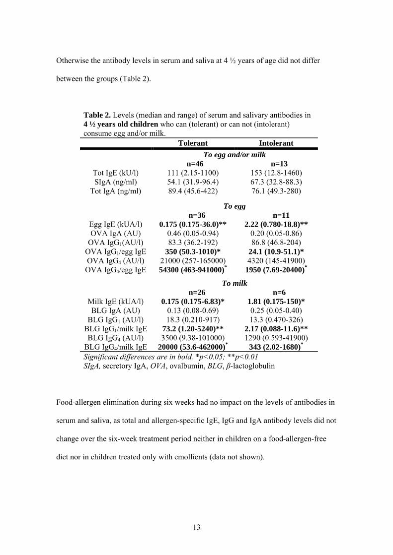
Otherwise the antibody levels in serum and saliva at 4 ½ years of age did not differ
between the groups (Table 2).
Table 2. Levels (median and range) of serum and salivary antibodies in 4 ½ years old children who can (tolerant) or can not (intolerant) consume egg and/or milk. Tolerant Intolerant To egg and/or milk
n=46 n=13 Tot IgE (kU/l) 111 (2.15-1100) 153 (12.8-1460) SIgA (ng/ml) 54.1 (31.9-96.4) 67.3 (32.8-88.3)
Tot IgA (ng/ml) 89.4 (45.6-422) 76.1 (49.3-280)
To egg n=36 n=11
Egg IgE (kUA/l) 0.175 (0.175-36.0)** 2.22 (0.780-18.8)** OVA IgA (AU) 0.46 (0.05-0.94) 0.20 (0.05-0.86)
OVA IgG1(AU/l) 83.3 (36.2-192) 86.8 (46.8-204) OVA IgG1/egg IgE 350 (50.3-1010)* 24.1 (10.9-51.1)* OVA IgG4 (AU/l) 21000 (257-165000) 4320 (145-41900) OVA IgG4/egg IgE 54300 (463-941000)* 1950 (7.69-20400)*
To milk n=26 n=6
Milk IgE (kUA/l) 0.175 (0.175-6.83)* 1.81 (0.175-150)* BLG IgA (AU) 0.13 (0.08-0.69) 0.25 (0.05-0.40)
BLG IgG1 (AU/l) 18.3 (0.210-917) 13.3 (0.470-326) BLG IgG1/milk IgE 73.2 (1.20-5240)** 2.17 (0.088-11.6)** BLG IgG4 (AU/l) 3500 (9.38-101000) 1290 (0.593-41900)
BLG IgG4/milk IgE 20000 (53.6-462000)* 343 (2.02-1680)* Significant differences are in bold. *p<0.05; **p<0.01 SIgA, secretory IgA, OVA, ovalbumin, BLG, β-lactoglobulin
Food-allergen elimination during six weeks had no impact on the levels of antibodies in
serum and saliva, as total and allergen-specific IgE, IgG and IgA antibody levels did not
change over the six-week treatment period neither in children on a food-allergen-free
diet nor in children treated only with emollients (data not shown).
13

DISCUSSION
The present study indicates that sensitized, eczematous children with high ratios of
IgG4/IgE to food allergens at an early age are more likely to consume the offending
foods at 4 ½ years of age, than children with low ratios. This could not be explained by
different antigen exposure in the two groups of children on inclusion, as the differences
were noted before any elimination diet of the infants had been instituted. High levels of
IgE to food allergens have earlier been shown to predict persistent food allergy [21, 22],
but in the present study this association could not be verified, possibly due to the low
numbers of children in the non-tolerant group. Instead high IgG4 antibody levels to food
allergens early in life foretell the ability for initially food SPT positive children to
consume the food at 4 ½ years of age.
Previous studies on immunotherapy with inhalant allergens indicate a protective role of
IgG4 antibodies, as the levels increase during treatment [23, 24]. IgG4 antibodies to
allergens have been proposed to act as blocking antibodies by competing with IgE for
allergen binding to IgE receptor expressing cells, such as mast cells and basophils [23]
and competition between IgE and IgG4 antibodies at the level of antigen-presenting cells
has been demonstrated in vitro as well [23]. The production of both IgE and IgG4
antibodies is upregulated by IL-4 from activated Th2 cells [9]. Interleukin-10, however,
which is secreted by e.g. regulatory T cells during immunotherapy [23], potently
suppresses IgE production, and simultaneously increases IgG4 production [10]. Another
immune response associated with protection from development of allergic disease is a
proposed modified Th2 immune response that includes high levels of IgG4 antibodies in
combination with a lack of IgE antibodies [11]. Clinical and immunological outcomes
14

after immunotherapy with food allergens are less investigated, but it has been suggested
that tolerance may be induced after oral administration of food allergens [5]. Recently,
it was shown that in food allergic children, 64% could at least tolerate daily doses of
actual food after a specific oral tolerance induction scheme, compared to 35% of food
allergic children on an elimination diet [25]. Our study may indicate that IgG4 may be a
valuable parameter to foretell which children that could benefit from administration of
food allergens instead of elimination diet, although this needs further evaluation.
The levels of IgG1 antibodies have also been shown to increase during immunotherapy,
at least during the beginning of treatment. However, other investigators have observed
increased levels of IgG1 to OVA [26] and BLG [27] in children with persisting
sensitization to food. In our study the levels of IgG1 antibodies to BLG and the ratios of
IgG1/IgE to BLG, tended to be higher on inclusion in children who could drink milk at 4
½ years of age compared to those children who could not. The expression of IgG1 is
promoted by IFNγ, and the antibody can form immune complexes with antigens, bind to
Fc receptors on lymphocytes and activate complement [28]. The favored IgG1 antibody
production in these children may depend on an immune response that is balanced
towards a Th1 milieu, possibly in combination with IgG4-inducing factors, e.g. IL-10.
High doses of allergens might favor the induction of Th1-type responses [29], and some
studies on airborne allergens suggest that exposure to the allergen may favor
immunological tolerance [30].
Serum antibodies to cow’s milk decrease during the first year of life in healthy children,
indicating immunological tolerance development to these proteins during continued
15

ingestion [8]. Furthermore, salivary IgA and serum IgG antibody responses to BLG
normally decrease with age in children, as previously reported by our group [7, 8, 15].
However, in this study we found no clear changes in antibody levels in eczematous,
sensitized children during their six-week treatment period with allergen elimination,
which is in line with a previous study reporting constant levels of serum antibodies
during an elimination diet for three weeks [31]. It can be speculated whether a treatment
period of three to six weeks is sufficient to ensure any changes in antibody levels. The
half-life of antibodies fluctuates, but may be up to several weeks, and antibody-
producing plasma cells may be much more long-lived than has been proposed earlier
(reviewed in [32]).
In the present study, we report that a small subgroup of children with negative food-
allergen SPT but with circulating IgE antibodies to egg and/or milk had higher IgG4/IgE
ratios than children with positive SPT to egg and/or milk. The negative outcome in SPT
in this sensitized subgroup may be explained by their high levels of IgG4 antibodies to
allergens competing with IgE for allergen binding to IgE receptor expressing cells and
thereby inhibiting mast cell degranulation [23]. Moreover, this response may be
associated with high levels of anti-inflammatory IL-10, which has been observed to
correlate negatively with the SPT wheal diameter [33].
A definite limitation of our study is that double-blind, placebo-controlled food
challenges (DBPCFC) were not routinely performed on inclusion, but were performed
later. Consequently, we can not prove that the sensitized children with eczema had food
allergy on inclusion. We can only state whether they tolerated the food items at 4 ½
16

years or not. The children classified as non-tolerant at 4 ½ years had either had a
positive DBPCFC or a recent reaction at accidental exposure. However, it is worth to
report that when analysing the three children with positive DBPCFC separately, we still
could observe lower levels of IgG4 and IgG4/IgE ratios in them, compared to children
who had introduced the eliminated food without problems (data not shown).
Incidentally, our results coincide with a recent study by Noh et al [34], reporting the
outcome of DBPCFC in older children, mean age 13.9 years. They found that the mean
IgE/IgG4 ratio was significantly higher in children with a positive challenge test,
compared to those with a negative test. Also, results from a recent study show that
elevated levels of cow’s milk specific IgG4 was associated with tolerance to milk in
atopic children and adults [35].
In summary, eczematous, food sensitized children with high levels of IgG4 and high
ratios of IgG4/IgE antibodies to food allergens early in life are more likely to consume
the offending food at 4 ½ years than children with low levels and ratios. Treatment with
allergen exclusion and/or skin care for six weeks did not significantly affect the levels
of serum IgE, IgG or salivary IgA antibodies in eczematous children.
17

ACKNOWLEDGEMENTS
We are grateful to all children and their parents who participated in this study. We also
wish to thank Anne-Marie Fornander, Kristina Warstedt and Ulrika Bengtsson for
excellent technical assistance and the participating nurses Birgit Burghauser, Christina
Helander, Christina Svensk, Elisabeth Andersson, Gunnel Bergsten, Lena Lindell,
Margareta Mattson and Monica Thunberg. The advice on language from Maurice
Devenney is gratefully acknowledged.
The Health Research Council in the South-east of Sweden, the Swedish Asthma and
Allergy Association’s Research Fund, the Swedish Research Council for Environment
Agricultural Sciences and Spatial Planning, the Swedish Samariten Foundation, The
Swedish Society of Medicine and County Council of Östergötland are acknowledged
for financial support.
18
REFERENCES
1. Johansson SGO, Bieber T, Dahl R, et al. Revised nomenclature for allergy for global use: Report of the nomenclature Review Committee of the World Allergy Organization, Oct 2003. J Allergy Clin Immunol 2004; 113:832-6.
2. Bruijnzeel-Koomen C, Ortolani C, Aas K, et al. Adverse reactions to food. European Academy of Allergology and Clinical Immunology 1995; 50:623-35.
3. Sicherer SH. Food allergy: When and how to perform oral food challenges. Pediatr Allergy Immunol 1999; 10:226-34.
4. Brand PLP, Vlieg-Boerstra BJ, Dubois AEJ. Dietary prevention of allergic disease in children: Are current recommendations really based on good evidence? Pediatr Allergy Immunol 2007; 18:475-9.
5. Niggemann B, Staden U, Rolinck-Werninghaus C, Beyer K. Specific oral tolerance induction in food allergy. Allergy 2006; 61:808-11.
6. Hattevig G, Kjellman B, Björkstén B. Appearance of IgE antibodies to ingested and inhaled allergens during the first 12 years of life in atopic and non-atopic children. Pediatr Allergy Immunol 1993; 4:182-6.
7. Jenmalm MC, Björkstén B. Development of immunoglobulin G subclass antibodies to ovalbumin, birch and cat during the first eight years of life in atopic and non-atopic children. Pediatr Allergy Immunol 1999; 10:112-21.
8. Jenmalm MC, Björkstén B. Exposure to cow's milk during the first three months of life is associated with increased levels of IgG subclass antibodies to β-lactoglobulin up to eight years. J Allergy Clin Immunol 1998; 102:671-8.
9. Ishizaka A, Sakiyama Y, Nakanishi M, Tomizawa K, Oshika E, Kojima K. The inductive effect of interleukin 4 on IgG4 and IgE synthesis in human peripheral blood lymphocytes. Clin Exp Allergy 1990; 79:392-6.
10. Satoguina J, Weyand E, Larbi J, Hoerauf A. T regulatory-1 cells induce IgG4 production by B cells: Role of IL-10. J Immunol 2005; 174:4718-26.
11. Platts-Mills TAE, Vaughan J, Squillace S, Woodfolk J, Sporik R. Sensitisation, asthma, and a modified Th2 response in children exposed to cat allergen: a population-based cross-sectional study. Lancet 2001; 357:752-6.
12. Juvonen P, Månsson M, Kjellman NIM, Björkstén B, Jakobsson I. Development of IgG and IgE antibodies to cow's milk proteins and ovalbumin after a temporary neonatal exposure to hydrolysed and whole cow's milk proteins. Pediatr Allergy Immunol 1999; 10:191-8.
13. van Asperen PP, Gleeson M, Kemp AS, et al. The relationship between atopy and salivary IgA deficiency in infancy. Clin Exp Immunol 1985; 62:753-7.
14. Gleeson M, Cripps AW, Clancy RL, Hensley MJ, Henry RL, Wlodarczyk JH. The significance of transient mucosal IgA deficiency on the development of asthma and atopy in children. In: Mestecky J, Russell MW, Jackson S, Michalek SM, Tlaskalová-Hogenová H, Stersl J, editors. Advances in Mucosal Immunology. New York: Plenum Press; 1996. p. 861-4.
15. Böttcher MF, Häggström P, Björkstén B, Jenmalm MC. Total and allergen-specific immunoglobulin A levels in saliva in relation to the development of allergy in infants up to 2 years of age. Clin Exp Allergy 2002; 32:1293-8.
16. Norrman G, Tomičić S, Böttcher MF, Oldaeus G, Strömberg L, Fälth-Magnusson K. Significant improvement of eczema with skin care and food elimination in small children. Acta Paediatr 2005; 94:1384-8.
19

17. Devenney I, Norrman G, Oldaeus G, Strömberg L, Fälth-Magnusson K. A new model for low-dose food challenge in children with allergy to milk or egg. Acta Paediatr 2006; 95:1133-9.
18. Jenmalm MC, Holt PG, Björkstén B. Maternal influence on IgG subclass antibodies to Bet v 1 during the first 18 months of life as detected with a sensitive ELISA. Int Arch Allergy Immunol 1997; 114:175-84.
19. Böttcher MF, Jenmalm MC, Björkstén B. Cytokine, chemokine and secretory IgA levels in human milk in relation to atopic disease and IgA production in infants. Pediatr Allergy Immunol 2003; 14:35-41.
20. Casas R, Böttcher MF, Duchén K, Björkstén B. Detection of IgA antibodies to cat, β-lactoglobulin, and ovalbumin allergens in human milk. J Allergy Clin Immunol 2000; 105:1236-40.
21. Saarinen KM, Pelkonen AS, Mäkelä MJ, Savilahti E. Clinical course and prognosis of cow's milk allergy are dependent on milk-specific IgE status. J Allergy Clin Immunol 2005; 116:869-75.
22. Vanto T, Helppilä S, Juntunen-Backman K, et al. Prediction of the development of tolerance to milk in children with cow's milk hypersensitivity. J Pediatr 2004; 144:218-22.
23. Till SJ, Francis JN, Nouria-Aria KT, Durham SR. Mechanisms of immunotherapy. J Allergy Clin Immunol 2004; 113.
24. Lue KH, Lin YH, Sun HL, Lu KH, Hsieh J-C, Chou M-C. Clinical and immunological effects of sublingual immunotherapy in asthmatic children sensitized to mites: a double-blind, randomized, placebo-controlled study. Pediatr Allergy Immunol 2006; 17:408-15.
25. Staden U, Rolinck-Werninghaus C, Brewe F, Wahn U, Niggemann B, Beyer K. Specific oral tolerance induction in food allergy in children: efficacy and clinical patterns of reaction. Allergy 2007; 62:1261-9.
26. Vance GHS, Thornton CA, Bryant TN, Warner JA, Warner JO. Ovalbumin-specific immunoglobulin G and subclass responses through the first 5 years of life in relation to duration of egg sensitization and the developmnet of asthma. Clin Exp Allergy 2004; 34:1542-9.
27. Sletten GBG, Halvorsen R, Egaas E, Halstensen TS. Changes in humoral responses to β-lactoglobulin in tolerant patients suggest a particular role for IgG4 in delayed, non-IgE-mediated cow's milk allergy Pediatr Allergy Immunol 2006; 17:435-43.
28. Getahun A, Heyman B. How antibodies act as natural adjuvants. Immunological Letters 2006; 104:38-45.
29. Secrist H, DeKruyff RH, Umetsu DT. Interleukin 4 production by CD4+ T cells from allergic individuals is modulated by antigen concentration and antigen-presenting cell type. J Exp Med 1995; 181:1081-9.
30. Perzanowski MS, Rönmark E, Platts-Mills TA, Lundbäck B. Effect of cat and dog ownership on sensitization and development of asthma among preteenage children. Am J Respir Crit Care Medicine 2002; 166:696-702.
31. Sloper KS, Wadsworth J, Brostoff J. Children with atopic eczema II: Immunological findings associated with dietary manipulations. QJM 1991; 80:695-705.
20

21
32. Radbruch A, Muehlinghaus G, Louger EO, et al. Competence and competition: the challenge of becoming a long-lived plasma cell. Nat Rev Immunol 2006; 6:741-50.
33. Macaubas C, Sly PD, Burton PR, et al. Regulation of T-helper cell responses to inhalant allergen during early childhood. Clin Exp Allergy 1999; 29:1223-31.
34. Noh G, Ahn H-S, Cho N-Y, Lee S, Oh J-W. The clinical significance of food specific IgE/IgG4 in food specific atopic dermatitis. Pediatr Allergy Immunol 2007; 18:63-70.
35. Ruiter B, Knol EF, Van Neerven RJJ, et al. Maintenance of tolerance to cow's milk in atopic individuals is characterized by high levels of specific immunoglobulin G4. Clin Exp Allergy 2007; 37:1103-10.