High - gut.bmj.com · sphincter with the levator ani and frequently Disruption ofthefibres ofthe...
4
Gut 1995; 37: 284-287 METHOD High resolution magnetic resonance imaging of the anal sphincter using an internal coil N M deSouza, W A Kmiot, R Puni, A S Hall, M Burl, C I Bartram, G M Bydder The Robert Steiner Magnetic Resonance Unit, Department of Radiology, Royal Postgraduate Medical School, London N M deSouza R Puni G M Bydder Department of Surgery, Royal Postgraduate Medical School, London W A Kmiot Hirst Research Centre, Elstree Way, Borehamwood A S Hall M Burl Department of Radiology, St Mark's Hospital, City Road, London C I Bartram Correspondence to: Mr W A Kmiot, Department of Surgery, Royal Postgraduate Medical School, Hammersmith Hospital, Du Cane Road, London W12 OHS. Accepted for rapid communication 29 March 1995 Abstract An internal receiver coil was used to obtain high resolution transverse and oblique coronal magnetic resonance images of the anal sphincter in five normal volunteers and five patients. The internal sphincter had a high signal intensity on T1 weighted, T2 weighted, and STIR sequences whereas the conjoined longitudinal muscle and external sphincter had a low signal inten- sity. The internal sphincter (but not the external sphincter) showed contrast enhancement after administration of intravenous gadopentetate dimeglumine. The oblique coronal plane was particularly useful for showing the thickness and the relations of the external sphincter. Sphinc- teric abscesses as well as muscle defects, hypertrophy, and atrophy were clearly shown. The coil was well tolerated by most subjects. It has considerable potential for improving the diagnosis of anorectal disease. (Gut 1995; 37: 284-287) Keywords: anal sphincter, magnetic resonance imaging, endoanal coil. Disease of the anal sphincter is a common and distressing problem. Anal endosonography is the imaging technique of choice for showing abnormalities in this region' 2 and has proved superior to conventional magnetic resonance imaging (MRI) when a whole body receiver coil is used.3 The quality of magnetic resonance images may be improved by the use of a receiver coil placed in close proximity to the region of interest. We therefore constructed a dedicated insertable coil for MRI of the anal sphincter and assessed its value in a pilot study of 10 subjects. Methods This study was approved by the research ethics committee of the Royal Postgraduate Medical School. Five adult volunteers (three male and two female, aged 31 to 68 years; median 49 years) were imaged. All volunteers were free of anorectal symptoms and had normal anal sphincter resting and squeeze tone on rectal examination. Five patients (three male and two female, aged 26 to 77 years, median 47) were also imaged. Two of these patients presented with purulent anal discharge due to fistula in ano, one with difficult defecation because of anal stenosis, and two with faecal incontinence (one because of sphincter disruption after obstetric trauma and the other idiopathic). Nine subjects were examined in the supine position. The lateral decubitus position was used for the patient with anal stenosis who could not lie supine. A cylindrical receiver coil 75 mm long and 9 mm in diameter was covered with a condom, placed within the anal canal and supported by an adjustable stand. Imaging was performed on either a 0.5 Tesla (T) Picker Asset or a 1.0 T Picker HPQ Vista MR scanner. Mildly TI weighted spin echo (SE 780/20 msec (TRITE)), T2 weighted spin echo (SE 2500/80 msec (TRITE)), and. STIR (short inversion time (TI) inversion recovery, IR 2500/30/110 (TRfIE/TI)) sequences were used. Transverse (perpendicular to the coil long axis) and oblique coronal (parallel to the coil long axis) 3 mm contiguous slices were obtained with a 192X 256 matrix and two to four signal acquisitions. A 10-12 cm field of view was used. Intravenous gadopentetate dimeglumine (Gadolinium- DTPA, 0 1 mmol/kg body weight) was given to one patient with anal discharge due to fistula in ano and another with liquid faecal incontinence. Results High resolution images of the anal sphincter were obtained in all subjects. The tissue signal was highest adjacent to the coil and decreased with distance from it. The useful imaging region extended at least 5 cm out from the centre of the coil in a plane transverse to the coil and included all components of the Relative signal intensities with different pulse sequences Musculus Anterior Conjoined External submucosae submucosal Internal longitudinal sphincter and Sequence Submucosa ali veins sphincter muscle levator ali T1 weighted spin echo Very high Moderate Low Intermediate Low Low T, weighted spin echo with contrast Very high Moderate High Very high Low Low T2 weighted spin echo Very high Moderate High High Low Low STIR Very high Moderate High High Low Low 284 on January 26, 2020 by guest. Protected by copyright. http://gut.bmj.com/ Gut: first published as 10.1136/gut.37.2.284 on 1 August 1995. Downloaded from
Transcript of High - gut.bmj.com · sphincter with the levator ani and frequently Disruption ofthefibres ofthe...

Gut 1995; 37: 284-287
METHOD
High resolution magnetic resonance imaging ofthe anal sphincter using an internal coil
N M deSouza, W A Kmiot, R Puni, A S Hall, M Burl, C I Bartram, G M Bydder
The Robert SteinerMagnetic ResonanceUnit, Department ofRadiology, RoyalPostgraduate MedicalSchool, LondonN M deSouzaR PuniG M Bydder
Department ofSurgery, RoyalPostgraduate MedicalSchool, LondonW A Kmiot
Hirst Research Centre,Elstree Way,BorehamwoodA S HallM Burl
Department ofRadiology, St Mark'sHospital, City Road,LondonC I Bartram
Correspondence to:MrW A Kmiot, Departmentof Surgery, RoyalPostgraduate MedicalSchool, HammersmithHospital, Du Cane Road,London W12 OHS.
Accepted for rapidcommunication29 March 1995
AbstractAn internal receiver coil was used to obtainhigh resolution transverse and obliquecoronal magnetic resonance images of theanal sphincter in five normal volunteersand five patients. The internal sphincterhad a high signal intensity on T1 weighted,T2 weighted, and STIR sequences whereasthe conjoined longitudinal muscle andexternal sphincter had a low signal inten-sity. The internal sphincter (but not theexternal sphincter) showed contrastenhancement after administration ofintravenous gadopentetate dimeglumine.The oblique coronal plane was particularlyuseful for showing the thickness and therelations ofthe external sphincter. Sphinc-teric abscesses as well as muscle defects,hypertrophy, and atrophy were clearlyshown. The coil was well tolerated by mostsubjects. It has considerable potential forimproving the diagnosis of anorectaldisease.(Gut 1995; 37: 284-287)
Keywords: anal sphincter, magnetic resonanceimaging, endoanal coil.
Disease of the anal sphincter is a common anddistressing problem. Anal endosonography isthe imaging technique of choice for showingabnormalities in this region' 2 and has provedsuperior to conventional magnetic resonance
imaging (MRI) when a whole body receiver coilis used.3 The quality of magnetic resonanceimages may be improved by the use of a receivercoil placed in close proximity to the region ofinterest. We therefore constructed a dedicatedinsertable coil for MRI of the anal sphincter andassessed its value in a pilot study of 10 subjects.
MethodsThis study was approved by the research ethicscommittee of the Royal Postgraduate Medical
School. Five adult volunteers (three male andtwo female, aged 31 to 68 years; median 49years) were imaged. All volunteers were free ofanorectal symptoms and had normal analsphincter resting and squeeze tone on rectalexamination. Five patients (three male and twofemale, aged 26 to 77 years, median 47) werealso imaged. Two of these patients presentedwith purulent anal discharge due to fistula inano, one with difficult defecation because ofanal stenosis, and two with faecal incontinence(one because of sphincter disruption afterobstetric trauma and the other idiopathic).Nine subjects were examined in the supine
position. The lateral decubitus position wasused for the patient with anal stenosis whocould not lie supine. A cylindrical receiver coil75 mm long and 9 mm in diameter was coveredwith a condom, placed within the anal canal andsupported by an adjustable stand. Imaging wasperformed on either a 0.5 Tesla (T) PickerAsset or a 1.0 T Picker HPQ Vista MR scanner.Mildly TI weighted spin echo (SE 780/20 msec(TRITE)), T2 weighted spin echo (SE 2500/80msec (TRITE)), and. STIR (short inversiontime (TI) inversion recovery, IR 2500/30/110(TRfIE/TI)) sequences were used. Transverse(perpendicular to the coil long axis) and obliquecoronal (parallel to the coil long axis) 3 mmcontiguous slices were obtained with a 192X256 matrix and two to four signal acquisitions.A 10-12 cm field ofview was used. Intravenousgadopentetate dimeglumine (Gadolinium-DTPA, 0 1 mmol/kg body weight) was given toone patient with anal discharge due to fistula inano and another with liquid faecal incontinence.
ResultsHigh resolution images of the anal sphincterwere obtained in all subjects. The tissue signalwas highest adjacent to the coil and decreasedwith distance from it. The useful imagingregion extended at least 5 cm out from thecentre of the coil in a plane transverse to thecoil and included all components of the
Relative signal intensities with different pulse sequences
Musculus Anterior Conjoined Externalsubmucosae submucosal Internal longitudinal sphincter and
Sequence Submucosa ali veins sphincter muscle levator ali
T1 weighted spin echo Very high Moderate Low Intermediate Low LowT, weighted spin echo with contrast Very high Moderate High Very high Low LowT2 weighted spin echo Very high Moderate High High Low LowSTIR Very high Moderate High High Low Low
284
on January 26, 2020 by guest. Protected by copyright.
http://gut.bmj.com
/G
ut: first published as 10.1136/gut.37.2.284 on 1 August 1995. D
ownloaded from
MRI of the anal sphincter
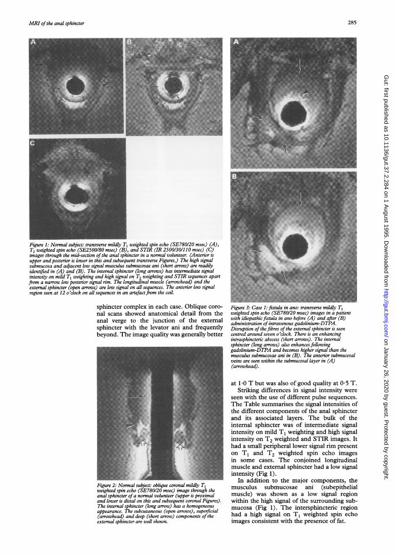
Figure 1: Normal subject: transverse mildly T1 weighted spin echo (SE780/20 msec) (A),T2 weighted spin echo (SE2S00/80 msec) (B), and STIR (IR 2500/30/11 0 msec) (C)images through the mid-section of the anal sphincter in a normal volunteer. (Anterior isupper and posterior is lower in this and subsequent transverse Figures.) The high signalsubmucosa and adjacent low signal musculus submucosae ani (short arrow) are readilyidentified in (A) and (B). The internal sphincter (long arrows) has intermediate signalintensity on mild T1 weighting and high signal on T2 weighting and STIR sequences apart,from a narrow low posterior signal rim. The longitudinal muscle (arrowhead) and theexternal sphincter (open arrows) are low signal on all sequences. The anterior low signalregion seen at 12 o'clock on all sequences in an artefactfrom the coil.
sphincter complex in each case. Oblique coro- Figure 3: Case 1: fistula in ano: transverse mildly T1nal scans showed anatomical detail from the weighted spin echo (SE780/20 msec) images in a patientanal verge to the junction of the external with idiopathic fistula in ano before (A) and after (B)administration of intravenous gadolinium-DTPA.sphincter with the levator ani and frequently Disruption of the fibres of the external sphincter is seenbeyond. The image quality was generally better centred around seven o'clock. There is an enhancing
intrasphincteric abscess (short arrows). The internalsphincter (long arrows) also enhances followinggadolinium-DTPA and becomes higher signal than themusculus submucosae ani in (B). The anterior submucosalveins are seen within the submucosal layer in (A)(arrowhead).
at 1-0 T but was also of good quality at 0 5 T.Striking differences in signal intensity were
seen with the use of different pulse sequences.The Table summarises the signal intensities ofthe different components of the anal sphincterand its associated layers. The bulk of theinteral sphincter was of intermnediate signalintensity on mild T1 weighting and high signal
w : 2fi t1 ! intensity on T2 weighted and STIR images. Ithad a small peripheral lower signal rim presenton T1 and T2 weighted spin echo imagesin some cases. The conjoined longitudinalmuscle and external sphincter had a low signalintensity (Fig 1).
In addition to the major components, theFigure 2: Normal subject- oblique coronal mildly T1 musculus submucosae ani (subepithelialweighted spin echo (SE780/20 msec) image through the musclus shosae am signal regionanal sphincter of a normal volunteer (upper is proximal muscle) was shown as a low signal regionand lower is distal on this and subsequent coronal Figures). within the high signal of the surrounding sub-The internal sphincter (long arrow) has a homogeneous mucosa (Fig 1). The intersphincteric regionappearance. The subcutaneous (open arrows), superficial(arrowhead) and deep (short arrow) components of the had a high signal on T1 weighted spin echoexternal sphincter are well shown. images consistent with the presence of fat.
285
on January 26, 2020 by guest. Protected by copyright.
http://gut.bmj.com
/G
ut: first published as 10.1136/gut.37.2.284 on 1 August 1995. D
ownloaded from
deSouza, Kmiot, Puni, Hall, Burl, Bartram, Bydder
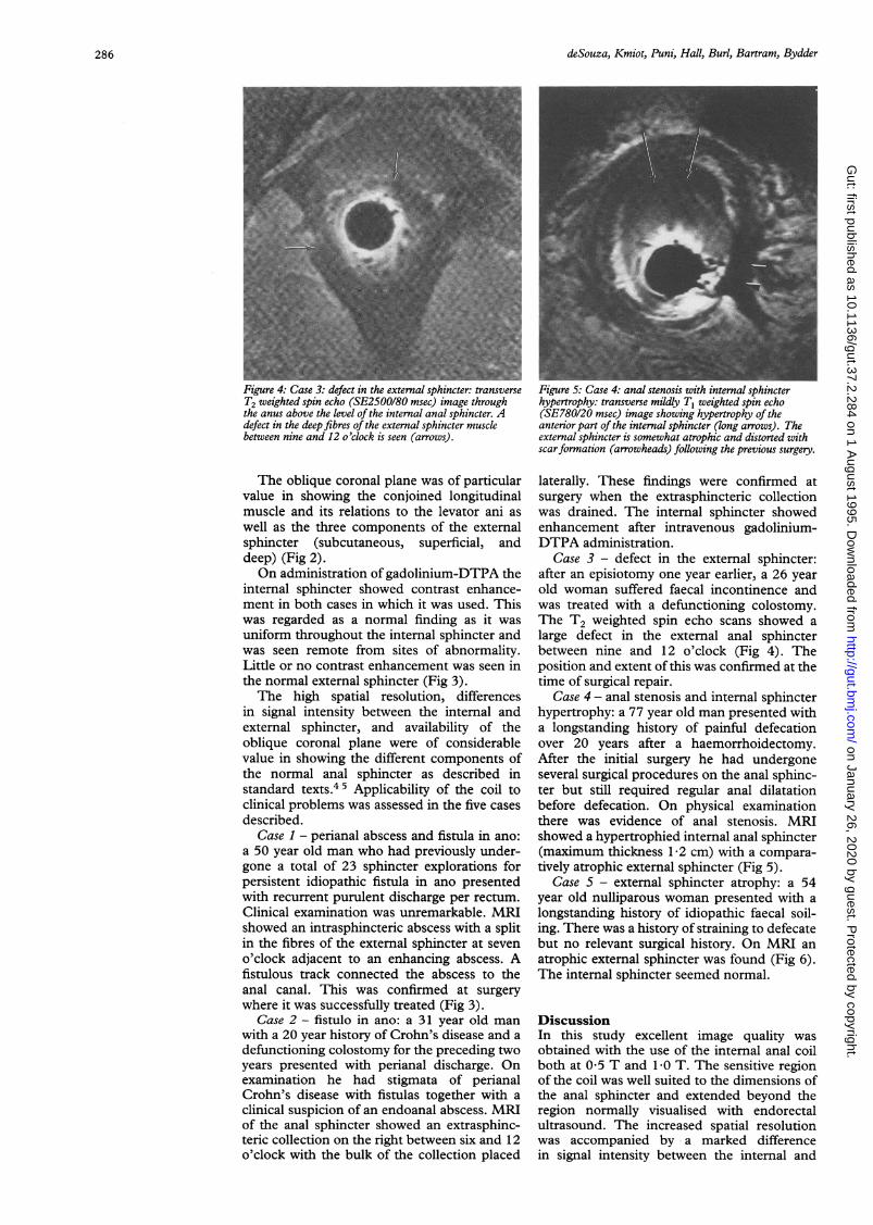
rigure 4: uase 3: aeject in tne external sphincter: transverse rigure .: Case 4: anal stenosis witl internal sphincterT2 weighted spin echo (SE2500/80 msec) image through hypertrophy: transverse mildly T1 weighted spin echothe anus above the level of the internal anal sphincter. A (SE780/20 msec) image showing hypertrophy of thedefect in the deep fibres of the external sphincter muscle anterior part of the internal sphincter (long arrows). Thebetween nine and 12 o'clock is seen (arrows). external sphincter is somewhat atrophic and distorted with
scarformation (arrowheads) following the previous surgery.
The oblique coronal plane was of particularvalue in showing the conjoined longitudinalmuscle and its relations to the levator ani aswell as the three components of the externalsphincter (subcutaneous, superficial, anddeep) (Fig 2).On administration ofgadolinium-DTPA the
internal sphincter showed contrast enhance-ment in both cases in which it was used. Thiswas regarded as a normal finding as it wasuniform throughout the internal sphincter andwas seen remote from sites of abnormality.Little or no contrast enhancement was seen inthe normal external sphincter (Fig 3).The high spatial resolution, differences
in signal intensity between the internal andexternal sphincter, and availability of theoblique coronal plane were of considerablevalue in showing the different components ofthe normal anal sphincter as described instandard texts.4 5 Applicability of the coil toclinical problems was assessed in the five casesdescribed.
Case 1 - perianal abscess and fistula in ano:a 50 year old man who had previously under-gone a total of 23 sphincter explorations forpersistent idiopathic fistula in ano presentedwith recurrent purulent discharge per rectum.Clinical examination was unremarkable. MRIshowed an intrasphincteric abscess with a splitin the fibres of the external sphincter at seveno'clock adjacent to an enhancing abscess. Afistulous track connected the abscess to theanal canal. This was confirmed at surgerywhere it was successfully treated (Fig 3).
Case 2 - fistulo in ano: a 31 year old manwith a 20 year history of Crohn's disease and adefunctioning colostomy for the preceding twoyears presented with perianal discharge. Onexamination he had stigmata of perianalCrohn's disease with fistulas together with aclinical suspicion of an endoanal abscess. MRIof the anal sphincter showed an extrasphinc-teric collection on the right between six and 12o'clock with the bulk of the collection placed
laterally. These findings were confirmed atsurgery when the extrasphincteric collectionwas drained. The internal sphincter showedenhancement after intravenous gadolinium-DTPA administration.
Case 3 - defect in the external sphincter:after an episiotomy one year earlier, a 26 yearold woman suffered faecal incontinence andwas treated with a defunctioning colostomy.The T2 weighted spin echo scans showed alarge defect in the external anal sphincterbetween nine and 12 o'clock (Fig 4). Theposition and extent of this was confirmed at thetime of surgical repair.
Case 4- anal stenosis and internal sphincterhypertrophy: a 77 year old man presented witha longstanding history of painful defecationover 20 years after a haemorrhoidectomy.After the initial surgery he had undergoneseveral surgical procedures on the anal sphinc-ter but still required regular anal dilatationbefore defecation. On physical examinationthere was evidence of anal stenosis. MRIshowed a hypertrophied internal anal sphincter(maximum thickness 1-2 cm) with a compara-tively atrophic external sphincter (Fig 5).
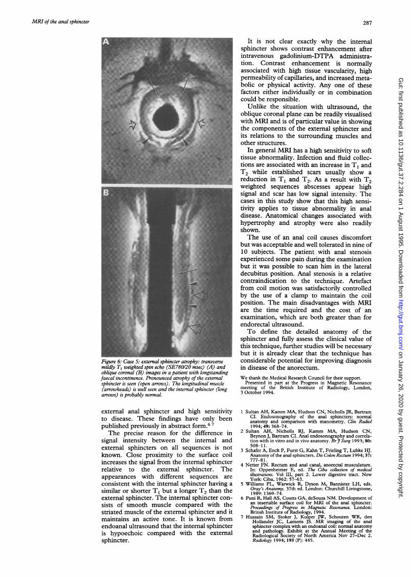
Case 5 - external sphincter atrophy: a 54year old nulliparous woman presented with alongstanding history of idiopathic faecal soil-ing. There was a history of straining to defecatebut no relevant surgical history. On MRI anatrophic external sphincter was found (Fig 6).The internal sphincter seemed normal.
DiscussionIn this study excellent image quality wasobtained with the use of the internal anal coilboth at 0.5 T and 1.0 T. The sensitive regionof the coil was well suited to the dimensions ofthe anal sphincter and extended beyond theregion normally visualised with endorectalultrasound. The increased spatial resolutionwas accompanied by a marked differencein signal intensity between the internal and
286
on January 26, 2020 by guest. Protected by copyright.
http://gut.bmj.com
/G
ut: first published as 10.1136/gut.37.2.284 on 1 August 1995. D
ownloaded from
MRI of the anal sphincter 287
:~~~~~~~~~~~~~~~~~~~~~~~~~~~~-- ---
4
Figure 6: Case 5: external sphincter atrophy: transversemildly T1 weighted spin echo (SE780/20 msec) (A) andoblique coronal (B) images in a patient with longstandingfaecal incontinence. Pronounced atrophy of the externalsphincter is seen (open arrows). The longitudinal muscle(arrowheads) is well seen and the internal sphincter (longarrows) is probably nornal.
external anal sphincter and high sensitivityto disease. These findings have only beenpublished previously in abstract form.6 7The precise reason for the difference in
signal intensity between the internal andexternal sphincters on all sequences is notknown. Close proximity to the surface coilincreases the signal from the internal sphincterrelative to the external sphincter. Theappearances with different sequences areconsistent with the internal sphincter having asimilar or shorter T1 but a longer T2 than theexternal sphincter. The internal sphincter con-sists of smooth muscle compared with thestriated muscle of the external sphincter and itmaintains an active tone. It is known fromendoanal ultrasound that the internal sphincteris hypoechoic compared with the externalsphincter.
It is not clear exactly why the internalsphincter shows contrast enhancement afterintravenous gadolinium-DTPA administra-tion. Contrast enhancement is normallyassociated with high tissue vascularity, highpermeability of capillaries, and increased meta-bolic or physical activity. Any one of thesefactors either individually or in combinationcould be responsible.
Unlike the situation with ultrasound, theoblique coronal plane can be readily visualisedwith MRI and is of particular value in showingthe components of the external sphincter andits relations to the surrounding muscles andother structures.
In general MRI has a high sensitivity to softtissue abnormality. Infection and fluid collec-tions are associated with an increase in T1 andT2 while established scars usually show areduction in T1 and T2. As a result with T2weighted sequences abscesses appear highsignal and scar has low signal intensity. Thecases in this study show that this high sensi-tivity applies to tissue abnormality in analdisease. Anatomical changes associated withhypertrophy and atrophy were also readilyshown.The use of an anal coil causes discomfort
but was acceptable and well tolerated in nine of10 subjects. The patient with anal stenosisexperienced some pain during the examinationbut it was possible to scan him in the lateraldecubitus position. Anal stenosis is a relativecontraindication to the technique. Artefactfrom coil motion was satisfactorily controlledby the use of a clamp to maintain the coilposition. The main disadvantages with MRIare the time required and the cost of anexamination, which are both greater than forendorectal ultrasound.To define the detailed anatomy of the
sphincter and fully assess the clinical value ofthis technique, further studies will be necessarybut it is already clear that the technique hasconsiderable potential for improving diagnosisin disease of the anorectum.We thank the Medical Research Council for their support.
Presented in part at the Progress in Magnetic Resonancemeeting of the British Institute of Radiology, London,3 October 1994.
1 Sultan AH, Kamm MA, Hudson CN, Nicholls JR, BartramCI. Endosonography of the anal sphincters: normalanatomy and comparison with manometry. Clin Radiol1994; 49: 368-74.
2 Sultan AH, Nicholls RJ, Kamm MA, Hudson CN,Beynon J, Bartram CI. Anal endosonography and correla-tion with in vitro and in vivo anatomy. BrJ7 Surg 1993; 80:508-11.
3 Schafer A, Enck P, Furst G, Kahn T, Frieling T, Lubke HJ.Anatomy of the anal sphincters. Dis Colon Rectum 1994; 37:777-81.
4 Netter FN. Rectum and anal canal, anorectal musculature.In: Oppenheimer E, ed. The Ciba collection of medicalillustrations. Vol III, part 2. Lower digestive tract. NewYork: Ciba, 1962: 57-63.
5 Williams PL, Warwick R, Dyson M, Bannister LH, eds.Gray's Anatomy. 37th ed. London: Churchill Livingstone,1989: 1369-74.
6 Puni R, Hall AS, Courts GA, deSouza NM. Development ofan insertable surface coil for MRI of the anal sphincter.Proceedings of Progress in Magnetic Resonance. London:British Institute of Radiology, 1994.
7 Hussain SM, Stoker J, Kulper JW, Schouten WR, denHollander JC, Lameris JS. MR imaging of the analsphincter complex with an endoanal coil: normal anatomyand pathology. Exhibit at the Annual Meeting of theRadiological Society of North America Nov 27-Dec 2.RadiologAy 1994; 193 (P): 445.
on January 26, 2020 by guest. Protected by copyright.
http://gut.bmj.com
/G
ut: first published as 10.1136/gut.37.2.284 on 1 August 1995. D
ownloaded from