
Embryonic Development VARIATIONS IN EMBRYONIC GERM LAYERS AND BODY CAVITY.

RESEARCH Open Access
HERV-H RNA is abundant in human embryonicstem cells and a precise marker for pluripotencyFederico A Santoni1,2*, Jessica Guerra2 and Jeremy Luban2,3*
Abstract
Background: Certain post-translational modifications to histones, including H3K4me3, as well as binding sites forthe transcription factor STAT1, predict the site of integration of exogenous gamma-retroviruses with great accuracyand cell-type specificity. Statistical methods that were used to identify chromatin features that predict exogenousgamma-retrovirus integration site selection were exploited here to determine whether cell type-specific chromatinmarkers are enriched in the vicinity of endogenous retroviruses (ERVs).
Results: Among retro-elements in the human genome, the gamma-retrovirus HERV-H was highly associated withH3K4me3, though this association was only observed in embryonic stem (ES) cells (p < 10-300) and, to a lesserextent, in induced pluripotent stem (iPS) cells. No significant association was observed in nearly 40 differentiatedcell types, nor was any association observed with other retro-elements. Similar strong association was observedbetween HERV-H and the binding sites within ES cells for the pluripotency transcription factors NANOG, OCT4, andSOX2. NANOG binding sites were located within the HERV-H 50LTR itself. OCT4 and SOX2 binding sites were within1 kB and 2 kB of the 50LTR, respectively. In keeping with these observations, HERV-H RNA constituted 2% of all polyA RNA in ES cells. As ES cells progressed down a differentiation pathway, the levels of HERV-H RNA decreasedprogressively. RNA-Seq datasets showed HERV-H transcripts to be over 5 kB in length and to have the structure50LTR-gag-pro-30LTR, with no evidence of splicing and no intact open reading frames.
Conclusion: The developmental regulation of HERV-H expression, the association of HERV-H with binding sites forpluripotency transcription factors, and the extremely high levels of HERV-H RNA in human ES cells suggest thatHERV-H contributes to pluripotency in human cells. Proximity of HERV-H to binding sites for pluripotencytranscription factors within ES cells might be due to retention of the same chromatin features that determined thesite of integration of the ancestral, exogenous, gamma-retrovirus that gave rise to HERV-H in the distant past.Retention of these markers, or, alternatively, recruitment of them to the site of the established provirus, may haveacted post-integration to fix the provirus within the germ-line of the host species. Either way, HERV-H RNA providesa specific marker for pluripotency in human cells.
Keywords: HERV-H, Endogenous retrovirus, Pluripotency, Long non-coding RNA, Embryonic stem cell, Inducedpluripotent stem cell
BackgroundVertebrate genomes contain retroviral sequences that arebelieved to be remnants of exogenous retroviral infectionfrom the distant past [1]. The genesis of these endogenousretroviruses (ERVs) necessitates establishment of provirus
by the ancestral, exogenous retrovirus within host germcells, such that these elements are maintained as heritablegenetic elements in the host species. Human endogenousretroviruses (HERVs) are not uncommon, and account forat least 8% of the total human DNA. By comparison withDNA sequences from exogenous retrovirus families, threemain classes of HERVs, gamma, beta, and delta have beenidentified [2]. Phylogenetic analyses identified HERV-K, abetaretrovirus, and HERV-H, a gammaretrovirus, as themost recent entries into the genomes of primates, 10 and25 million years ago, respectively [3,4].
* Correspondence: [email protected]; [email protected] of Genetic Medicine and Development, University of Geneva, 1rue Michel-Servet, Geneva CH-1211, Switzerland2Department of Microbiology and Molecular Medicine, University of Geneva,1211, Geneva 4, SwitzerlandFull list of author information is available at the end of the article
© 2012 Santoni et al.; licensee BioMed Central Ltd. This is an Open Access article distributed under the terms of the CreativeCommons Attribution License (http://creativecommons.org/licenses/by/2.0), which permits unrestricted use, distribution, andreproduction in any medium, provided the original work is properly cited.
Santoni et al. Retrovirology 2012, 9:111http://www.retrovirology.com/content/9/1/111

Though some HERVs are transcriptionally active, mostretroviral sequences in the human genome are corruptedby mutations or successive insertion of transposable ele-ments; most HERVs lack intact open reading frames forviral protein production and no autonomously replicatingHERV has been identified. As a result, HERVs are gener-ally considered non-functional, junk DNA. In some cases,though, HERVs make significant - even essential - contri-butions to the normal physiology of the host species [5-8].HERVs have also been implicated in the development ofpathological conditions [9-14]. Interestingly, HERVs canbe activated by exogenous retrovirus infection [15] and, inreciprocal fashion, the immune response to these elementscan influence the outcome of infection by pathogenic, ex-ogenous retroviruses such as HIV-1 [16,17].Exogenous retroviruses integrate into locations through-
out the host chromosomal DNA in a quasi-random fashion(reviewed extensively in [18]). Previously, we developedstatistical tools to identify association between the sites ofprovirus establishment and chromosome marks – as deter-mined by chromatin immuno-precipitation with massivelyparallel DNA sequencing (ChIPSeq) [18,19]. From thisanalysis we identified chromatin modifications (H3K4me3,H3K4me1, and H3K9ac) and binding sites for transcriptionfactors (STAT1) that predict the site of integration forgamma-retroviruses with great accuracy in a cell-type spe-cific manner [18,19]. Precise markers such as these werenot identified for other classes of retrovirus such as thelentivirus HIV-1 [18]. Here, the same statistical methodswere exploited to determine whether any ERVs retain asso-ciation with cell type-specific chromatin features thatmight have determined the site of integration in the distantpast, or that served to fix the provirus in the genetic patri-mony of the host species.
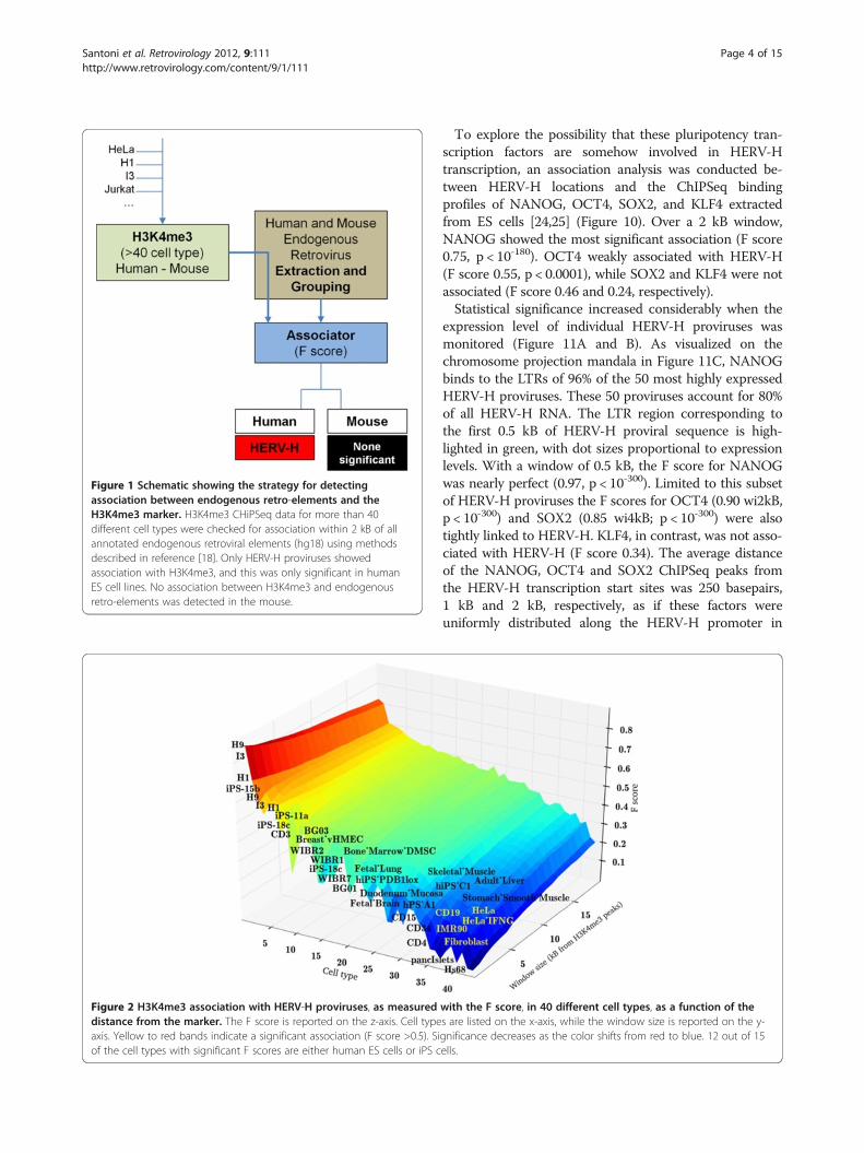
ResultsSearch for chromatin features near the site of ERVsPreviously observed, high-level association betweengamma-retrovirus integration sites and particular epi-genetic markers [18] prompted a quest to find associ-ation between any known endogenous retroviral elementin the human and mouse genomes, and the cell-typespecific localization of particular chromosomal features.For this purpose, ChIPSeq profiles were evaluated frommore than 40 different human and mouse cell types, in-cluding ES cells, iPS cells, monocytes, HeLa cells, CD4+T cells, and CD34+ hematopoietic cells (Table 1).H3K4me3 was used in the initial analysis because of theavailability of ChIPSeq datasets from a large number ofcell types for this marker. The ERV dataset was com-piled using all endogenous, LTR-containing elementsannotated via RepeatMasker on the human referencegenome hg18 (UCSC) or the mouse reference genomemm9 (UCSC).
Figure 1 shows this analysis schematically. The blockAssociator is a computational module fed by ChIPSeqprofiles and LTR loci. Among all LTRs, only HERV-Hshowed a significant association with H3K4me3, thoughthis association was only with some human cell types; thelocation of the endogenous gamma-retrovirus was asso-ciated with H3K4me3 profiles in ES cells (F-score >0.8;p < 10-300, by Fisher exact test) and, to a lesser extent,in iPS cells (F-score >0.7; p < 10-100). F-scores > 0.5 areconsidered significant with 1.0 maximal [18], so thesevalues are highly significant. In contrast, no retroviralelement in the mouse was found to be associated withH3K4me3.The data were assessed by means of Hierarchical Clus-
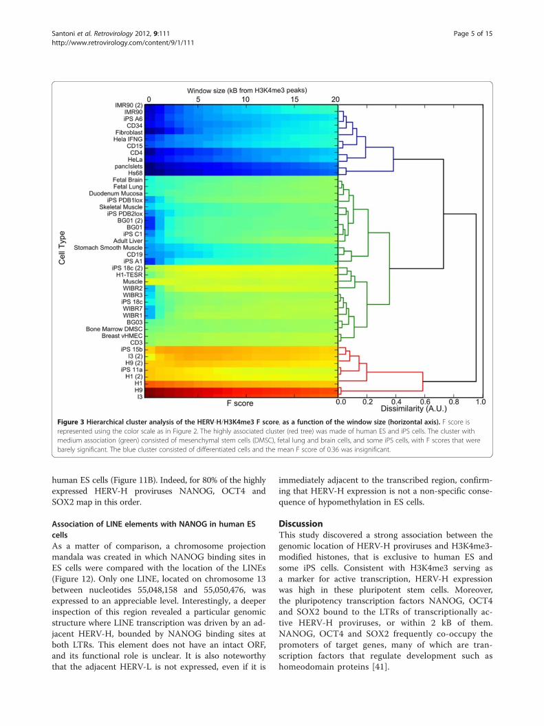
tering. Each association profile was calculated as a func-tion of the distance between HERV-H and the nearestH3K4me3 marker, discretized with steps of 0.5 kB. Thespecific cells are listed on the x axis of Figure 2, and thewindow size in kilobases is shown on the y-axis. TheEuclidean distance between these profiles was used todiscriminate among clusters. The algorithm identifiedthree main clusters of high, medium and low association(represented in red, green and blue in Figure 3). Thecluster with the highest association (red) was populatedby ES cells (H1, H9 and I3) and iPS cells (iPS-15b andiPS-11a), with a mean F score of 0.74. The cluster withmedium association (green) was populated by bone mar-row mesenchymal stem cells (DMSC), breast cancer cells(vHMEC), fetal lung cells and fetal brain cells; it had Fscores that were just barely significant. Some iPS cells(A6, PDB1lox, 18c, C1) fall within this cluster, thus con-firming that many iPS cells have epigenetic profiles dis-tinct from those of ES cells [29]. The blue clusterconsisted of differentiated cells, including CD4+ T cells,fibroblasts, pancreatic islet cells, and HeLa cells; themean F score of 0.36 was insignificant. The associationof HERV-H with the post-translational histone modifica-tion H3K4me3 correlated strongly with the degree of celldifferentiation, a relationship that was clearly visibleusing chromosome projection mandalas (Figure 4).
HERV-H expression in ES cellsGiven the remarkable, pluripotent stem cell-specific associ-ation of HERV-H with H3K4me3, a marker for transcrip-tionally active promoters [30,31], HERV-H expressionwould be expected to be higher in ES and iPS cells than indifferentiated cell types. Consistent with this possibility,HERV-H was not associated with H3K27me3 (F score 0.2),a marker for transcriptional repression in ES cells [32]. EN-CODE Project RNA-seq data sets [33] of paired 75 nucleo-tide reads from human ES cells and 6 differentiated celltypes were assessed for HERV-H RNA peaks. In H1 humanES cells, HERV-H RNA accounted for 2% of the totalRNA. This extraordinary level of HERV-H expression was
Santoni et al. Retrovirology 2012, 9:111 Page 2 of 15http://www.retrovirology.com/content/9/1/111

confirmed with RNA-seq data [34] from H9, another EScell line. For comparison, expression of the younger andbetter-conserved beta-retrovirus HERV-K was 1000-foldlower than HERV-H in H1 ES cells. HERV-H RNA was100-lower in HeLa cells and more than 100-fold lowerin K562 myelogenous leukemia cells, GSM12878 lympho-blastoid cells, HepG2 hepatocellular liver carcinoma cells,human umbilical vein endothelial cells (HUVEC), andNHEK epidermal keratinocytes. BRD2, a gene that isexpressed at nearly the same level in all these cell typeswas 25-fold lower in expression than HERV-H in ES cells(Figure 5).Almost all the HERV-H RNA expressed in ES cells
had the structure 50LTR-gag-pro-30LTR with deletion ofthe pol and env regions, and no intact open readingframes. HERV-H fragments containing pol and envsequences were barely detected (Figure 6).It has been reported that human ES cell DNA is hypo-
methylated with respect to differentiated cell lines, andthat this global effect releases all endogenous elements,such as SINEs, LINEs, and HERVs, from transcriptionalsilencing [35]. To determine if HERV-H expression inES cells is simply a result of this global trend, the RNA-seq data was used to compare the expression level of allrepetitive elements in ES cells. Compared to LINEs andSINEs, HERV-H was by far the most expressed repetitiveelement in human ES cells, accounting for nearly alltranscription of the HERV family (Figure 7).It is also important to consider that expression of en-
dogenous retro-elements might be influenced by localeffects from adjacent transcriptional units. The differ-ence between the expression level of the repetitive ele-ments and the expression level of the surroundingsequences was assessed (see Methods for details). By thismeasure, HERV-H exhibited transcriptional specificity
comparable to that of conventional genes, in terms ofspecific/unspecific transcription ratio (0.85 vs. 0.86),while LINEs and SINEs had a transcription ratio of 0.06and 0.03, respectively (Figure 7).
HERV-H expression over the course of human ES celldifferentiationThe disparity between the high transcriptional level ofHERV-H in human ES cells, and the near absence ofHERV-H transcription in differentiated cells prompted anassessment of HERV-H expression as ES cells differentiate.To accomplish this, raw data was analyzed from a 4-pointtime course experiment (http://www.ncbi.nlm.nih.gov/geo/query/acc.cgi?acc=GSE20301) in which RNA was collectedfrom human ES cells in the undifferentiated state (N0), earlyinitiation (N1), neural progenitor (N2), and pre-glial cell(N3) stages [26]. High HERV-H transcriptional levels wereconfirmed in the undifferentiated N0 stage, and HERV-Hlevels decreased progressively with a magnitude that corre-lated with the differentiation time-point (Figure 8).
HERV-H and pluripotency transcription factorsWhen ectopically expressed in particular combinations,the transcription factors NANOG, OCT4, and SOX2 arecapable of reprogramming mature somatic cells into pluri-potent stem cells [36-39]. Conversely, the expression ofthese factors decreases as cells differentiate [40]. TheRNA-seq data sets utilized above that measure the dynam-ics of RNA in ES cells as they differentiate into neural pro-genitors was analyzed for expression of HERV-H and forthese transcription factors. HERV-H RNA levels correlatedwell with those of NANOG and OCT4 (Figure 9). SOX2was more stably expressed during differentiation than wasNANOG or OCT4 and its expression did not correlatewith that of HERV-H.
Table 1 Data used for analysis of endogenous retroviruses
Data type GEO accession number Factor Cell type or tissue Reference
Human
ChIPSeq, GSE16256 H3K4me3, H3K27me3 H1, H9, I3, iPS, IMR90 [20]
ChIPSeq, GSE22499 H3K4me3 BG01/03, WIBR1/2/7, hiPSA6/C1, Fibroblast [21]
ChIPSeq, GSE15353 H3K4me3 HeLa w/o IFN [22]
ChIPSeq, GSE19465 H3K4me3 hiPS-11a/18c/15b/20b, duodenum mucosa, BM-MSC,smoothmuscle, adult liver, fetal lung, fetal brain, CD34, CD3, CD15,CD19, Pancreatic Islets
[23]
ChIPSeq, GSE20650 NANOG,OCT4, KLF4 H1 [24]
ChIPSeq, GSE18292 SOX2 H1 [25]
RNASeq, GSE23316 H1, HeLa, K562 UCSC ENCODE Project
RNASeq, GSE20301 H1, differentiated H1 [26]
Mouse
ChIPSeq, GSE22075 Mouse H3K4me3 ES, LSK cells [27]
ChIPSeq, GSE12241 Mouse H3K4me3 ES, MEF [28]
Santoni et al. Retrovirology 2012, 9:111 Page 3 of 15http://www.retrovirology.com/content/9/1/111
To explore the possibility that these pluripotency tran-scription factors are somehow involved in HERV-Htranscription, an association analysis was conducted be-tween HERV-H locations and the ChIPSeq bindingprofiles of NANOG, OCT4, SOX2, and KLF4 extractedfrom ES cells [24,25] (Figure 10). Over a 2 kB window,NANOG showed the most significant association (F score0.75, p < 10-180). OCT4 weakly associated with HERV-H(F score 0.55, p < 0.0001), while SOX2 and KLF4 were notassociated (F score 0.46 and 0.24, respectively).Statistical significance increased considerably when the
expression level of individual HERV-H proviruses wasmonitored (Figure 11A and B). As visualized on thechromosome projection mandala in Figure 11C, NANOGbinds to the LTRs of 96% of the 50 most highly expressedHERV-H proviruses. These 50 proviruses account for 80%of all HERV-H RNA. The LTR region corresponding tothe first 0.5 kB of HERV-H proviral sequence is high-lighted in green, with dot sizes proportional to expressionlevels. With a window of 0.5 kB, the F score for NANOGwas nearly perfect (0.97, p < 10-300). Limited to this subsetof HERV-H proviruses the F scores for OCT4 (0.90 wi2kB,p < 10-300) and SOX2 (0.85 wi4kB; p < 10-300) were alsotightly linked to HERV-H. KLF4, in contrast, was not asso-ciated with HERV-H (F score 0.34). The average distanceof the NANOG, OCT4 and SOX2 ChIPSeq peaks fromthe HERV-H transcription start sites was 250 basepairs,1 kB and 2 kB, respectively, as if these factors wereuniformly distributed along the HERV-H promoter in
Figure 1 Schematic showing the strategy for detectingassociation between endogenous retro-elements and theH3K4me3 marker. H3K4me3 CHiPSeq data for more than 40different cell types were checked for association within 2 kB of allannotated endogenous retroviral elements (hg18) using methodsdescribed in reference [18]. Only HERV-H proviruses showedassociation with H3K4me3, and this was only significant in humanES cell lines. No association between H3K4me3 and endogenousretro-elements was detected in the mouse.
Figure 2 H3K4me3 association with HERV-H proviruses, as measured with the F score, in 40 different cell types, as a function of thedistance from the marker. The F score is reported on the z-axis. Cell types are listed on the x-axis, while the window size is reported on the y-axis. Yellow to red bands indicate a significant association (F score >0.5). Significance decreases as the color shifts from red to blue. 12 out of 15of the cell types with significant F scores are either human ES cells or iPS cells.
Santoni et al. Retrovirology 2012, 9:111 Page 4 of 15http://www.retrovirology.com/content/9/1/111
human ES cells (Figure 11B). Indeed, for 80% of the highlyexpressed HERV-H proviruses NANOG, OCT4 andSOX2 map in this order.
Association of LINE elements with NANOG in human EScellsAs a matter of comparison, a chromosome projectionmandala was created in which NANOG binding sites inES cells were compared with the location of the LINEs(Figure 12). Only one LINE, located on chromosome 13between nucleotides 55,048,158 and 55,050,476, wasexpressed to an appreciable level. Interestingly, a deeperinspection of this region revealed a particular genomicstructure where LINE transcription was driven by an ad-jacent HERV-H, bounded by NANOG binding sites atboth LTRs. This element does not have an intact ORF,and its functional role is unclear. It is also noteworthythat the adjacent HERV-L is not expressed, even if it is
immediately adjacent to the transcribed region, confirm-ing that HERV-H expression is not a non-specific conse-quence of hypomethylation in ES cells.
DiscussionThis study discovered a strong association between thegenomic location of HERV-H proviruses and H3K4me3-modified histones, that is exclusive to human ES andsome iPS cells. Consistent with H3K4me3 serving asa marker for active transcription, HERV-H expressionwas high in these pluripotent stem cells. Moreover,the pluripotency transcription factors NANOG, OCT4and SOX2 bound to the LTRs of transcriptionally ac-tive HERV-H proviruses, or within 2 kB of them.NANOG, OCT4 and SOX2 frequently co-occupy thepromoters of target genes, many of which are tran-scription factors that regulate development such ashomeodomain proteins [41].
Figure 3 Hierarchical cluster analysis of the HERV-H/H3K4me3 F score, as a function of the window size (horizontal axis). F score isrepresented using the color scale as in Figure 2. The highly associated cluster (red tree) was made of human ES and iPS cells. The cluster withmedium association (green) consisted of mesenchymal stem cells (DMSC), fetal lung and brain cells, and some iPS cells, with F scores that werebarely significant. The blue cluster consisted of differentiated cells and the mean F score of 0.36 was insignificant.
Santoni et al. Retrovirology 2012, 9:111 Page 5 of 15http://www.retrovirology.com/content/9/1/111

These observations strongly support the hypothesis thatHERV-H transcripts play a role in human pluripotency andthat this role is finely regulated by three of the most import-ant transcription factors in ES cells. In addition to the bind-ing of NANOG, OCT4, and SOX2 to the HERV-Hpromoter, HERV-H RNA decreased as ES cells differen-tiated, in a manner that was proportional to the expressionof NANOG and OCT4. Conversely, HERV-H RNA was un-detectable in primary fibroblasts but increased enormouslyafter forced re-programming to generate pluripotent stem
cells (unpublished data provided by Audrey Letourneauand Stylianos Antonarakis). HERV-H, then, can beexploited as a reliable marker of ES cell pluripotency, aswell as an indicator of the degree of “stemness” of iPS cellsas they are generated from fibroblasts.HERV-H transcripts are 5 to 6 kB in length and lack
open reading frames. We can only speculate about thefunction of these lncRNAs. They might, for example, serveto soak-up miRNAs that promote differentiation, as hasbeen shown with linc-MD1 in muscle differentiation [42]
Figure 5 Cumulative expression of all HERV-H proviruses in human H1 ES cells (hESC), HeLa cells, or K562 cells, compared toexpression of HERV-K and BRD2, a constitutive gene with the same expression level in all three cell types. In human ES cells, HERV-H isexpressed 1000-fold higher than HERV-K and 25-fold higher than BRD2. HERV-H expression is barely detectable in HeLa, and no significant HERV-H expression was detected in K562 cells. RNASeq data for this analysis were from reference [33].
Figure 4 Chromosome projection mandalas showing the proximity of each HERV-H provirus to the nearest site of H3K4Me3 on thechromosome, in human I3 ES cells, iPS-15b cells, and HeLa cells. Each dot on the mandala indicates an HERV-H provirus, as described inreference [18]. The angular distance around the mandala indicates the linear position of each provirus on the indicated chromosome. The radialdistance from the perimeter indicates the distance of the provirus from the nearest H3K4Me3 site, in log scale from 0 to 1 megabase. Blue dotsare HERV-H proviruses within 2 kB from the nearest marker. Red dots are proviruses >2 kB away from the nearest H3K4Me3 site. The associationstrength (F score) is written under each Mandala. F score > 0.5 constitutes a significant association.
Santoni et al. Retrovirology 2012, 9:111 Page 6 of 15http://www.retrovirology.com/content/9/1/111

or the PTENP1 pseudogene in the regulation of PTENand growth suppression [43]. They might bind to chroma-tin and act as a scaffold for the local recruitment of pluri-potency transcription factors, similar to other lncRNAslike HOTAIR for histone modification complexes [44] andXist in the context of X-chromosome inactivation [44-46].Alternatively, HERV-H might counteract retrovirus spreadby interfering with packaging of retroviral genomic RNA[47,48] or by soaking up miRNAs that are required forretrovirus transduction.
The study here failed to identify chromatin markersthat associate with endogenous retro-elements in mice.This was somewhat surprising given the many endogen-ous retro-elements in this species, including endogenousgamma-retroviruses, some of which are intact and func-tional [8]. It was also surprising because exogenousgamma-retroviruses have the same integration site pre-ferences in mouse cells as they have in human cells [18].MLV integration sites are associated with the H3K4me3profile in mouse embryonic fibroblasts (F score = 0.83;
Figure 6 Mapping of RNA-seq reads from H1 human ES cells on a schematic of the HERV-H provirus. The quantity of each RNA read wasnormalized to the reads corresponding to the 50 LTR. Only RNA fragments corresponding to 50LTR-gag-pro-30LTR were expressed to a significantlevel in human ES cells.
Figure 7 HERV-H expression accounts for nearly all HERV expression in human ES cells and is a not a non-specific consequence ofwide-spread hypo-methylation in these cells. Quantitation of the RNA-seq reads from H1 ES cells, broken down according to the LINES, SINES,all HERVs, the nearly 1,000 HERV-H proviruses, and conventional genes. HERV-H RNA accounted for nearly all the HERV RNA in human ES cells,and 2% of total RNA. Specific vs. non-specific expression was determined by comparing the expression level of each element to the surroundingsequences.
Santoni et al. Retrovirology 2012, 9:111 Page 7 of 15http://www.retrovirology.com/content/9/1/111

p < 10-100). Similar results with murine hematopoieticstem cells (F score of 0.81; p < 10-100) indicate that, as inhuman cells, the association strength is cell-type dependent.One possible explanation for the failure to identify chro-
matin markers associated with endogenous murine retro-viruses is species-specific differences in the recruitment ofthe transcriptional silencing machinery. In murine EScells, for example, a sequence-specific DNA-binding pro-tein, ZNF809 [49], recruits TRIM28 and other compo-nents of the cellular machinery that silence MLV [50].ZNF809 has no orthologue in humans; perhaps ZNF809arose as a result of selective pressure exerted by murinespecific gamma-retroviruses during evolution.
Previous work demonstrated that when exogenous retro-viruses integrate they home to sites of H3K4me3 [18].Similarly, the association of endogenous gamma-retrovirusHERV-H with H3K4me3 suggests that when human andsimian germ cells were bombarded with the HERV-H an-cestor 15 to 30 millions years ago, these ancient retro-viruses integrated in proximity to H3K4me3-markedchromatin. These proviruses might then have retainedthese cell-type specific marks as they became fixed in theprimate genome. Alternatively, unmethylated HERV-HLTRs might have recruited chromatin remodeling factorsand induced H3K4me3 modification of the viral promoterafter integration had occurred.
Figure 8 HERV-H expression correlates with differentiation status. HERV-H expression as H1 human ES cells differentiate down a pathwaytowards neural progenitors and early glial cells. Black bars indicate unspecific expression; white bars represent specific expression, adjusted forexpression as described in Figure 7. BRD2 has the same expression level at each stage of differentiation and was used to normalize HERV-H RNAlevels.
Figure 9 HERV-H expression levels correlate with those of pluripotency transcription factors NANOG and OCT4 as human ES cellsmove down a differentiation pathway. N0, undifferentiated ES cells. N1, early initiation stage of differentiation. N2, neural progenitor stage.OCT4 and NANOG are positively correlated with HERV-H (ρ = 0.95, ρ = 0.84, respectively). SOX2 shows no correlation.
Santoni et al. Retrovirology 2012, 9:111 Page 8 of 15http://www.retrovirology.com/content/9/1/111

Analysis of the DNA surrounding HERV-H pro-viruses failed to clarify which of these two scenarios ismore likely. Additionally, search for epigenetic markerslike H3K4me3 in syntenic regions in the mouse gen-ome was attempted to determine if these chromatin
marks are conserved across the species and predate theentry of HERV-H into the primate genome. DNA sur-rounding HERV-H proviruses in the human genomewas aligned to the mouse genome (using the tool Lift-Over, http://genome.ucsc.edu/cgi-bin/hgLiftOver) after
Figure 10 Modified chromosome projection mandalas depicting NANOG, OCT4, SOX2 and KLF4 associations with HERV-H proviruses.Mandalas were generated as described in reference [18] and Figure 4 above. The green ring indicates the HERV-H 50 LTR region (500 nucleotidesfrom the transcriptional start site). Dot size is proportional to the expression level of each single provirus. NANOG is bound to the 50LTR of almostall highly expressed retroviruses. OCT4 shows a significant association. SOX2 binds all expressed HERV-H in a region between 1 KB and 2 KB whileKLF4 does not show any significant association pattern.
Santoni et al. Retrovirology 2012, 9:111 Page 9 of 15http://www.retrovirology.com/content/9/1/111

Figure 11 (See legend on next page.)
Santoni et al. Retrovirology 2012, 9:111 Page 10 of 15http://www.retrovirology.com/content/9/1/111

(See figure on previous page.)Figure 11 Ordered spacing of pluripotency transcription factors, binding to the HERV-H 50 LTR in human ES cells. (A) Associationstrength (F score) of NANOG, OCT4 and SOX2 binding sites with the 50 most highly expressed HERV-H proviruses (accounting for 80% of totalHERV-H expression), as a function of distance from the HERV-H transcription start site (TSS). Maxima in F score indicate the distance of greatestassociation. (B) Average distance of NANOG (red), OCT4 (blue) and SOX2 (green) to HERV-H TSS is shown schematically. As expected from auniform distribution model, the average distance is half of the distance between maximal association and TSS. (C) Chromosome ProjectionMandala combining NANOG, OCT4 and SOX2 with respect to 50 HERV-H proviruses. The three embryonic transcription factors bind with thesame order (NANOG-OCT4-SOX2) to the promoter region of the most expressed HERV-Hs.
Figure 12 (A) Chromosome projection mandala depicting the association between NANOG and expressed LINEs in human ES cells. Onlyone LINE (large blue dot on chromosome 13) was expressed to high level in these cells. (B) The genomic region around the LINE on chromosome 13is shown with the UCSC Genome Browser, where the linear chromosome is mapped on the horizontal axis. The position of the adjacent HERV-H andHERV-L proviruses is shown. Four biological replicates confirm HERV-H and LINE expression in human ES cells while no expression was detected inK562 cells (only one of the 4 replicates is shown). The direction of transcription was determined by strand-specific sequencing [33]. NANOG bound toboth LTRs (represented with black squares along the LTR row) in the adjacent HERV-H. The adjacent HERV-L was not transcribed.
Santoni et al. Retrovirology 2012, 9:111 Page 11 of 15http://www.retrovirology.com/content/9/1/111

excision of all repetitive elements (performed withRepeatMasker, http://www.repeatmasker.org/). Nothinginformative was found by measuring the association ofmouse H3K4me3 with these syntenic regions and com-paring these values with those obtained using controlloci.P300 and H3K27ac bind intra-species conserved
regions and co-localize in embryonic-specific enhancers[34,51]. These markers are also associated with HERV-H in human ES cells, lying within 4 kB of 80% ofHERV-H proviruses (F score 0.95; p < 10-300). It seemsunlikely that an exogenous retrovirus would be capableof recruiting these factors by exploiting random con-served regions around its integration site. This suggeststhat a pre-existing layer of epigenetic markers favoredintegration of HERV-H into particular host loci andthat these features are still preserved millions of yearslater.
ConclusionsAmong retroelements in the human genome, the en-dogenous gammaretrovirus HERV-H is extraordinary forits high level expression in embryonic stem cells, inwhich it makes up 2% of all polyadenylated RNA. Thehuman genome has ~1,000 copies of HERV-H, and themajority of the HERV-H RNA is encoded by a subsetof 50 of these. HERV-H expression decreases as ES cellslose pluripotency, to the point where its expressionis undetectable in fibroblasts. Consistent with thisexpression pattern, HERV-H is also expressed to highlevel in many iPS cells, though expression in some iPScells is more modest; this heterogeneity may reflectreported differences in the epigenetic profile of manyiPS cells, when compared with ES cells [29]. This sug-gests, then, that HERV-H RNA offers a relatively strin-gent marker for human pluripotency that would beworth monitoring during the generation of new iPSlines. The HERV-H RNAs in ES cells average about 5kB in length and encode no protein. It, therefore,seems likely that HERV-H RNA contributes to pluri-potency by acting as a chromatin-associated structuralelement or by acting as a microRNA decoy.
MethodsStatistical analysis of association between chromatinmarkers and retro-elementsAssociation with a given marker was defined as thepresence of the endogenous proviral DNA within afixed distance (usually 2 kilobases) from the nearestmarker on the linear sequence of the chromosome. Un-like exogenous retroviruses, the endogenous virus isalready integrated. Therefore, to restore the conditionsbefore integration, the distance from the j-esim markerMj with peaks in loci {mj0, . . ., mjN} and the i-esim
provirus Vi spanning along the loci {Vis, Vi
e} has beencalculated as
d Vi;Mj� � ¼ min Vi
s �Mj
�� ��; Vie �Mj
�� ��� �
¼ min Vic �Mj
�� ��� �� Vei � Vs
i
2
where Vci ¼ Vs
i þVei
2 is the central locus of the provirus.As a control dataset, we randomly selected 100000
genomic locations. Association strength was measuredwith the statistical method based on the F score, as pre-viously described in [18].Formally the Fβ-score is defined as the β-weighted
harmonic mean of Precision(P) and Sensitivity(R):Fβ≡ 1þ β2
� �PR
β2PþR.
Here β = 0.5 to give more weight to Precision than toSensitivity. This balances type I and type II errors byadjusting for the high rate of False Positives inherent tothe examination of large datasets for genome-wide bind-ing sites according to statistical significance (F scorebased statistics and comparison with other measureshave been extensively discussed in [18]).Markers with F scores ranging between 0.5 and 1 were
considered to be associated with endogenous integrationsites.
RNASeq data analysisHERV-H is present in more than 1000 “imperfect” cop-ies in the human genome and its transcripts share anumber of short conserved regions (each around100 bp). Therefore, deep sequencing of those transcriptsyields reads (25-75 bp long sequences) which perfectlyalign to several genomic loci. Indeed, multireads map-ping is still a challenging process [52]. The strategyadopted here was to perform the alignment of uniquelymapped single- and paired-end reads and to reassign themultiple-mapped reads in function of the expressionlevel of the surrounding (context) region [26]. HERV-Hexpression was evaluated in term of “Reads Per Kilobaseper Million mapped reads (RPKM)” with the standardformula Er ¼ K Nr
LrNT[53] where Nr is the number of
reads mapping onto the r transcript, Lr is the length (inkB) of the r-esim transcript and NT is the total numberof reads K = 106.Alignments of RNASeq generated reads have been per-
formed with a two-step procedure. First we used Bowtie[54] on raw data, admitting up to two mismatches for eachalignment, and then we discriminated between uniquemapping reads and multireads, 80% and 20% of the totalnumber of reads respectively. As expected, many multi-reads matched with repeated elements. Ignoring themwould have resulted in a potential underestimation of theexpression of endogenous retroviruses and, in general, of all
Santoni et al. Retrovirology 2012, 9:111 Page 12 of 15http://www.retrovirology.com/content/9/1/111

repetitive elements. Therefore, we adopted a probabilisticassignment based on the amount of reads that univocallymap onto the surrounding regions (context), as describedin more detail in the next paragraph. This evaluation is alsouseful to establish if a repeated element is expressly tran-scribed or if it is part of another structure (i.e. a gene).
Probabilistic alignment of multiple mapping readsThe uniquely mapping read was defined as a short se-quence generated by high throughput sequencing that canbe aligned to a single genomic region s. Accordingly, wedefined the multiread as a sequence r that aligns to a setof M regions {S1,. . .,SM}. For each region si the context re-gion is ci =Ci/(Ci \ si) being Ci a genomic region of nnucleotides encompassing si. The assumption that theamount of reads is proportional to the amount of actualmRNA implies that the set of multireads is distributed onthe reference genome accordingly to the amount ofuniquely mapping reads aligned to the context regions.Consider RD sð Þ as the function giving the number ofreads of the dataset D that map univocally to the genomicregion s described by the tuple (chr, start, end). Therefore,the probability of the read r to be actually part of themRNA generated from the region si is estimated as:
PD r∈ sið Þ ¼ RD cið ÞXM
j
RD cj� �
:
Eventually, the set of multireads mapping to the same Mregions is then partitioned to {S1,. . .,SM} accordingly to PD (si).
SpecificityThe first axiom of high throughput sequencing asserts thatthe number of reads aligned to a specific genomic region isproportional to how much RNA has been generated by thisregion within the cell. At this point it is worth observingthat repeated element (RE) sequences might be present inthe RNA just because they are part of longer mRNAs.Since we expect that elements having a specific bio-
logical function are independently transcribed, weattempted to distinguish between RE specifically expressedwith their own promoter from those that are part oflonger RNAs. The number of reads mapped to a region scan be naively modeled as a linear combination of specificreads T(s), unspecific reads U(s) and additional zero-meannoise σ2 that account for all other experimental and non-systematic fluctuations that can randomly influence theoutput of the sequencing process. Formally:
R sð Þ ¼ U Sð Þ þ T sð Þ þ σ2
where R sð Þ , as before, is the function giving the numberof reads assigned to the region s.
Therefore, the mean number of reads in the region sis:
E T sð Þ½ � ¼ R sð Þ � U sð Þ:In order to estimate U(s), it is possible to count the
number of reads mapped to the context region c as pre-viously shown. Therefore we set
U sð Þ ¼ R cð Þ2
;
and we eventually adopt the following approximation tocorrect for the non-specificity of transcription:
E T sð Þ½ �≅R sð Þ �R cð Þ2
AbbreviationsHERV: Human endogenous retrovirus; ES: Embryonic stem; iPS: Inducedpluripotent stem; LTR: Long terminal repeat; lncRNA: Long non-coding RNA.
Competing interestsThe authors declare that they have no competing interests.
Authors’ contributionsFS and JL conceived and designed the experiments. FS, JG, and JL analyzedthe data. FS and JL wrote the paper. All authors read and approved the finalmanuscript.
AcknowledgementsThe authors wish to thank Audrey Letourneau and Stylianos Antonarakis forproviding unpublished RNA-seq data. This work was supported by NIDA/NIH/USA Grant DP1DA034990 and Swiss National Science Foundation grant3100A0-128655 to J.L.
Author details1Department of Genetic Medicine and Development, University of Geneva, 1rue Michel-Servet, Geneva CH-1211, Switzerland. 2Department ofMicrobiology and Molecular Medicine, University of Geneva, 1211, Geneva 4,Switzerland. 3Program in Molecular Medicine and Biochemistry & MolecularPharmacology, University of Massachusetts Medical School, 373 PlantationStreet, Biotech 2, Suite 319, Worcester, MA 01605, USA.
Received: 5 December 2012 Accepted: 16 December 2012Published: 20 December 2012
References1. Stoye JP: Studies of endogenous retroviruses reveal a continuing
evolutionary saga. Nat Rev Microbiol 2012, 10:395–406.2. Weiss RA: The discovery of endogenous retroviruses. Retrovirology
2006, 3:67.3. Subramanian RP, Wildschutte JH, Russo C, Coffin JM: Identification,
characterization, and comparative genomic distribution of the HERV-K(HML-2) group of human endogenous retroviruses. Retrovirology2011, 8:90.
4. Stengel A, Roos C, Hunsmann G, Seifarth W, Leib-Mosch C, Greenwood AD:Expression profiles of endogenous retroviruses in Old World monkeys. JVirol 2006, 80:4415–4421.
5. Mi S, Lee X, Li X, Veldman GM, Finnerty H, Racie L, LaVallie E, Tang XY,Edouard P, Howes S, et al: Syncytin is a captive retroviral envelopeprotein involved in human placental morphogenesis. Nature 2000,403:785–789.
6. Vargas A, Moreau J, Landry S, LeBellego F, Toufaily C, Rassart E, Lafond J,Barbeau B: Syncytin-2 plays an important role in the fusion of humantrophoblast cells. J Mol Biol 2009, 392:301–318.
7. Mangeney M, Renard M, Schlecht-Louf G, Bouallaga I, Heidmann O, Letzelter C,Richaud A, Ducos B, Heidmann T: Placental syncytins: genetic disjunction
Santoni et al. Retrovirology 2012, 9:111 Page 13 of 15http://www.retrovirology.com/content/9/1/111

between the fusogenic and immunosuppressive activity of retroviralenvelope proteins. Proc Natl Acad Sci U S A 2007, 104:20534–20539.
8. Coffin J, Hughes S, Varmus H: Retroviruses. New York: Cold Spring HarborLaboratory Press; 1997.
9. Muster T, Waltenberger A, Grassauer A, Hirschl S, Caucig P, Romirer I,Fodinger D, Seppele H, Schanab O, Magin-Lachmann C, et al: Anendogenous retrovirus derived from human melanoma cells. Cancer Res2003, 63:8735–8741.
10. Kolson DL, Gonzalez-Scarano F: Endogenous retroviruses and multiplesclerosis. Ann Neurol 2001, 50:429–430.
11. Nexo BA, Christensen T, Frederiksen J, Moller-Larsen A, Oturai AB, Villesen P,Hansen B, Nissen KK, Laska MJ, Petersen TS, et al: The etiology of multiplesclerosis: genetic evidence for the involvement of the humanendogenous retrovirus HERV-Fc1. PLoS One 2011, 6:e16652.
12. Petersen T, Moller-Larsen A, Thiel S, Brudek T, Hansen TK, Christensen T:Effects of interferon-beta therapy on innate and adaptive immuneresponses to the human endogenous retroviruses HERV-H and HERV-W,cytokine production, and the lectin complement activation pathway inmultiple sclerosis. J Neuroimmunol 2009, 215:108–116.
13. Karlsson H, Schroder J, Bachmann S, Bottmer C, Yolken RH: HERV-W-relatedRNA detected in plasma from individuals with recent-onsetschizophrenia or schizoaffective disorder. Mol Psychiatry 2004, 9:12–13.
14. Young GR, Eksmond U, Salcedo R, Alexopoulou L, Stoye JP, Kassiotis G:Resurrection of endogenous retroviruses in antibody-deficient mice.Nature 2012, 491:774–778.
15. Garrison KE, Jones RB, Meiklejohn DA, Anwar N, Ndhlovu LC, Chapman JM,Erickson AL, Agrawal A, Spotts G, Hecht FM, et al: T cell responses to humanendogenous retroviruses in HIV-1 infection. PLoS Pathog 2007, 3:e165.
16. SenGupta D, Tandon R, Vieira RG, Ndhlovu LC, Lown-Hecht R, Ormsby CE,Loh L, Jones RB, Garrison KE, Martin JN, et al: Strong human endogenousretrovirus-specific T cell responses are associated with control of HIV-1in chronic infection. J Virol 2011, 85:6977–6985.
17. van der Kuyl AC: HIV infection and HERV expression: a review.Retrovirology 2012, 9:6.
18. Santoni FA, Hartley O, Luban J: Deciphering the code for retroviralintegration target site selection. PLoS Comput Biol 2010, 6:e1001008.
19. Santoni FA: EMdeCODE: a novel algorithm capable of reading words ofepigenetic code to predict enhancers and retroviral integration sites andto identify H3R2me1 as a distinctive mark of coding versus non-codinggenes. Nucl Acid Res 2012, in press.
20. Hawkins RD, Hon GC, Lee LK, Ngo Q, Lister R, Pelizzola M, Edsall LE, Kuan S,Luu Y, Klugman S, et al: Distinct epigenomic landscapes of pluripotentand lineage-committed human cells. Cell Stem Cell 2010, 6:479–491.
21. Guenther MG, Frampton GM, Soldner F, Hockemeyer D, Mitalipova M,Jaenisch R, Young RA: Chromatin structure and gene expressionprograms of human embryonic and induced pluripotent stem cells. CellStem Cell 2010, 7:249–257.
22. Robertson G, Hirst M, Bainbridge M, Bilenky M, Zhao Y, Zeng T, EuskirchenG, Bernier B, Varhol R, Delaney A, et al: Genome-wide profiles of STAT1DNA association using chromatin immunoprecipitation and massivelyparallel sequencing. Nat Methods 2007, 4:651–657.
23. Bernstein BE, Stamatoyannopoulos JA, Costello JF, Ren B, Milosavljevic A,Meissner A, Kellis M, Marra MA, Beaudet AL, Ecker JR, et al: The NIHroadmap epigenomics mapping consortium. Nat Biotechnol 2010,28:1045–1048.
24. Kunarso G, Chia NY, Jeyakani J, Hwang C, Lu X, Chan YS, Ng HH, Bourque G:Transposable elements have rewired the core regulatory network ofhuman embryonic stem cells. Nat Genet 2010, 42:631–634.
25. Lister R, Pelizzola M, Dowen RH, Hawkins RD, Hon G, Tonti-Filippini J, NeryJR, Lee L, Ye Z, Ngo QM, et al: Human DNA methylomes at baseresolution show widespread epigenomic differences. Nature 2009,462:315–322.
26. Wu JQ, Habegger L, Noisa P, Szekely A, Qiu C, Hutchison S, Raha D, EgholmM, Lin H, Weissman S, et al: Dynamic transcriptomes during neuraldifferentiation of human embryonic stem cells revealed by short, long,and paired-end sequencing. Proc Natl Acad Sci U S A 2010, 107:5254–5259.
27. Adli M, Zhu J, Bernstein BE: Genome-wide chromatin maps derivedfrom limited numbers of hematopoietic progenitors. Nat Methods 2010,7:615–618.
28. Mikkelsen TS, Ku M, Jaffe DB, Issac B, Lieberman E, Giannoukos G, AlvarezP, Brockman W, Kim TK, Koche RP, et al: Genome-wide maps of
chromatin state in pluripotent and lineage-committed cells. Nature2007, 448:553–560.
29. Bock C, Kiskinis E, Verstappen G, Gu H, Boulting G, Smith ZD, Ziller M, CroftGF, Amoroso MW, Oakley DH, et al: Reference maps of human ES and iPScell variation enable high-throughput characterization of pluripotent celllines. Cell 2011, 144:439–452.
30. Ruthenburg AJ, Allis CD, Wysocka J: Methylation of lysine 4 on histone H3:intricacy of writing and reading a single epigenetic mark. Mol Cell 2007,25:15–30.
31. Barski A, Cuddapah S, Cui K, Roh TY, Schones DE, Wang Z, Wei G, ChepelevI, Zhao K: High-resolution profiling of histone methylations in the humangenome. Cell 2007, 129:823–837.
32. Bernstein BE, Mikkelsen TS, Xie X, Kamal M, Huebert DJ, Cuff J, Fry B, MeissnerA, Wernig M, Plath K, et al: A bivalent chromatin structure marks keydevelopmental genes in embryonic stem cells. Cell 2006, 125:315–326.
33. Celniker SE, Dillon LA, Gerstein MB, Gunsalus KC, Henikoff S, Karpen GH,Kellis M, Lai EC, Lieb JD, MacAlpine DM, et al: Unlocking the secrets of thegenome. Nature 2009, 459:927–930.
34. Rada-Iglesias A, Bajpai R, Swigut T, Brugmann SA, Flynn RA, Wysocka J: Aunique chromatin signature uncovers early developmental enhancers inhumans. Nature 2011, 470:279–283.
35. Tada T, Tada M: Toti-/pluripotential stem cells and epigeneticmodifications. Cell Struct Funct 2001, 26:149–160.
36. Takahashi K, Yamanaka S: Induction of pluripotent stem cells from mouseembryonic and adult fibroblast cultures by defined factors. Cell 2006,126:663–676.
37. Yu J, Vodyanik MA, Smuga-Otto K, Antosiewicz-Bourget J, Frane JL, Tian S,Nie J, Jonsdottir GA, Ruotti V, Stewart R, et al: Induced pluripotent stemcell lines derived from human somatic cells. Science 2007, 318:1917–1920.
38. Chambers I, Silva J, Colby D, Nichols J, Nijmeijer B, Robertson M, Vrana J,Jones K, Grotewold L, Smith A: Nanog safeguards pluripotency andmediates germline development. Nature 2007, 450:1230–1234.
39. Mitsui K, Tokuzawa Y, Itoh H, Segawa K, Murakami M, Takahashi K,Maruyama M, Maeda M, Yamanaka S: The homeoprotein Nanog isrequired for maintenance of pluripotency in mouse epiblast and ES cells.Cell 2003, 113:631–642.
40. Boiani M, Scholer HR: Regulatory networks in embryo-derived pluripotentstem cells. Nat Rev Mol Cell Biol 2005, 6:872–884.
41. Boyer LA, Lee TI, Cole MF, Johnstone SE, Levine SS, Zucker JP, Guenther MG,Kumar RM, Murray HL, Jenner RG, et al: Core transcriptional regulatorycircuitry in human embryonic stem cells. Cell 2005, 122:947–956.
42. Cesana M, Cacchiarelli D, Legnini I, Santini T, Sthandier O, Chinappi M,Tramontano A, Bozzoni I: A long noncoding RNA controls muscledifferentiation by functioning as a competing endogenous RNA. Cell2011, 147:358–369.
43. Poliseno L, Salmena L, Zhang J, Carver B, Haveman WJ, Pandolfi PP: Acoding-independent function of gene and pseudogene mRNAsregulates tumour biology. Nature 2010, 465:1033–1038.
44. Tsai MC, Manor O, Wan Y, Mosammaparast N, Wang JK, Lan F, Shi Y, SegalE, Chang HY: Long noncoding RNA as modular scaffold of histonemodification complexes. Science 2010, 329:689–693.
45. Tattermusch A, Brockdorff N: A scaffold for X chromosome inactivation.Hum Genet 2011, 130:247–253.
46. Chaumeil J, Le Baccon P, Wutz A, Heard E: A novel role for Xist RNA in theformation of a repressive nuclear compartment into which genes arerecruited when silenced. Genes Dev 2006, 20:2223–2237.
47. Song X, Wang B, Bromberg M, Hu Z, Konigsberg W, Garen A: Retroviral-mediated transmission of a mouse VL30 RNA to human melanoma cellspromotes metastasis in an immunodeficient mouse model. Proc NatlAcad Sci U S A 2002, 99:6269–6273.
48. Meric C, Goff SP: Characterization of Moloney murine leukemia virusmutants with single-amino-acid substitutions in the Cys-His box of thenucleocapsid protein. J Virol 1989, 63:1558–1568.
49. Wolf D, Goff SP: Embryonic stem cells use ZFP809 to silence retroviralDNAs. Nature 2009, 458:1201–1204.
50. Wolf D, Goff SP: TRIM28 mediates primer binding site-targeted silencingof murine leukemia virus in embryonic cells. Cell 2007, 131:46–57.
51. Creyghton MP, Cheng AW, Welstead GG, Kooistra T, Carey BW, Steine EJ,Hanna J, Lodato MA, Frampton GM, Sharp PA, et al: Histone H3K27acseparates active from poised enhancers and predicts developmentalstate. Proc Natl Acad Sci U S A 2011, 107:21931–21936.
Santoni et al. Retrovirology 2012, 9:111 Page 14 of 15http://www.retrovirology.com/content/9/1/111

52. Pepke S, Wold B, Mortazavi A: Computation for ChIP-seq and RNA-seqstudies. Nat Methods 2009, 6:S22–S32.
53. Mortazavi A, Williams BA, McCue K, Schaeffer L, Wold B: Mapping andquantifying mammalian transcriptomes by RNA-Seq. Nat Methods 2008,5:621–628.
54. Langmead B, Trapnell C, Pop M, Salzberg SL: Ultrafast and memory-efficient alignment of short DNA sequences to the human genome.Genome Biol 2009, 10:R25.
doi:10.1186/1742-4690-9-111Cite this article as: Santoni et al.: HERV-H RNA is abundant in humanembryonic stem cells and a precise marker for pluripotency.Retrovirology 2012 9:111.
Submit your next manuscript to BioMed Centraland take full advantage of:
• Convenient online submission
• Thorough peer review
• No space constraints or color figure charges
• Immediate publication on acceptance
• Inclusion in PubMed, CAS, Scopus and Google Scholar
• Research which is freely available for redistribution
Submit your manuscript at www.biomedcentral.com/submit
Santoni et al. Retrovirology 2012, 9:111 Page 15 of 15http://www.retrovirology.com/content/9/1/111


