Hereditary primary lateral sclerosis and progressive ...
12
Vol.:(0123456789) 1 3 Journal of Neurology https://doi.org/10.1007/s00415-019-09235-x ORIGINAL COMMUNICATION Hereditary primary lateral sclerosis and progressive nonfluent aphasia José Gazulla 1 · Isidro Ferrer 2 · Silvia Izquierdo‑Alvarez 3 · Sara Alvarez 4 · Rocío Sánchez‑Alcudia 4 · María Bestué‑Cardiel 5 · María Seral 5 · Isabel Benavente 6 · Esther Sierra‑Martínez 1 · José Berciano 7 Received: 27 January 2019 / Revised: 1 February 2019 / Accepted: 5 February 2019 © Springer-Verlag GmbH Germany, part of Springer Nature 2019 Abstract Objective To report a kindred with an association between hereditary primary lateral sclerosis (PLS) and progressive non- fluent aphasia. Patients and methods Six members from a kindred with 15 affected individuals spanning three generations, suffered from spasticity without muscle atrophy or fasciculation, starting in the lower limbs and spreading to the upper limbs and bulbar musculature, followed by effortful speech, nonfluent language and dementia, in 5 deceased members. Disease onset was during the sixth decade of life, or later. Cerebellar ataxia was the inaugural manifestation in two patients, and parkinsonism, in another. Results Neuropathological examination in two patients demonstrated degeneration of lateral corticospinal tracts in the spinal cord, without loss of spinal, brainstem, or cerebral motor neurons. Greater loss of corticospinal fibers at sacral and lumbar, rather than at cervical or medullary levels was demonstrated, supporting a central axonal dying-back pathogenic mechanism. Marked reduction of myelin and nerve fibers in the frontal lobes was also present. Argyrophilic grain disease and primary age-related tauopathy were found in one case each, and considered incidental findings. Genetic testing, including exome sequencing aimed at PLS, ataxia, hereditary spastic paraplegia, and frontotemporal lobe dementia, triplet-repeated primed polymerase chain reaction aimed at dominant spinocerebellar ataxias, and massive sequencing of the human genome, yielded negative results. Conclusion A central distal axonopathy affecting the corticospinal tract, exerted a pathogenic role in the dominantly inher- ited PLS-progressive nonfluent aphasia association, described herein. Further molecular studies are needed to identify the causative mutation in this disease. Keywords Primary lateral sclerosis · Motor neuron disease · Progressive nonfluent aphasia · Cerebellar ataxia · Parkinsonism · Dominant inheritance · Argyrophylic grain disease · Primary age-related tauopathy · Central distal axonopathy Electronic supplementary material The online version of this article (https://doi.org/10.1007/s00415-019-09235-x) contains supplementary material, which is available to authorized users. * José Gazulla [email protected]; [email protected] 1 Department of Neurology, Hospital Universitario Miguel Servet. Isabel la Católica, 1-3, 50009 Zaragoza, Spain 2 Department of Pathology and Experimental Therapeutics, University of Barcelona, IDIBELL-Bellvitge University Hospital, CIBERNED. Hospitalet de LLobregat, Barcelona, Spain 3 Section of Genetics, Department of Clinical Biochemistry, Hospital Universitario Miguel Servet, Zaragoza, Spain 4 NIMGenetics, Cantoblanco, Madrid, Spain 5 Section of Neurology, Hospital San Jorge, Huesca, Spain 6 Department of Clinical Neurophysiology, Hospital San Jorge, Huesca, Spain 7 Department of Neurology, Hospital Universitario Marqués de Valdecilla (IDIVAL), University of Cantabria, CIBERNED, Santander, Spain
Transcript of Hereditary primary lateral sclerosis and progressive ...

Vol.:(0123456789)1 3
Journal of Neurology https://doi.org/10.1007/s00415-019-09235-x
ORIGINAL COMMUNICATION
Hereditary primary lateral sclerosis and progressive nonfluent aphasia
José Gazulla1 · Isidro Ferrer2 · Silvia Izquierdo‑Alvarez3 · Sara Alvarez4 · Rocío Sánchez‑Alcudia4 · María Bestué‑Cardiel5 · María Seral5 · Isabel Benavente6 · Esther Sierra‑Martínez1 · José Berciano7
Received: 27 January 2019 / Revised: 1 February 2019 / Accepted: 5 February 2019 © Springer-Verlag GmbH Germany, part of Springer Nature 2019
AbstractObjective To report a kindred with an association between hereditary primary lateral sclerosis (PLS) and progressive non-fluent aphasia.Patients and methods Six members from a kindred with 15 affected individuals spanning three generations, suffered from spasticity without muscle atrophy or fasciculation, starting in the lower limbs and spreading to the upper limbs and bulbar musculature, followed by effortful speech, nonfluent language and dementia, in 5 deceased members. Disease onset was during the sixth decade of life, or later. Cerebellar ataxia was the inaugural manifestation in two patients, and parkinsonism, in another.Results Neuropathological examination in two patients demonstrated degeneration of lateral corticospinal tracts in the spinal cord, without loss of spinal, brainstem, or cerebral motor neurons. Greater loss of corticospinal fibers at sacral and lumbar, rather than at cervical or medullary levels was demonstrated, supporting a central axonal dying-back pathogenic mechanism. Marked reduction of myelin and nerve fibers in the frontal lobes was also present. Argyrophilic grain disease and primary age-related tauopathy were found in one case each, and considered incidental findings. Genetic testing, including exome sequencing aimed at PLS, ataxia, hereditary spastic paraplegia, and frontotemporal lobe dementia, triplet-repeated primed polymerase chain reaction aimed at dominant spinocerebellar ataxias, and massive sequencing of the human genome, yielded negative results.Conclusion A central distal axonopathy affecting the corticospinal tract, exerted a pathogenic role in the dominantly inher-ited PLS-progressive nonfluent aphasia association, described herein. Further molecular studies are needed to identify the causative mutation in this disease.
Keywords Primary lateral sclerosis · Motor neuron disease · Progressive nonfluent aphasia · Cerebellar ataxia · Parkinsonism · Dominant inheritance · Argyrophylic grain disease · Primary age-related tauopathy · Central distal axonopathy
Electronic supplementary material The online version of this article (https ://doi.org/10.1007/s0041 5-019-09235 -x) contains supplementary material, which is available to authorized users.
* José Gazulla [email protected]; [email protected]
1 Department of Neurology, Hospital Universitario Miguel Servet. Isabel la Católica, 1-3, 50009 Zaragoza, Spain
2 Department of Pathology and Experimental Therapeutics, University of Barcelona, IDIBELL-Bellvitge University Hospital, CIBERNED. Hospitalet de LLobregat, Barcelona, Spain
3 Section of Genetics, Department of Clinical Biochemistry, Hospital Universitario Miguel Servet, Zaragoza, Spain
4 NIMGenetics, Cantoblanco, Madrid, Spain5 Section of Neurology, Hospital San Jorge, Huesca, Spain6 Department of Clinical Neurophysiology, Hospital San Jorge,
Huesca, Spain7 Department of Neurology, Hospital Universitario Marqués de
Valdecilla (IDIVAL), University of Cantabria, CIBERNED, Santander, Spain

Journal of Neurology
1 3
Introduction
Primary lateral sclerosis (PLS) is a rare spastic spinobulbar neurodegenerative disease, differentiated from amyotrophic lateral sclerosis (ALS) by preservation of lower motor neu-rons. Primary progressive aphasia (PPA) is a variant of fron-totemporal dementia (FTLD); to our knowledge, the asso-ciation between hereditary PLS and progressive nonfluent aphasia, a subtype of PPA, has been rarely reported in the medical literature. Here, we describe a kindred with such an association, which spanned three generations.
Patients and methods
The pedigree of the family under study appears in Fig. 1, comprising 15 affected individuals over three generations. There was no familial consanguinity. Six affected individu-als were examined by clinical neurologists, three of them (cases III-7, III-12, and III-21) by one of us (JG). We, there-fore, describe in detail the clinical data of these patients, making a tabular summary of the remaining three patients.
Informed consents from surviving patient III-21 and relatives of deceased patients were obtained for all tests performed, and for publication. The study was in accord-ance with the Declaration of Helsinki and the local ethics committee.
Clinical studies
Case III-21 [proband] A 65-year-old woman experienced progressively unsteady gait as of age 56, accompanied by dysarthria and episodes of choking; socially inappropriate
remarks, emotional lability and irritability were noted later by family members.
On examination, she was alert and oriented. Spontane-ous language consisted of short sentences with syntactic errors. Confrontation naming revealed phonemic parapha-sia, single-word comprehension and sentence repetition being preserved. Speech was explosive, slow and effortful, with disrupted prosody. Spasticity was found in the hips and knees, Ashworth scale grade 2 [50], together with hyperac-tive limb and jaw muscle stretch reflexes, and ankle clonus; plantar responses were indifferent. Limb muscle and tongue bulk and strength were normal, without fasciculation. Stance was wide-based, Romberg test was negative, gait was ataxo-spastic requiring occasional support, and tandem gait was impossible. There was final tremor and slight dysmetria in the finger-to-nose and heel-to-shin manoeuvres (see vide-otapes). Sensation was intact to pinprick, joint position and vibration. Eye movements were full, with no nystagmus, and ocular pursuit was smooth.
Neuropsychological testing performed 7 and 9 years after disease onset, using the Wisconsin Card Sorting Test, Boston Naming Test, Trail Making Tests A and B, Tower of London Test, Semantic Fluency Test (animals), Phone-mic Verbal Fluency test, and the Barcelona Test, revealed a progressive decrease of speed in information processing, phonemic verbal fluency, and executive function, with perse-veration and rigidity. Phonemic paraphasia was increasingly detected. A slight impairment in episodic memory did not worsen between tests, while attention, orientation to time and place, calculation, insight, and bidimensional construc-tive praxis were preserved. Globally, these results outlined a frontosubcortical deterioration profile.
Case III-7 A previously healthy woman developed wide-based gait at the age of 70 years, accompanied by hand dysmetria. Muscle stiffness appeared in the legs and
Fig. 1 Pedigree of the family. Horizontal bars under numbers indicate examined subjects; arrow indicates proband; slash lines indicate deceased individual. Squares are male and circles are female. Filled symbols indicate affected subjects, and empty symbols, unaffected subjects

Journal of Neurology
1 3
later in the arms, together with extremely painful spasms in the limbs and trunk, which abated spontaneously after 5 years. Joint contractures developed, and she became bed-ridden. Pseudobulbar affect, dysarthria, dysphagia, urinary and fecal incontinence subsequently ensued.
Twelve years after disease onset, progressively hesitant, halting, and scarce spontaneous language appeared, fol-lowed by apathy, diminished social interest and memory impairment. The patient died aged 85.
On examination 5 months before death, spontaneous language was limited to very short sentences with syntac-tic errors; she repeated single words and followed two-step commands. There was a spastic quadriparesis with flexion contractures at the elbows, wrists, hips, and knees; gait and limb coordination could not be tested. Tongue and limb muscle volume was normal, without fasciculation. Limb and jaw tendon reflexes were hyperactive, with ankle clonus, and plantar responses were extensor. Pain and vibration sense was preserved. Eye movements were full on vertical and lateral gaze, no nystagmus was observed, and ocular pursuit was saccadic. Speech was effortful and hypophonic, and snout and grasp reflexes were present.
Case III-12 A 62-year-old man developed hypomimia, bradikynesia and slow gait. On examination, left-sided cogwheel rigidity, slow left hand and foot tapping, and decreased arm sway during gait were noted. Parkinson’s disease was diagnosed and treated with rotigotin and pramipexole, without improvement. Two years later, pro-gressive stiffness in the legs appeared, which spread to the arms, causing frequent and extremely painful spasms in the limbs and trunk (Ashworth scale grade 4, Tardieu scale grade 4, Penn Spasm Frequency Scale grade 3) [25, 52], temporarily relieved by intrathecal baclofen. Dysar-thria, dysphagia, and sphincter incontinence ensued, and he became wheelchair-bound 9 years after disease onset. At age 66, a progressively scarce, stuttering, and effort-ful speech started, followed by apathy, emotional lability, ideas of prejudice, bouts of verbal and physical aggres-siveness, and memory loss. Generalized spasticity without muscle atrophy was found on examination at age 69, and muscle strength was grade 4/5 [44]. Mini-Mental State Examination [19] score was 26/30, which fell to 20/30 over 1 year, and he was diagnosed with PLS and FTLD. Death came at age 72.
On neurological examination at age 70, he was bedridden, alert with no spontaneous language, verbal answers limited to short sentences. Extreme spasticity was present in upper and lower limbs. Limb muscle and tongue bulk were normal, without fasciculation. Tendon reflexes were brisk, spontane-ous ankle clonus was present, and plantar responses were extensor. Muscle strength was grade 4/5 [44].Stance and gait were impossible. Pain sensation was preserved. Facial hypomimia and fixed stare were observed. Extraocular
movements were full and ocular pursuit was saccadic, with-out nystagmus. Speech was effortful and hypophonic.
Tabular summary of clinical studies
Clinical data of all 6 examined individuals are summarized in Table 1. Age of onset varied from 52 to 70 years (median 60). There were 5 deceased patients at ages ranging between 70 and 85 years (median 75); median disease duration was 15 years (range 10–25). Pyramidal semeiology initiated with spastic gait was the inaugural manifestation, which extended to the upper limbs and lower cranial nerve muscu-lature, gradually causing quadriparesis with painful muscle spasms and pseudobulbar palsy. In no case was fasciculation or muscle atrophy observed. In addition, cases III-7 and III-21 showed clear static and kinetic cerebellar ataxia, and case III-21 exhibited parkinsonism.
In a variable period of the clinical course, all six patients developed an alteration of language, initially consisting of effortful, halting, and dysprosodic speech, with phonemic paraphasia and reduced verbal fluency appearing later. With disease progression, apathy, followed by memory and ori-entation difficulties, mutism and dementia, appeared in five cases. Cognitive deterioration following a frontosubcortical pattern was detected in case III-21.
Electrophysiological studies
In case III-16, an electromyographic study performed 7 years after disease onset, revealed normal motor unit action potentials and absence of spontaneous activity in the deltoid, triceps brachii, first dorsal interosseous, quadriceps, medial gastrocnemius, and tibialis anterior muscles.
In case III-21, nerve conduction and electromyographic studies performed 6 years after disease onset revealed nor-mal findings; no spontaneous muscle activity was recorded. Sensory evoked potentials were normal.
Imaging studies
MRI findings in case III-12 showed thinning of the trunk of the corpus callosum, dilation of supratentorial ventricles, and widening of central and frontoparietal sulci. Subcortical hyperintensities were detected in both central regions. Tem-poral and occipital lobes were normal in appearance (Fig. 2).
In case III-21, MRIs performed 6 and 7 years after dis-ease onset revealed thinning of the corpus callosum, ventric-ular dilation and progressive dilation of frontoparietal and central sulci; the remaining intracranial structures displayed a normal appearance.
An amyloid positron emission tomography-computerized tomography (PET-CT) with florbetapir did not show abnor-mal tracer binding.

Journal of Neurology
1 3
Neuropathological studies
Autopsy studies were performed on cases III.7 and III.12.Case III.7 Macroscopically, the brain showed marked
atrophy of the frontal lobes due to severely reduced white matter, with atrophy of the corpus callosum and ventricular dilation; the temporal lobes were normal (Fig. 3a).
Microscopic examination revealed marked reduction of myelin and nerve fibers, as revealed by myelin, myelin basic protein and neurofilament antibody stains. The primary motor cortex showed preservation of Betz neurons and mild astrogliosis (Fig. 3b).
A marked reduction of myelin and nerve fibers was found in the pyramidal tracts of the lumbar spinal cord,
Table 1 Clinical manifestations of the kindred under study
F female, M male
Case II-6 Case III-7 Case III-9 Case III-12 Case III-16 Case III-21
Age at onset (years) 52 70 55 62 58 56Age at death (years) 77 85 75 72 70 −Disease duration (years) 25 15 20 10 12 9Sex F F M M M FSpasticity + + + + + +Brisk tendon reflexes + + + + + +Ankle clonus Not mentioned + + + + +Plantar responses Extensor bilateral Extensor bilateral Extensor bilateral Extensor bilateral Extensor bilateral IndifferentMuscle atrophy − − − − − −Fasciculation − − − − − −Ataxia Not mentioned + − − − +Parkinsonism Not mentioned − − + − −Dysarthria/dysphagia + + + + + +Urinary incontinence + + + + + −Nonfluent speech + + + + + Impaired pho-
nemic verbal fluency
Apathy + + + + + −Other behavioural symp-
toms− − − Emotional labil-
ity, delusional ideas
agressiveness
- Disinhibitionemotional lability
Dementia + + + + + −
Fig. 2 MRI study in case III-12. a Cranial MRI axial T2-weighted image showing frontoparietal atrophy and sulcal enlargement, espe-cially of the central sulci (SC). b Coronal T2-FLAIR image shows subtle subcortical hyperintensity underlying the dilated intrapari-
etal sulci (IS), and atrophy of the gyrus parietalis superior (GPS). c Midsagittal T2-weighted image showing atrophy of the trunk of the corpus callosum (CC), and enlargement of the marginal branch of the cingulated (MBCS) and precentral sulci (PS)

Journal of Neurology
1 3
the only level available for study (Fig. 3c). Spinal motor neurons were preserved, with a few axonal balloonings in the anterior horn. In addition, abundant 4Rtau pre-tangles and grains, and a few tangles were observed in the hip-pocampus and entorhinal cortex, accompanied by thorn-shaped astrocytes and abundant oligodendrocytes with tau deposits conforming coiled bodies, in the temporal white matter (Fig. 3d). Abnormal TDP-43, α-synuclein or β-amyloid deposits were not detected.
Case III.12 The primary motor cortex displayed normal neuronal populations (Fig. 4a–c), and so did other regions of the frontal and parietal cortices.
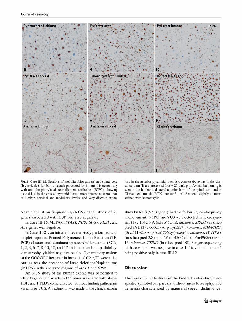
Degeneration of the lateral and, to a lesser extent, ante-rior corticospinal fascicles with axon and myelin loss, moderate astrogliosis, and absence of microglial response, macrophages or inflammatory cells, was the main altera-tion in the spinal cord. Degeneration was much more marked at sacral and lumbar, than at cervical spinal cord and medullary levels (Fig. 5a–d). No neuronal loss or cell body alteration was noted in the anterior horns of the spi-nal cord, except for a few ballooned axons in the ante-rior horn in the distal spinal cord and in Clarke’s column (a normal finding with aging in this region, Fig. 5g, h)). Other spinal cord fascicles were not affected; myelin and
nerve fibers were preserved in the spinal roots and dorsal tracts (Figs. 4d–g, 5e, f).
The cerebellum showed moderate loss of Purkinje cells with occasional torpedoes and empty baskets (Fig. 4j). Mod-erate neuronal loss was observed in the bulbar olive (greater than corresponding to age), accompanied by moderate astro-cytosis but no reactive microglia. No alteration was found in the nucleus of the hypoglossal nerve, motor nucleus of the vagus nerve, oculomotor and pontine nuclei, red nuclei, colliculi or substantia nigra.
Only one section each of the anterior caudate, putamen, and pulvinar nucleus was available for study; no neuronal alterations were seen.
Neurofibrillary tangles (NFT) composed of 4Rtau and 3Rtau were observed in the hippocampus and entorhinal cor-tex (Fig. 4h, i). Abnormal TDP-43, α-synuclein or β-amyloid deposits were not observed in any region.
To summarize, the neuropathological studies showed: (1) degeneration of the lateral corticospinal tracts, with an axonal dying-back pattern in case III.12; (2) preservation of spinal, brainstem and cortical motor neurons; (3) severe atro-phy of the frontal white matter and corpus callosum in case III-7; (4) cerebello-olivary degeneration, mainly affecting Purkinje cells in case III.12; (5) argyrophilic grain disease
Fig. 3 Autopsy features in Case III-7. a Macroscopic coronal section showing atrophy of the frontal lobes, reduction in the white matter of the centrum semiovale and corpus callosum, enlargement of lateral ventricles and normal temporal lobes. b Microscopic section of the motor cortex (MC) showing preservation of Betz neurons (Klüver-Barrera; bar = 45 µm). c In this transverse section of the lateral pyramidal tract, note marked reduction of axons in the lateral pyrami-
dal tract (anti-neurofilament 200 kDa antibody, clone RT97; bar = 25 µm). d Section of the hippocampus (CA1 region) showing neu-rons with deposits of hyperphosphorylated tau and grains (Phospho-Tau [Ser202, Thr205] Monoclonal Antibody AT8; bar = 225 µm). Sections processed for immunohistochemistry are slightly counter-stained with hematoxylin

Journal of Neurology
1 3
(AGD) stage III in case III-7; and (6) primary age-related tauopathy (PART) in case III-12.
Genetic testing
In case III-7, an exome panel aimed at PLS (24 genes) did not detect pathogenic variants, or variants of unknown sig-nificance (VUS). In addition, microarray-based Compara-tive Genomic Hybridization (aCGH) 400K was performed to detect deletions in genes involved in PLS, especially TBK1, with negative results.
Finally, massive sequencing of the human genome was carried out to identify genomic variants: (1) single nucle-otide (SNV) and insertion–deletion (INDEL) variants included in exonic regions, 3´UTR and 5′UTR regions, and
all intron variants described as probably pathogenic or path-ogenic in the ClinVar database (v20150330); and (2) copy number variations in exonic, intronic or intergenic regions. No pathogenic variants associated with the object of consul-tation were identified. However, a low-frequency population VUS was identified in a gene that has compatible signs with the phenotype under study, with which it was not possible to establish an association: p.(Pro656Thr), exon 17, MED25 [NM_030973.3:c.1966C > A], missense variant, carrier in heterozygosis.
In case III-12, MLPA (Multiplex Ligation-dependent Probe Amplification) aimed at the hereditary spastic paraparesis (HSP) genes SPAST, NIPA1 (SPG6), SPG7, REEP1 (SPG31), and ALT1 (SPG3A) was negative. A
Fig. 4 Histological features in Case III-12. a–c Sections of primary motor cortex (MC) stained with hematoxylin and eosin (HE), mye-lin basic protein (MBP), and glial fibrillary acidic protein (GFAP), showing neuronal preservation and absence of reactive astrocytosis (bar = 45 µm). Transverse sections of the spinal cord at the cervi-cal level showing preservation of the dorsal columns (d) and loss of myelinated fibers in the lateral pyramidal tract (e) (Klüver-Barrera; bar = 45 µm). f, g Note absence of alterations in anterior (ant root)
or dorsal roots of the spinal cord (Klüver-Barrera; bar = 100 µm). h–j Neurofibrillary degeneration visualized with AT8 antibody in the entorhinal cortex (EC) and CA1 region of the hippocampus (CA1, bar = 100 µm); section of the cerebellar cortex (bar = 25 µm) showing axonal ballooning, as revealed with anti-phosphorylated neurofila-ment antibodies (RT97). Sections processed for immunohistochemis-try are slightly counterstained with hematoxylin
Journal of Neurology
1 3
Next Generation Sequencing (NGS) panel study of 27 genes associated with HSP was also negative.
In Case III-16, MLPA of SPAST, NIPA, SPG7, REEP, and ALT genes was negative.
In Case III-21, an initial molecular study performed with Triplet-repeated Primed Polymerase Chain Reaction (TP-PCR) of autosomal dominant spinocerebellar ataxias (SCA) 1, 2, 3, 6, 7, 8, 10, 12, and 17 and dentatorubral–pallidoluy-sian atrophy, yielded negative results. Dynamic expansions of the GGG GCC hexamer in intron 1 of C9orf72 were ruled out, as was the presence of large deletions/duplications (MLPA) in the analyzed regions of MAPT and GRN.
An NGS study of the human exome was performed to identify genomic variants in 145 genes associated with ataxia, HSP, and FTLD/exome directed, without finding pathogenic variants or VUS. An extension was made to the clinical exome
study by NGS (5713 genes), and the following low-frequency allelic variants (< 1%) and VUS were detected in heterozygo-sis: (1) c.134C > A (p.Pro45Gln), missense, SPAST (in silico pred 3/8); (2) c.666C > A (p.Tyr222*), nonsense, MMACHC; (3) c.5118C > A (p.Asn1706Lys) exon 40, missense, (4) ITPR1 (in silico pred 2/8); and (5) c.1486C > T (p.Pro496Ser) exon 13, missense, TTBK2 (in silico pred 1/8). Sanger sequencing of these variants was negative in case III-16, variant number 4 being positive only in case III-12.
Discussion
The core clinical features of the kindred under study were spastic spinobulbar paresis without muscle atrophy, and dementia characterized by inaugural speech disturbance.
Fig. 5 Case III-12. Sections of medulla oblongata (a) and spinal cord (b cervical; c lumbar; d sacral) processed for immunohistochemistry with anti-phosphorylated neurofilament antibodies (RT97), showing axonal loss in the crossed pyramidal tract, more intense at sacral than at lumbar, cervical and medullary levels, and very discrete axonal
loss in the anterior pyramidal tract (e); conversely, axons in the dor-sal column (f) are preserved (bar = 25 µm). g, h Axonal ballooning is seen in the lumbar and sacral anterior horn of the spinal cord and in Clarke’s column (i) (RT97, bar = 45 µm). Sections slightly counter-stained with hematoxylin

Journal of Neurology
1 3
Disease onset was in the sixth decade of life or later, and inheritance was autosomal dominant with complete penetrance.
The motor manifestations exhibited by this family fulfilled the clinical diagnostic criteria of PLS [56]: (1) insidious onset of spastic paresis, beginning in the lower extremities; (2) onset in the fifth decade or later; (3) grad-ually progressive course (duration ≥ 3 years; (4) clinical findings limited to corticospinal dysfunction; and (5) sym-metrical distribution, ultimately developing spastic spin-obulbar paresis. Criterion number 3 (absence of family history) was not met.
Juvenile PLS may be sporadic but also inherited in a recessive mode, the causative mutant gene being ALS2 on chromosome 2q33.1, which is also responsible for infan-tile-onset ascending spastic paralysis and a juvenile form of ALS [25, 46, 51]. Erb claimed that PLS could be heredi-tary [18], albeit recent examples of adult onset autosomal dominant PLS are limited to a kinship with compound heterozygote mutations in SPG7, and a French–Canadian family, in which a locus was identified on chromosome 4 [15, 69, 73]. Adult onset PLS is not caused by muta-tions in ALS2 [5], but apparently sporadic cases have been related to mutations in SPG7, SPG11, DCTN1, PARK2, and expansions in C9orf72 [47, 72]. Isolated instances of PLS may occur within FTLD [21] and ALS pedigrees [4, 55, 58], such as those associated with mutations in TBK1 and UBQLN2, respectively [72]. None of the mentioned variants was found in this study.
PLS is rare, comprising about 5% of motor neuron dis-ease [MND] patients presenting at specialized neuromus-cular clinics [49]. The controversy about whether PLS is a subtype of ALS or a discrete entity continues to this day [22, 26]: revised El Escorial criteria consider PLS a restricted ALS phenotype [40], while other authors maintain PLS is a distinct syndrome [13].
Our patients had no lower motor neuron signs; in conse-quence, electrophysiological testing was performed only in cases III-16 and III-21, in which electromyographic studies were, respectively, normal, 7 and 6 years after disease onset.
The manifestations displayed by this kindred fell into the category of PLS-plus (complicated PLS): “predomi-nant upper motor neuron signs plus clinical, laboratory, or pathological evidence of dementia, parkinsonism, or sen-sory abnormalities”, because “spastic spinobulbar paralysis without muscle atrophy, visible fasciculations or evidence of denervation on electromyography at ≥ 4 years from dis-ease onset” [22, 39, 62], together with dementia or cognitive decline, was present in every case.
Case III-12 associated dopamine agonist therapy-resist-ant parkinsonism with PLS, as reported in PLS-plus [20, 22, 41, 62]. Although the substantia nigra was normal on pathological examination, assessment of the basal ganglia
could not be performed in depth, precluding any potential clinicopathological correlation.
Cerebellar ataxia has not been considered a component of PLS-plus, its presence making it necessary to differentiate PLS-plus from multiple system atrophy [20, 40]. However, cerebellar intention tremor and unsteady gait were reported in a family with MND and dementia [two members express-ing PLS], in correlation with loss of Purkinje cells [54]. Patients III-7 and III-21 exhibited inaugural unsteady gait and dysmetria, although no histological abnormalities were present in the cerebellum in the first case. Contrariwise, cer-ebello-olivary degeneration was found in case III-12, in spite of an absence of ataxic signs. The presence of cerebellar ataxia in this context could be related to factors that modify the expression of the causative gene and induce differing clinical and pathological manifestations, as demonstrated in hereditary ataxia [61].
In spite of preserved bladder function being considered part of PLS [56], urinary incontinence was present in five of our cases and urinary urgency in the other, as reported in PLS [39, 47], the cause being detrusor hyperreflexia and a spastic internal vesical sphincter [59].
PLS is considered to spare intellect, although impairment in executive function, verbal memory, psychomotor speed and verbal fluency (similar to ALS and progressive mus-cular atrophy) has been identified in up to 49% of patients [8, 12, 53]. In one study, total score on the Addenbrooke’s Cognitive Examination was abnormal in 21 patients with PLS (mean 82.5/100, cutoff value 88), the most prominent deficits being reduced verbal fluency, and deficiencies in memory, language and visuospatial function [1].
PLS is not believed to form part of the FTLD–MND com-plex; however, this association has been reported before [14, 29]. In one series, 3.3% of PLS cases developed FTLD of the behavioural or semantic variants [12]. Rapid and severe cog-nitive decline appeared years after progressive UMN signs, with memory impairment never being an early symptom, as observed in this kindred, in which similar time courses of evolution and progression of symptoms, suggest a com-mon pathogenic origin for motor and cognitive symptoms. Amyloid was absent in pathological material [1], as in our neuropathological and florbetapir PET-CT studies, ruling out Alzheimer’s disease as the present illness’ cause.
Every investigated member in this kin suffered from dementia starting up to 12 years after motor symptom onset, except for patient III-21 (who only exhibited frontosubcor-tical deterioration after 8 years of disease). Early speech (halting, stuttering and effortful speech) and language dis-turbances (agrammatism, decreased verbal fluency and pho-nemic paraphasia) preceded apathy, emotional lability, and memory impairment. These manifestations were evaluated against clinical criteria for PPA, and a nonfluent/agrammatic variant was diagnosed [23]. Aggressiveness and delusional

Journal of Neurology
1 3
ideas occurred in case III-12, and social disinhibition and emotional lability in case III-21; however, clinical diagnostic criteria for behavioural variant FTLD were not met [9, 23].
Nonfluent PPA has been related to potential development of ALS and other motor syndromes [16, 30, 64], as found in this family. It was mentioned in association with PLS in one case, but no clinical description was provided [34]. Deposition of tau protein predominating in the left posterior frontoinsular cortex is the most likely pathological basis [30, 33], although in this study, tau deposition was found in the form of AGD in case III-7, and of PART in case III-12.
AGD is an age-related 4R tauopathy. It may overlap with other neurodegenerative disorders and is second only to Alz-heimer’s disease in frequency, although no association with the APOE ε4 genotype has been reported. It is character-ized by three main pathological features: argyrophilic grains, oligodendrocytic coiled bodies, and neuronal cytoplasmatic pre-tangles in the medial temporal lobes, as found in case III-7. AGD lacks distinctive clinical features and may mani-fest as a slowly progressive amnestic syndrome; however, up to 59% of cases with pathologically demonstrated AGD were cognitively normal [31, 57].
PART refers to the presence of age-related 3R and 4R tau-positive NFT in neurons, without extension beyond the temporal neocortex. Whether PART represents early Alz-heimer’s disease remains controversial, as the frequency of association with APOE ε4 is low [33, 57]. A diagnosis of PART requires a Braak NFT stage ≤ IV (usually III or lower), and a Thal beta-amyloid phase ≤ 2. PART is clas-sified pathologically as definite or possible, depending on whether beta-amyloid is absent or present, respectively [10, 32]. Clinically, it may be divided into asymptomatic (no cognitive decline or dementia), or symptomatic (cognitive decline or dementia present); [10]; case III-12 can be clas-sified as definite, symptomatic PART.
Definite PART may go either with normal intellect, mild cognitive impairment or dementia, as no precise clinical features have been outlined for it. Depression, increasing Braak stage, and presence of other tau pathologies or neu-rodegenerative diseases have been associated with definite, symptomatic PART [3].
AGD and PART do not affect the left frontoinsular cortex involved in nonfluent PPA, and have not been previously associated with PLS in the medical literature; therefore, they must be considered incidental findings in the present kindred.
Neuropathological examination of cases with PLS and dementia have revealed corticospinal tract degeneration, preservation of spinal motor neurons and loss of cerebral cortex neurons, which displayed either ubiquitin-positive tau [66], ubiquitin-positive [36, 48], TDP-43 [29, 34, 37], neurofilament-immunoreactive neuronal inclusions [42], dendritic ballooning [65], or diffuse beta-amyloid plaques
[17]. Every one of these cases was sporadic, and the data provided were insufficient to define a dementing syndrome characteristic of PLS. None of the aforementioned cortical changes was found in our autopsied cases.
A diagnosis of autopsy-proven PLS in this study was corroborated by degeneration of corticospinal tracts in the spinal cord and bulbar pyramids without neuron loss, gliosis, Bunina bodies, ubiquitin or TDP-43 inclusions, in spinal motor neurons [22]. The more pronounced corti-cospinal fiber loss at sacral and lumbar, rather than at cer-vical and medullary levels found in case III-12, supports by pathological means a dying-back pathogenic mecha-nism, in which central axons degenerate following a dis-tal-to-proximal gradient, as previously proposed for PLS [11, 74]. Moreover, preservation of upper motor neuron cell bodies indicated that a cerebral cortical neuronopathy could not be claimed as causal in cases III-7 and III-12.
Marked loss of myelin and nerve fibers in the frontal and parietal lobes without deposition of abnormal mate-rial, was found in case III-7. Taken together, these data suggest that white matter degeneration centered on axonal motor and language networks could be a primary patho-logic alteration in this kindred [27, 34, 67].
Central distal axonopathies have been implied as causal mechanisms in HSP, lathyrism, Friedreich’s ataxia, ALS and clioquinol intoxication [38, 68, 74]. A primary cen-tral axonopathy that affects language structures has been proposed for progressive nonfluent aphasia on the basis of white matter damage on MRI [60], as found in case III.12.
MRI demonstrated dilation of central sulci in this series [as reported in PLS], enlarged frontoparietal sulci and lat-eral ventricles, bilateral white matter abnormalities in the corona radiata and atrophy of the corpus callosum; the last two have been associated with PLS and PPA [2, 35, 56, 63]. In addition, impairment in memory, executive and language test scores in PLS, has been found to correlate with white matter tract damage on diffusion-tensor MRI [7, 45].
The main differential diagnosis in this kindred would be FTLD–MND, excluded by absence of lower motor neuron clinical, electromyographic and pathological signs, and a mean survival time of 196.8 months, approximately equiva-lent to 217 ± 22.4 months in one series [53]. This is in con-trast with FTLD–MND, whose mean duration was estimated as 2–3 years [29], and 38.5 ± 4.5 months [28]. SPG4, SPG7, and SPG11 were also considered in the differential diagnosis [6, 69, 71], as well as SCA1 and SCA17 in cases III-7 and III-21, because these exhibited cerebellar ataxia in addition to spasticity and cognitive decline [43, 70]. All these dis-eases were ruled out by genetic analysis. Furthermore, no potentially pathogenic gene mutations were identified on NGS whole exome sequencing, covering 5.713 genes, and whole genome sequencing.

Journal of Neurology
1 3
In summary, a kindred with a hitherto undescribed, domi-nantly inherited variant of PLS and progressive nonfluent aphasia, in which the corticospinal tracts were affected by a central distal axonopathy, and cortical motor neurons were preserved, is presented. Further molecular studies are needed to identify the causative mutation in this disease, to offer accurate genetic counseling to the family under study.
Acknowledgements Our thanks to Dr. Antonio Oliveros-Juste for remission of patients; Drs. Ana Vela and Jordi Aldomá for MR images; Mr. Juan Ramón Solans, Drs. Ariadna Fernández-Sanz and Jesús Aladrén-Sangrós for help with the videtapes; Mrs. María Jesús Chopo for obtention of DNA samples; Mrs. Teresa Sopeña Biarge and Mrs. Mar González-Cantalejo from the Medical Library of Hospital Universitario Miguel Servet, for bibliographical research; and Dr. Jorge Alfaro for assistance in the pathological studies.
Compliance with ethical standards
Conflicts of interest None.
Ethical standards All human studies have been approved by the appro-priate ethics committee and have therefore been performed in accord-ance with the ethical standards laid down in the 1964 Declaration of Helsinki.
References
1. Agarwal S, Highton-Williamson E, Caga J, Matamala JM, Dhar-madasa T, Howells J et al (2018) Primary lateral sclerosis and the amyotrophic lateral sclerosis-frontotemporal dementia spectrum. J Neurol 265:1819–1828
2. Agosta F, Galantucci S, Magnani G, Marcone A, Martinelli D, Antonietta Volontè M et al (2015) MRI signatures of the fron-totemporal lobar degeneration continuum. Hum Brain Mapp 36:2602–2614
3. Besser LM, Crary JF, Mock C, Kukull WA (2017) Comparison of symptomatic and asymptomatic persons with primary age-related tauopathy. Neurology 89:1707–1715
4. Brugman F, Wokke JH, Vianney de Jong JM, Franssen H, Faber CG, Van den Berg LH (2005) Primary lateral sclerosis as a phe-notypic manifestation of familial ALS. Neurology 64:1778–1779
5. Brugman F, Eymard-Pierre E, van den Berg LH, Wokke JH, Gauthier-Barichard F, Boespflug-Tanguy O (2007) Adult-onset primary lateral sclerosis is not associated with mutations in the ALS2 gene. Neurology 69:702–704
6. Brugman F, Veldink JH, Franssen H, de Visser M, de Jong JM, Faber CG et al (2009) Differentiation of hereditary spastic parapa-resis from primary lateral sclerosis in sporadic adult-onset upper motor neuron syndromes. Arch Neurol 66:509–514
7. Canu E, Agosta F, Galantucci S, Chiò A, Riva N, Silani V et al (2013) Extramotor damage is associated with cognition in primary lateral sclerosis patients. PLoS One 8:e82017
8. Caselli RJ, Smith BE, Osborne D (1995) Primary lateral sclerosis: a neuropsychological study. Neurology 45:2005–2009
9. Chare L, Hodges JR, Leyton CE, McGinley C, Tan RH, Kril JJ et al (2014) New criteria for frontotemporal dementia syndromes: clinical and pathological diagnostic implications. J Neurol Neu-rosurg Psychiatry 85:865–870
10. Crary JF, Trojanowski JQ, Schneider JA, Abisambra JF, Abner EL, Alafuzoff I et al (2014) Primary age-related tauopathy
(PART): a common pathology associated with human aging. Acta Neuropathol 128:755–766
11. Deng HX, Zhai H, Fu R, Shi Y, Gorrie GH, Yang Y et al (2007) Distal axonopathy in an alsin-deficient mouse model. Hum Mol Genet 16:2911–2920
12. de Vries BS, Rustemeijer LMM, van der Kooi AJ, Raaphorst J, Schröder CD, Nijboer TCW et al (2017) A case series of PLS patients with frontotemporal dementia and overview of the literature. Amyotroph Lateral Scler Frontotemporal Degener 18:534–548
13. de Vries BS, Rustemeijer LMM, Bakker LA, Schröder CD, Veldink JH, van den Berg LH et al (2019) Cognitive and behavioural changes in PLS and PMA: challenging the con-cept of restricted phenotypes. J Neurol Neurosurg Psychiatry 90:141–147
14. Doran M, Enevoldson TP, Ghadiali EJ, Larner AJ (2005) Mills syndrome with dementia: broadening the phenotype of FTD/MND. J Neurol 252:846–847
15. Dupré N, Valdmanis PN, Bouchard JP, Rouleau GA. Autosomal dominant primary lateral sclerosis. Neurology 68:1156–7
16. Elahi FM, Miller BL (2017) A clinicopathologic approach to the diagnosis of dementia. Nat Rev Neurol 13:457–476
17. Engel PA, Grunnet M (2000) Atypical dementia and spastic para-plegia in a patient with primary lateral sclerosis and numerous neocortical beta amyloid plaques: new disorder or Alzheimer’s disease variant? J Geriatr Psychiatry Neurol 13:60–64
18. Erb W (1902) Spastic and syphilitic spinal paralysis. Lancet 2:969–974
19. Folstein M, Folstein SE, McHugh PR (1975) “Mini-Mental State” a practical method for grading the cognitive state of patients for the clinician. J Psychiatr Res 12:189–198
20. Gilbert RM, Fahn S, Mitsumoto H, Rowland LP (2010) Parkinson-ism and motor neuron diseases: twenty-seven patients with diverse overlap syndromes. Mov Disord 25:1868–1875
21. Gómez-Tortosa E, Van der Zee J, Ruggiero M, Gijselinck I, Este-ban-Pérez J, García-Redondo A et al (2017) Familial primary lateral sclerosis or dementia associated with Arg573Gly TBK1 mutation. J Neurol Neurosurg Psychiatry 88:996–997
22. Gordon PH, Cheng B, Katz IB, Pinto M, Hays AP, Mitsumoto H, Rowland LP (2006) The natural history of primary lateral sclero-sis. Neurology 66:647–653
23. Gorno-Tempini ML, Hillis AE, Weintraub S, Kertesz A, Mendez M, Cappa SF et al (2011) Classification of primary progressive aphasia and its variants. Neurology 76:1006–1014
24. Haugh AB, Pandyan AD, Johnson GR (2006) A systematic review of the Tardieu Scale for the measurement of spasticity. Disabil Rehabil 28:899–907
25. Helal M, Mazaheri N, Shalbafan B, Malamiri RA, Dilaver N, Buchert R et al (2018) Clinical presentation and natural history of infantile-onset ascending spastic paralysis from three families with an ALS2 founder variant. Neurol Sci 39:1917–1925
26. Hudson AJ, Kiernan JA, Muñoz DG, Pringle CE, Brown WF, Ebers GC (1993) Clinicopathological features of primary lateral sclerosis are different from amyotrophic lateral sclerosis. Brain Res Bull 30:359–364
27. Iwata NK, Kwan JY, Danielian LE, Butman JA, Tovar-Moll F, Bayat E et al (2011) White matter alterations differ in pri-mary lateral sclerosis and amyotrophic lateral sclerosis. Brain 134:2642–2655
28. Josephs KA, Knopman DS, Whitwell JL, Boeve BF, Parisi JE, Petersen RC et al (2005) Survival in two variants of tau-negative frontotemporal degeneration: FTLD-U vs. FTLD-MND. Neurol-ogy 65:645–647
29. Josephs KA, Dickson DW (2007) Frontotemporal lobar degenera-tion with upper motor neuron disease/primary lateral sclerosis. Neurology 69:1800–1801

Journal of Neurology
1 3
30. Josephs KA, Duffy JR (2008) Apraxia of speech and nonfluent aphasia: a new clinical marker for corticobasal degeneration and progressive supranuclear palsy. Curr Opin Neurol 21:688–692
31. Josephs KA (2017) Current understanding of neurodegenera-tive diseases associated with the protein tau. Mayo Clin Proc 92:1291–1303
32. Josephs KA, Murray ME, Tosakulwong N, Whitwell JL, Knopman DS, Machulda MM et al (2017) Tau aggregation influences cogni-tion and hippocampal atrophy in the absence of beta-amyloid: a clinico-imaging-pathological study of primary age-related tauopa-thy (PART). Acta Neuropathol 133:705–715
33. Knopman DS, Nestor PJ (2017) Beyond clinical syndromes in primary progressive aphasia. Seeking etiologic diagnoses. Neurol-ogy 88:2244–2245
34. Kobayashi Z, Tsuchiya K, Arai T, Yokota O, Yoshida M, Shi-momura Y et al (2010) Clinicopathological characteristics of FTLD-TDP showing corticospinal tract degeneration but lacking lower motor neuron loss. J Neurol Sci 298:70–77
35. Kolind S, Sharma R, Knight S, Johansen-Berg H, Talbot K, Turner MR (2013) Myelin imaging in amyotrophic and primary lateral sclerosis. Amyotroph Lateral Scler Frontotemporal Degener 14:562–573
36. Konagaya M, Sakai M, Matsuoka Y, Konagaya Y, Hashizume Y (1998) Upper motor neuron predominant degeneration with frontal and temporal lobe atrophy. Acta Neuropathol 96:532–536
37. Kosaka T, Fu YJ, Shiga A, Ishidaira H, Tan CF, Tani T et al (2012) Primary lateral sclerosis: upper-motor-predominant amyotrophic lateral sclerosis with frontotemporal lobar degeneration–immuno-histochemical and biochemical analyses of TDP-43. Neuropathol-ogy 32:373–384
38. Lamarche JB, Lemieux B, Lieu HB. The neuropathology of “typi-cal” Friedreich’s ataxia in Quebec (1984). Can J Neurol Sci 11(4 Suppl): 592–600
39. Le Forestier N, Maisonobe T, Piquard A, Rivaud S, Crevier-Buch-man L, Salachas F et al (2001) Does primary lateral sclerosis exist? A study of 20 patients and a review of the literature. Brain 124:1989–1999
40. Ludolph A, Drory V, Hardiman O, Nakano I, Ravits J, Robbere-cht W et al (2015) A revision of El escorial criteria. Amyotroph Lateral Scler Frontotemporal Degener 16:291–292
41. Mabuchi N, Watanabe H, Atsuta N, Hirayama M, Ito H, Fukatsu H et al (2004) Primary lateral sclerosis presenting parkinsonian symptoms without nigrostriatal involvement. J Neurol Neurosurg Psychiatry 75:1768–1771
42. Mackenzie IR, Feldman H (2004) Neurofilament inclusion body disease with early onset frontotemporal dementia and primary lateral sclerosis. Clin Neuropathol 23:183–193
43. Matilla-Dueñas A, Goold R, Giunti P (2008) Clinical, molecular, and pathophysiological insights into spinocerebellar ataxia type 1. Cerebellum 7:106–114
44. Medical Research Council (1976) Aids to the examination of the peripheral nervous system. Her Majesty’s Stationery Office, London
45. Meoded A, Kwan JY, Peters TL, Huey ED, Danielian LE, Wiggs E et al (2013) Imaging findings associated with cognitive perfor-mance in primary lateral sclerosis and amyotrophic lateral scle-rosis. Dement Geriatr Cogn Dis Extra 3:233–250
46. Mintchev N, Zamba-Papanicolaou E, Kleopa KA, Christodoulou K (2009) A novel ALS2 splice-site mutation in a Cypriot juvenile-onset primary lateral sclerosis family. Neurology 72:28–32
47. Mitsumoto H, Nagy PL, Gennings C, Murphy J, Andrews H, Goetz R et al (2015) Phenotypic and molecular analyses of pri-mary lateral sclerosis. Neurol Genet 1:e3
48. Mochizuki A, Komatsuzaki Y, Iwamoto H, Shoji S (2004) Fron-totemporal dementia with ubiquitinated neuronal inclusions presenting with primary lateral sclerosis and parkinsonism:
clinicopathological report of an autopsy case. Acta Neuropathol 107:377–380
49. Murphy MJ, Grace GM, Tartaglia MC, Orange JB, Chen X, Rowe A et al (2008) Cerebral haemodynamic changes accompa-nying cognitive impairment in primary lateral sclerosis. Amyo-troph Lateral Scler 9:359–368
50. Pandyan AD, Johnson GR, Price CI, Curless RH, Barnes MP, Rodgers H (1999) A review of the properties and limitations of the Ashworth and modified Ashworth Scales as measures of spasticity. Clin Rehabil 13:373–383
51. Panzeri C, De Palma C, Martinuzzi A, Daga A, De Polo G, Bre-solin N et al (2006) The first ALS2 missense mutation associ-ated with JPLS reveals new aspects of alsin biological function. Brain 129:1710–1719
52. Penn RD, Savoy SM, Corcos D, Latash M, Gottlieb G, Parke B et al (1989) Intrathecal baclofen for severe spinal spasticity. N Engl J Med 320:1517–1521
53. Piquard A, Le Forestier N, Baudoin-Madec V, Delgadillo D, Salachas F, Pradat PF et al (2006) Neuropsychological changes in patients with primary lateral sclerosis. Amyotroph Lat Scler 7:150–160
54. Polvikoski TM, Murray A, Harper PS, Neal JW (2003) Famil-ial motor neuron disease with dementia: phenotypic varia-tion and cerebellar pathology. J Neurol Neurosurg Psichiatry 74:1516–1520
55. Praline J, Guennoc AM, Vourc’h P, De Toffol B, Corcia P (2010) Primary lateral sclerosis may occur within familial amyotrophic lateral sclerosis pedigrees. Amyotroph Lat Scler 11:154–156
56. Pringle CE, Hudson AJ, Muñoz DG, Kiernan JA, Brown WF, Ebers GC (1992) Primary lateral sclerosis. Clinical features, neu-ropathology and diagnostic criteria. Brain 115:495–520
57. Rodriguez RD, Suemoto CK, Molina M, Nascimento CF, Leite RE, de Lucena Ferretti-Rebustini RE, et al (2016) Argyrophilic grain disease: demographics, clinical, and neuropathological features from a large autopsy study. J Neuropathol Exp Neurol 75:628–635
58. Rosen DR, Siddique T, Patterson D, Figlewicz DA, Sapp P, Hen-tati A et al (1993) Mutations in Cu/Zn superoxide dismutase gene are associated with familial amyotrophic lateral sclerosis. Nature 362:59–62
59. Russo LS (1982) Clinical and electrophysiological studies in pri-mary lateral sclerosis. Arch Neurol 39:662–664
60. Schwindt GC, Graham NL, Rochon E, Tang-Wai DF, Lobaugh NJ, Chow TW et al (2013) Whole-brain white matter disruption in semantic and nonfluent variants of primary progressive aphasia. Hum Brain Mapp 34:973–984
61. Schut JW, Haymaker W (1951) Hereditary ataxia. A pathologic study of five cases of common ancestry. J Neuropathol Clin Neu-rol 1:183–213
62. Singer MA, Statland JM, Wolfe GI, Barohn RJ (2007) Primary lateral sclerosis. Muscle Nerve 35:291–302
63. Smith CD (2002) Serial findings in a case of primary lateral scle-rosis. Neurology 57:647–649
64. Strong MJ, Abrahams S, Goldstein LH, Woolley S, Mclaughlin P, Snowden J et al (2017) Amyotrophic lateral sclerosis—frontotem-poral spectrum disorder (ALS-FTSD): revised diagnostic criteria. Amyotroph Lateral Scler Frontotemporal Degener 18:153–174
65. Sugihara H, Horiuchi M, Kamo T, Fujisawa K, Abe M, Sakiy-ama T et al (1999) A case of primary lateral sclerosis taking a prolonged clinical course with dementia and having an unusual dendritic ballooning. Neuropathology 19:77–84
66. Tan CF, Kakita A, Piao YS, Kikugawa K, Endo K, Tanaka M et al (2003) Primary lateral sclerosis: a rare upper-motor-predom-inant form of amyotrophic lateral sclerosis often accompanied by frontotemporal lobar degeneration with ubiquitinated neuronal

Journal of Neurology
1 3
inclusions? Report of an autopsy case and a review of the litera-ture. Acta Neuropathol 105:615–620
67. Tartaglia MC, Laluz V, Rowe A, Findlater K, Lee DH, Kennedy K et al (2009) Brain atrophy in primary lateral sclerosis. Neurology 72:1236–1241
68. Thomas PK, Schaumburg HH, Spencer PS, Kaeser HE, Pallis CA, Rose FC et al (1984) Central distal axonopathy syndromes: newly recognized models of naturally occurring human degenera-tive disease. Ann Neurol 15:313–315
69. Valdmanis PN, Dupré N, Rouleau GA (2008) A locus for pri-mary lateral sclerosis on chromosome 4ptel-4p16.1. Arch Neurol 65:383–386
70. Van Gaalen J, Giunti P, van de Warrenburg BP (2011) Movement disorders in spinocerebellar ataxias. Mov Disord 26:792–800
71. Vázquez-Costa JF, Bataller L, Vílchez JJ (2016) Primary lateral sclerosis and hereditary spastic paraplegia in sporadic patients. An important distinction in descriptive studies. Ann Neurol 80:169–170
72. Yamashita S, Ando Y (2015) Genotype-phenotype relationship in hereditary amyotrophic lateral sclerosis. Transl Neurodegener 3:13
73. Yang Y, Zhang L, Lynch DR, Lukas T, Ahmeti K, Sleiman PM et al (2016) Compound heterozygote mutations in SPG7 in a fam-ily with adult-onset primary lateral sclerosis. Neurol Genet 2:e60
74. Zhai P, Pagan F, Statland J, Butman JA, Floeter MK (2003) Pri-mary lateral sclerosis. A heterogeneous disorder composed of dif-ferent subtypes? Neurology 60:1258–1265