Hepatology Dr. Mohamed Ismail Hepatology Clinical ... Clinical presentation of liver disease...
11
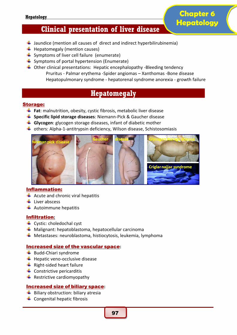
97 Hepatology Dr. Mohamed Ismail Clinical presentation of liver disease Jaundice (mention all causes of direct and indirect hyperbilirubinemia) Hepatomegaly (mention causes) Symptoms of liver cell failure (enumerate) Symptoms of portal hypertension (Enumerate) Other clinical presentations: Hepatic encephalopathy -Bleeding tendency Pruritus - Palmar erythema -Spider angiomas – Xanthomas -Bone disease Hepatopulmonary syndrome - hepatorenal syndrome anorexia - growth failure Hepatomegaly Storage: Fat: malnutrition, obesity, cystic fibrosis, metabolic liver disease Specific lipid storage diseases: Niemann-Pick & Gaucher disease Glycogen: glycogen storage diseases, infant of diabetic mother others: Alpha-1-antitrypsin deficiency, Wilson disease, Schistosomiasis Inflammation: Acute and chronic viral hepatitis Liver abscess Autoimmune hepatitis Infiltration: Cystic: choledochal cyst Malignant: hepatoblastoma, hepatocellular carcinoma Metastases: neuroblastoma, histiocytosis, leukemia, lymphoma Increased size of the vascular space : Budd-Chiari syndrome Hepatic veno-occlusive disease Right-sided heart failure Constrictive pericarditis Restrictive cardiomyopathy Increased size of biliary space : Biliary obstruction: biliary atresia Congenital hepatic fibrosis Chapter 6 Hepatology
Transcript of Hepatology Dr. Mohamed Ismail Hepatology Clinical ... Clinical presentation of liver disease...
97
Hepatology Dr. Mohamed Ismail
Clinical presentation of liver disease
Jaundice (mention all causes of direct and indirect hyperbilirubinemia) Hepatomegaly (mention causes) Symptoms of liver cell failure (enumerate) Symptoms of portal hypertension (Enumerate) Other clinical presentations: Hepatic encephalopathy -Bleeding tendency
Pruritus - Palmar erythema -Spider angiomas – Xanthomas -Bone disease Hepatopulmonary syndrome - hepatorenal syndrome anorexia - growth failure
Hepatomegaly
Storage:
Fat: malnutrition, obesity, cystic fibrosis, metabolic liver disease Specific lipid storage diseases: Niemann-Pick & Gaucher disease Glycogen: glycogen storage diseases, infant of diabetic mother others: Alpha-1-antitrypsin deficiency, Wilson disease, Schistosomiasis
Inflammation:
Acute and chronic viral hepatitis Liver abscess Autoimmune hepatitis
Infiltration:
Cystic: choledochal cyst Malignant: hepatoblastoma, hepatocellular carcinoma Metastases: neuroblastoma, histiocytosis, leukemia, lymphoma
Increased size of the vascular space: Budd-Chiari syndrome Hepatic veno-occlusive disease Right-sided heart failure Constrictive pericarditis Restrictive cardiomyopathy
Increased size of biliary space: Biliary obstruction: biliary atresia Congenital hepatic fibrosis
Chapter 6 Hepatology
98
Hepatology Dr. Mohamed Ismail
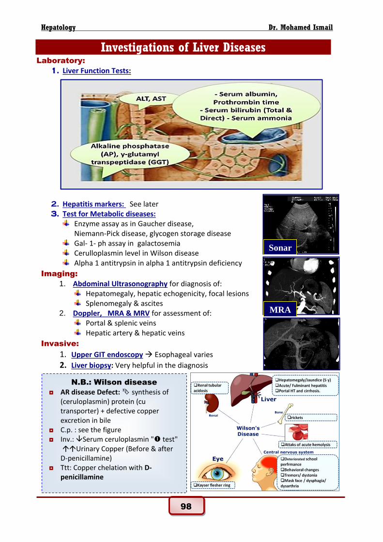
Investigations of Liver Diseases Laboratory:
1. Liver Function Tests:
2. Hepatitis markers: See later 3. Test for Metabolic diseases:
Enzyme assay as in Gaucher disease, Niemann-Pick disease, glycogen storage disease
Gal- 1- ph assay in galactosemia Cerulloplasmin level in Wilson disease Alpha 1 antitrypsin in alpha 1 antitrypsin deficiency
Imaging:
1. Abdominal Ultrasonography for diagnosis of: Hepatomegaly, hepatic echogenicity, focal lesions Splenomegaly & ascites
2. Doppler, MRA & MRV for assessment of: Portal & splenic veins Hepatic artery & hepatic veins
Invasive:
1. Upper GIT endoscopy Esophageal varies
2. Liver biopsy: Very helpful in the diagnosis
MRA
Sonar
N.B.: Wilson disease
◘ AR disease Defect: synthesis of (ceruloplasmin) protein (cu transporter) + defective copper excretion in bile
◘ C.p. : see the figure ◘ Inv.: Serum ceruloplasmin " test"
Urinary Copper (Before & after D-penicillamine)
◘ Ttt: Copper chelation with D-penicillamine
99
Hepatology Dr. Mohamed Ismail
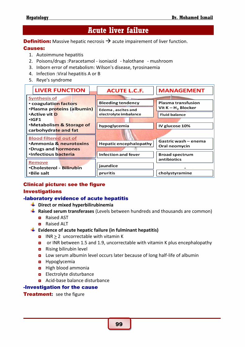
Acute liver failure
Definition: Massive hepatic necrosis acute impairement of liver function.
Causes:
1. Autoimmune hepatitis 2. Poisons/drugs :Paracetamol - isoniazid - halothane - mushroom 3. Inborn error of metabolism: Wilson's disease, tyrosinaemia 4. Infection :Viral hepatitis A or B 5. Reye's syndrome
Clinical picture: see the figure
Investigations
-laboratory evidence of acute hepatitis
Direct or mixed hyperbilirubinemia Raised serum transferases (Levels between hundreds and thousands are common) ◘ Raised AST ◘ Raised ALT
Evidence of acute hepatic failure (in fulminant hepatitis) ◘ INR > 2 uncorrectable with vitamin K ◘ or INR between 1.5 and 1.9, uncorrectable with vitamin K plus encephalopathy ◘ Rising bilirubin level ◘ Low serum albumin level occurs later because of long half-life of albumin ◘ Hypoglycemia ◘ High blood ammonia ◘ Electrolyte disturbance ◘ Acid-base balance disturbance
-Investigation for the cause Treatment: see the figure
100
Hepatology Dr. Mohamed Ismail
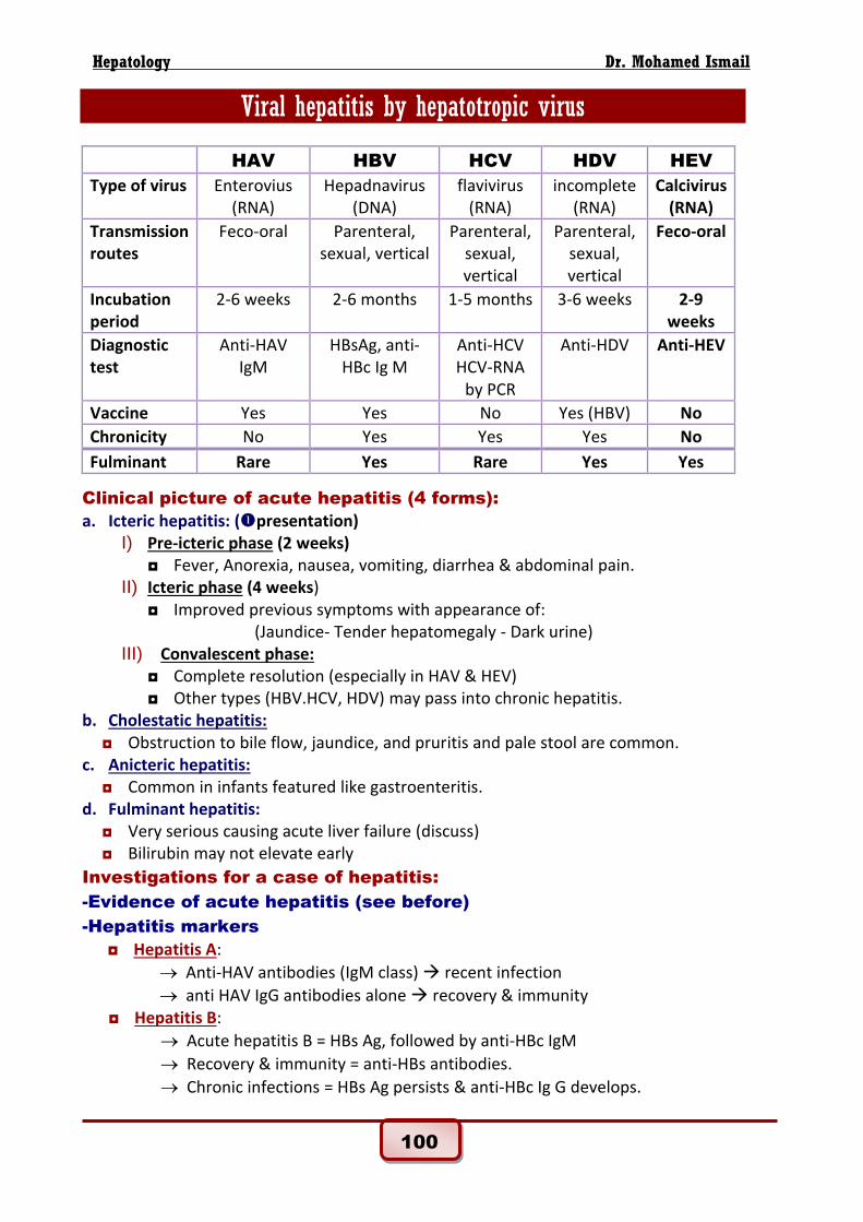
Viral hepatitis by hepatotropic virus
HAV HBV HCV HDV HEV
Type of virus Enterovius (RNA)
Hepadnavirus (DNA)
flavivirus (RNA)
incomplete (RNA)
Calcivirus (RNA)
Transmission routes
Feco-oral Parenteral, sexual, vertical
Parenteral, sexual, vertical
Parenteral, sexual, vertical
Feco-oral
Incubation period
2-6 weeks 2-6 months 1-5 months 3-6 weeks 2-9 weeks
Diagnostic test
Anti-HAV IgM
HBsAg, anti-HBc Ig M
Anti-HCV HCV-RNA
by PCR
Anti-HDV Anti-HEV
Vaccine Yes Yes No Yes (HBV) No
Chronicity No Yes Yes Yes No
Fulminant Rare Yes Rare Yes Yes
Clinical picture of acute hepatitis (4 forms):
a. Icteric hepatitis: (presentation) I) Pre-icteric phase (2 weeks)
◘ Fever, Anorexia, nausea, vomiting, diarrhea & abdominal pain. II) Icteric phase (4 weeks)
◘ Improved previous symptoms with appearance of: (Jaundice- Tender hepatomegaly - Dark urine)
III) Convalescent phase: ◘ Complete resolution (especially in HAV & HEV) ◘ Other types (HBV.HCV, HDV) may pass into chronic hepatitis.
b. Cholestatic hepatitis: ◘ Obstruction to bile flow, jaundice, and pruritis and pale stool are common.
c. Anicteric hepatitis: ◘ Common in infants featured like gastroenteritis.
d. Fulminant hepatitis: ◘ Very serious causing acute liver failure (discuss) ◘ Bilirubin may not elevate early
Investigations for a case of hepatitis:
-Evidence of acute hepatitis (see before)
-Hepatitis markers
◘ Hepatitis A:
Anti-HAV antibodies (IgM class) recent infection
anti HAV IgG antibodies alone recovery & immunity ◘ Hepatitis B:
Acute hepatitis B = HBs Ag, followed by anti-HBc IgM
Recovery & immunity = anti-HBs antibodies.
Chronic infections = HBs Ag persists & anti-HBc Ig G develops.
101
Hepatology Dr. Mohamed Ismail
◘ Hepatitis C:
Anti-HCV antibody = exposure to infection ( does not denote recovery)
HCV RNA by PCR technique denotes viremia.
Viral load can be assessed by quantitative PCR for treatment purposes.
Prevention of viral hepatits 1. General preventive measures
Hepatitis A: ◘ Isolation of acute cases from day care, school during the infectious period
(7 days after onset of jaundice). ◘ Strict hand washing, particularly after changing diapers and before preparing
or serving food is also important. Hepatitis B: ◘ Prevention of parenteral transmission by
1. strict screening of blood and blood products, 2. strict regulations and instrument sterilization for all procedures 3. Prevention of perinatal transmission during delivery of infected mothers.
Hepatitis C: Same precautions as hepatitis B.
2. Vaccination
Hepatitis A: A potent inactivated hepatitis A vaccine is available. Hepatitis B: ◘ Routine vaccination of all newborns ◘ Hepatitis B vaccine was included in program of vaccinations in Egypt since
1993. ◘ Vaccination should also target high risk groups:
e.g. thalassemics and hemophilics receiving repeated blood and blood products.
◘ Patients with chronic liver disease must also be protected from superadded hepatitis A or B infections.
Hepatitis C: No available vaccine Hepatitis D: Vaccination against hepatitis B.
Chronic hepatitis
Definition: Continuing inflammatory liver disease > than 6 months.
Causes:
1. Autoimmune hepatitis 2. Chronic infection: HBV, HDV, and HCV 3. Chronic inflammatory bowel disease (ulcerative colitis – sclerosing cholangitis) 4. Drug-induced: Rifampicin, isoniazid, nitrofurantoin 5. Errors of metabolism as Wilson disease – alpha 1 antitrypsin - cystic fibrosis. 6. Non alcoholic fatty liver disease
Incidence: Most common cause in developed countries Presentation: Obese children – lethargy right upper quadrant pain
102
Hepatology Dr. Mohamed Ismail
Clinical picture
A. Acute hepatitis which fails to resolve within 6 month B. Insidiously diagnosed in an asymptomatic child or present with
◘ Firm hepatomegaly ◘ Hepatosplenomegaly ◘ Symptoms of chronic liver cell failure:
Jaundice – bleeding – Ascites – spiders – palmer erythema C. Other symptoms of the cause
◘ Neurological changes in Wilson ◘ Skin rash, arthritis and hemolytic anemia in autoimmune hepatitis ◘ GIT manifestation in ulcerative colitis
Investigation
◘ Liver function test : abnormal ◘ Abdominal ultrasound: assess liver size and texture – spleen – Ascites ◘ Liver biopsy to assess
Degree of inflammation Stage of fibrosis
◘ Test for the cause Hepatitis markers for hepatitis B,C Positive autoantibodies, e.g. antinuclear antibodies (ANAs) or
liver/kidney microsomal antibodies (LKMs) in autoimmune hepatitis Cerulloplasmin and urinary copper in Wilson disease
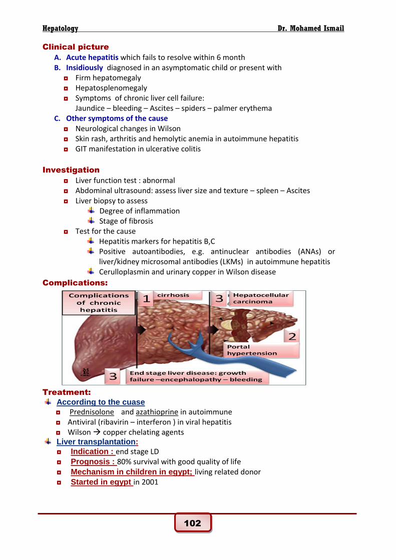
Complications:
Treatment:
According to the cuase
◘ Prednisolone and azathioprine in autoimmune ◘ Antiviral (ribavirin – interferon ) in viral hepatitis ◘ Wilson copper chelating agents
Liver transplantation:
◘ Indication : end stage LD ◘ Prognosis : 80% survival with good quality of life ◘ Mechanism in children in egypt; living related donor ◘ Started in egypt in 2001
103
Hepatology Dr. Mohamed Ismail
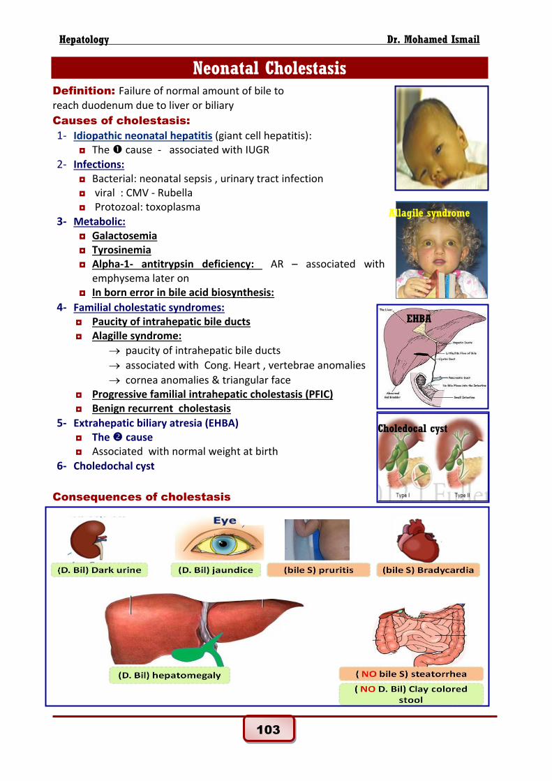
Neonatal Cholestasis
Definition: Failure of normal amount of bile to reach duodenum due to liver or biliary
Causes of cholestasis:
1- Idiopathic neonatal hepatitis (giant cell hepatitis): ◘ The cause - associated with IUGR
2- Infections: ◘ Bacterial: neonatal sepsis , urinary tract infection ◘ viral : CMV - Rubella ◘ Protozoal: toxoplasma
3- Metabolic: ◘ Galactosemia ◘ Tyrosinemia ◘ Alpha-1- antitrypsin deficiency: AR – associated with
emphysema later on ◘ In born error in bile acid biosynthesis:
4- Familial cholestatic syndromes: ◘ Paucity of intrahepatic bile ducts ◘ Alagille syndrome:
paucity of intrahepatic bile ducts
associated with Cong. Heart , vertebrae anomalies
cornea anomalies & triangular face ◘ Progressive familial intrahepatic cholestasis (PFIC) ◘ Benign recurrent cholestasis
5- Extrahepatic biliary atresia (EHBA) ◘ The cause ◘ Associated with normal weight at birth
6- Choledochal cyst
Consequences of cholestasis
Allagile syndrome
EHBA
Choledocal cyst
104
Hepatology Dr. Mohamed Ismail
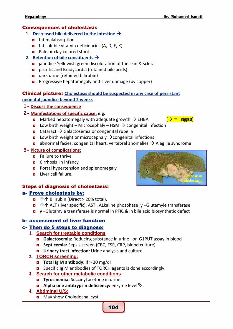
Consequences of cholestasis
1. Decreased bile delivered to the intestine ◘ fat malabsorption ◘ fat soluble vitamin deficiencies (A, D, E, K) ◘ Pale or clay colored stool.
2. Retention of bile constituents ◘ jaundice Yellowish green discoloration of the skin & sclera ◘ pruritis and Bradycardia (retained bile acids) ◘ dark urine (retained bilirubin) ◘ Progressive hepatomegaly and liver damage (by copper)
Clinical picture: Cholestasis should be suspected in any case of persistant neonatal jaundice beyond 2 weeks
1- Discuss the consequence
2- Manifestations of specific cause: e.g. ◘ Marked hepatomegaly with adequate growth EHBA ( = suggest) ◘ Low birth weight – Microcephaly – HSM congenital infection ◘ Cataract Galactosemia or congenital rubella ◘ Low birth weight or microcephaly congenital infections ◘ abnormal facies, congenital heart, vertebral anomalies Alagille syndrome
3- Picture of complications: ◘ Failure to thrive ◘ Cirrhosis in infancy ◘ Portal hypertension and splenomegaly ◘ Liver cell failure.
Steps of diagnosis of cholestasis:
a- Prove cholestasis by:
◘ Bilirubin (Direct > 20% total). ◘ ALT (liver specific), AST , ALkaline phosphase ,γ –Glutamyle transferase ◘ γ –Glutamyle transferase is normal in PFIC & in bile acid biosynthetic defect
b- assessment of liver function
c- Then do 5 steps to diagnose:
1. Search for treatable conditions
◘ Galactosemia: Reducing substance in urine or G1PUT assay in blood ◘ Septicemia: Sepsis screen (CBC, ESR, CRP, blood culture). ◘ Urinary tract infection: Urine analysis and culture.
2. TORCH screening:
◘ Total Ig M antibody: if > 20 mg/dl ◘ Specific Ig M antibodies of TORCH agents is done accordingly
3. Search for other metabolic conditions
◘ Tyrosinemia: Succinyl acetone in urine. ◘ Alpha one antitrypsin deficiency: enzyme level.
4. Abdminal U/S:
◘ May show Choledochal cyst
105
Hepatology Dr. Mohamed Ismail
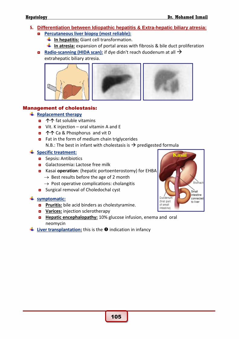
5. Differentiation between Idiopathic hepatitis & Extra-hepatic biliary atresia:
◘ Percutaneous liver biopsy (most reliable): In hepatitis: Giant cell transformation. In atresia: expansion of portal areas with fibrosis & bile duct proliferation
◘ Radio-scanning (HIDA scan): if dye didn't reach duodenum at all extrahepatic biliary atresia.
Management of cholestasis:
Replacement therapy ◘ fat soluble vitamins ◘ Vit. K injection – oral vitamin A and E ◘ Ca & Phosphorus and vit D ◘ Fat in the form of medium chain triglycerides
N.B.: The best in infant with cholestasis is predigested formula
Specific treatment: ◘ Sepsis: Antibiotics ◘ Galactosemia: Lactose free milk ◘ Kasai operation: (hepatic portoenterostomy) for EHBA
Best results before the age of 2 month
Post operative complications: cholangitis ◘ Surgical removal of Choledochal cyst
symptomatic: ◘ Pruritis: bile acid binders as cholestyramine. ◘ Varices: injection sclerotherapy ◘ Hepatic encephalopathy: 10% glucose infusion, enema and oral
neomycin Liver transplantation: this is the indication in infancy
Kasai
pro
106
Hepatology Dr. Mohamed Ismail
Portal hypertension
Definition: Portal venous pressure exceeds 12 mmHg (Normal: 5-10 mmHg)
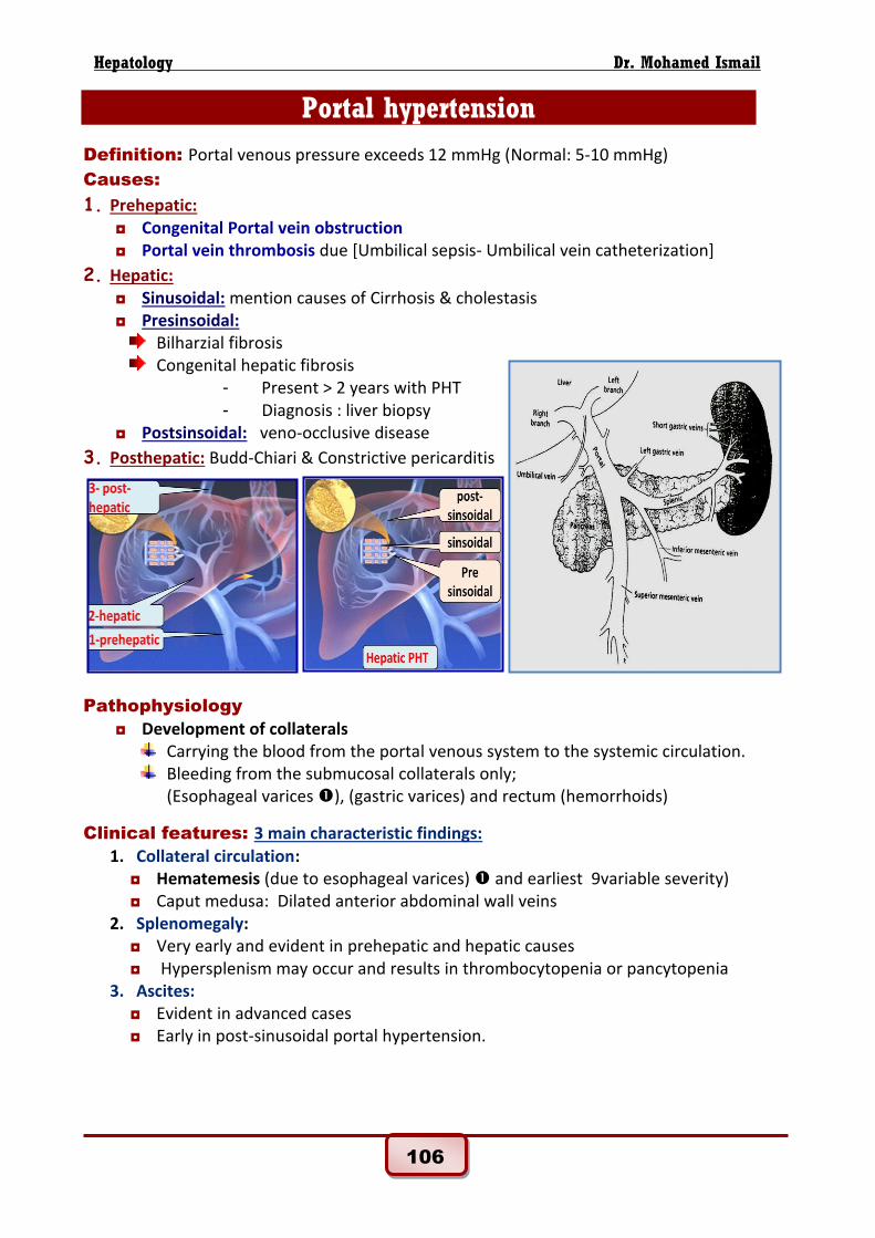
Causes:
1. Prehepatic: ◘ Congenital Portal vein obstruction ◘ Portal vein thrombosis due [Umbilical sepsis- Umbilical vein catheterization]
2. Hepatic: ◘ Sinusoidal: mention causes of Cirrhosis & cholestasis ◘ Presinsoidal:
Bilharzial fibrosis Congenital hepatic fibrosis
- Present > 2 years with PHT - Diagnosis : liver biopsy
◘ Postsinsoidal: veno-occlusive disease
3. Posthepatic: Budd-Chiari & Constrictive pericarditis
Pathophysiology
◘ Development of collaterals Carrying the blood from the portal venous system to the systemic circulation. Bleeding from the submucosal collaterals only;
(Esophageal varices ), (gastric varices) and rectum (hemorrhoids)
Clinical features: 3 main characteristic findings:
1. Collateral circulation: ◘ Hematemesis (due to esophageal varices) and earliest 9variable severity) ◘ Caput medusa: Dilated anterior abdominal wall veins
2. Splenomegaly: ◘ Very early and evident in prehepatic and hepatic causes ◘ Hypersplenism may occur and results in thrombocytopenia or pancytopenia
3. Ascites: ◘ Evident in advanced cases ◘ Early in post-sinusoidal portal hypertension.
107
Hepatology Dr. Mohamed Ismail
Investigations
◘ Upper GIT endoscopy for detection of esophageal varices ◘ Abdominal ultrasonography and Doppler ultrasonography:
Demonstrate the direction of flow within the portal system the patency of the portal vein and Presence of portosystemic collaterals.
◘ CT angiography and MR venography (demonstrate vessel patency) ◘ Liver function test ◘ Investigation for the cause
Hepatitis markers Autoimmune screening TMS- sweet chloride test Liver biopsy
Management:
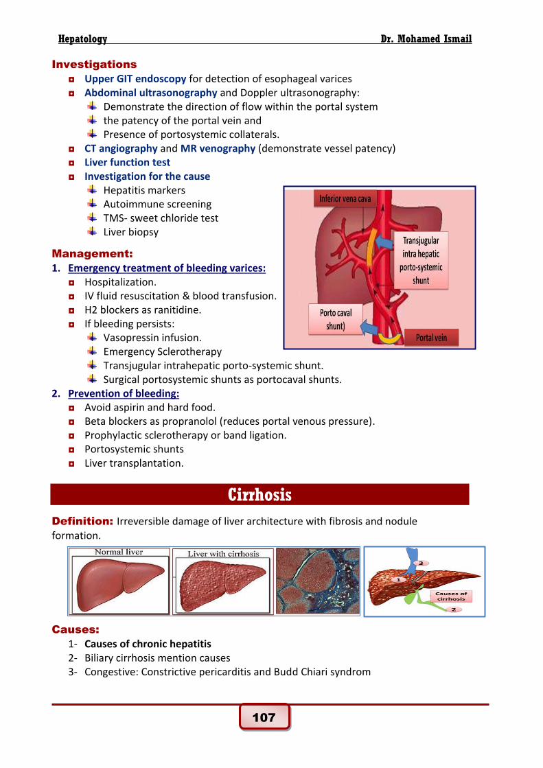
1. Emergency treatment of bleeding varices: ◘ Hospitalization. ◘ IV fluid resuscitation & blood transfusion. ◘ H2 blockers as ranitidine. ◘ If bleeding persists:
Vasopressin infusion. Emergency Sclerotherapy Transjugular intrahepatic porto-systemic shunt. Surgical portosystemic shunts as portocaval shunts.
2. Prevention of bleeding: ◘ Avoid aspirin and hard food. ◘ Beta blockers as propranolol (reduces portal venous pressure). ◘ Prophylactic sclerotherapy or band ligation. ◘ Portosystemic shunts ◘ Liver transplantation.
Cirrhosis
Definition: Irreversible damage of liver architecture with fibrosis and nodule formation.
Causes:
1- Causes of chronic hepatitis 2- Biliary cirrhosis mention causes 3- Congestive: Constrictive pericarditis and Budd Chiari syndrom