Hepatolithiasis - SUNY Downstate Medical Center · choledochal cysts or malignant biliary tumors....
62
HEPATOLITHIASIS ALIREZA SADEGHI, MD KINGS COUNTY HOSPITAL CENTER SUNY DOWNSTATE MEDICAL CENTER
Transcript of Hepatolithiasis - SUNY Downstate Medical Center · choledochal cysts or malignant biliary tumors....

HEPATOLITHIASIS
ALIREZA SADEGHI, MD
KINGS COUNTY HOSPITAL CENTERSUNY DOWNSTATE MEDICAL CENTER

Surgical Management of HEPATOLITHIASIS
ALIREZA SADEGHI, MDSUNY DOWNSTATE MEDICAL CENTER

Overview
• HEPATOLITHIASIS:
– Historical Perspectives– Definition– Epidemiology & Natural
History– Etiology of Biliary Stones– Classification Systems– Symptoms– Diagnosis– Treatment Regiments– Conclusion

Historical Perspectives
• Vachell & Stevens (1906)– First published report of Intrahepatic Calculi
• By 1930, multiple descriptions– Recurrent Pyogenic Cholangitis
– Hong Kong Disease
– Biliary Obstruction Syndrome of the Chinese
– Oriental Cholangitis
– Oriental Infestational Cholangitis
– Intrahepatic Lithiasis
Vachell HR, Stevens WM: Case of Intrahepatic Calculi. Br Med J1906;1:434-36

Definition
• Calculi or concretions– located proximal to the confluence of the
right and left hepatic ducts• Stones are present in Right and/or Left
hepatic ducts and/or their tributaries irrespective of the coexistence of choledocholithiasis or cholecystolithiasis
Nakayama F. Intrahepatic Stones; Blumgart’s Surgery of the Liver & Biliary Tract. 765-774. 1994

Epidemiology
• Occurs mainly in Southeast Asia:– China, Korea, Japan, Taiwan, Philippines, Vietnam,
Thailand, Malaysia, Brazil, Indonesia and India
– Incidence reported to be 20-30% of all patients undergoing surgery for gallstone disease.
– Low incidence in Europe and North America
– With increased immigration more cases are being reported in the North American institutions however there is little experience in treating the condition
Nakayama F. Intrahepatic Stones; Blumgart’s Surgery of the Liver & Biliary Tract. 765-774. 1994

Epidemiology
• Statistics
Nakayama F. Intrahepatic Stones; Blumgart’s Surgery of the Liver & Biliary Tract. 765-774. 1994

Epidemiology
• Incidence– Equal gender distribution
– Affects patients in 3rd to 7th decades on life
– More common in rural areas of Southeast Asia among the lower socio-economic class
– Overall incidence in East Asia is declining• China & Taiwan: 20%
• Japan: 2.2%
Nakayama F. Intrahepatic Stones; Blumgart’s Surgery of the Liver & Biliary Tract. 765-774. 1994
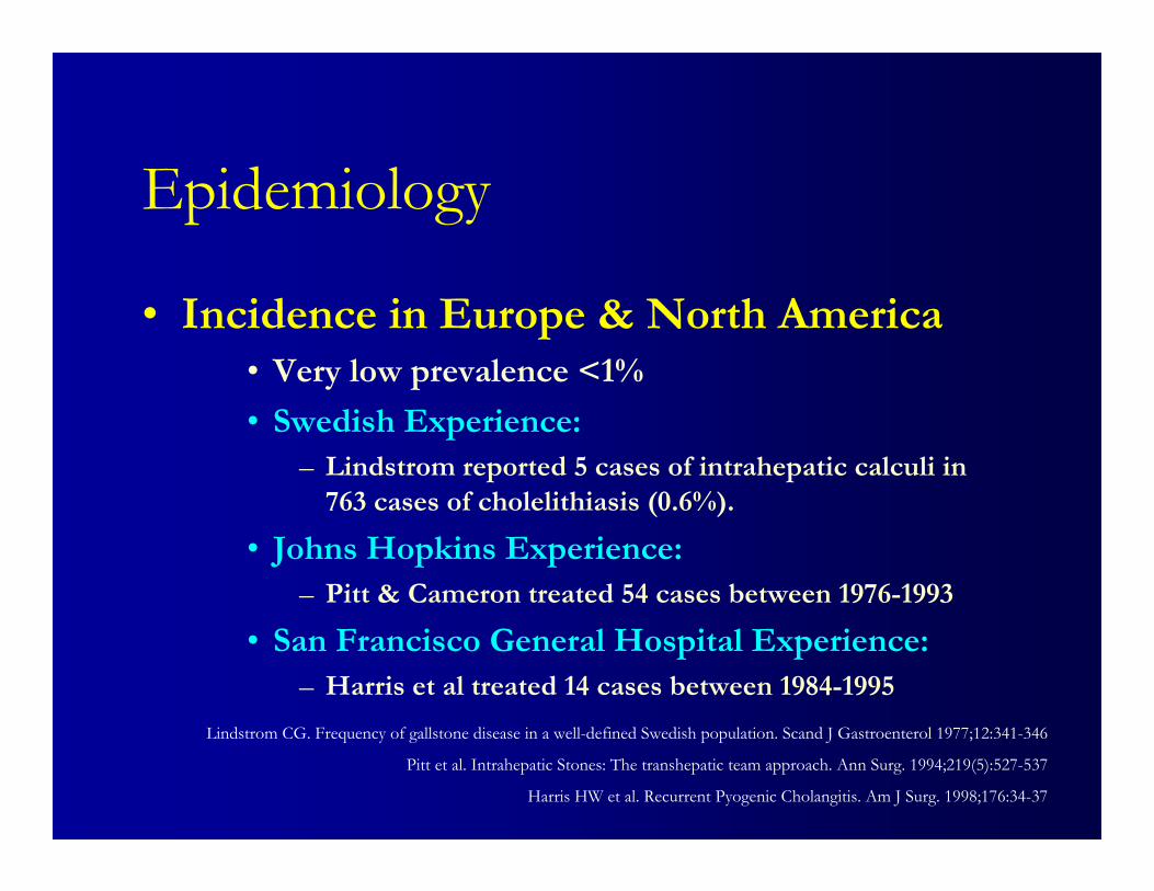
Epidemiology
• Incidence in Europe & North America• Very low prevalence <1%
• Swedish Experience:– Lindstrom reported 5 cases of intrahepatic calculi in
763 cases of cholelithiasis (0.6%).
• Johns Hopkins Experience:– Pitt & Cameron treated 54 cases between 1976-1993
• San Francisco General Hospital Experience:– Harris et al treated 14 cases between 1984-1995
Lindstrom CG. Frequency of gallstone disease in a well-defined Swedish population. Scand J Gastroenterol 1977;12:341-346
Pitt et al. Intrahepatic Stones: The transhepatic team approach. Ann Surg. 1994;219(5):527-537
Harris HW et al. Recurrent Pyogenic Cholangitis. Am J Surg. 1998;176:34-37

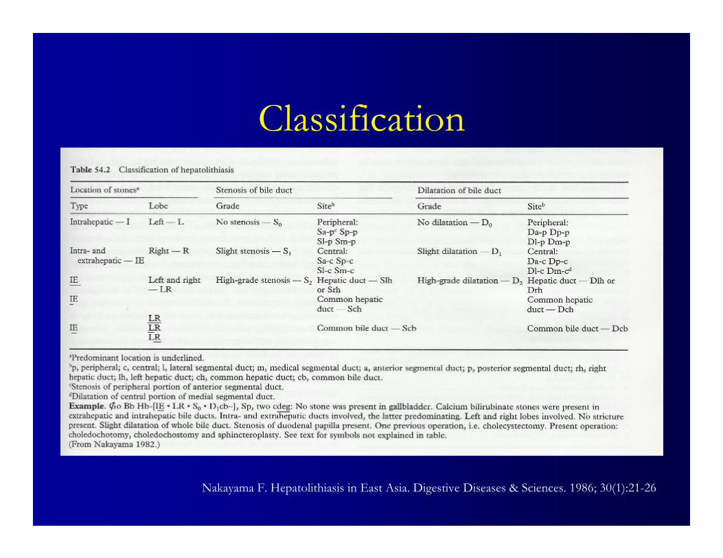
Classification
• Most recent system: Nakayama in 1982• Presence of stones in the intra-hepatic bile ducts: I
• Presence of stones in the intra & extra-hepatic bile ducts: IE
• Right & Left hepatic ducts distal to the confluence
• Segmental ducts are divided into central & peripheral
– Central: Ducts of first and second branches
Nakayama F. Hepatolithiasis in East Asia. Digestive Diseases & Sciences. 1986; 30(1):21-26

Classification
Nakayama F. Hepatolithiasis in East Asia. Digestive Diseases & Sciences. 1986; 30(1):21-26

Classification
• Nakayama: 1982– Strictures (S): decrease in the diameter of bile
ducts relative to adjacent parts.• S0: Absence of stricture
• S1: Slight stricture (Diameter > 2mm)
• S2: Marked stricture (Diameter < 2 mm)
Nakayama F. Hepatolithiasis in East Asia. Digestive Diseases & Sciences. 1986; 30(1):21-26
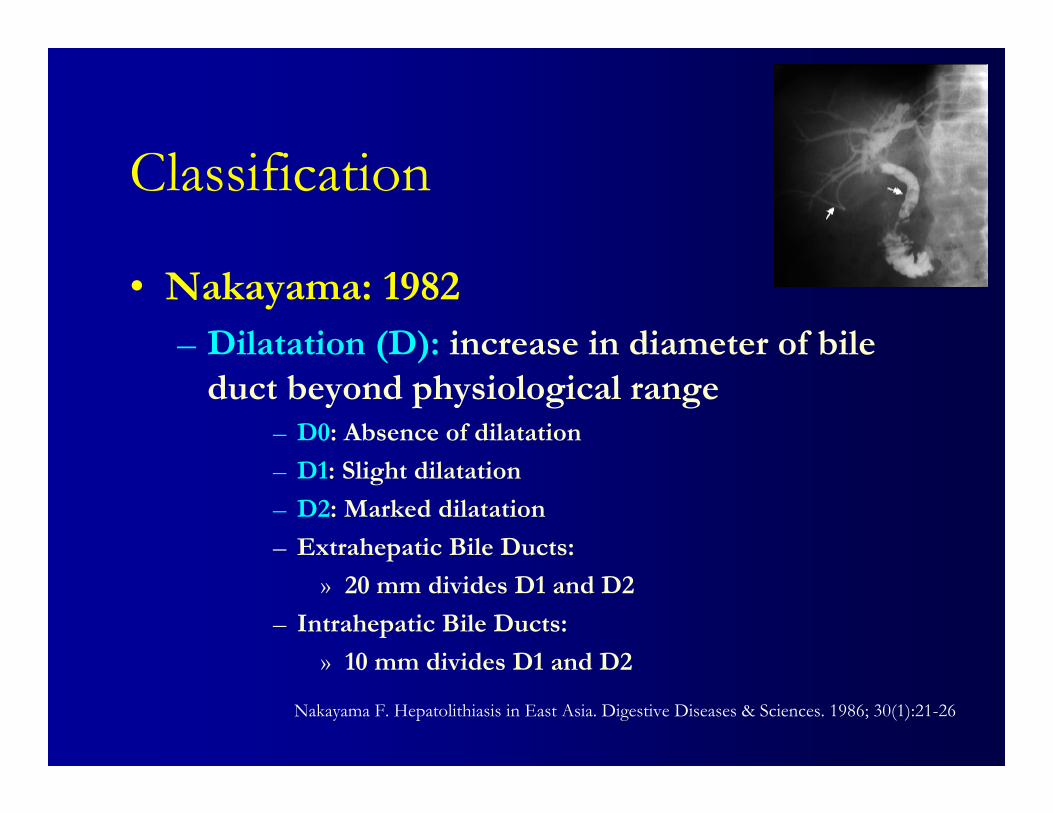
Classification
• Nakayama: 1982– Dilatation (D): increase in diameter of bile
duct beyond physiological range– D0: Absence of dilatation
– D1: Slight dilatation
– D2: Marked dilatation
– Extrahepatic Bile Ducts:
» 20 mm divides D1 and D2
– Intrahepatic Bile Ducts:
» 10 mm divides D1 and D2
Nakayama F. Hepatolithiasis in East Asia. Digestive Diseases & Sciences. 1986; 30(1):21-26
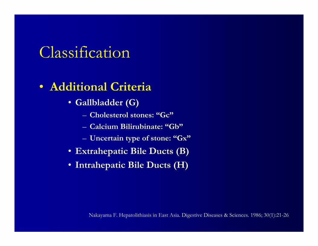
Classification
• Additional Criteria• Gallbladder (G)
– Cholesterol stones: “Gc”
– Calcium Bilirubinate: “Gb”
– Uncertain type of stone: “Gx”
• Extrahepatic Bile Ducts (B)
• Intrahepatic Bile Ducts (H)
Nakayama F. Hepatolithiasis in East Asia. Digestive Diseases & Sciences. 1986; 30(1):21-26
Classification
Nakayama F. Hepatolithiasis in East Asia. Digestive Diseases & Sciences. 1986; 30(1):21-26

Etiology
• Intrahepatic Lithiasis– East vs. West
– In the west is generally thought to be secondary to stones originating in the gall bladder or primarily resulting from benign strictures, sclerosing cholangitis, choledochal cysts or malignant biliary tumors.
– In the East it is regarded as a separate entity altogether. The majority of cases are associated with recurrent pyogenic cholangitis in regions with parasitic infestations
Rao et al. Intrahepatic Stones – an etiological quagmire. Indian J Gastroenterology 2004;23:201-2

Etiology
• Ethnic vs. Environmental factors• Survey by Nakayama looked at the proportion of
HL in different provinces of China as compared to that of HL in Japan:
– Taiwan 53.1%– Hong Kong 3.1%– Singapore 1.7%– Japan 4.1%
• In spite of a similar ethnic background (Chinese descent) the relative proportion of HL is marked different in Taiwan, Hong Kong & Singapore
Nakayama F. Hepatolithiasis in East Asia. Digestive Diseases & Sciences. 1986; 30(1):21-26
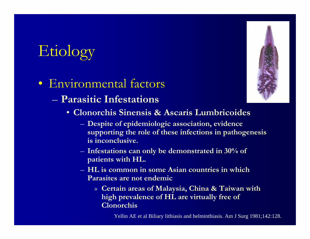
Etiology
• Environmental factors– Parasitic Infestations
• Clonorchis Sinensis & Ascaris Lumbricoides– Despite of epidemiologic association, evidence
supporting the role of these infections in pathogenesis is inconclusive.
– Infestations can only be demonstrated in 30% of patients with HL.
– HL is common in some Asian countries in which Parasites are not endemic
» Certain areas of Malaysia, China & Taiwan with high prevalence of HL are virtually free of Clonorchis
Yellin AE et al Biliary lithiasis and helminthiasis. Am J Surg 1981;142:128.

Etiology
• Parasites• Theory:
– Analysis of stones have shown debris & ova which may have served as a nidus for stone formation
– This is probably incidental rather than causative and may merely be an association with stones in endemic areas of infestation
Yellin AE et al Biliary lithiasis and helminthiasis. Am J Surg 1981;142:128.

Etiology
• Environmental factors– Bacterial Infections
• Human bile is Sterile under normal circumstances• Incidence of Bacteria in bile of patients with HL is almost
100% in most series• Bacteria commonly found:
– Klebsiella sp– E. Coli– Pseudomonas sp– Entercoccous sp– Bacteroides sp
• Entry route: access biliary tree by injury from a parasitic infection
Sheen-Chen SM et al. Bacteriology & Antimicrobial choices in Hepatolithiasis. AJIC 2000;28:298-301
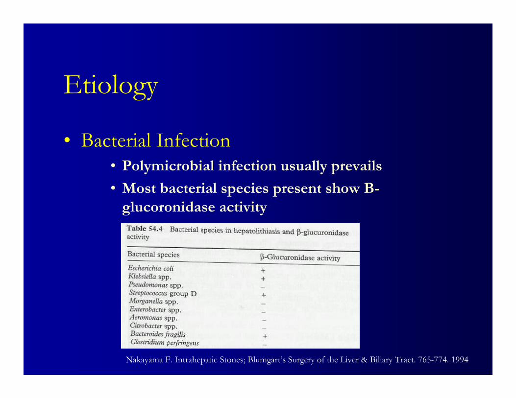
Etiology
• Bacterial Infection• Polymicrobial infection usually prevails
• Most bacterial species present show B-glucoronidase activity
Nakayama F. Intrahepatic Stones; Blumgart’s Surgery of the Liver & Biliary Tract. 765-774. 1994

Etiology
• Bacterial infection• B-Glucuronidase is responsible for catalyzing the
hydrolysis of direct bilirubin to the indirect unconjugated form
• Unconjugated bilirubin is water-insoluble & combines with calcium bilirubinate to form Calcium Bilirubinate (PIGMENT) stones which comprise the majority of the cases of Hepatolithiasis (80%)
• Cholesterol & mixed stones are increasing (10%) as the western diet invades Asia
Nakayama F. Intrahepatic Stones; Blumgart’s Surgery of the Liver & Biliary Tract. 765-774. 1994

Etiology
Nakayama F. Intrahepatic Stones; Blumgart’s Surgery of the Liver & Biliary Tract. 765-774. 1994

Etiology
• Bile Stasis
• Main cause of Intrahepatic Stones in the West
• Stenosis & Strictures– Benign strictures
» Postoperative: Most Common Etiology
– Sclerosing Cholangitis
– Choledochal cysts
– Malignant biliary tumors
Rao et al. Intrahepatic Stones – an etiological quagmire. Indian J Gastroenterology 2004;23:201-2
Etiology
• Other factors– Diet
• Asian diet:– high in carbohydrates and low in fat & protein.
• Saturated fats causes CCK release & relaxation of the sphincter of Oddi
– Low fat diet leads to biliary stasis
• Low protein diet– Low level of Glucaro-1.4-lactone which is an inhibitor
of B-Glucuronidase, potentiating the deconjungation reaction
Rao et al. Intrahepatic Stones – an etiological quagmire. Indian J Gastroenterology 2004;23:201-2

Etiology
• Rare causes:– Iatrogenic Factors
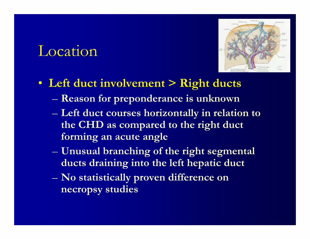
Location
• Left duct involvement > Right ducts– Reason for preponderance is unknown– Left duct courses horizontally in relation to
the CHD as compared to the right duct forming an acute angle
– Unusual branching of the right segmental ducts draining into the left hepatic duct
– No statistically proven difference on necropsy studies

Pathophysiology
• Recurrent Pyogenic Cholangitis• Recurrent bouts of cholangitis
• Inflammatory changes in the bile ducts leading to further stricture formation and causing bile stasis
• Walls of the ducts are thickened and fibrosed
• Hepatic parenchyma associated with stones within the intrahepatic ducts show mild to severe atrophy and fibrosis leading to cirrhosis
Nakayama F. Intrahepatic Stones; Blumgart’s Surgery of the Liver & Biliary Tract. 765-774. 1994

Natural History
• If left untreated Hepatolithiasis leads to• Recurrent pyogenic cholangitis
• Progressive biliary strictures
• Formation of liver abscesses
• Atrophy of the affected liver
• Secondary biliary cirrhosis
• Portal Hypertension
• Cholangiocarcinoma
Nakayama F. Intrahepatic Stones; Blumgart’s Surgery of the Liver & Biliary Tract. 765-774. 1994
Jan YY et al. Surgical treatment of Hepatolithiasis: Long-term results. Surgery 1996;120:509-514
Kusano T et al. Natural Progression of Untreated Hepatolithiasis. J Clin Gastroenterol 2001;33(2):114-17

Natural History
• 122 patients with CT & CT-Cholangiography findings of Hepatolithiasis– 14/122 became symptomatic at mean 3.4 yrs– Symptoms:
• Recurrent abdominal pain, hepatic abscesses, lobar atrophy, cholangitis & cholangiocarcinoma
• Lobar Atrophy: Major role in development symptoms
– 13/14 (93%) of symptomatic group– 14/108 (13%) of asymptomatic group
Kusano T et al. Natural Progression of Untreated Hepatolithiasis. J Clin Gastroenterol 2001;33(2):114-17

Natural History
• 122 patients with CT & CT-Cholangiography findings of Hepatolithiasis– Summary:
• Recurrence of HL in the atrophic liver causes persistent chronic inflammation which leads to formation of strictures and more HL and possibly cholangiocarcinoma.
• Lobar Atrophy is a risk factor for cholangiocarcinoma & hepatectomy is indicated
Kusano T et al. Natural Progression of Untreated Hepatolithiasis. J Clin Gastroenterol 2001;33(2):114-17
Natural History
• Cholangiocarcinoma & Hepatolithiasis– Incidence: 2.36% - 10 %– Proposed mechanisms
• Prolonged irritation of biliary epithelium by calculi• Long term exposure to bile & its products• Repeated infections• Metabolic byproducts of bacteria in the biliary tree• Dynamic irritation by unstable bile flow
– Bile Stasis, reflux & turbulence
Nakanuma Y et al. Are Hepatolithiasis and cholangiocarcinoma etiologically related? Virchows Arch 1985;406: 45-58.
Jan YY et al. Surgical treatment of Hepatolithiasis: Long-term results. Surgery 1996;120:509-514.

Cholangiocarcinoma & Hepatolithiasis
• China
– Sixteen of 20 (80%) of patients with peripheral intrahepatic cholangiocarcinoma had associated intrahepatic stones.
Chen MF, Jan YY et al. Clinical experience in 20 hepatic resections for peripheral cholangiocarcinoma. Cancer 1989;64:2226-2232
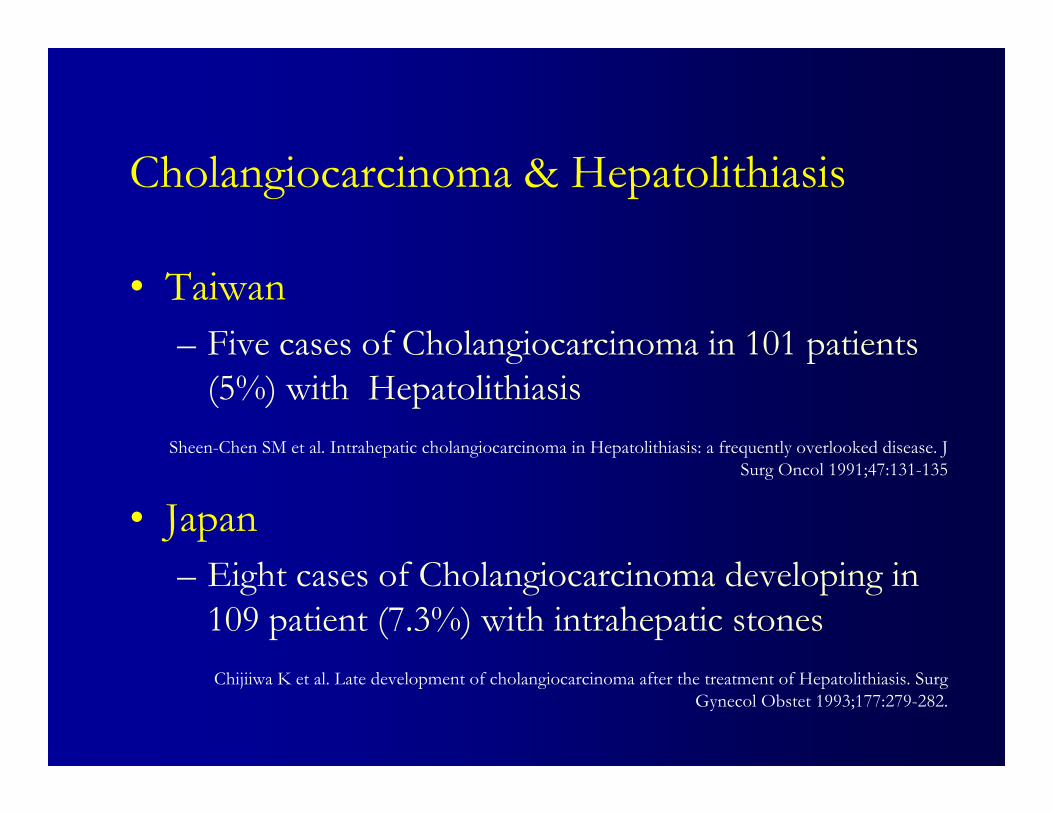
Cholangiocarcinoma & Hepatolithiasis
• Taiwan– Five cases of Cholangiocarcinoma in 101 patients
(5%) with HepatolithiasisSheen-Chen SM et al. Intrahepatic cholangiocarcinoma in Hepatolithiasis: a frequently overlooked disease. J
Surg Oncol 1991;47:131-135
• Japan– Eight cases of Cholangiocarcinoma developing in
109 patient (7.3%) with intrahepatic stonesChijiiwa K et al. Late development of cholangiocarcinoma after the treatment of Hepatolithiasis. Surg
Gynecol Obstet 1993;177:279-282.

Cholangiocarcinoma & Hepatolithiasis
• Hong Kong– Ten of 103 (10%) patients of undergoing
hepatectomy for Hepatolithiasis had cholangiocarcinoma
– In addition 3 patients developed cholangiocarcinoma at interval of 7 – 36 months after the first operation for Hepatolithiasis
Chen DW et al. Immediate and long-term outcomes of hepatectomy for Hepatolithiasis. Surgery 2004;135:386-93.

Cholangiocarcinoma & Hepatolithiasis
• Hong Kong:– Follow up of 91
patients.– 5 yr survival
• Group I: 9%• Group II: 93%
Conclusion:Cholangiocarcinoma is an independent prognostic factor of the survival of patients with Hepatolithiasis who underwent hepatectomy
Chen DW et al. Immediate and long-term outcomes of hepatectomy for Hepatolithiasis. Surgery 2004;135:386-93.

Symptoms
• No pathognomonic symptoms• RUQ Pain• Fever• Bouts of Cholangitis• Less frequently jaundice• Abnormal LFTs with elevation of Alk Phos• In study of 54 patients at Johns Hopkins
– 1. Cholangitis 67%– 2. Abdominal Pain 63%– 3. Jaundice 39%
Rao et al. Intrahepatic Stones – an etiological quagmire. Indian J Gastroenterology 2004;23:201-2
Pitt HA, Cameron JL et al. Intrahepatic Stones; The Transhepatic Team Approach. Annals of Surgery 1994;219: 527-37
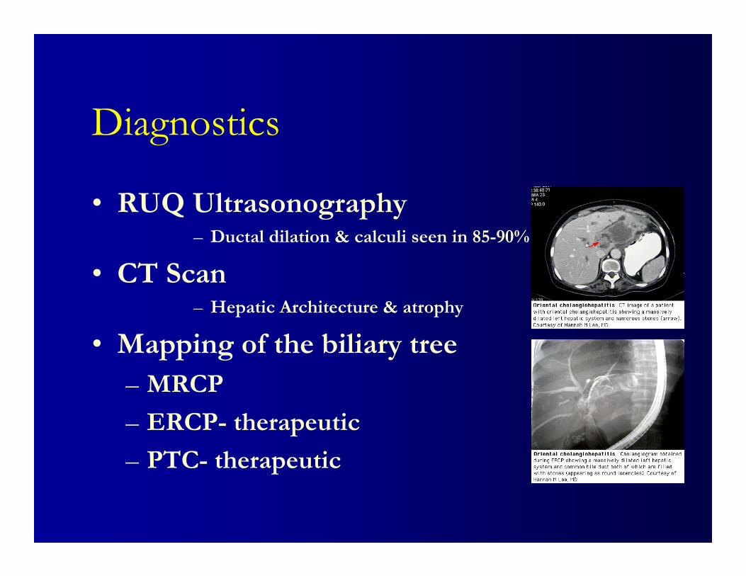
Diagnostics
• RUQ Ultrasonography– Ductal dilation & calculi seen in 85-90%
• CT Scan– Hepatic Architecture & atrophy
• Mapping of the biliary tree– MRCP
– ERCP- therapeutic
– PTC- therapeutic

Treatment
• Aims of treatment– Prevention of liver damage by early clearance of stones and
elimination of bile stasis• Removal of stones• Removal of strictured bile ducts• Providing good drainage of bile
– Minimizing bacterial infection
• Resection of source of recurrent infection & biliary stasis• Removal of cholangiocarcinoma• Removal of atrophic liver• Removal of hepatic abscess
– Residual stones should be able to spontaneously enter the GI tractPitt HA, Cameron JL et al. Intrahepatic Stones; The Transhepatic Team Approach. Annals of Surgery
1994;219: 527-37.

Treatment
• Team Approach to treatment– Combined Interventional Radiological,
Gastroenterological & Surgical Regimen using the Transhepatic approach
• Percutaneous Placement of transhepatic access catheters
• Surgery for underlying biliary disease & stone removal
• Postoperative percutaneous choledochoscopy & residual or recurrent stone removal through transhepatic stents
Pitt HA, Cameron JL et al. Intrahepatic Stones; The Transhepatic Team Approach. Annals of Surgery 1994;219: 527-37.

Treatment
• Percutaneous Options– In the Jaundice Patient
– PTC with PT Biliary Drainage (PTBD):• Sinus tract dilated to 18 Fr to ease introduction of
– Choldechoscope
– Basket/Balloon catheters/Stents
– Microwave, Electrohydraulic lithotripsy and Laser probes for stone fragmentation & piecemeal removal
Nakayama F. Intrahepatic Stones; Blumgart’s Surgery of the Liver & Biliary Tract. 765-774. 1994

Treatment
• Percutaneous Transhepatic Choledoscopic removal of intrahepatic stones– Retrospective study of 79 patients with early
diagnosed intrahepatic stones over 8 years• Success rate of 76.8 %• Removal of right sided stones was difficult• Cholangitis recurred in 1/3 of patients within 3-5
years• Intrahepatic strictures was the major determinant for
recurrence of stones and symptoms.Cheung MT et al. Percutaneous Transhepatic Choledoscopic removal of intrahepatic stones. Brit J Surg
2003;90:1409-1415
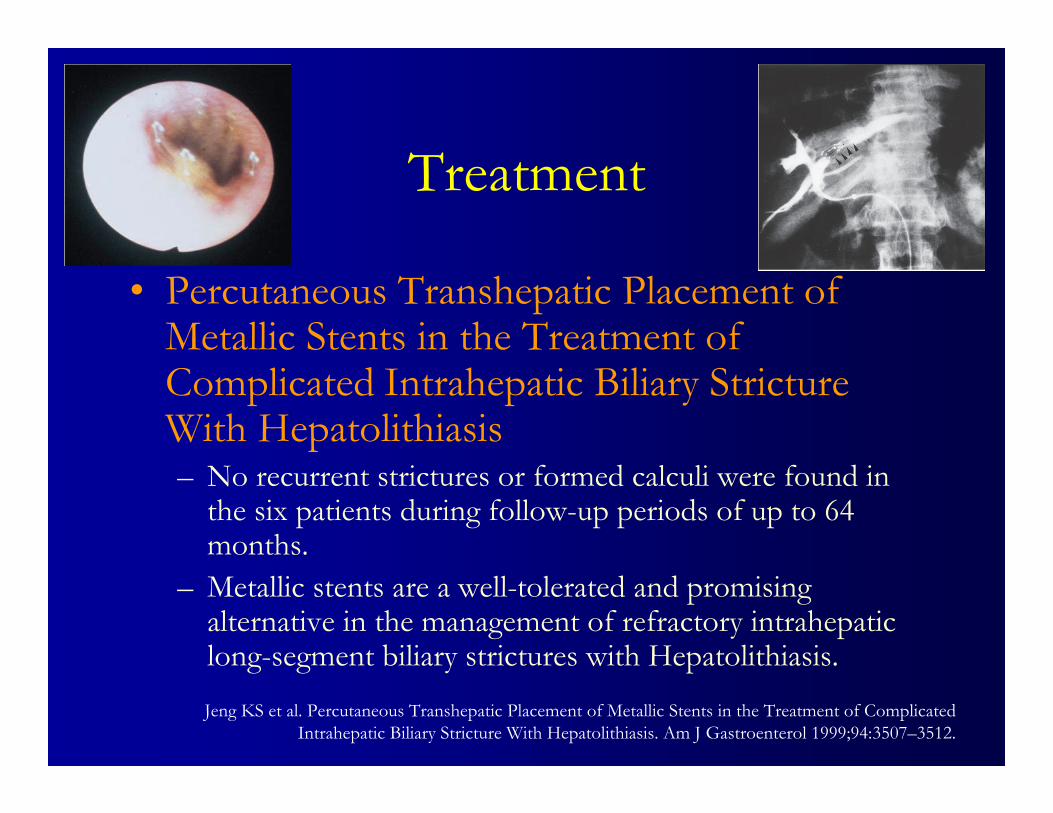
Treatment
• Percutaneous Transhepatic Placement of Metallic Stents in the Treatment of Complicated Intrahepatic Biliary Stricture With Hepatolithiasis– No recurrent strictures or formed calculi were found in
the six patients during follow-up periods of up to 64 months.
– Metallic stents are a well-tolerated and promising alternative in the management of refractory intrahepatic long-segment biliary strictures with Hepatolithiasis.Jeng KS et al. Percutaneous Transhepatic Placement of Metallic Stents in the Treatment of Complicated
Intrahepatic Biliary Stricture With Hepatolithiasis. Am J Gastroenterol 1999;94:3507–3512.

Treatment
• Endoscopic Retrograde Approach– Absence of Jaundice
– Removal of both Intra & Extrahepatic biliary stones
• Introduction of basket/balloon catheters
• Avoids injury to the hepatic parenchyma
• Difficult technique with high failure rates
Nakayama F. Intrahepatic Stones; Blumgart’s Surgery of the Liver & Biliary Tract. 765-774. 1994

Treatment
• Extracorporeal Shock Wave Lithotripsy– Limited success if there is no predetermined
evacuation route for the stone fragments– Frequent presence of bile duct strictures makes
removal difficult in spite of successful fragmentation
– Stone clearance rate of up to 95% reported– When combined with ERCP and Electrohydraulic
lithotripsy.Nakayama F. Intrahepatic Stones; Blumgart’s Surgery of the Liver & Biliary Tract. 765-774. 1994
Binmoeller KF et al. Treatment of difficult bile duct stones using mechanical, electrohydraulic and extracorporeal shock wave lithotripsy. Endoscopy1993 Mar;25(3):201-6

Treatment
• Choledochostomy Approach– Previous CBD exploration with T-tube
placement with tract maturation– Insertion of balloon/basket catheters– Choledochofibrescopy– Limited to the Left hepatic ducts– Difficult to reach the Right biliary tree
• Unfavorable angle
Nakayama F. Intrahepatic Stones; Blumgart’s Surgery of the Liver & Biliary Tract. 765-774. 1994

Treatment
• Postoperative Choledoscopic Removal of Intrahepatic Stones via a T-tube tract– Retrospective study of 44 patients in Hong Kong
• Successful procedure in 22/44 patients• Presence of Stricture associated with higher failure
rates (P=0.002)• Invaluable access route but a limited procedure when
strictures present
Cheung MT. Postoperative Choledoscopic Removal of Intrahepatic Stones via a T-tube tract Brit J Surg 1997;84:1224-28

Treatment
• Surgical Options– Based on presence of I or IE Hepatolithiasis
• Choledochotomy with CBD Exploration• Choledochostomy with drainage• Hepatico/Cholangioenterostomy • Placement of Access Loops• Hepatic Resection
– Lobectomy (L>R)– Segmental Resection
• Hepatic Transplantation
Nakayama F. Intrahepatic Stones; Blumgart’s Surgery of the Liver & Biliary Tract. 765-774. 1994

Treatment
• Access Loop Procedures– Provide continuous postoperative access to the biliary tree for
residual/recurrent stone retraction– Percutaneous/Cutaneous
• Permanent cutaneous access– Hepaticocutaneous jejunosotomy
» Side effects: bile leakage, cutaneous excoriation, electrolyte imbalances
• Percutaneous access– Subparietal hepaticojejunal access
– Endoscopic• Interposition jejunal segment between hepatic hilum & duodenum• Side to side Roux-en-Y jejuno-duodenal access loop
Rao et al. Intrahepatic Stones – an etiological quagmire. Indian J Gastroenterology 2004;23:201-2
Beckingham IJ et al. Subparietal hepaticojejunal access loop for the long-term management of intrahepatic stones. Brit J Surg 1998;85:1360-63

Treatment
• Access Loops
Beckingham IJ et al. Subparietal hepaticojejunal access loop for the long-term management of intrahepatic stones. Brit J Surg 1998;85:1360-63
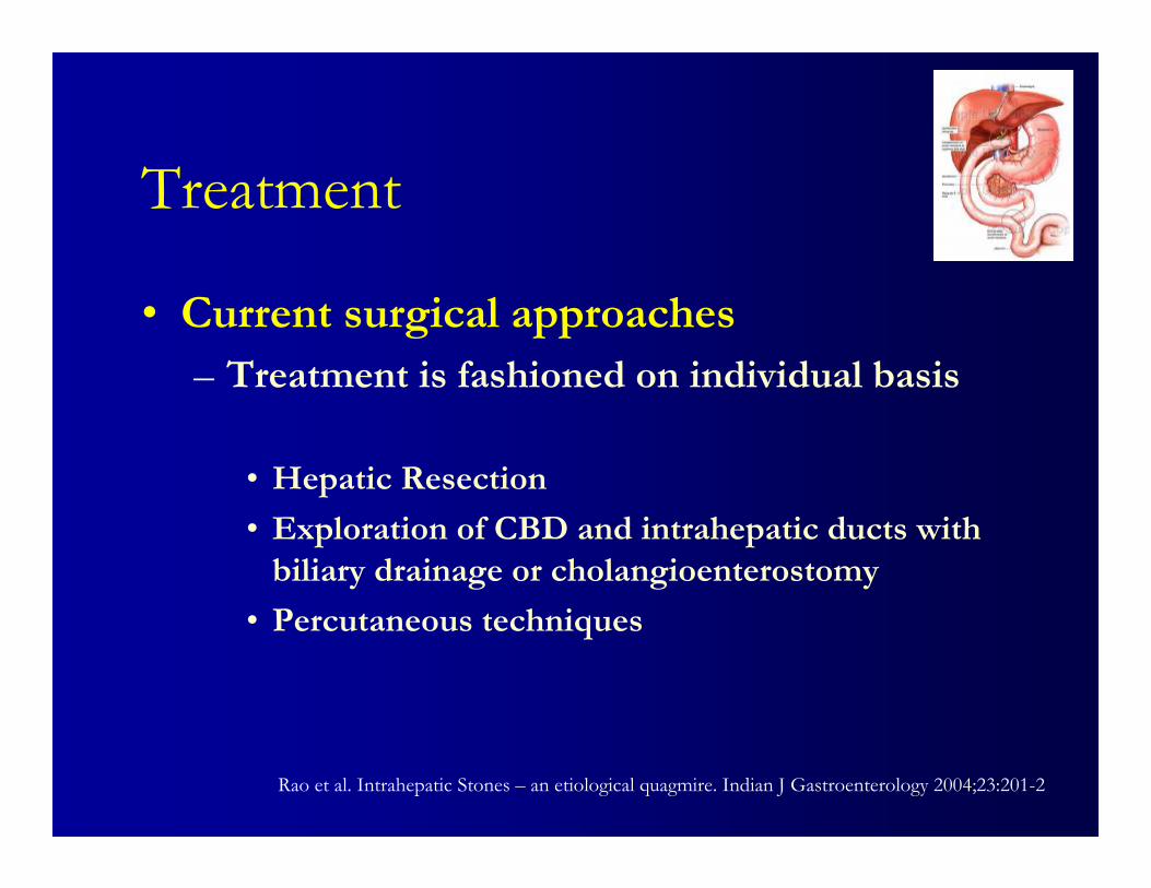
Treatment
• Current surgical approaches– Treatment is fashioned on individual basis
• Hepatic Resection
• Exploration of CBD and intrahepatic ducts with biliary drainage or cholangioenterostomy
• Percutaneous techniques
Rao et al. Intrahepatic Stones – an etiological quagmire. Indian J Gastroenterology 2004;23:201-2

Treatment
• Indications for Hepatic Resection– Advantage
• Removal of all stones along with pathologic bile ducts Includingthe carcinomatous bile ducts
– Atrophic & Fibrotic / Abscess of a liver segment or lobe• Left > Right
– Possibility of concomitant cholangiocarcinoma– Localized intrahepatic calculi with irreversible biliary
strictures
Nakayama F. Intrahepatic Stones; Blumgart’s Surgery of the Liver & Biliary Tract. 765-774. 1994
Otani K et al. Comparison of Treatments for Hepatolithiasis: Hepatic resection versus Cholangioscopic Lithotomy. J Am Coll Surg 1999;189:177-182
Fan ST et al. Treatment of Hepatolithiasis: Improvement of result by a systemic approach. Surgery 1991;109:474-480

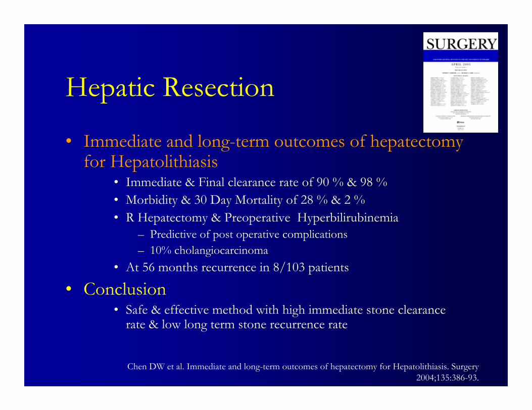
Hepatic Resection
• Immediate and long-term outcomes of hepatectomy for Hepatolithiasis– Retrospective study of 103 patients over
13 years in Hong Kong
Hepatic Resection
• Immediate and long-term outcomes of hepatectomy for Hepatolithiasis
• Immediate & Final clearance rate of 90 % & 98 %• Morbidity & 30 Day Mortality of 28 % & 2 %• R Hepatectomy & Preoperative Hyperbilirubinemia
– Predictive of post operative complications– 10% cholangiocarcinoma
• At 56 months recurrence in 8/103 patients
• Conclusion• Safe & effective method with high immediate stone clearance
rate & low long term stone recurrence rate
Chen DW et al. Immediate and long-term outcomes of hepatectomy for Hepatolithiasis. Surgery 2004;135:386-93.

Left Hepatic Resection
• The surgical treatment of isolated left-sided Hepatolithiasis
• Retrospective analysis of 128 patients with HL isolated to the left hepatic ducts over 22 years in China
• Concomitant strictures were present in 72% of patients
Sun WB et al. The surgical treatment of isolated left-sided Hepatolithiasis: A 22 year experience. Surgery 2000;127-493-7

Left Hepatic Resection
• Left Hepatic lobectomy is the most effective treatment to remove all lesions, stones, strictures or potential cholangiocarcinoma
Sun WB et al. The surgical treatment of isolated left-sided Hepatolithiasis: A 22 year experience. Surgery 2000;127-493-7
Hepatic Resection vs. PTCSL
• Comparison of Treatments for Hepatolithiasis: Hepatic Resection Versus Cholangioscopic Lithotomy– Retrospective review of 54 patients over 16 years in
Japan– 26 patients under went Hepatic Resection
• Mainly Left sided stones– 28 patients under went PTCSL
• Mainly Right and some B/L stones• Average of 6 treatments per patient
Otani K et al. Comparison of Treatments for Hepatolithiasis: Hepatic Resection Versus Cholangioscopic Lithotomy. J Am Coll Surg 1999;189:177-182

Hepatic Resection vs. PTCSL
Otani K et al. Comparison of Treatments for Hepatolithiasis: Hepatic Resection Versus Cholangioscopic Lithotomy. J Am Coll Surg 1999;189:177-182
Hepatic Resection vs. PTCSL
• Rate of complete removal of intrahepatic stones was similarly high in the hepatic resection and the PTCSL groups
• Complication and 5-year survival rates were comparable• Recurrence rate was significantly higher in the PTCSL
group• Conclusion:
– Localized Hepatolithiasis with strictures is bettermanaged by hepatic resection
– Resection of the dominant segment followed by POCSL is also recommended for bilateral stones.
Otani K et al. Comparison of Treatments for Hepatolithiasis: Hepatic Resection Versus Cholangioscopic Lithotomy. J Am Coll Surg 1999;189:177-182

Johns Hopkins Experience
• Intrahepatic Stones:Transhepatic Team Approach– Retrospective study of 54 patients over 18 years
• 2 hepatic resection in total due to earlier presentation• PTBD routinely done all pts & achieved complete removal
in 14 patients • 40 pts underwent surgery for stone clearance & stricture
treatment with bilioenteric anastamosis and access loops• Postoperative percutaneous procedures were required in
20/40 pts for residual stones & persistent intrahepatic stones.
• Multidisciplinary team approach– Recurrence of rate of 20%– Final Stone clearance rate of 94%
Pitt HA, Cameron JL et al. Intrahepatic Stones; The Transhepatic Team Approach. Annals of Surgery 1994;219: 527-37.

Hong Kong Experience
• Treatment of Hepatolithiasis: improvement of result by a systematic approach– Retrospective review of 127 patients over 6 years
– Recurrence rate independent of biliary drainage– Stone recurrence of 15.8 % which is lowest ever
reported» Highest reported rate 70%
– Complete stone clearance rate of 89.9 %– Treatment of recurrence facilitated through
permanent cutaneous access loopsFan ST et al. Treatment of hepatolithiasis:improvement of result by a systemic approach. Surgery
1991;109:474-80

Conclusion
• HL is complex disease which requires a transhepatic team approach individualized for each patient according to the site of involvement & presence of strictures.
• Multidisciplinary approach including surgical, endoscopic and interventional radiological techniques has the best outcome.