Hepatocellular Carcinoma, Possibly Disseminated by ... · Hepatocellular Carcinoma, Possibly...
18
Yamanashi Med. J. 8(4), l63-l80, l99S Three Indonesian Cases ofIntraperitoneal Hepatocellular Carcinoma, Possib by Spontaneous Tumor Masayuki YAMAMoTo, Yoshihiro AKAHANEi), Takao AiN Kaoru NAGAKoRi, Hideki F{ju" IV{as Yoshiro MATsuMoTo, and Hiros The First Dapartment ofSungeay, i)The Fixs't Dopartmenl ofMed 2)President of Yamanashi Medical U Abstract: We report 3 Indonesian patients with intraper caused by spontaneous rupture of hepatocellular carcino expansively growing nodular metastatic tumors and all nodu case. Tke first case ttnderwent lateral segmentectomy in S cases underwent liver resection for primary HCC in YMU ho seven intraperitoneal tumors, ranging from approximate together with skin metastasis, the other two cases hacl a large omentum; the tumors measured 6.5 cm and 13 cm in maximal d case, there were Ro intrahepatic metastases detected. Since t rare amongJapanese HCC patients, we report these 8 cases an distribution of HCC recurrence patterns in Japan. Key words: Hepatocellular carcinoma, SpontaReot}s rup metastasls INTReDuc'rloN Peritoneal dissemination of hepatocellular carcinoma (HCC) is not a commeR type of recurrence. The Liver Cancer S£udy Group of Japan has reported the prevalence of peri- toneal rr}etastasis as about 16.3%i), but in most cases disseminated tumors were multiple, either diffuse or sporadic, and not resectable. IR this paper, we report three cases of in- traperitoneal and extrahepatic development of resectable HCC tumor nodules which had developed beneath the greater omentum; ene in each ef two patients and 7 in one patient. L. Received Septerr}ber 9, l993 Accepted December 1, l993 Tamaho, Nakakoma, Yamanashi 409-38, Japan Coinciden£ally, all 3 cases w who uRderwent stirgical re tumors iR Yamanashi Medic (YMU) Hospital. This pa£tem has not been seen among l5 patients who ttnderwent liv YMU Hospital during the pas report these cases and possibl recurrence pattern are discuss study. ' CASER£poR'rs Case 1: A 33-year-old male, sian, living in Surabaya,Java, and 70 kg in weight. Blood £ype His father was a successful the patient had been educate
Transcript of Hepatocellular Carcinoma, Possibly Disseminated by ... · Hepatocellular Carcinoma, Possibly...
Yamanashi Med. J. 8(4), l63-l80, l99S
Three Indonesian Cases ofIntraperitoneal Development of Metastatic
Hepatocellular Carcinoma, Possibly Disseminated
by Spontaneous Tumor Rupture
Masayuki YAMAMoTo, Yoshihiro AKAHANEi), Takao AiNoTAi),Jun ITAKuRA, Hiroshi KoHNo,
Kaoru NAGAKoRi, Hideki F{ju" IV{asayuki FtuiNoi),
Yoshiro MATsuMoTo, and Hiroshi SuzuKi2)
The First Dapartment ofSungeay, i)The Fixs't Dopartmenl ofMedicine, Yamanashi Medical Unive?sdy, and
2)President of Yamanashi Medical University
Abstract: We report 3 Indonesian patients with intraperitonial tumor development possibly
caused by spontaneous rupture of hepatocellular carcinoma (HCC). Each case presented with
expansively growing nodular metastatic tumors and all nodules were surgically removed in each
case. Tke first case ttnderwent lateral segmentectomy in Shanghai, while the second and third
cases underwent liver resection for primary HCC in YMU hospital. Although the first case had
seven intraperitoneal tumors, ranging from approximately 1 to 6 cm in maximal diameter,together with skin metastasis, the other two cases hacl a large solitary tumor, covered by the greater
omentum; the tumors measured 6.5 cm and 13 cm in maximal diameter, respectively. In the tl}ird
case, there were Ro intrahepatic metastases detected. Since this kind of rec"rrence pattern is very
rare amongJapanese HCC patients, we report these 8 cases and discuss the presei}t findings on the
distribution of HCC recurrence patterns in Japan.
Key words: Hepatocellular carcinoma, SpontaReot}s rupture, lndonesian, Intraperi{oReal
metastasls
INTReDuc'rloN
Peritoneal dissemination of hepatocellular
carcinoma (HCC) is not a commeR type of
recurrence. The Liver Cancer S£udy Group of
Japan has reported the prevalence of peri-toneal rr}etastasis as about 16.3%i), but in most
cases disseminated tumors were multiple,either diffuse or sporadic, and not resectable.
IR this paper, we report three cases of in-
traperitoneal and extrahepatic development of
resectable HCC tumor nodules which haddeveloped beneath the greater omentum; ene
in each ef two patients and 7 in one patient.
L.Received Septerr}ber 9, l993
Accepted December 1, l993Tamaho, Nakakoma, Yamanashi 409-38, Japan
Coinciden£ally, all 3 cases were Indonesians
who uRderwent stirgical removal of these
tumors iR Yamanashi Medical Universi£y(YMU) Hospital. This pa£tem of recurrence
has not been seen among l50 Japanese HCC
patients who ttnderwent liver resectieR in
YMU Hospital during the past 10 years. We
report these cases and possible causes of this
recurrence pattern are discussed in the present
study.
' CASER£poR'rs
Case 1: A 33-year-old male, Chinese Indone-
sian, living in Surabaya,Java, 174 cm in height
and 70 kg in weight. Blood £ype A, Rh positive.
His father was a successful businessman and
the patient had been educated in the U.S.A.
164 M. Yamamoto et al.
Liver dysfunction was first noted at the age of
24 years while in the U.S.A. and he wasdiagnosed as a carrier of hepatitis B virus
(HBV). His mother was also an HBV carrier.
In January 1990, he developed sudden pain
in the abdomen and underwent emergencylaparotomy in Shanghai, China. At surgery,
hemoperitoneum was found and about oneliter of blood was suctioned. Under the di-
agnosis of spontaneous rupture of HCC in the
lateral segment, he underwent lateral segmen-
tectomy, but another large tumor was detected
in the right lobe during surgery and was not
resected. His family sought suitable treatment
for the remnant liver in Shanghai, the U.S.A.
and Indonesia, but everywhere additional liver
resection was not recommended. In May, one
subcutaneous tumor appeared at the lower
presternal region and the patient decided to
come to YMU Hospital in August, for ex--
amination and possible tumor removal. In
July, selective angiography of the liver (SAG)
perfbrmed in Surabaya revealed more than 2
large tumors in the right lobe and transcathe-
ter hepatic arteial infusion (TAI) with 10 mg
mitomycin (MMC) and 20 mg doxorubicin(adriamycin-ADR) with lipiodol was per-formed selectively to the right lobe.
On admission to YMU Hospital, there was
nojaundice, vascular spider, or palmar erythe-
ma, but gynecomastia was observed. At the
upper end of the upper median abdominal
surgical scar a protruding tumor, 6.5 × 6.0 ×
3.5 cm, was seen (Fig. 1). The tumor was solid
and fixed to the rectus abdominal muscle. The
abdomen was flat and soft and there was no
tenderness. The liver was not palpable.
Blood examination on admission did not
reveal anemia, but there were slight increases
in GOT (88 Ull) and GPT (59 U/l). TheClinical Stage2) of accompanying liver cirrhosis
was Stage I: Alb, 4.3 gldl; rl-. Bil, 1.2 mgldl; PT,
93.1 %; IC GRis, 5.6 %; no ascites. Virus marker
analysis was positive for HBsAg, anti-HBe,
anti-HBc, and anti-HCV. Tumor markeranalysis detected higher Ievels of AFP (103
nglmt) and PIVKA-II (8.3 AUfml).
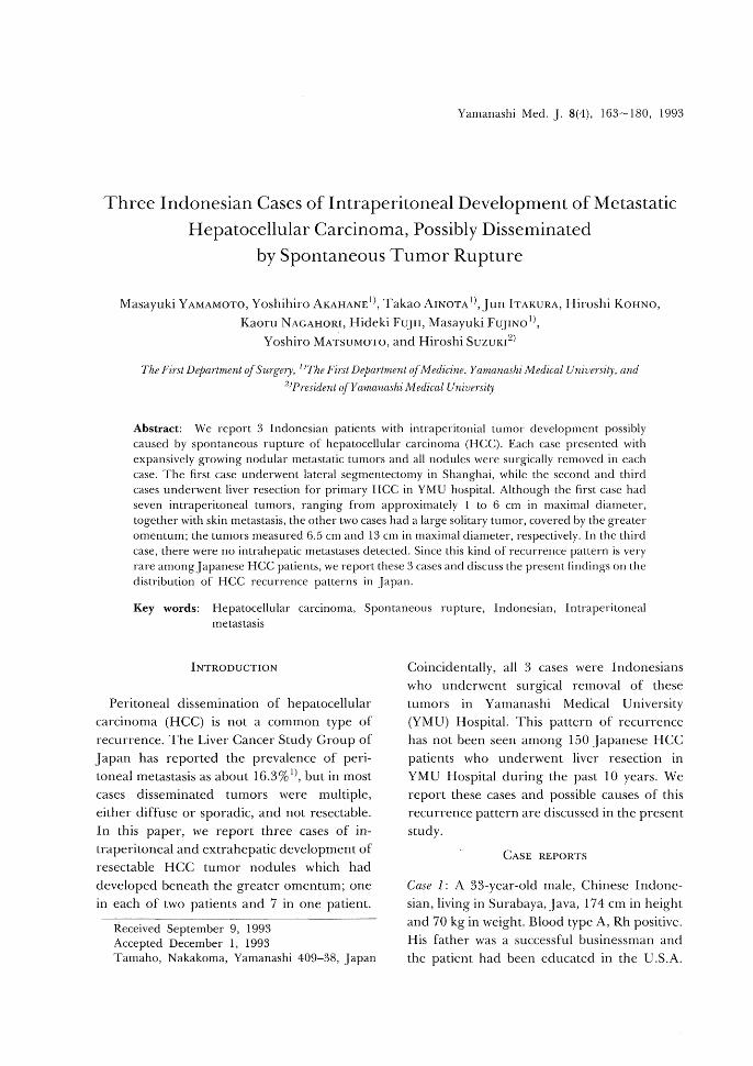
Abdominal CT revealed low density areaswith lipiodol deposits in the right Iobe (Fig. 1 ),
suggesting a large HCC with central necrosis.
Fig.
/"・
n)・
A >ietik sa
l,s,(
1. CT indicated a skin metastasis and large lipidol deposit in HCC occupying the right
lobe.
Intraperitoneal Metastasis ofIndonesian HCC 165
Splenomegaly and gallstones were alsoobserved. US revealed at least 3 hyperechoic
masses, 6.0 × 7.0 cm, 6.5 × 6.1 cm, 8.7 × 7.4
cm, in the right lobe but no echogenic mass in
the left lobe. Many gallstones were also de-
tected.
After examination, physicians in the First
Department of Medicine persuaded the pa-
tient and his family to return to Indonesia to
continue receiving TAI treatment in Indonesia
as he had before coming to Japan, since there
was no indication that tumor removal would
extend his survival period. However, the fami-
ly was eager for him to be treated in Japan and
the doctors then referred him to our Surgical
Department. Since his liver dysfunction was
not severe and no liver tumor was found in the
remnant region of the left lobe, resection of
the tumor in the right lobe as well as the
metastatic tumor to the skin was possible,
although it was not certain whether these
treatment would extend his survival period.
On September 20, 1990, he underwentsurgery. The skin tumor, about 7 cm indiameter with a clear border, was resected. It
was hard and infiltrated the anterior sheath of
the rectus abdominal muscle. At laparotomy, a
smal! quantity of serous ascites was observed.
In addition, 7 tumor nodules were found in
the abdominal cavity; 2 (6.0 x 4.3 × 4.0 cm,
1.8 × 1.7 × 1.1 cm) (Figs. 2 and 3a) covered by
the greater omentum, one (3.7 × 3.3 × 8.0 cm)
adhered to the ascending colon, one (2.8 × 2.5
× 1.8 cm) to the transverse colon, one (1.4 ×
1.2 × 1.2 cm) to the ileum, one (1.6 × 1.4 × 1.4
cm) to thejejunum, and one to the parietal
peritoneum (1.2 × 1.2 × I.2 cm). There were
no other small and diffuse peritoneal dissemi-
nations as seen in adenocarcinoma. All of the
tumors adhered to the greater omentum. They
seemed to be due to peritoneal dissemination
during tumor rupture. All were excised surgi-
cally. Macroscopically, all intraperitoneal
tumors were yellowish and compact, and the
borders were clearly demarcated. The in-
trahepatic tumor (12 × 7 cm by perioperative
US) was not removed but wedge resection ofa
small piece of the liver including a small
metastatic nodule was performed. For subse-
quent TAI, cannulation to the hepatic artery
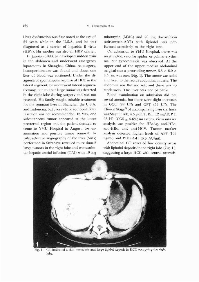
Fig. 2. Case 1: One of the extrahepatic abdominal tumors (indicated by arrow)
on the preoperative CT, retrospectively.
was recognized
166 M. Yamamoto et al.
a
'r;'b7
ee・
,.c- E"x" S.il3lai '.'- k--ii.
g3k
.lageK
:ip
.iscst
b" ICS'
"wlepte '
15t. k ptS
., f;{ 'i"t,・,,,,ct"7pt',"i 'iwa -,
i-:{.t-.t?Si¥lllitlSillil;;.:y$W・"",ii・,tk'"N il/lskV
'e
,gei
fits r,¥g
gh,#t1t..,.
-tsx
.,,,..tl$i-l ・,{isi,.s}iiii{
deL kgk
esr
3bFig. 3.
ee
tsstk
ceew
liijxiil".',.ttslllliee1 illileell ssJIEiili;locx"sp:'`sltigE'mewa:mpec"i;'X
mp. ajtw .wiilii?Sii
k-.. 'Z}eX22
ag er・・u.idr.
.Cl ma
.trKX
te
2
wo g2$ tw" ge %
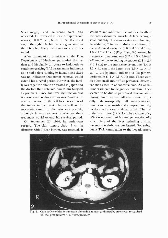
Case 1: Macroscopic (a) and microscopic (b) findings of the largest extrahepatic
abdominal tumor. Histologically the tumor was moderately differentiated hepatocellu-
lar carcinoma, covered by thin capsule (HE, original magnification ×40).
Intraperitoneal Metastasis ofIndonesian HCC 167
through the gastroduodenal artery and its
connection to a subcutaneous port in the
abdominal wall were performed together with
cholecystectomy. None of the lymph nodes
along the gastroduodenal ligament suggested
metastasls.
Microscopically, all tumors were composed
of thick trabecular-type and solid-type tumor
cells, accompanied by giant cells and clear cells;
moderately differentiated HCC (Edmondson
Grade III) (Fig. 3b). One of the seven tumor
nodules was located at the subserosal layer of
the jejunum and was the only tumor where
hematogenic metastasis may have been possi-
ble. All the other tumors were considered
disseminated tumors.
After his return to Indonesia, although TAI
was performed, he died in December 1990.
Case 2: A 68-year-old male, a retired business-
man, 156.5 cm in height and 57.5 kg in weight,
Indonesian living in Jakarta, Java. Blood type
B, Rh positive.
He had a history of blood infusion many
years ago, but the details were obscure. He
underwent appendectomy at the age of 33
years. Since 1978, he had been followed for
liver dysfunction. In January 1990, AFP in-
creased to 649 nglml and on March 26, CT
performed in Indonesia revealed a low density
area in the anterior-superior region (S8) of the
liver. He had only slight discomfort in the
upper abdomen. He was admitted to the First Department of
Medicine on April 6. There was no anemia or
jaundice and no vascular spider or palmar
erythema. An elastic hard liver was palpable
for 9 cm on the median line and 5 cm on the
right middle clavicular line. The edge of the
liver was dull and the surface was irregular.
The spleen was palpable to a two-finger width.
Besides HCC accompanied by Iiver cirrhosis
and gallstones, he had hypertension, asthma
bronchialis, a slight glucose intolerance and
esophageal varices. Biochemical examination
indicated liver dysfunction, such as high GOT
(128 U/l) and GPT (141 Ull), but the accom-
panying liver cirrhosis was Clinical Stage I;
Alb, 8.6 gldl; T. Bil, 1.1 mgldl; PT%, 114.896;
ICGRis, 17.6%; no ascites. Virus marker
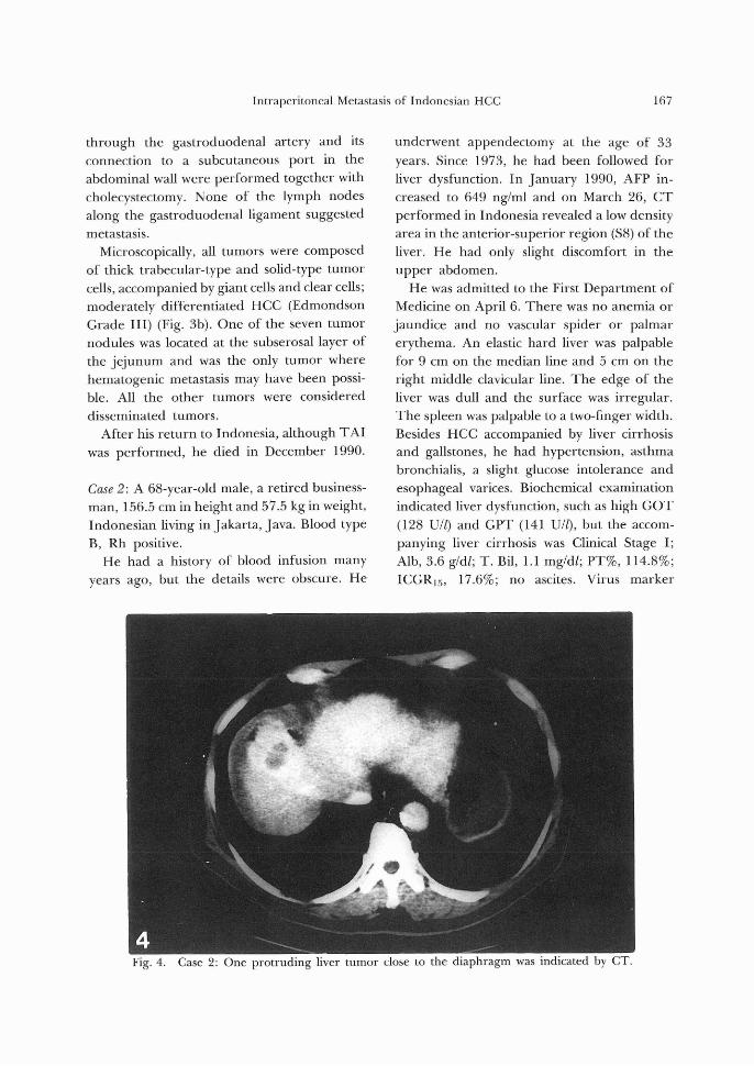
Fig. 4. Case 2: One protrudmg liver tumor close to the diaphragm was indicated by CT.
168 M. Yamamoto et al.
5a w
k
2
lifffs -- .st.,.,,,, ¥ss.wa
"."
.ew/kkg,xe,"ee'"/ec,,,,,,s・ ,¥,?ISii'tt,sstt'g.,
Ht:・eqes ・
!xge
' " s.. t"
aus'-Xss n.
.S .e -K ge ・: ktKst. .i- rVn
Y/,,$.stiS,2.eea itsi4r
v-ev w"C
t---
H- t-1
pti'4c v}
'g
,".w,.,,,tilili.t9i X, ;F・,tts.if/t.3bspti'ii)iii"$sit/ts.p."-eStse
, tsix siiSir.
te ,ktw'
・ kSA tlf -pt .
re
. Mt AVif '{
- "ts ' "iN:i¥ie
t
1
li.r., As.`;
"sM.-
"-xt'ep" tst
xjillll'
Fig. 5.
gx
.
- "v.. ・vx
rg,.-ipL),P,t`.
'
st
'
VM
k "t
"
S s. ` F =. .--
t
- ti
$zas
es ,}y.f. i. ,. .e'`s-- ki}Igsls $k'gi
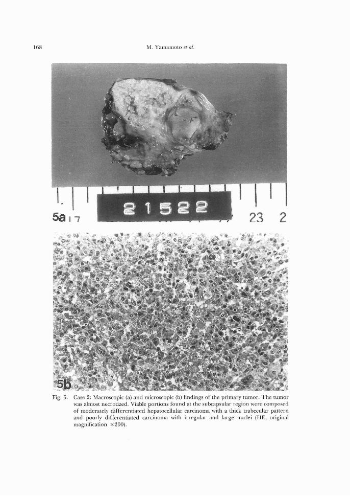
wht vNb kMh-.n .- F whCase 2: Macroscopic (a) and microscopic (b) findings of the primary tumor. The tumor
was almost necrotized. Viable portions found at the subcapsular region were composed
of moderately differentiated hepatocellular carcinoma with a thick trabecular pattern
and poorly differentiated carcinoma with irregular and large nuclei (HE, origmal
magnification ×200).
Intraperitoneal Metastasis ofIndonesian HCC 169
analysis was kegative for HBsAg, HBeAg, and
anti-HCV. AFP was 1818 nglml and PIVKA-II
was Iess than O.06 AUIml.
On SAG performed on April 16, a S8 tumer
irrigated by the anterior-superior branch of
the right hepatic artery was recogltized. Vascu-
lar invasien was negative. Frem the right
hepatic artery, the transca£heter arterial embo-
lization (TAE) was performed using 4e mgADR, 10 ml Iipiodol and Spongel@. Then, he
was transferred to the First Department ef
Surgery on May 1. On May 15, 1990, after his general coltdition
and liver function had improved following
TAE, he underwent partial liver resection of
the S8 tumor (Fig. 4) and cholecystectomy.
The cirrhetic liver surface of the right lobe
adhered tightly to the diaphragm, but the
greater omentum gathered between the tumor
and the diaphragm. Cautious dissection of the
strongly fibrous adhesion gave access to the
tumor, but the protruding tumor adhered
tightly to the diaphragm suggesting a post-
rupture state. In the surgical specimen, the 3.0
× 2.0 × S.5 cm tumor was a single nodulartype with infiltration ofthe surrounding tissue,
and without capsule formatiolt. Serosal in-
filtration was positive. The macroscopic Stage
(TNM)2) was II. Microscopically, most of the
tumor was necrotic probably due to preopera-
tive TAE. The viable portion was moderate}y
to poorly differentiated HCC (Fig. 5). Micro-
scepic invasion of the vasculature was positive,
and tumor infiltration of the connective tissue
adhering to the diaphragm was positive.According to surgical findings and macro- and
microscopic analysis, the tumor was apparently
in a status after tumor rupture.
After liver resection, AFP decreased to l4
nglml on May 28 and IO nglml on June ll.
TAI with 60 mg epirubicin and 8 ml Iipiodol
was performed on June 25. Lipiodol CT did
not indica£e lipiodol deposits in the remnantliver.
After discharge, he returned £o Indonesia.
In March 1991, AFP increased £o more than
1,eOO ng!ml and CT revealed a large mass in
£he right lobe. He was readmitted to the First
Department of Medicine en May 20, I991.
On admission his body weight was 56 kg.
General condition was good. There was noanemia orjaundice. In the upper right abdo-
minal region, a reund tumor mass, about 5 cm
in diameter, free from the liver, was palpable.
It was elastic firm with tendemess.
On admission, accompanying liver cirrhosis
was Clinical Stage II: Alb, 8.4 g/dl; T. Bil, 1.3
mgfdl; PT%, 85.1%; ICGRis, 27.0 %; noascites. Tumor marker analysis detected ele-
vated AFP (May 27, 78,OOO nglr[tl) and PIV-
KA-II (May 27, 48.4 AUIml).
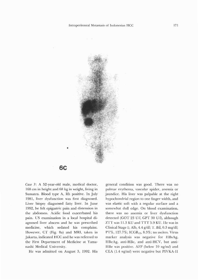
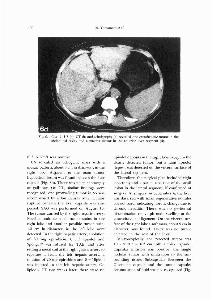
US, CT and scintigraphy revealed one ex-
trahepatic tumor in the abdominal cavity (Figs.
6-a, -b, -c), and a massive tumor occupying the
anterior segment of the right lobe (Fig. 6-d).
SAG on May 27 revealed a large tumor stain in
the right lobe and small stains in the left lobe.
One extrahepatic tumor stain, about 7 cm in
diameter, fed by the gastroduedenal artery
was also recognized. Portography did notindicate the right branch of the portal vein. To
the right hepatic artery, 40 mg epirubicin and
6 ml lipiodol were infused. Eltdoscopy re-
vealed that the blue esophageal varices had
progressed, compared with those before the
initial surgery.
He was transferred £o the First Department
of Surgery on June 10, 1991. From the above
observations, the liver tumor was considered
unresectable, but the rapidly growing abdo-
minal tumor was removed to prevent rupture
and also to allow subsequent TAI treatment to
focus exclusively on the Iiver. On June 20,
1991, extirpation of the extrahepatic abdomin-
al tumor and caRnulation to the right hepatic
artery for TAI were performed. Slight ascites
was recognized. The abdominal tumor was
covered by the greater omentum and hadslight adhesion to the transverse colon. There
were no e{her tumors or peritoBeal dissemiRa-
tiolt detec£ed. There was no lymph nodemetastasis observed. Macroscopic TNM Stage
170 M. Yamamoto et al.
was Stage IV-B.
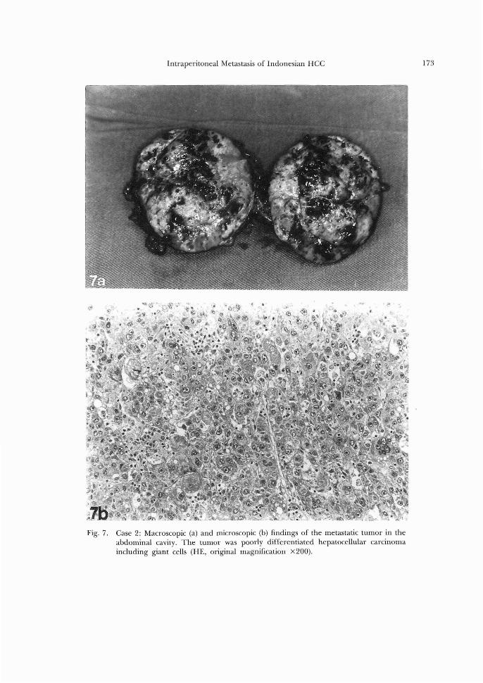
The tumor was 6.5 x 6.0 × 5.5 cm, roundand elastic hard. Thick tumor capsule had not
developed. Microscopically, the tumor was a
metastatic nodule of HCC, containing abun-
dant polynuclear giant cells (poorly differenti-
ated HCC) (Fig. 7). Examination of ascites did
not reveal any cancer cells.
Postoperative weekly TAI using 1O mg ADR
and 1 ml lipiodol through the port, sub-
cutaneously connected to the hepatic cannula,
was started from the second week and con-
tinued after his return to Indonesia, but he
died within 6 months.
Intraperitoneal Metastasis of Indonesian HCC I71
e
wt
3i5rst
.t/tctff-
.t
.ilts
/・
'・$isli・.
-kek..
xee
, g8
''"'"rc"' 's・
"・i!S・ ', ' , Sr..
t. .. ./ t'!iit ' "
did ptgtt・
・3. ,1:・・
ti"' i -ZiiE' ffi'ia-
6C
Case 3: A 52-year-old male, medical doctor,
168 cm in height and 68 kg in weight, living in
Sumatra. Blood type A, Rh positive. In July
1981, liver dysfunction was first diagnosed.
Liver biopsy diagnosed fatty liver. In June
1992, he felt epigastric pain and distension in
the abdomen. Acidic food exacerbated his
pain. US examination in a local hospital di-
agnosed liver abscess and he was prescribed
medicine, which sedated his complaint.
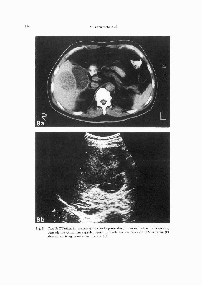
However, CT (Fig. 8a) and MRI, takefi in
Jakarta, indicated HCC and he was referred to
the First Department of Medicine at Yama-
nashi Medical University.
He was admitted on August 3, 1992. His
general condition was good. There was no
palmar erythema, vascular spider, anemia or
jaundice. His liver was palpable at the right
hypochondrial region to one finger width, and
was elastic soft with a regular surface and a
somewhat dull edge. On blood examination,
there was no anemia or liver dysfunction
detected (GOT 23 U14 GPT 30 Ull), although
ZTT was 11.3 KU and TTT 5.9 KU. He was inClinical Stage i; Alb, 4.4 g/dl; T. Bil, O.3 mgldt;
PT96, 127.796; ICGRis, 8.3%; no ascites. Virus
marker analysis was negative for HBsAg,
HBeAg, anti-HBc, and anti-HCV, but anti・-
HBe was positive. AFP (below 10 ng!ml) and
CEA (1.4 nglml) were negative but PIVKA-II
172 M. Yamamoto et al.
ttt'
'
k・;
s
Fig. 6. Case 2: US
abdominal
' es, ,. -,.
・s kSpt ,
''
rvll$p ' ,' "'t'/s・・
t(r
i$i?i,$rrn
.v"
n-
tt kN-..L t¥.-
-;"'r
ff
(a), CT (b) and scintigraphy (c) revealed one extrahepatic tumor in the
cavity and a massive tumor in the anterior liver segment (d).
(O.3 AUIml) was positive.
US revealed an echogenic mass with amosaic pattern, about 8 cm in diameter, in the
right lobe. Adjacent to the main tumorhypoechoic lesion was found beneath the liver
capsule (Fig. 8b). There was no splenomegaly
or gallstone. On CT, similar findings were
recognized; one protruding tumor in S5 was
accompanied by a low density area. Tumor
rupture beneath the Iiver capsule was sus-
pected. SAG was performed on August 10.The tumor was fed by the right hepatic artery.
Possible multiple small tumor stains in the
right lobe and another possible tumor stain,
1.5 cm in diameter, in the left lobe were
detected. In the right hepatic artery, a solution
of 60 mg epirubicin, 6 ml lipiodol andSpongel@ was infused for TAE, and aftersetting a metal coil at the right gastric artery to
separate it from the left hepatic artery, a
solution of 20 mg epirubicin and 2 ml lipidol
was irijected to the left hepatic artery. On
lipiodol CT two weeks later, there were no
lipiodol deposits in the right lobe except in the
clearly detected tumor, but a faint lipiodol
deposit was detected on the visceral surface of
the lateral segment.
Therefore, the surgical plan included right
lobectomy and a partial resection of the small
lesion in the lateral segment, if confirmed at
surgery. At surgery on September 4, the liver
was dark red with small regenerative nodules
but not hard, indicating fibrotic change due to
chronic hepatitis. There was no peritoneal
dissemination or lymph node swelling at the
gastroduodenal ligament. On the visceral sur-
face ofthe right lobe a soft mass, about 9 cm in
diameter, was found. There was no tumordetected in the rest of the liver.
Macroscopically, the resected tumor was
10.5 × 9.7 × 6.3 cm with a thick capsule.
Capsular invasion was positive; the single
nodular tumor with infiltration to the sur-
rounding tissue. Subcapsular (between the
Glissonian capsule and the tumor capsule)
accumulation of fluid was not recognized (Fig.
Intraperitoneal Metastasis ofIndonesian HCC 173
kee6ee
S
・'Yl Si'
,{.-,is{ .
i3.Xiill
g{s.`M・ ,a;lr・S" ''/igek'$gecK
ts・ tt 'ases''''I'': pttv'''''
ttlii/F/,ik"t2.$X" k・tm." ,S"geiiil&,S.t,$,,,avggRi,ii}ft,if,,%,,&tt.k
of.;l8t){,w4:pt'1 ",z;gesew7
t.ies"g.k
ue&eags '
gg s gsgig
"ieepa¥IV2giis$
s(
ge,,gkeees,,yssg.scfimpAg?ee ...eq.."st)l eegek., ss
}rw
{sts $l'litr{
Fig. 7. Case 2: Macroscopic (a) and microscopic (b) findings of the metastatic tumor in the
abdominal cavity. The tumor was poorly differentiated hepatocellular carcinoma
including giant cells (HE, original magnification ×200).
I74 M. Yamamoto et al.
Fig. 8. Case 3: CT taken inJakarta (a) indicated a protruding tumor in the liver. Subcapsular,
beneath the Glissonian capsule, liquid accumulation was observed. US in Japan (b)
showed an image similar to that on CT.
Intraperltoneal Metastasis of Indonesian Hα 175
嫉
’二、
警・
一ズ〉妻趨
’脳弓琴寮綴、’,許
噂炉’
横ら、
・無蜘
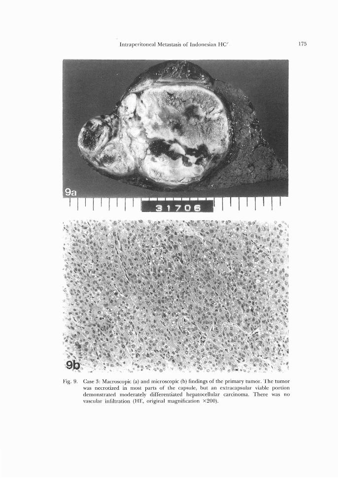
Fig.9 Case 3:Macroscopic(a)and microscoplc(b)5ndings of the primary tumor. The tumor
was necrotized in most parts of the capsule, but an extracapsular viable portlon
demonstrated moderately dlfferentlated hepatocellular carcinoma There was no vascular lnfiltration(HE, orlgmal magni丘cation×200).
176 M. Yamamoto et al.
9a). Macroscopic TNM Stage was Stage II,since tumor rupture is not considered in this
classification. Microscopically, the main tumor
was almost necrotized but moderately diffe-
rentiated HCC remained at the subcapsular
region as well as at the extracapsular infiltra-
tion area (Fig. 9b).
On October 2, before discharge from the
hospital, another TAI was perfbrmed using 30
mg ADR and 5 ml lipiodol to the remnant left
lobe, although PIVKA-II had decreased after
surgery to O.06 on September 17.
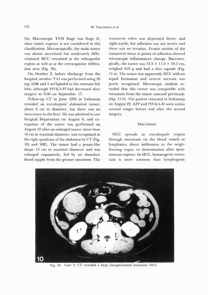
Follow-up CT in June 1998 in Indonesia
revealed an extrahepatic abdominal tumor,
about 6 cm in diameter, but there was norecurrence in the liver. He was admitted to our
Surgical Department on August 6, and ex-
tirpation of the tumor was performed on
August 12 after an enlarged tumor, more than
10 cm in maximal diameter, was recognized in
the right quadrant of the abdomen by CT (Fig.
10) and MRI. The tumor had a potato-like
shape 13 cm in maximal diameter and was
enlarged expansively, fed by an abundant
blood supply from the greater omentum. The
transverse colon was depressed down- and
right-wards, but adhesion was not severe and
there was no invasion. Frozen section of the
connective tissue at points of adhesion showed
microscopic inflammatory change. Macrosco-
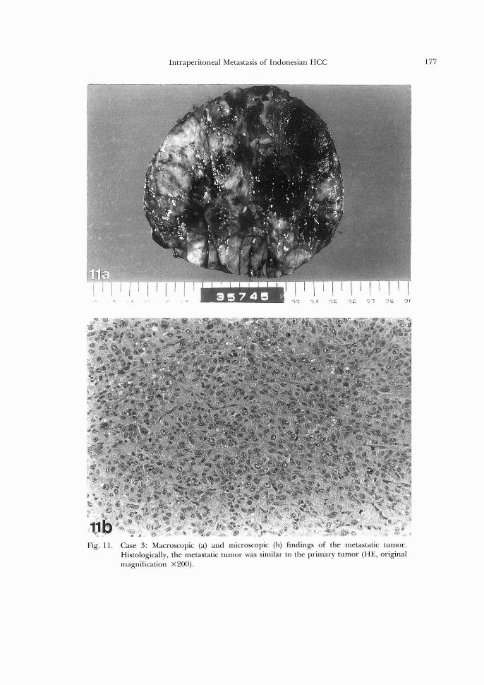
pically, the tumor was 13.5 × 11.0 × 10.5 cm,
weighed 410 g and had a thin capsule (Fig.
11-a). The tumer was apparently HCC with no
septal formation and central necrosis was
partly recognized. Microscopic analysis re-
vealed that this tumor was compatible with
metastasis from the tumor resected previously
(Fig. 11-b). The patient returned to Indonesia
on August 22. AFP and PIVKA-II were within
normal ranges before and after the second
surgery.
DIscussloN
HCC spreads to extrahepatic organsthrough metastasis via the blood vessels or
lymphatics, direct infiltration to the neigh-
bouring organ, or dissemination after spon-
taneous rupture. In HCC, hematogenic metas-
tasis is more common than Iymphogenic
Fig. 10. Case 8: CT revealed a large intraperitoneal metastatlc HCC.'
177Intraperltoneal Metastasis of Indonesian HCC
178 M. Yamamoto et al.
metastasis. Vascular invasien, together with
the size of the tumor, larger or smaller than 2
cm, and Rumber of tumers, single or mu}tiple,
are factors in determining the macroscopic
TNM Stage2). Clinically, }ymph node metasta-
sis is not common in the progession of HCC,
al£hough about 30 to 35% of autepsy casesshowed lymph node metastasis3). Except for
adjacent lymph node metastasis at the per£a
hepatis, Iymph node metastasis is treated as a
kind of distant organ metastasis (M factor) in
the TNM classification sys£em; lymph Rodemetastasis in the hepatoduodenal ligament is
registered as Ml (distant organ metastasis
positive). The Liver Cancer Study Group ef
Japan (LCSG) reported that, among HCC
patieRts who underwent laparo£omy between1988 and 1989, 2649 patients did not have
lymph node metastasis while 76 patients had
lymph node metastasis, and S49 pa£ients werenot reportedi). In the metastatic cases, 52
metastasized lymph nodes were not adjacen£
and, therefore, considered Ml.
Direct infiltration to the neighbouring organ
is more frequently observed to the diaphragm,
especially after TAE to a large HCC near the
diaphragm. Usually HCC has an expansive
growing form and direct invasion to theduedenum or colon is rare. In Case 2 operative
findings indicated that the previous spon-
taneous rupture and adhesion to the di-
aphragm occurred before TAE. Peritoneal dissemination does net occur fre-
quently in HCC, compared with £hat in adeRo-
carcinoma of other iRtraperitoneal organs, and
therefore, peritoneal dissemiHation er spon-
taneous rupture of HCC, its possible cause, is
not an important fac£or in determining the
TNM classificatioR. Actua}ly, distant organ
metastasis of HCC is not rare. On autopsy
ana}ysis, the LCSG also reported that 435 of
918 cases (47.4%) had metastasis to the lungs,
15I of 881 cases (17.1%) to the intraperitoneal
organs, 144 of 885 cases (16.3%) to the
periteneum, ll9 of 893 cases (I3.3%) to the
adrenal glands, le8 of 853 cases (12.7 %) to the
bone, 7 of 884 cases (O.8 %) to the skin, and 149
of790 cases (18.9%) to other organsi). Howev-
er, in 1803 HCC patients with recurrence after
liver resection, I448 (80.3%) had intrahepatic
metastasis, 134 (7.4%) to the lungs, 105 (5.8%)
to the bone, 39 (O.9%) to the lymph nodes, 23
(1.1%) te the peritoneum, 19 (1.l%) £o thebrain, 16 (O.9%) to the adrenal glands, and I9
(I.1%) to other organs. Peritoneal metastasis
comprised 6.5% of extrahepatic metastases.
Thus, in our clinical experience distant organ
metastasis of HCC is rare. In particular,
peritoneal metastasis usually demonstra£es a
disseminated form, diffuse or sporadic, or
coexists with other distaRt organ metastases.
Therefore, it is very rare for HCC patients
with peritoneal metastasis to be referred to
surgeons for resection of such tumors.
However, intraperitoneal me£astatic tumors
in this report were all reund and expansively
growing nodular types, most of which were
covered by the omentum, free from ether
organs, and all were resectable. In Case 1,
tumor cells were apparently scattered in the
incisional wound during the surgical proce-
dure and developed as skin metastases. The
abdominal tumors in all 8 cases may also have
been scattered to the abdominal cavity during
tumor rupture and developed, fed by new
vasculature from the omentum. Although in
Cases 2 and 3 no hemoperitoneum or severe
pain was experienced, their surgical and mac-
roscopic findings persuade us that tumor
rupture occurred. In Case 1, these multiple
tumors may not be an unusual metastatic form
of advanced HCC case af£er tumor rupture,but in Cases 2 and 3, the large extrahepatic
recurrent tumor was solitary and covered by
the greater omentum, from which nourishing
arteries developed. How tumor cells delivered
to the periteReum after tumor rupture de-
veloped to a solitary large recurrent tumer
remains unclear. Growth may be explaiRed as
the result of a decrease in the immune re-
sponse of the host, but £hat does no£ explain
why the tumor was solitary. Moreover, in Case
Intraperitoneal Metastasis of Indonesian HCC l79
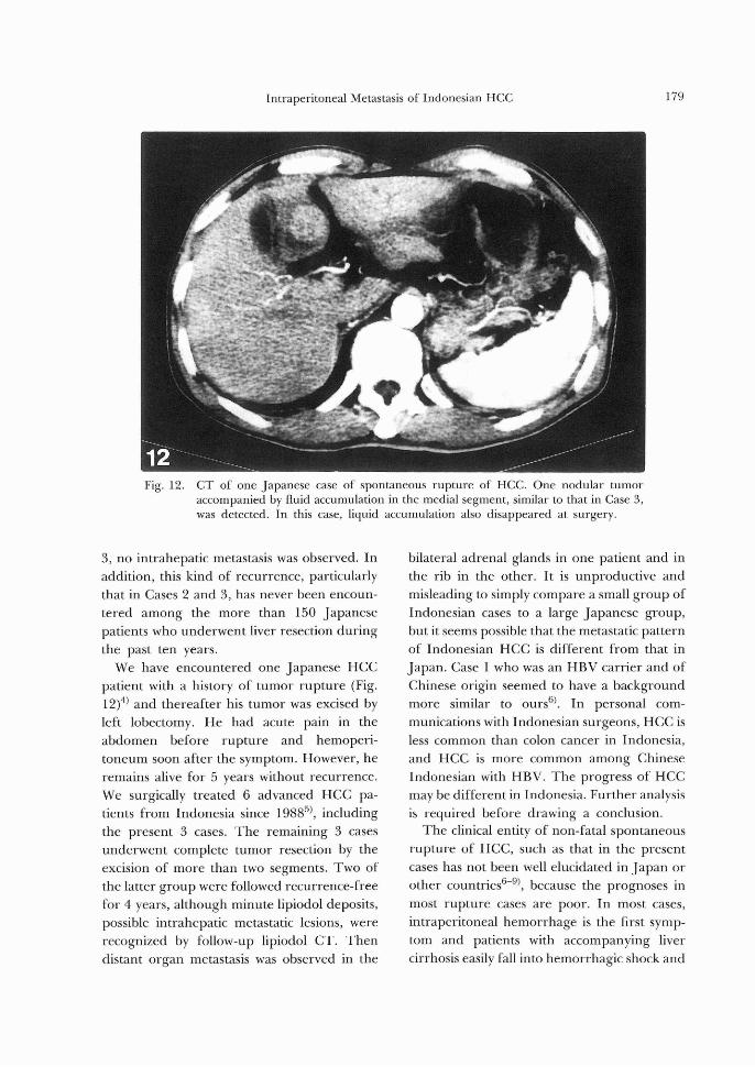
Fig.
'("['ISN.
'sesf-,lii '1/l.¥,・,.. ,,
pt" S・,k'ig'es'ee'
:fp;i$t: i:iis:"・"i} n:i・} i. .,
4ser・ r:f'・・'
'";・・. '-" i:ajil
N
12. CT of one Japanese case of spontaneous rupture of HCC. One nodular tumor accompanied by fluid accumulation in the medial segment, similar to that in Case 3,
was detected. In this case, liquid accumulation also disappeared at surgery.
8, no intrahepatic metastasis was observed. In
addition, this kind of recurrence, particularly
that in Cases 2 and 3, has never been encoun-
tered among the more than 150 Japanesepatients who underwent liver resection during
the past ten years.
We have encountered one Japanese HCCpatient with a history of tumor rupture (Fig.
12)4) and thereafter his tumor was excised by
left lobectomy. He had acute pain in the
abdomen before rupture and hemoperi-toneum soon after the symptom. However, he
remains alive for 5 years without recurrence.
We surgically treated 6 advanced HCC pa-tients from Indonesia since I9885), including
the present 3 cases. The remaining 3 cases
underwent complete tumor resection by the
excision of more than two segments. Two of
the latter group were followed recurrence-free
for 4 years, although minute lipiodol deposits,
possible intrahepatic metastatic lesions, were
recognized by fo11ow-up lipiodol CT. Then
distant organ metastasis was observed in the
bilateral adrenal glands in one patient and in
the rib in the other. It is unproductive and
misleading to simply compare a small group of
Indonesian cases to a large Japanese group,
but it seems possible that the metastatic pattern
of Indonesian HCC is different from that in
Japan. Case 1 who was an HBV carrier and of
Chinese origin seemed to have a background
more similar to ours6). In personal com-
munications with Indonesian surgeons, HCC is
less common than colon cancer in Indonesia,
and HCC is more common among ChineseIndonesian with HBV. The progress of HCCmay be differentin Indonesia. Further analysis
is required before drawing a conclusion.
The clinical entity of non-fatal spontaneous
rupture of HCC, such as that in the present
cases has not been well elucidated in Japan or
other countries6-9), because the prognoses in
most rupture cases are poor. In most cases,
intraperitoneal hemorrhage is the first symp-
tom and patients with accompanying livercirrhosis easily fall into hemorrhagic shock and
180 M. Yamamoto et al.
die without sufficient resuscitation trea£ments
or further surgical treatment, and very few
cases are referred to specialized hospitals for
surgery. Therefore, survival rates following
resuscitation varied, and there are no clear
reports on recurrence patterns after complete
removal of the liver tumors. In the analysis of
LCSG with respect to the cause of death in
HCC patients, tumor rupture accounts fer 9 to
lI% of deaths. Similar or slightly highr rates
were registered in other Asian6)'8-iO) and
Africanii) countries, while a very low rate was
reported among CaucasiansiO・i2).
The present intraperitoneal nodular rnetas-
tasis is not the same as peritoneal dissemina-
tion which often develops from adenocarcino-
ma in an abdominal ergan. All tumors de-
veloped expansively in the abdominal cavity.
The Iarge tumor in Case 3 depressed the
transverse colon but did no£ infiltrate to it.
There was Ro microscopic metastasis detected
in the surrounding tissue. Since extirpation of
these tumors was not difficult and if Case 3 can
survive lenger, such intraperitoneal metastatic
tumors should be resected. However at pre-
sent, Ro data are available on the results of
aggressive surgical maneuvers.
REFERENCES
1) The Liver Cancer Study Group ofJapan. The leth report of the follow-up study of primary
liver cancer in Japan, The Liver Cancer study
Group of Japan, Kyoto, 1992.
2) Yamamoto M, Sugahara K. Overview of the general rules for the clinical and pathological
study of primary liver cancer in Japan. In:
Tobe T, Kameda H, Okudaira M, Ohto M, et al., eds. Primary liver cancer in Japan. Tokyo:
Springer-Verlag, l992: 385-392.
S) LiverCancerStedyGroupofJapan:Summary ofthe data from a follew-up study by the Liver
Cancer Study ofJapan: same as in Reference
[2]: 445-453.
4) Yamamoto M, Mogaki M, Sugahara K. Spon-
taneous rupture of primary liver cancer. Nihonrinsho 1988: 46: 208-216.
5) Yamamoto M, Akahaue Y, Ainota T, et al. Experience with IndoResian patienzs who had
hepatocellular carcinorna. Bulletin of Yama-
nashi Med Univ 199S' 10: 38-44. , 6) Ker CG. Hepatocellular carcinoma iR Taiwan:
same as in Reference [2]: 4Il-419. 7) Baiasegaram M. Rupture of liver cell carcino-
ma. Aust NZ J Surg 1968; 37: S82-337.
8) Ong GB, Taw JL. Spon£aneous rupture of hepatocellular carcinoma. Br Med J 1972; 4:
146-149. 9) Van Landingham SB, HendricksJC, Roberts JW. Spontaneous rupture of hepatocellular carcinoma. J Surg Oncol }985; 29: l29-131.
10) Inouye AA, Whelan T.} Jr. Primary liver cancer; A review of 205 cases in Hawaii. AmJ
Surg 1979; 138: 58-61.
11) SteinerPE.Canceroftheliverandcirrhosisin rrrans-Saharan Africa and the United States of
America. Cancer l960; 13: 1085-1166.
I2) El-Domeini AA, Huvos AG, Goldsmith HS, Foot FW. Primary malignant tumors of the liver. Cancer 1971; 27: 7-11.