Hepatocellular Adenoma Immuno-Histochemistry · Who Classification of tumours of digestive system...
36
Hepatocellular Adenoma Immuno-Histochemistry Patrick Martin Envoi Specialist Pathologists
Transcript of Hepatocellular Adenoma Immuno-Histochemistry · Who Classification of tumours of digestive system...
Hepatocellular Adenoma Immuno-Histochemistry
Patrick Martin
Envoi Specialist Pathologists
Envoi
Prof Andrew Clouston
The A team
What are HCAs
• Benign lesions of the liver
• Barthelmes & Tait “Most important benign tumour of the liver
• First described by Edmondson 1958
• Linked by Baum to OCP in 1973
• Macroscopically
Palpable lesion
Range in colour from White-Yellow-Brown
Grow up to 30cm in diameter
What are HCAs
• Microscopically
• Lack normal hepatic parenchyma
• Tracts and hepatic veins are absent
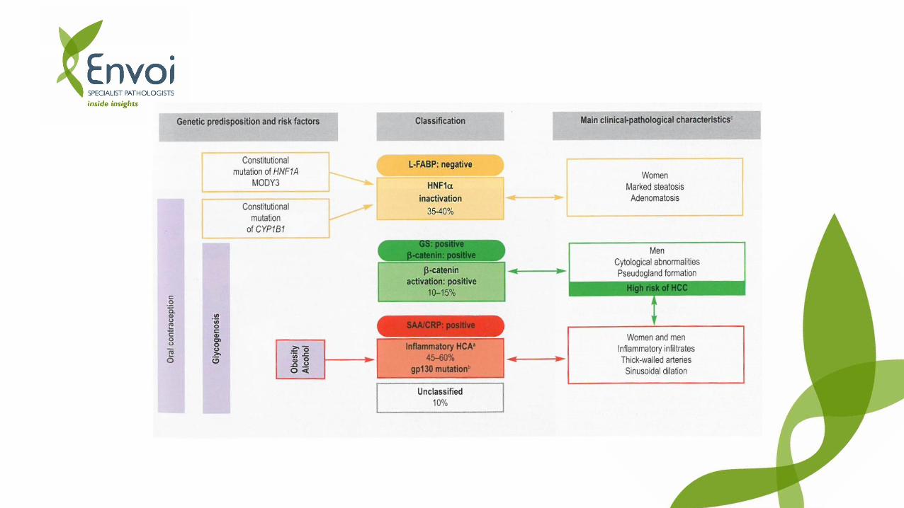
• Subclassifed into 4 groups
1. HCA, inflammatory
2. HCA, HNF1 alpha inactivated
3. HCA, beta catenin activated
4. HCA, unclassified
Epidemiology
Who Classification of tumours of digestive system states:
• Incidence of HCA is 3-4 per 100 000 in Europe and North America
• Lower in Asia
• 85% cases occur in young women
• Rare in children, men and elderly
• At Envoi we have seen 121 HCAs from 99 patients
Etiology
• Major risk factor for HCA is exposure to oestrogenic and androgenic steroids
• 80% of young women with HCA have been on the contraceptive pill.
• Risk increases with duration of use
• Prevalence is declining with lower oestrogen pills being available
• Lesions usually shrink after stopping use of contraceptives and post menopause
• Most men have been users of anabolic steroids for body building
Etiology cont’d
• Non-hormonal risk factors:
• Glycogenosis type 1 (Von Gerke disease)
• Glycogenosis type 3 (Forbes disease)
• Galactosaemia
• Tyrosinaemia
• Familial polyposis coli
• Hepatic iron overload with β-thalassemia
• Obesity
Clinical features
• Abdominal pain
• Abdominal mass
• Intraperitoneal haemorrhage (20-25% of cases have significant haemorrhage). Risk is higher when tumours >5cm
• Abnormal LFTs
• Incidental liver mass during radiology
• Can present as single or multiple lesions (>10 “adenomatosis”)
Importance of classification
• Whilst benign there are a small proportion (4%) of HCAs which develop into HCCs
• Depending on HCA subtype the risk of transformation varies (typically increases in patients with glycogenosis or steroid use)
• Risk of Bleed
• Decision to operate
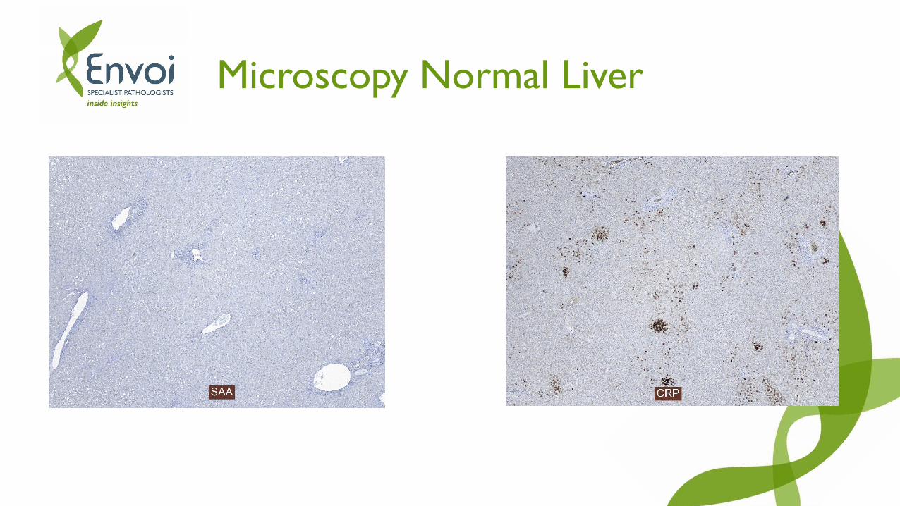
Microscopy Normal Liver
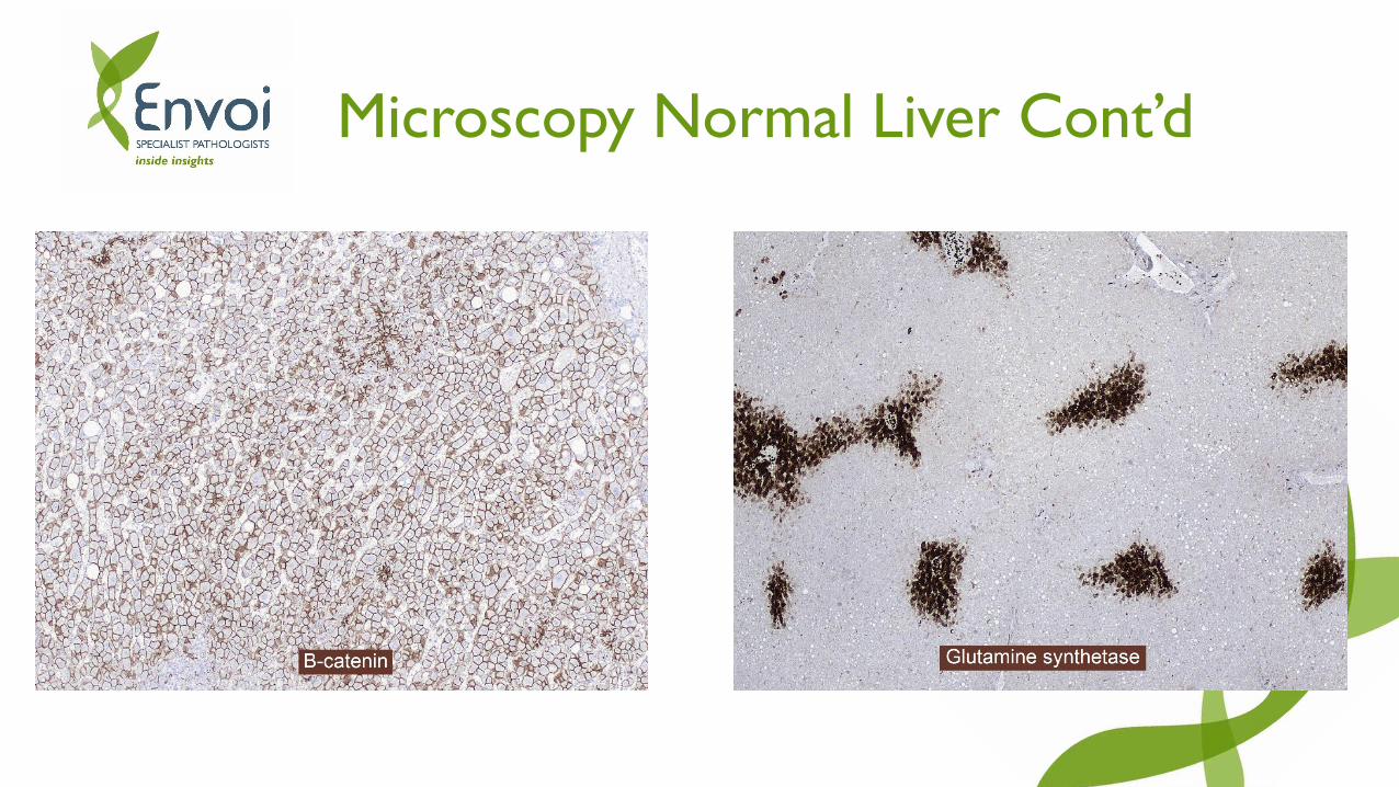
Microscopy Normal Liver Cont’d
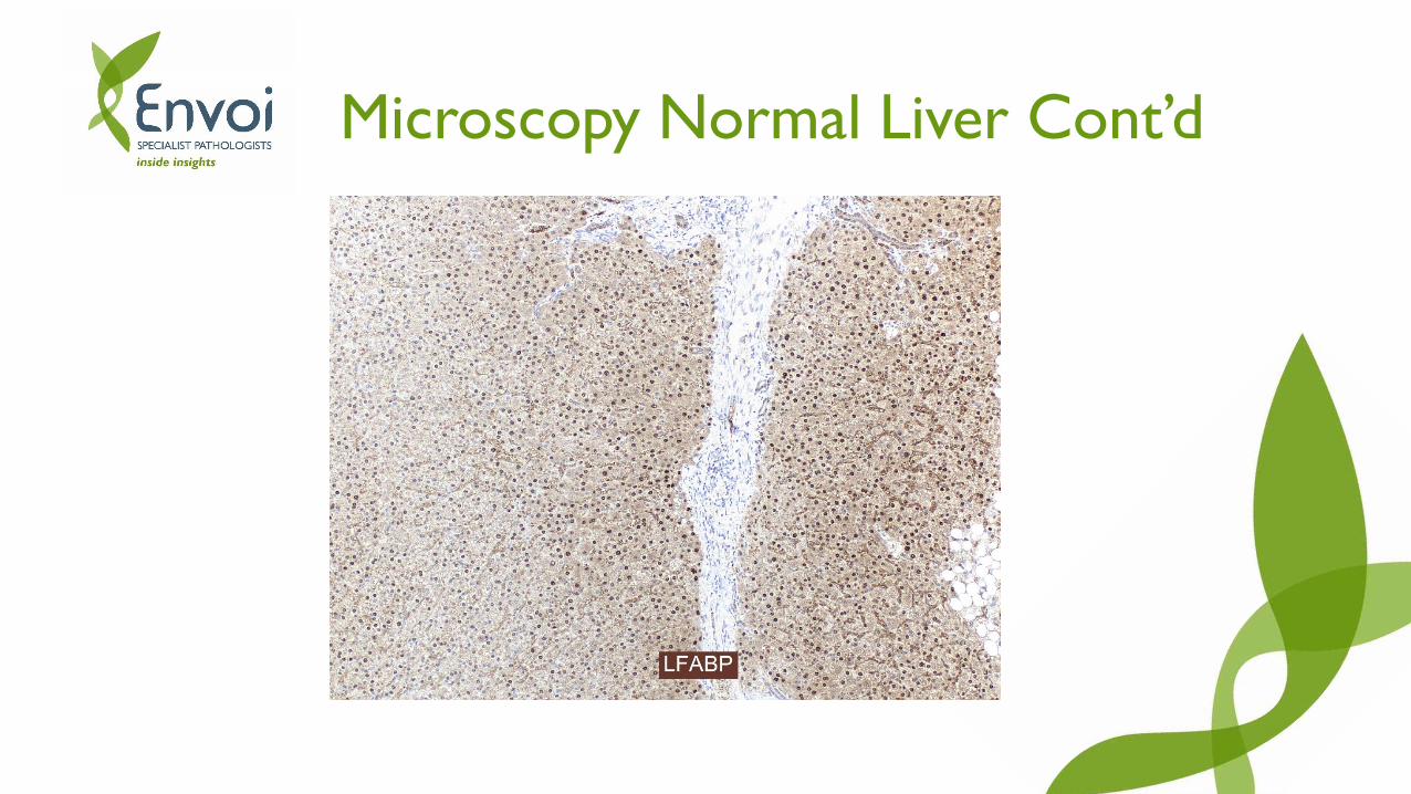
Microscopy Normal Liver Cont’d
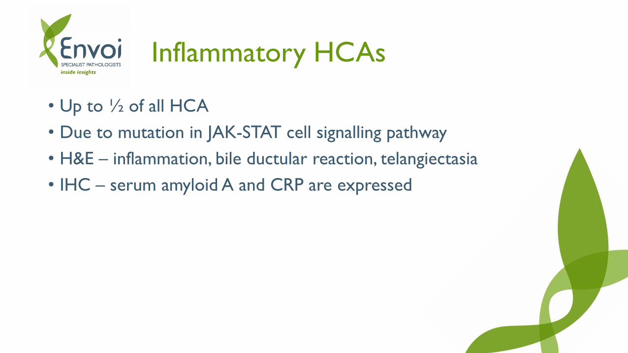
Inflammatory HCAs
• Up to ½ of all HCA
• Due to mutation in JAK-STAT cell signalling pathway
• H&E – inflammation, bile ductular reaction, telangiectasia
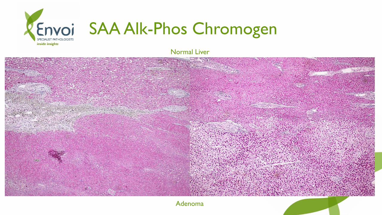
• IHC – serum amyloid A and CRP are expressed
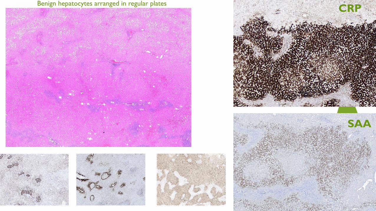
SAA
CRPBenign hepatocytes arranged in regular plates
Normal Liver
Adenoma
SAA Alk-Phos Chromogen
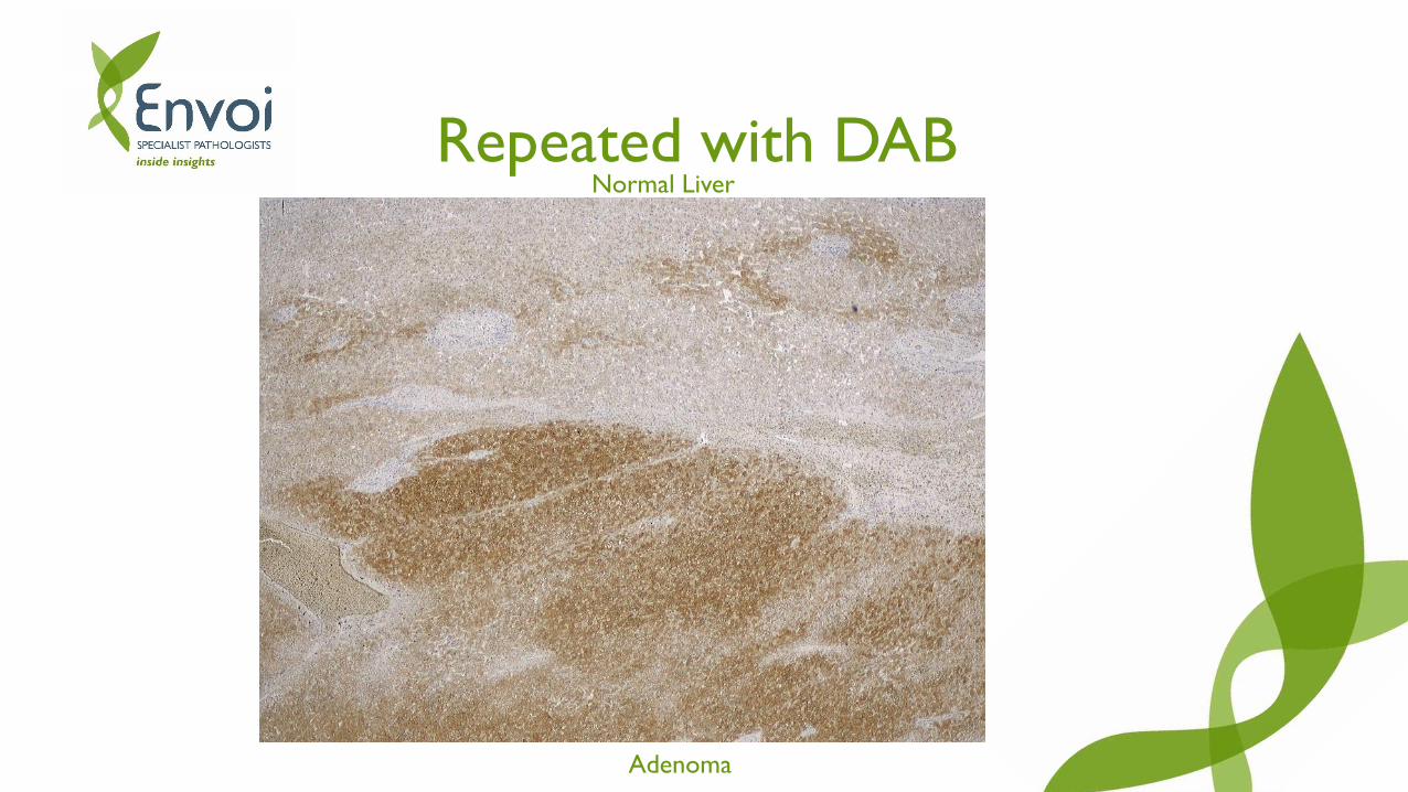
Normal Liver
Adenoma
Repeated with DAB
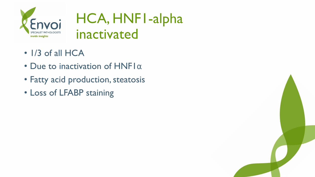
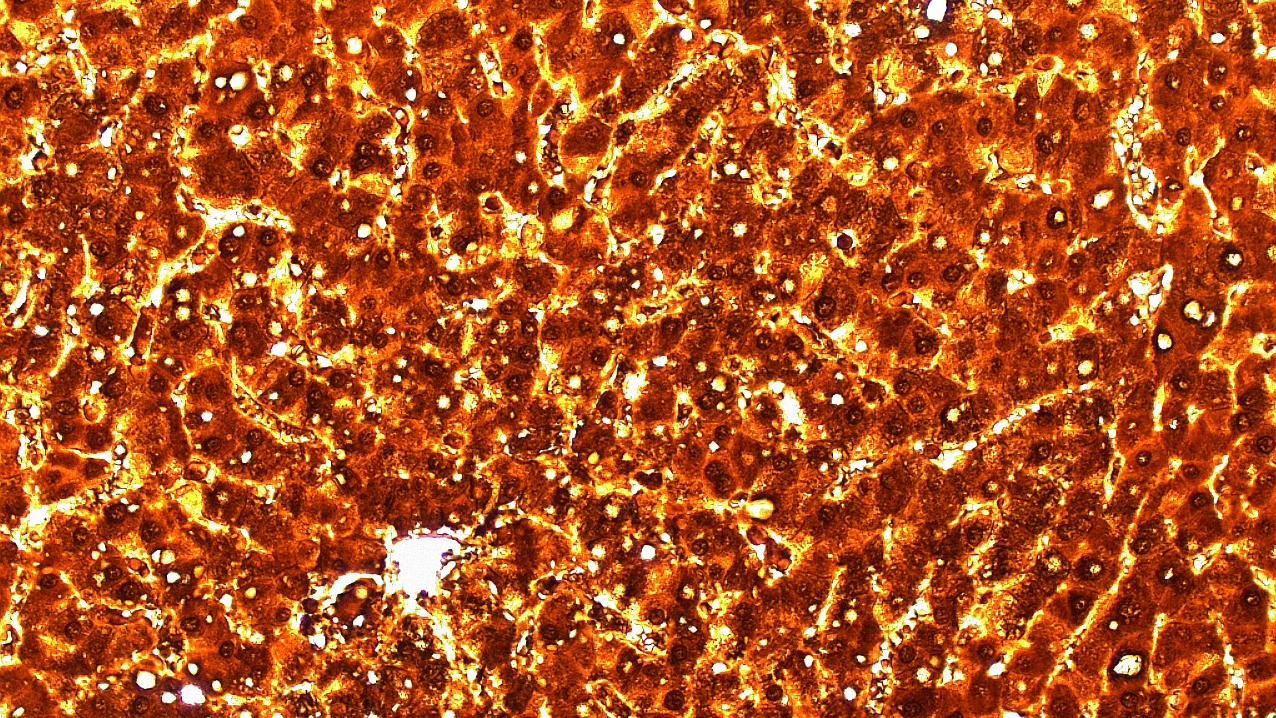
HCA, HNF1-alpha inactivated
• 1/3 of all HCA
• Due to inactivation of HNF1α
• Fatty acid production, steatosis
• Loss of LFABP staining
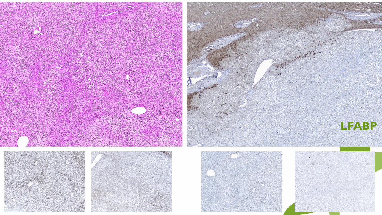
LFABP
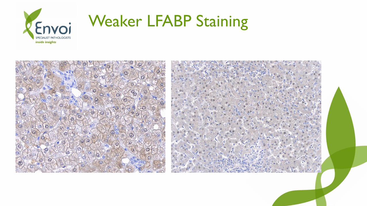
Weaker LFABP Staining
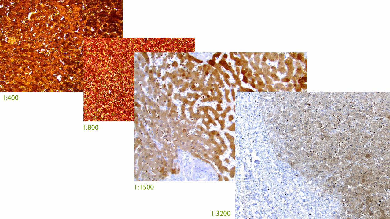
1:400
1:800
1:1500
1:3200
HCA, beta catenin activated
• 10-15% of HCA
• 4% risk of malignant transformation
• Can show cholestasis and architectural and cytologic atypia
• Mutation in gene for b-catenin – WNT pathway activation
• Nuclear staining for B-catenin and diffuse staining for glutamine synthetase
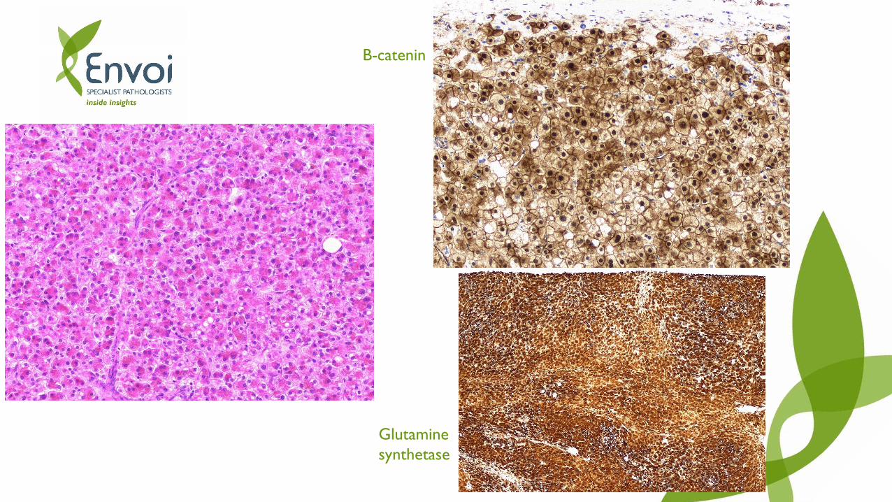
B-catenin
Glutamine
synthetase
Well differentiated HCC
• Well differentiated HCC can be difficult to differentiate from a dysplastic nodule or HCA
• Accuracy improves when 2 of these 3 are positive• HSP70
• Glypican 3
• Beta catenin
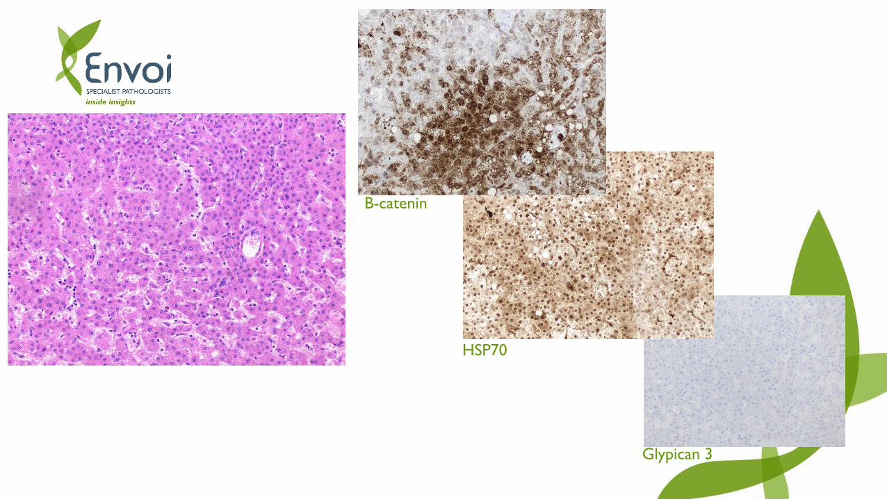
HSP70
Glypican 3
B-catenin
HCA, unclassified
• Up to 10% of cases
• No known mutation or morphologic pattern
• No abnormal IHC staining
Markers of hepatocellular differentiation
• Poorly differentiated HCC can be difficult to distinguish from other poorly differentiated malignancies – melanoma, carcinoma
• New markers have been developed to identify tumours with hepatocellular differentiation• Arginase-1
• HepPar1
• Glypican3
• CD10, pCEA, AFP
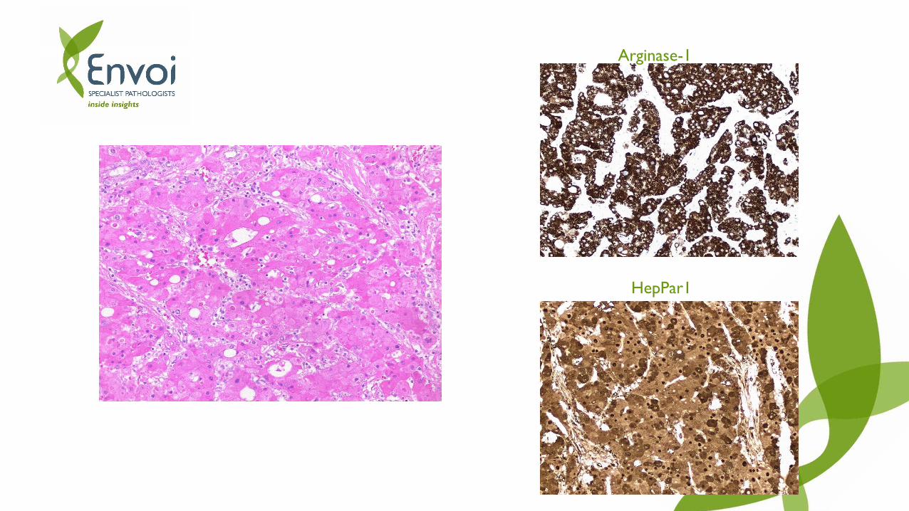
HepPar1
Arginase-1
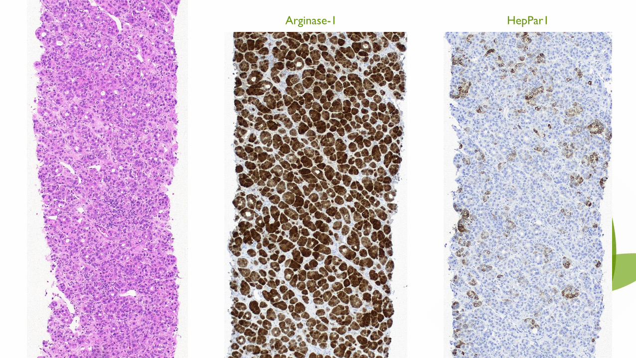
Arginase-1 HepPar1
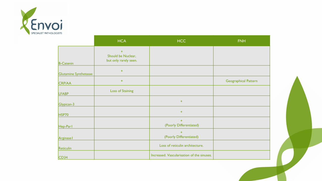
HCA HCC FNH
B-Catenin
+
Should be Nuclear,
but only rarely seen.
Glutamine Synthetasae+
CRP/AA+ Geographical Pattern
LFABPLoss of Staining
Glypican-3+
HSP70+
Hep-Par1
+
(Poorly Differentiated)
Arginase1
+
(Poorly Differentiated)
ReticulinLoss of reticulin architecture.
CD34Increased. Vascularisation of the sinuses.
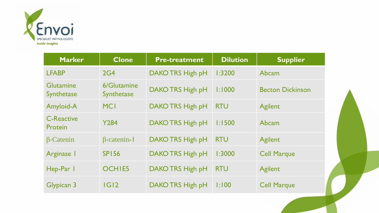
Marker Clone Pre-treatment Dilution Supplier
LFABP 2G4 DAKO TRS High pH 1:3200 Abcam
Glutamine
Synthetase
6/Glutamine
SynthetaseDAKO TRS High pH 1:1000 Becton Dickinson
Amyloid-A MC1 DAKO TRS High pH RTU Agilent
C-Reactive
ProteinY284 DAKO TRS High pH 1:1500 Abcam
β-Catenin β-catenin-1 DAKO TRS High pH RTU Agilent
Arginase 1 SP156 DAKO TRS High pH 1:3000 Cell Marque
Hep-Par 1 OCH1E5 DAKO TRS High pH RTU Agilent
Glypican 3 1G12 DAKO TRS High pH 1:100 Cell Marque
Future Work
• Ass-1
• Initially used to try to classify unclassified HCA
• Shows strong link to risk of bleed 67.4%
• Also shown in some of the classified HCAs
• Tumours showed activation of Sonic Hedgehog Pathway

Many thanks to Dr Greg Miller
References
Whitmer, B. A., et al, 2015. Hepatocellular Adenoma. Medscape.
Bioulac-Sage, P., Cubel, G., Balabaud, C., 2011 Pathological diagnosis of hepatocellular adenoma in clinical practice. Diagnostic Histology 17:12, p. 521-529.
Barthelmes, L., Tait, I, S., 2005. Liver Cell Adenoma and Liver Cell Adenomatosis. HPB, 7, p. 186-196.
Geller, SA., Dhall, D., Alsabeh, R., 2008. Application of Immunohistochemistry to Liver and Gastrointestinal Neoplasms, Arch Pathol Lab Med 132, p. 490-499
Web, 2017. Benign Liver Tumors, American Liver Foundation