![Glucagon-Like Peptide-1 Mediates the Protective Effect of ...Glucagon-Like Peptide-1 Mediates the Protective Effect of the Dipeptidyl Peptidase IV Inhibitor on ... diseases[],leadstoaprogressive,irreversiblelossoffunc-tional](https://static.fdocuments.net/doc/165x107/6115cef534302b72db1aeeca/glucagon-like-peptide-1-mediates-the-protective-effect-of-glucagon-like-peptide-1.jpg)
Hepatitis C virus modulates signal peptide peptidase to ...
12
Hepatitis C virus modulates signal peptide peptidase to alter host protein processing Junki Hirano a , Sachiyo Yoshio b , Yusuke Sakai c , Li Songling d , Tatsuya Suzuki a , Yumi Itoh a , He Zhang a , David Virya Chen a , Saori Haga a , Hiroko Oomori e , Takahiro Kodama f , Yusuke Maeda g,h , Yoshihiro Ono i , Yu Takahashi i , Daron M. Standley d,h , Masahiro Yamamoto h,j , Kohji Moriishi k , Kyoji Moriya l , Tatsuya Kanto b , Tetsuo Takehara f , Kazuhiko Koike l , Yoshiharu Matsuura g,h,1 , and Toru Okamoto a,h,1 a Institute for Advanced Co-Creation Studies, Osaka University, Osaka, 565-0871, Japan; b The Research Center for Hepatitis and Immunology, National Center for Global Health and Medicine, Chiba, 272-8516, Japan; c Department of Veterinary Pathology, Yamaguchi University, Yamaguchi, 753-8515, Japan; d Department of Genome Informatics, Osaka University, Osaka, 565-0871, Japan; e Core Instrumentation Facility, Osaka University, Osaka, Japan; f Department of Gastroenterology and Hepatology, Graduate School of Medicine, Osaka University, Osaka, Japan; g Department of Molecular Virology, Research Institute for Microbial Diseases, Osaka University, Osaka, 565-0871, Japan; h Center for Infectious Disease Education and Research, Osaka University, Osaka, 565-0871, Japan; i Department of Hepato-Pancreatic-Biliary Surgery, Japanese Foundation for Cancer Research, Tokyo, 135-8550, Japan; j Department of Immunoparasitology, Osaka University, Osaka, 565-0871, Japan; k Department of Microbiology, Faculty of Medicine, University of Yamanashi, Yamanashi, 409-3898, Japan; and l Department of Gastroenterology, Graduate School of Medicine, The University of Tokyo, Tokyo, 113-8655, Japan Edited by Peter Palese, Icahn School of Medicine at Mount Sinai, New York, NY, and approved April 22, 2021 (received for review December 21, 2020) Immunoevasins are viral proteins that prevent antigen presentation on major histocompatibility complex (MHC) class I, thus evading host immune recognition. Hepatitis C virus (HCV) evades immune surveillance to induce chronic infection; however, how HCV-infected hepatocytes affect immune cells and evade immune recognition re- mains unclear. Herein, we demonstrate that HCV core protein func- tions as an immunoevasin. Its expression interfered with the maturation of MHC class I molecules catalyzed by the signal peptide peptidase (SPP) and induced their degradation via HMG-CoA reduc- tase degradation 1 homolog, thereby impairing antigen presentation to CD8 + T cells. The expression of MHC class I in the livers of HCV core transgenic mice and chronic hepatitis C patients was impaired but was restored in patients achieving sustained virological response. Finally, we show that the human cytomegalovirus US2 protein, pos- sessing a transmembrane region structurally similar to the HCV core protein, targets SPP to impair MHC class I molecule expression. Thus, SPP represents a potential target for the impairment of MHC class I molecules by DNA and RNA viruses. HCV | MHC class I | signal peptide peptidase | antigen presentation H epatitis C virus (HCV) infection is strongly associated with the development of liver steatosis, cirrhosis, hepatocellular carcinoma (HCC) (1), and extrahepatic manifestations, such as type 2 diabetes, mixed cryoglobulinemia, and non-Hodgkin lym- phoma (2). Approximately 80% of patients with HCV develop chronic infections, whereas only 5% of those infected with hepa- titis B virus acquire chronic infections (3). Although immune cells (i.e., T cells, NK cells, and dendritic cells) have been shown to be functionally impaired in patients with chronic hepatitis C (CHC) (4), how HCV-infected hepatocytes affect immune cells remains unclear. In CHC patients, the administration of direct-acting antivirals dramatically improves sustained virological response (SVR) (5); however, liver disease is not ameliorated due to prolonged liver damage (6). In addition, the elimination of HCV does not fully restore immune cell proliferation and function (7, 8), suggesting that liver damage caused by HCV infection may affect the res- toration of immune cell function. Thus, the characterization of viral and host factors involved in immune modulation during HCV infection is necessary to decrease the risk of HCC development in CHC patients after achieving SVR. HCV belongs to the genus Hepacivirus (family Flaviviridae) and harbors a positive-sense, single-stranded RNA genome; its viral RNA is translated into a single polyprotein of ∼3,000 amino acids that is subsequently processed into 10 viral proteins through cleavage by host and viral proteases (9). The core protein is the first to be translated and cleaved off the polyprotein by host signal peptidase. The signal sequence in the C-terminal region of the im- mature core protein is further processed by the host protease, signal peptide peptidase (SPP), before maturation (10). We previously demonstrated that SPP is essential for stable expression of the core protein (Fig. 1A, Upper). Moreover, we reported that SPP inhibition induces proteasome-dependent degradation of the core protein, mediated by an E3 ligase, a 3:8 chromosomal translocation in he- reditary renal cancer (TRC8) (11) (Fig. 1A, Lower), and efficiently suppresses production of infectious HCV particles (12). SPP has also been reported to cleave cellular proteins, such as human leu- kocyte antigen-A (HLA-A), heme oxygen-1 (HO-1), X-box binding protein 1u (XBP1u), and prolactin (13–16). However, the biological significance of these cleavage events is not fully understood. Several viruses possess immunoevasins to inhibit antigen pre- sentation on MHC class I molecules. For instance, the Epstein– Barr virus encoding nuclear antigen 1 inhibits antigen presentation on MHC class I molecules via a Glycine–Alanine repeat (17, 18). The herpes simplex virus (HSV) encoding ICP47 and the human cytomegalovirus (HCMV) encoding US6 modulate TAP functions Significance The mechanism by which hepatitis C virus (HCV) evades immune surveillance and causes chronic infection is unclear. We demon- strate here that HCV core protein interferes with the maturation of major histocompatibility complex (MHC) class I catalyzed by signal peptide peptidase (SPP) and induces degradation via HMG- CoA reductase degradation 1 homolog. In addition, we found that the core protein transmembrane domain is homologous to the human cytomegalovirus US2 protein, whose transmembrane region also targets SPP to impair MHC class I molecule expression in a similar manner. Therefore, our data suggest that SPP repre- sents a potential target for the impairment of MHC class I mol- ecules by DNA and RNA viruses. Author contributions: Y. Matsuura and T.O. designed research; J.H., S.Y., Y.S., L.S., T.S., Y.I., H.Z., D.V.C., S.H., H.O., Y. Maeda, D.M.S., and T.O. performed research; T. Kodama, Y.O., Y.T., M.Y., K. Moriishi, K. Moriya, T.T., and K.K. contributed new reagents/analytic tools; J.H., S.Y., Y.S., L.S., T.S., Y.I., H.Z., D.V.C., S.H., H.O., Y. Maeda, D.M.S., T. Kanto, and T.O. analyzed data; and Y. Matsuura and T.O. wrote the paper. The authors declare no competing interest. This article is a PNAS Direct Submission. This open access article is distributed under Creative Commons Attribution-NonCommercial- NoDerivatives License 4.0 (CC BY-NC-ND). 1 To whom correspondence may be addressed. Email: [email protected] or [email protected]. This article contains supporting information online at https://www.pnas.org/lookup/suppl/ doi:10.1073/pnas.2026184118/-/DCSupplemental. Published May 25, 2021. PNAS 2021 Vol. 118 No. 22 e2026184118 https://doi.org/10.1073/pnas.2026184118 | 1 of 12 MICROBIOLOGY Downloaded by guest on January 26, 2022
Transcript of Hepatitis C virus modulates signal peptide peptidase to ...

Hepatitis C virus modulates signal peptide peptidase toalter host protein processingJunki Hiranoa, Sachiyo Yoshiob, Yusuke Sakaic, Li Songlingd, Tatsuya Suzukia, Yumi Itoha, He Zhanga, David Virya Chena,Saori Hagaa, Hiroko Oomorie, Takahiro Kodamaf, Yusuke Maedag,h, Yoshihiro Onoi, Yu Takahashii,Daron M. Standleyd,h, Masahiro Yamamotoh,j, Kohji Moriishik, Kyoji Moriyal, Tatsuya Kantob
, Tetsuo Takeharaf,Kazuhiko Koikel, Yoshiharu Matsuurag,h,1, and Toru Okamotoa,h,1
aInstitute for Advanced Co-Creation Studies, Osaka University, Osaka, 565-0871, Japan; bThe Research Center for Hepatitis and Immunology, NationalCenter for Global Health and Medicine, Chiba, 272-8516, Japan; cDepartment of Veterinary Pathology, Yamaguchi University, Yamaguchi, 753-8515, Japan;dDepartment of Genome Informatics, Osaka University, Osaka, 565-0871, Japan; eCore Instrumentation Facility, Osaka University, Osaka, Japan;fDepartment of Gastroenterology and Hepatology, Graduate School of Medicine, Osaka University, Osaka, Japan; gDepartment of Molecular Virology,Research Institute for Microbial Diseases, Osaka University, Osaka, 565-0871, Japan; hCenter for Infectious Disease Education and Research, Osaka University,Osaka, 565-0871, Japan; iDepartment of Hepato-Pancreatic-Biliary Surgery, Japanese Foundation for Cancer Research, Tokyo, 135-8550, Japan; jDepartmentof Immunoparasitology, Osaka University, Osaka, 565-0871, Japan; kDepartment of Microbiology, Faculty of Medicine, University of Yamanashi, Yamanashi,409-3898, Japan; and lDepartment of Gastroenterology, Graduate School of Medicine, The University of Tokyo, Tokyo, 113-8655, Japan
Edited by Peter Palese, Icahn School of Medicine at Mount Sinai, New York, NY, and approved April 22, 2021 (received for review December 21, 2020)
Immunoevasins are viral proteins that prevent antigen presentationon major histocompatibility complex (MHC) class I, thus evadinghost immune recognition. Hepatitis C virus (HCV) evades immunesurveillance to induce chronic infection; however, how HCV-infectedhepatocytes affect immune cells and evade immune recognition re-mains unclear. Herein, we demonstrate that HCV core protein func-tions as an immunoevasin. Its expression interfered with thematuration of MHC class I molecules catalyzed by the signal peptidepeptidase (SPP) and induced their degradation via HMG-CoA reduc-tase degradation 1 homolog, thereby impairing antigen presentationto CD8+ T cells. The expression of MHC class I in the livers of HCV coretransgenic mice and chronic hepatitis C patients was impaired butwas restored in patients achieving sustained virological response.Finally, we show that the human cytomegalovirus US2 protein, pos-sessing a transmembrane region structurally similar to the HCV coreprotein, targets SPP to impair MHC class I molecule expression. Thus,SPP represents a potential target for the impairment of MHC class Imolecules by DNA and RNA viruses.
HCV | MHC class I | signal peptide peptidase | antigen presentation
Hepatitis C virus (HCV) infection is strongly associated withthe development of liver steatosis, cirrhosis, hepatocellular
carcinoma (HCC) (1), and extrahepatic manifestations, such astype 2 diabetes, mixed cryoglobulinemia, and non-Hodgkin lym-phoma (2). Approximately 80% of patients with HCV developchronic infections, whereas only 5% of those infected with hepa-titis B virus acquire chronic infections (3). Although immune cells(i.e., T cells, NK cells, and dendritic cells) have been shown to befunctionally impaired in patients with chronic hepatitis C (CHC)(4), how HCV-infected hepatocytes affect immune cells remainsunclear.In CHC patients, the administration of direct-acting antivirals
dramatically improves sustained virological response (SVR) (5);however, liver disease is not ameliorated due to prolonged liverdamage (6). In addition, the elimination of HCV does not fullyrestore immune cell proliferation and function (7, 8), suggestingthat liver damage caused by HCV infection may affect the res-toration of immune cell function. Thus, the characterization ofviral and host factors involved in immune modulation during HCVinfection is necessary to decrease the risk of HCC development inCHC patients after achieving SVR.HCV belongs to the genus Hepacivirus (family Flaviviridae) and
harbors a positive-sense, single-stranded RNA genome; its viralRNA is translated into a single polyprotein of ∼3,000 amino acidsthat is subsequently processed into 10 viral proteins throughcleavage by host and viral proteases (9). The core protein is thefirst to be translated and cleaved off the polyprotein by host signal
peptidase. The signal sequence in the C-terminal region of the im-mature core protein is further processed by the host protease, signalpeptide peptidase (SPP), before maturation (10). We previouslydemonstrated that SPP is essential for stable expression of the coreprotein (Fig. 1A, Upper). Moreover, we reported that SPP inhibitioninduces proteasome-dependent degradation of the core protein,mediated by an E3 ligase, a 3:8 chromosomal translocation in he-reditary renal cancer (TRC8) (11) (Fig. 1A, Lower), and efficientlysuppresses production of infectious HCV particles (12). SPP hasalso been reported to cleave cellular proteins, such as human leu-kocyte antigen-A (HLA-A), heme oxygen-1 (HO-1), X-box bindingprotein 1u (XBP1u), and prolactin (13–16). However, the biologicalsignificance of these cleavage events is not fully understood.Several viruses possess immunoevasins to inhibit antigen pre-
sentation on MHC class I molecules. For instance, the Epstein–Barr virus encoding nuclear antigen 1 inhibits antigen presentationon MHC class I molecules via a Glycine–Alanine repeat (17, 18).The herpes simplex virus (HSV) encoding ICP47 and the humancytomegalovirus (HCMV) encoding US6 modulate TAP functions
Significance
The mechanism by which hepatitis C virus (HCV) evades immunesurveillance and causes chronic infection is unclear. We demon-strate here that HCV core protein interferes with the maturationof major histocompatibility complex (MHC) class I catalyzed bysignal peptide peptidase (SPP) and induces degradation via HMG-CoA reductase degradation 1 homolog. In addition, we foundthat the core protein transmembrane domain is homologous tothe human cytomegalovirus US2 protein, whose transmembraneregion also targets SPP to impair MHC class I molecule expressionin a similar manner. Therefore, our data suggest that SPP repre-sents a potential target for the impairment of MHC class I mol-ecules by DNA and RNA viruses.
Author contributions: Y. Matsuura and T.O. designed research; J.H., S.Y., Y.S., L.S., T.S.,Y.I., H.Z., D.V.C., S.H., H.O., Y. Maeda, D.M.S., and T.O. performed research; T. Kodama,Y.O., Y.T., M.Y., K. Moriishi, K. Moriya, T.T., and K.K. contributed new reagents/analytictools; J.H., S.Y., Y.S., L.S., T.S., Y.I., H.Z., D.V.C., S.H., H.O., Y. Maeda, D.M.S., T. Kanto, andT.O. analyzed data; and Y. Matsuura and T.O. wrote the paper.
The authors declare no competing interest.
This article is a PNAS Direct Submission.
This open access article is distributed under Creative Commons Attribution-NonCommercial-NoDerivatives License 4.0 (CC BY-NC-ND).1To whom correspondence may be addressed. Email: [email protected] [email protected].
This article contains supporting information online at https://www.pnas.org/lookup/suppl/doi:10.1073/pnas.2026184118/-/DCSupplemental.
Published May 25, 2021.
PNAS 2021 Vol. 118 No. 22 e2026184118 https://doi.org/10.1073/pnas.2026184118 | 1 of 12
MICRO
BIOLO
GY
Dow
nloa
ded
by g
uest
on
Janu
ary
26, 2
022
to inhibit the translocation of antigenic peptides to the endoplasmicreticulum (ER) lumen (19–21). Adenoviruses encode E3-19Kprotein to retain MHC class I molecules within the ER, whereashuman immunodeficiency viruses encode Nef to induce the trans-location of MHC class I molecules from the trans-Golgi network tothe lysosome, thereby blocking their cell surface expression (22, 23).Meanwhile, Ebola virus possesses glycoprotein that interact withMHC class I molecules expressed on the cell surface to preventtheir antigen presentation via steric shielding (24). Moreover, theviral E3 ligase K3 encoded by murine γ-herpes virus and the gly-coproteins US2 and US11 encoded by HCMV induce the degra-dation of MHC class I molecules (25–28).HLA-A, a substrate of SPP, is a member of the classical
major histocompatibility complex (MHC) class I gene family.Intracellular peptides derived from pathogens are loaded onMHC class I molecules presenting on cell surfaces and arerecognized by the CD8+ T cells. In this study, we investigatedwhether SPP substrates induce degradation following SPPinhibition.
ResultsSPP Is Crucial for the Expression of MHC Class I Molecules. We con-structed bicistronic lentiviral vector expressing SPP substrates(Fig. 1B) and found that the core protein expression was sup-pressed in SPPKO Huh7 cells, as reported previously (Fig. 1C).Expression of HLA-A was reduced in SPPKO cells (Fig. 1D),whereas that of HO-1, XBP1u, and prolactin showed no signif-icant difference between the wild-type (WT) and SPPKO cells(Fig. 1 E–G). Moreover, SPP deficiency did not impact the ex-pression of other membrane proteins, such as transferrin receptor1 (TFR1, SI Appendix, Fig. S1A). Taken together, these resultssuggest that HLA-A expression is dependent on SPP.HLA-A, -B, and -C are classical MHC class I molecules, which
are highly polymorphic proteins and are widely expressed in alltissues, whereas HLA-E, -F, and -G are nonclassical MHC class Imolecules, which are usually nonpolymorphic proteins and showrestricted expression patterns. To examine whether the expressionof other MHC class I molecules is also dependent on SPP, these
Fig. 1. SPP is essential for the stable expression of MHC I molecules. (A) Schematic representation of the maturation steps (Upper) and the degradationpathway (Lower) of the HCV core protein. (B) The structure of the bicistronic vector used. (C–G) WT, SPPKO, and SPPKO Huh7 cells expressing SPP werelentivirally transduced with the core protein (C), HLA-A (D), HO-1 (E), XBP1u (F), and prolactin (G). (H and I) Lentiviral vectors encoding HLA-B (H) or HLA-C (I) weretransduced in WT, SPPKO, and SPPKO Huh7 cells expressing SPP. (J) SPPKO Huh7 cells were transduced with the lentivirus vector expressing SPP and SPPKO Huh7cells exogenously expressing gradual levels of SPP were established. (K) SPPKO Huh7 cells expressing various concentrations of SPP were lentivirally expressedHLA-A. (L–N) WT and sgSPP HEK293T (L), HepG2 (M), and HeLa cells (N) were infected with the lentivirus expressing HLA-A. (O) HEK293T cells were transfectedwith HA-tagged SPP and FLAG-tagged core or EE-tagged HLA-A. The immunoprecipitated samples (IP) and whole-cell lysates (WCL) were subjected to sodiumdodecyl-sulfate polyacrylamide gel electrophoresis and immunoblotting. The data shown in C–O are representative of three independent experiments.
2 of 12 | PNAS Hirano et al.https://doi.org/10.1073/pnas.2026184118 Hepatitis C virus modulates signal peptide peptidase to alter host protein processing
Dow
nloa
ded
by g
uest
on
Janu
ary
26, 2
022

molecules were expressed in SPPKO Huh7 cells. We found thatexpression of HLA-B and -C was impaired in SPPKO Huh7 cells(Fig. 1 H and I) but that of HLA-E, -F, and -G was not (SI Ap-pendix, Fig. S1B). These data suggest that the expression of clas-sical MHC class I molecules is dependent on SPP.Next, we generated SPPKO Huh7 cells exogenously expressing
gradual levels of SPP (Fig. 1J). The expression of HLA-A andthe core protein was restored by SPP expression (Fig. 1K and SIAppendix, Fig. S1C). In addition, we confirmed the impairedexpression of MHC class I molecules in HEK293T, HepG2, andHeLa cells expressed single-guided (sg) SPP and Cas9 (Fig. 1 L–Nand SI Appendix, Fig. S1D). Finally, coimmunoprecipitation assayrevealed that SPP specifically interacted with the core protein andHLA-A but not with TFR1 (Fig. 1O and SI Appendix, Fig. S1E).Collectively, our data revealed that SPP is a regulator of classicalMHC class I molecules.
Proteolytic Activity of SPP Regulates the Expression of MHC Class I.To examine the effect of SPP inhibitors (29) on the expression ofHLA-A, Huh7 cells stably expressing HLA-A were treated withvarious concentrations of these inhibitors. Treatment with YO-01027, LY-411575, and RO-4929097 but not DAPT, which is aninactive relevant to YO-01027, efficiently suppressed HLA-Aexpression in a dose-dependent manner (Fig. 2A). In addition,YO-01027 treatment suppressed the expression of HLA-B and-C but not of HO-1, XBP-1u, and prolactin (Fig. 2B and SIAppendix, Fig. S2 A–C). The impairment of HLA-A expressionby YO-01027 was also confirmed in HEK293T, HepG2, andHeLa cells (Fig. 2C). These data suggest that the inhibition ofthe protease activity of SPP impairs the expression of MHCclass I molecules.SPP has two putative protease active sites at Asp219 and
Asp265. We found that HLA-A expression was only restored inSPPKO Huh7 cells expressing WT SPP, not those expressing themutant variant (Fig. 2 D and E). We also examined SPPKO Huh7cells producing immature and uncleaved HLA-A and found thatHLA-A fused with FLAG and HA at the N and C terminus, re-spectively (FLAG-HLA-A-HA, Fig. 2F), was expressed in SPPKOHuh7 cells as well as in those expressing SPP. Although uncleavedHLA-A was not observed in either cell line, treatment with aproteasome inhibitor Ac-Leu-Leu-Nle-Aldehyde (ALLN) mark-edly restored the expression of uncleaved HLA-A in SPPKOHuh7 cells (Fig. 2G). These data suggest that immature HLA-A isquickly degraded by proteasomes.We then investigated the cell surface expression of MHC class
I molecules in SPPKO cells using flow cytometry. Expression ofendogenous MHC class I molecules, not TFR1, was impaired inSPPKO Huh7 cells; however, it was restored by exogenous ex-pression of SPP (Fig. 2H and SI Appendix, Fig. S2D). Moreover,treatment with YO-01027, not DAPT, also impaired expression ofendogenous MHC class I molecules (SI Appendix, Fig. S2E).Furthermore, the stability of MHC class I molecules was impairedin SPPKO Huh7; however, it was restored via exogenous expres-sion of SPP (Fig. 2I). Collectively, these data suggest that theproteolytic activity of SPP is required for the stable expression ofMHC class I molecules.The molecular mechanism underlying the degradation of im-
mature MHC class I molecules in SPPKO cells was also elucidated.β2Μ is essential for stable expression of MHC class I molecules(30) (Fig. 2J). Moreover, we observed that β2M overexpressioncaused a slight enhancement of HLA-A levels in WT Huh7 cellsbut not in SPPKO Huh7 cells (Fig. 2K). Additionally, although theHLA-A messenger RNA (mRNA) levels were comparable be-tween WT and SPPKO Huh7 cells (SI Appendix, Fig. S2F), ex-pression of MHC class I molecules was restored by treatment withALLN (SI Appendix, Fig. S2G). Previous studies have reported thatTRC8 and HMG-CoA reductase degradation 1 homolog (HRD1)
are functional E3 ligases for MHC class I molecules (25, 31, 32).The core protein expression was clearly restored in sgTRC8/SPPKOHuh7 cells but not in sgHRD1/SPPKO Huh7 cells (Fig. 2L).Meanwhile, loss of HRD1 but not TRC8 in SPPKO Huh7 cellsinhibited the degradation of HLA-A, -B, and -C (Fig. 2 M and N).Expression of endogenous MHC class I molecules was also restoredin sgHRD1/SPPKO Huh7 cells (SI Appendix, Fig. S2H). Addition-ally, treatment with PNGase F, which is an enzyme to removeN-linked glycosylation, revealed that the glycosylation status ofimmature MHC class I molecules in SPPKO Huh7 cells was similarto that of SPPKO Huh7 cells expressing SPP (SI Appendix, Fig.S2I). Meanwhile, immunoprecipitation results showed that SPPinteracted with both TRC8 and HRD1 but not with green fluo-rescent protein (GFP) or TFR1 (Fig. 2O and SI Appendix, Fig. S1Eand S2J). Collectively, these data suggest that immature MHC classI molecules produced in SPPKO cells are recognized and that theirdegradation is induced by HRD1.Considering that we have also previously shown that TRC8-
mediated degradation of the immature core represents a type ofER quality control (12), we next examined the effect of immatureHLA-A on ER stress. While deficiency of SPP did not contributeto the induction of ER stress (SI Appendix, Fig. S2K), the immaturecore protein produced in sgTRC8/SPPKO Huh7 cells but not theimmature HLA-A produced in sgHRD1/SPPKO Huh7 cells in-duced ER stress (SI Appendix, Fig. S2L), suggesting that inductionof ER stress by immature SPP substrate occurs in a substrate-specific manner.
SPP Regulates Antigen Presentation on MHC Class I Molecules. Next,we determined whether SPP deficiency functionally impairs anti-gen presentation on MHC class I molecules. Although IFN-γproduction was dependent on the coculture with CD8+ T cells andthe addition of OVA peptide (Fig. 3A and SI Appendix, Fig. S2M),it was significantly impaired in the supernatants of SPP−/− mouseembryonic fibroblasts (MEFs) and CD8+ T cell coculture system(Fig. 3B). In addition, the exogenous expression of WT SPP butnot of mutant SPP restored IFN-γ production in SPP−/− MEFs(Fig. 3B). Furthermore, treatment of WT MEFs with YO-01027but not with DAPT suppressed IFN-γ production (Fig. 3C). Col-lectively, these results suggest that proteolysis of MHC class Imolecules by SPP plays a crucial role in the antigen presentationto activate CD8+ T cells.As the expression of both SPP and MHC class I molecules was
impaired in liver-specific SPP-knockout mice (SPPLKO, Fig. 3 D–F),we sought to determine the effects of SPP deficiency on liverpathogenicity. Liver sections of 2-mo-old SPPLKOmice showed noapparent abnormality. Moreover, no obvious steatosis, macro-phage, CD3+T cell infiltration, or activated satellite cells was ob-served (Fig. 3G). We also confirmed that the transcriptional levelsof inflammatory genes were comparable between the WT andSPPLKO mice (Fig. 3H). Hence, loss of SPP does not appear toimpact the expression of inflammatory genes or tissue structure;however, it does impair the expression of MHC class I molecules.
HCV Core Protein Promotes the Degradation of MHC Class I Molecules.Next, we examined the involvement of the HCV core protein onMHC class I expression and found that the HCV core proteinsuppress HLA-A expression, whereas that of Japanese encepha-litis virus, which matures independently of SPP (33), had no effect(Fig. 4 A and B). Furthermore, we observed that HCV infectionsignificantly reduced HLA-A expression in Huh7 cells expressingHLA-A (Fig. 4C).We also examined the effects of the HCV genotypes on im-
paired HLA-A expression. Each of the core proteins derived fromthe seven genotypes (GT1 to GT7) exhibited impaired HLA-Aexpression. Among these, the effects of core proteins derivedfrom GT3, GT4, and GT6 were the most potent (SI Appendix,
Hirano et al. PNAS | 3 of 12Hepatitis C virus modulates signal peptide peptidase to alter host protein processing https://doi.org/10.1073/pnas.2026184118
MICRO
BIOLO
GY
Dow
nloa
ded
by g
uest
on
Janu
ary
26, 2
022

Fig. 2. SPP cleavage is required for the stable expression of MHC class I molecules. (A) Huh7 cells stably expressing HLA-A were treated with YO-01027, LY-411575, RO-4929097, and DAPT. (B) Huh7 cells stably expressing HLA-B (Upper) or HLA-C (Lower) were treated with YO-01027. (C) HEK293T (Upper), HepG2(Middle), and HeLa (Lower) cells stably expressing HLA-A were treated with YO-01027. (D) SPPKO Huh7 cells were infected with the lentivirus expressing HA-tagged SPP (WT), SPP D219A (mutant 1; M1), SPP D265A (mutant 2; M2), or SPP D219/D265A (mutant 3; M3). (E) SPPKO Huh7 cells stably expressing WT or SPPmutants were infected with the lentivirus expressing HLA-A. (F) Structure of the N-terminal FLAG-tagged and C-terminal HA-tagged HLA-A (FLAG-HLAA-HA)and GFP under the control of the ubiquitin promoter. (G) SPPKO and SPPKO Huh7 cells expressing SPP were infected with the lentivirus expressing HLA-A andtreated with ALLN. (H) Surface expression of MHC class I molecules in WT (Left), SPPKO (Middle), and SPPKO Huh7 cells expressing SPP (Right) were deter-mined using flow cytometry. (I) Stability of HLA-A was examined by treatment with cycloheximide (CHX) and ALLN in SPPKO (Left), and SPPKO Huh7 cellsexpressing SPP (Right) (J) WT and sgβ2M Huh7 cells and (K) WT and SPPKO Huh7 cells expressing β2M were infected with the lentivirus expressing HLA-A.HLA-A expression was detected using immunoblotting 48 h post-transduction using anti-HA antibodies. (L and M) The sgTRC8/SPPKO and sgHRD1/SPPKOHuh7 cells were infected with the lentivirus expressing (L) the core protein and (M) HLA-A. (N) The sgHRD1/SPPKO Huh7 cells were infected with lentivirusexpressing HLA-B or -C. (O) HEK293T cells were transfected with HA-tagged SPP and Glu-Glu (EE)-tagged TRC8 or EE-tagged HRD1. The immunoprecipitatedsamples (IP) and whole-cell lysates (WCL) were subjected to sodium dodecyl-sulfate polyacrylamide gel electrophoresis and immunoblotting. The data shownin A–E, G, and J–O are representative of three independent experiments; those in H and I are representative of two independent experiments.
4 of 12 | PNAS Hirano et al.https://doi.org/10.1073/pnas.2026184118 Hepatitis C virus modulates signal peptide peptidase to alter host protein processing
Dow
nloa
ded
by g
uest
on
Janu
ary
26, 2
022

Fig. S3A). Furthermore, an experiment using cycloheximideshowed that HLA-A stability was impaired by exogenous ex-pression of the core protein (Fig. 4D). Next, to examine whetherHLA-A was degraded by HRD1, the core protein was expressedin Huh7 or sgHRD1 Huh7 cells stably expressing HLA-A, -B, or
-C. While expression of the core protein reduced the expressionHLA-A, -B, and -C (Fig. 4E), these effects were restored bysgHRD1 transduction (Fig. 4F). Thus, our data suggest that theHCV core protein induces HRD1-mediated degradation of MHCclass I molecules during HCV infection.
Fig. 3. SPP is required for antigen presentation to CD8+ T cells. (A) Schematic representation of the antigen presentation assay. (B) SPP+/+ MEFs, SPP−/− MEFs,SPP−/− MEFs expressing SPP, and SPP−/− MEFs expressing SPP D219/265A (mutant 3; M3) were treated with the OVA peptide (1.0 nM) for 7 h and coculturedwith OVA-specific CD8+ T cells for 72 h. (C) SPP+/+ MEFs were treated with YO-01027 or DAPT (10 μM) for 24 h and cocultured with OVA-specific CD8+ T cell. (D)SPP expression of WT or SPPLKO male mice (n = 3, 2-mo old). (E) The liver lysates in Dwere subjected to immunoblotting, and the (F) signal intensities of eachband were quantified. The relative expression of MHC class I molecules was normalized to that of actin. (G) Liver sections were stained using hematoxylineosin (HE), oil red O (ORO), Iba1 (also named Daintain/AIF-1), CD3, and smooth muscle actin (SMA) (Scale bars, 50 μm). (H) mRNA levels were determined usingqPCR (n = 4). The data are representative of two (B and C) or three (D–H) independent experiments and are presented as the mean ± SD. Significance (*P <0.05; **P < 0.01; n.s., not significant) was determined using Student’s t test (n = 3 in B, C, and F; n = 4 in H).
Hirano et al. PNAS | 5 of 12Hepatitis C virus modulates signal peptide peptidase to alter host protein processing https://doi.org/10.1073/pnas.2026184118
MICRO
BIOLO
GY
Dow
nloa
ded
by g
uest
on
Janu
ary
26, 2
022

Lastly, we investigated how the core protein induces MHC classI molecule degradation. Immunoprecipitation analysis revealedthat SPP could specifically interact with HLA-A and the coreprotein (Figs. 1O and 4G; lanes 3 and 4), while interactions be-tween SPP and HLA-A was impaired by the interaction betweenSPP and the core protein (Fig. 4G, lanes 5 and 6), indicating thatthe core protein suppressed the interaction between SPP andMHC class I molecules. In addition, expression of the core proteinproduced uncleaved HLA-A as shown in Fig. 2F (SI Appendix, Fig.S3B). Taken together, our data suggest that the core protein di-rectly binds to SPP and attenuates SPP activity, thus producingimmature MHC class I molecules and inducing HRD1-mediateddegradation. Because SPP is expected to have other substrates inaddition to those we tested in Fig. 1, we speculate that the HCV-core–mediated modulation of SPP activity may globally alterprotein processing.
HCV Core Protein Antagonizes the MHC Class I Antigen PresentationMachinery. We found that the expression of endogenous MHCclass I molecules but not TFR1 was down-regulated by the
expression of core protein (Fig. 5A and SI Appendix, Fig. S3C).Moreover, IFN-γ produced by the CD8+ T cells cocultured withMEFs expressing the core protein was significantly decreasedcompared with those cultured with MEFs expressing GFP(Fig. 5B). In the livers of CoreTg mice, the expression of MHC Iclass I molecules was significantly impaired (Fig. 5 C and D).Consistent with the MEF results, IFN-γ production by CD8+
T cells cocultured with the hepatocytes of CoreTg mice wassignificantly impaired compared with that of WT mice (Fig. 5E).We next investigated the liver samples of CHC patients who un-derwent surgical resection. Similar to the results described above,expression of MHC class I molecules in the livers of CHC patientswas significantly impaired compared with normal livers (Fig. 5F),while core protein levels did not correlate with that of MHC class Imolecules. Next, we examined the effects of HCV elimination onthe expression of MHC class I molecules in the livers of CHC pa-tients and other hepatic diseases. Although the expression of MHCclass I molecules in the SVR group was slightly higher comparedto that of the control group, no statistically significant differenceswere observed between this group (Fig. 5G) and the control and
Fig. 4. Core protein impairs the interaction between SPP and MHC class I molecules and induces the degradation of MHC class I molecules. (A and B) Huh7cells stably expressing HLA-A were transfected with 0.1, 0.2, 0.4, or 0.8 μg of the (A) pCAG OSF-HCV or (B) pCAG FLAG-JEV core. (C) Huh7 cells stably expressingHLA-A were infected with HCV at a multiplicity of infection of 5.0 or 10.0 and incubated for 96 h. (D) Huh7 cells stably expressing HLA-A were transduced withthe core protein. Stability of HLA-A was evaluated by treatment with cycloheximide (CHX) and ALLN. (E) Huh7 cells stably expressing HLA-A (Left), HLA-B(Middle), and HLA-C (Right) were transfected with pCAG OSF-HCV core. (F) The WT or sgHRD1 Huh7 cells stably expressing HLA-A (Left), HLA-B (Middle), andHLA-C (Right) were transfected with pCAG OSF-HCV core. (G) HEK293T cells were transfected with HLA-A-EE, FLAG-core, and SPP-HA D219/265A (M3). Theimmunoprecipitated samples (IP) and whole-cell lysates (WCL) were subjected to sodium dodecyl-sulfate polyacrylamide gel electrophoresis and immuno-blotting. The data are representative of three independent experiments.
6 of 12 | PNAS Hirano et al.https://doi.org/10.1073/pnas.2026184118 Hepatitis C virus modulates signal peptide peptidase to alter host protein processing
Dow
nloa
ded
by g
uest
on
Janu
ary
26, 2
022
nontumor tissues of those with nonalcoholic steatohepatitis or al-coholic hepatitis (Fig. 5H). Collectively, these data suggest thatHCV infection specifically impaired the expression of MHC class Imolecules thereby evading immune system recognition, which mightbe associated with the promotion of viral survival.
SPP Is a Common Target for the Down-Regulation of MHC Class IMolecules by Viruses. SPP has been suggested to interact withthe HCMV-encoded US2 protein and is involved in the degra-dation of MHC class I molecules (34). Therefore, we comparedthe roles of HCMV US2 and the HCV core protein in the down-regulation of MHC class I molecules. Results show that HLA-Aexpression was significantly decreased following US2 expression(Fig. 6 A and B). Immunoprecipitation results further revealed thatSPP with mutated protease active sites exhibited enhanced inter-action with US2 compared with WT SPP (Fig. 6C), suggesting thatthe proteolytic activity of SPP participates in the interaction be-tween US2 and SPP. Moreover, alignment using HHpred revealedthat the transmembrane region of HCV core protein was partiallyshared with that of US2 (Fig. 6D). In addition, in silico modeling ofHCV core protein and HCMV US2 revealed similar helix struc-ture, suggesting that US2 is a potential target of SPP (Fig. 6E). Todetermine whether US2 is a substrate of SPP, US2 fused with HAand FLAG at the N and C terminus, respectively (Fig. 6F), wasexpressed in HEK293T cells and treated with YO-01027. Althoughthe molecular size of US2 showed no change following YO-01027treatment (Fig. 6G), that of the HCV core protein was higher. Inaddition, uncleaved HCV core protein was detected by YO-01027treatment (Fig. 6H). These data suggest that US2 is not cleaved bySPP. In US2-expressing Huh7 cells, we observed severely impairedexpression of the mature HLA-A detected by the anti-HA anti-body, and ALLN treatment enhanced the expression of immatureHLA-A detected (Figs. 2F and 6I). These data suggest that US2expression inhibited the cleavage of HLA-A by SPP.Next, we examined whether US2-mediated HLA-A degradation
is dependent on TRC8 or HRD1. HLA-A expression was restoredin sgTRC8 Huh7 cells (Fig. 6J), suggesting that US2 expressioninduced the TRC8-dependent HLA-A degradation as previouslyreported (25). Furthermore, immunoprecipitation results revealedthat SPP interacted with TRC8 and US2 (SI Appendix, Fig. S3D).In contrast to the core protein, US2 expression did not affect theinteraction between SPP and HLA-A (Fig. 6K). This indicates thatthe HCV core, as well as HCMV-derived US2, target SPP for thedegradation of MHC class I molecules, albeit with differentmolecular mechanisms.Overall, we demonstrated the viral strategies, utilizing SPP,
for the down-regulation of MHC class I molecules. HCMV US2interacts with SPP to inhibit the maturation of MHC class Imolecules via SPP inhibition, thereby inducing TRC8-dependentdegradation without cleavage by SPP (Fig. 6L). Meanwhile, HCVcore protein interacts with SPP and inhibits the maturation ofMHC class I molecules through cleavage by SPP, thus inducingHRD1-dependent degradation (Fig. 6M). These two viruses utilizeSPP to down-regulate MHC I molecules and evade immune rec-ognition by CD8+ T cells (Fig. 6N).
DiscussionSPP cleaves the core proteins of viruses belonging to the generaPestivirus and Hepacivirus and is critical for core protein matu-ration. We have previously demonstrated its key role in the for-mation of infectious particles and liver pathogenesis of HCV (12,29). Although SPP cleaves cellular proteins (13–16), to the best ofour knowledge, there is no report on its role in the expression ofcellular substrates. Therefore, we investigated whether other SPPsubstrates also undergo degradation due to SPP inhibition. Wealso examined whether the expression of the core protein inter-feres with their maturation.
Among the SPP substrates, MHC class I molecules, namelyHLA-A, -B, and -C but not -E, -F, and -G, were degraded by SPPinhibition, indicating that SPP is essential for the maturation ofclassical MHC class I molecules. HLA-A, -B, and -C are report-edly cleaved by SPP, the cleaved products are then loaded onHLA-E and play a role in “self” recognition by immune cells, suchas NK cells (13). We further showed that SPP inhibition inducesthe HRD1-mediated degradation of classical MHC class I mole-cules and that SPP is important for antigen presentation to CD8+
T cells. Once the signal peptides of the N terminus of HLAs arecleaved by the signal peptidase, mature HLAs can be producedbefore cleavage by SPP. However, it remains unclear as to why thesignal peptide cleaved by SPP is critical for HLA maturation.Although this peptide is loaded on HLA-E for “self” recognitionto inhibit NK cells, our data suggest that it also stabilizes matureMHC class I molecules.The expression of mature MHC class I molecules is tightly
regulated at the transcriptional and post-translational levels. Forexample, the binding immunoglobulin protein, calnexin, calreti-culin, and the protein disulfide isomerase family A member 3 arerequired for MHC class I expression (35–38). In addition, theinteraction between MHC class I molecules and β2M is essentialfor subsequent formation of the peptide-loading complex, com-prising a transporter associated with antigen processing (TAP)and Tapasin (39). In this study, we have revealed that the cata-lytic cleavage by SPP is required for the expression of MHC classI molecules.The degradation of MHC class I molecules has been extensively
studied. Cells lacking β2M induce the HRD1-dependent degra-dation of MHC I molecules (31, 40). Furthermore, the HLA-B27of MHC class I molecules is strongly associated with ankylosingspondylitis (41). HLA-B27 is prone to induce its misfolding,resulting in its degradation (42). In addition, the homeostaticiron regulator (HFE) is also a member of the MHC class Iprotein family. The HFE containing the Cys282 mutation to Thy(HFE-C282Y) has impaired surface expression and is stronglyassociated with the development of hereditary hemochromatosis(43). Interestingly, both the HLA-B27 and HFE-C282Y are de-graded by HRD1 (31). Although it remains unclear whether themachinery for HRD1-mediated degradation is similar to that ofHLA-A, -B, and -C (i.e., via SPP inhibition) we revealed thatimmature MHC class I molecules produced via SPP inhibitionutilized HRD1 as a ubiquitin ligase similar to HLA-B27 and HEF-C282Y. The pathological consequences of the down-regulationof classical MHC class I molecules by SPP inhibition should beexplored in future studies.Our data indicated that the HCV core protein inhibited the
cleavage of MHC class I molecules and induced their degradationsimilar to US2, thus strongly suggesting that the core proteinfunctions as an immunoevasin. US2, a glycoprotein possessing atype I transmembrane domain, interacts with SPP (34) and in-duces the TRC8-dependent degradation of MHC class I mole-cules (25). We revealed that the active sites of SPP are involved ininteraction with US2, while US2 inhibits the proteolysis of MHCclass I molecules to induce TRC8-dependent degradation. Al-though it remains unclear how the tertiary complex of SPP,MHC class I molecules, and US2 or the core protein is formedat the ER, SPP may be a suitable target for the suppression ofMHC class I molecules in DNA and RNA viruses. Moreover,the core protein modulates other SPP substrates, which mightbe involved in other cellular functions, such as HCV-inducedliver pathogenesis.In addition, SPP cleaves the viral proteins of the Bunyamwera
virus and HSV (glycoprotein K) (44–46) and plays an importantrole in their propagation. Therefore, the biological significanceof using the host SPP in these viruses should be further investi-gated. Pestivirus and Hepacivirus utilize SPP for the maturationof the core protein. Interestingly, the expression of MHC class I
Hirano et al. PNAS | 7 of 12Hepatitis C virus modulates signal peptide peptidase to alter host protein processing https://doi.org/10.1073/pnas.2026184118
MICRO
BIOLO
GY
Dow
nloa
ded
by g
uest
on
Janu
ary
26, 2
022

molecules was reduced in cells infected with bovine diarrheavirus and border disease virus (47, 48). Thus, the core protein hasbeen proposed to play a key role in the impairment of the MHCclass I expression in cells infected with Pestivirus. Further studiesare warranted to identify the virus-specific differences in thematuration between the core protein by SPP and the viral proteasein the Flaviviridae family.The down-regulation of MHC class I molecules by SPP inhi-
bition or the expression of the core protein might impair “self”recognition and activate NK cells. We demonstrated that thedegradation of classical MHC class I molecules was induced inSPPKO cells and core-expressing cells. NKG2D is a major ac-tivating receptor expressed in NK cells and recognizes theMHC class I polypeptide–related sequence A, MHC class Ipolypeptide–related sequence B, and ULBP1-6 rather than theclassical MHC class I molecules in humans (49). The heterodimerof CD94 and NKG2A of NK cells recognizes HLA-E to inhibit theactivation of NK cells (49). Therefore, not only classical MHCclass I molecules but also other molecules (i.e., nonclassical MHCclass I molecules) participate in the regulation of NK cells. Weobserved that the livers of SPPLKO mice did not exhibit inflam-mation. Moreover, those of β2M−/− mice, which lack the cellsurface expression of MHC class I molecules, exhibited no in-flammation (50). Although some of the nonclassical MHC class I
molecules can mature independently of β2M, how SPP contributesto the regulation of NK cells requires further investigation.In summary, we showed that SPP is required for the stable ex-
pression of MHC class I molecules and that their catalytic cleavageby SPP is crucial for antigen presentation to, and subsequent rec-ognition by, CD8+ T cells. We also demonstrated how the HCVcore protein evades antigen presentation on MHC class I mole-cules; particularly, SPP inhibition and the expression of the coreprotein induced the HRD-1–dependent degradation of MHC classI molecules. Finally, we confirmed the impairment of MHC class Imolecules in CoreTg mice and patients infected with HCV. Ourfindings illustrate an immune evasion strategy of HCV to avoidrecognition by host (SI Appendix, Fig. S4). Specifically, the coreprotein was found to interfere with the interaction between SPPand HLA-A, thereby inducing accumulation of immature HLAsand subsequent HRD1-dependent degradation by proteasomes.
Materials and MethodsCell Lines and Virus. Human hepatoma cell lines (i.e., Huh7, Huh7.5.1, andHepG2), human embryonic kidney cell line (HEK293T), and HeLa cells derivedfrom cervical carcinoma cells were obtained from the National Institute ofInfectious Disease. MEFs were generated from E14.5 embryos as describedpreviously (12). The SPP, TRC8, and SPP/TRC8 knockout Huh7 cells and MEFswere previously described (12). All cell lines were maintained in Dulbecco’smodified Eagle’s medium supplemented with 10% fetal bovine serum (FBS),
Fig. 5. Core protein impairs antigen presentation on MHC I molecules. (A) Endogenous expression of TFR1 (Upper) or HLA-A, -B, and -C (Lower) in Huh7 cellsexpressing glutathione S-transferase (GST) or the core protein was analyzed using flow cytometry. (B) The WT MEFs expressing GFP or the core protein weretreated with OVA peptides (1.0 nM) and cocultured with OVA-specific CD8+ T cells for 72 h. (C) Liver lysates from WT and CoreTg male mice (n = 4, 2-mo old)and (D) the expression of MHC class I molecules. (E) Hepatocytes derived from WT or CoreTg mice were treated with OVA peptides and cocultured with OVA-specific CD8+ T cells. (F) Nontumor samples from normal livers (normal) and HCV-positive livers (HCV+) (n = 4). Quantification of the expression of MHC class Imolecules (Left) and their normalization (Right). (G) Nontumor samples from normal livers (normal) and livers of SVR patients (HCV-SVR) (n = 4; Left). (H)Nontumor samples from normal livers (normal) and livers of patients with nonalcoholic steatohepatitis (NASH) or alcoholic hepatitis (AH) (n = 3; Left). IFN-γlevels were determined using enzyme-linked immunosorbent assay. Liver lysates were subjected to immunoblotting, and the expression of MHC class Imolecules was quantified and normalized to that of actin. The data are representative of two independent experiments and are presented as the mean ± SDshown in (n = 3 in B, E, and H; n = 4 in D and F–H. Significance (*P < 0.05; **P < 0.01; n.s., not significant) was determined using Student’s t test.
8 of 12 | PNAS Hirano et al.https://doi.org/10.1073/pnas.2026184118 Hepatitis C virus modulates signal peptide peptidase to alter host protein processing
Dow
nloa
ded
by g
uest
on
Janu
ary
26, 2
022
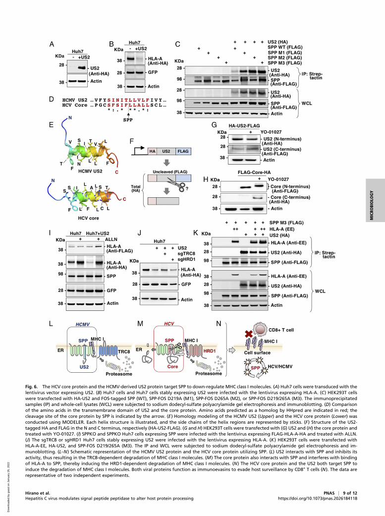
Fig. 6. The HCV core protein and the HCMV-derived US2 protein target SPP to down-regulate MHC class I molecules. (A) Huh7 cells were transduced with thelentivirus vector expressing US2. (B) Huh7 cells and Huh7 cells stably expressing US2 were infected with the lentivirus expressing HLA-A. (C) HEK293T cellswere transfected with HA-US2 and FOS-tagged SPP (WT), SPP-FOS D219A (M1), SPP-FOS D265A (M2), or SPP-FOS D219/265A (M3). The immunoprecipitatedsamples (IP) and whole-cell lysates (WCL) were subjected to sodium dodecyl-sulfate polyacrylamide gel electrophoresis and immunoblotting. (D) Comparisonof the amino acids in the transmembrane domain of US2 and the core protein. Amino acids predicted as a homolog by HHpred are indicated in red; thecleavage site of the core protein by SPP is indicated by the arrow. (E) Homology modeling of the HCMV US2 (Upper) and the HCV core protein (Lower) wasconducted using MODELER. Each helix structure is illustrated, and the side chains of the helix regions are represented by sticks. (F) Structure of the US2-tagged HA and FLAG in the N and C terminus, respectively (HA-US2-FLAG). (G and H) HEK293T cells were transfected with (G) US2 and (H) the core protein andtreated with YO-01027. (I) SPPKO and SPPKO Huh7 cells expressing SPP were infected with the lentivirus expressing FLAG-HLA-A-HA and treated with ALLN.(J) The sgTRC8 or sgHRD1 Huh7 cells stably expressing US2 were infected with the lentivirus expressing HLA-A. (K) HEK293T cells were transfected withHLA-A-EE, HA-US2, and SPP-FOS D219/265A (M3). The IP and WCL were subjected to sodium dodecyl-sulfate polyacrylamide gel electrophoresis and im-munoblotting. (L–N) Schematic representation of the HCMV US2 protein and the HCV core protein utilizing SPP. (L) US2 interacts with SPP and inhibits itsactivity, thus resulting in the TRC8-dependent degradation of MHC class I molecules. (M) The core protein also interacts with SPP and interferes with bindingof HLA-A to SPP, thereby inducing the HRD1-dependent degradation of MHC class I molecules. (N) The HCV core protein and the US2 both target SPP toinduce the degradation of MHC class I molecules. Both viral proteins function as immunoevasins to evade host surveillance by CD8+ T cells (N). The data arerepresentative of two independent experiments.
Hirano et al. PNAS | 9 of 12Hepatitis C virus modulates signal peptide peptidase to alter host protein processing https://doi.org/10.1073/pnas.2026184118
MICRO
BIOLO
GY
Dow
nloa
ded
by g
uest
on
Janu
ary
26, 2
022
100 U/mL penicillin, and 100 μg/mL streptomycin. HCV derived from the JFH-1strain containing adaptive mutations in E2, p7, and NS2 (51) was prepared byserial passages of Huh7.5.1 cells as previously described (29).
Mice. SPPLKO mice were generated by mating SPPfl/fl mice (12) and Alb-Cretransgenic mice, which express the Cre recombinase gene under the albuminpromoter (52). CoreTG (53) and OT-I Tg mice (54) have been previously de-scribed. Mice (8 to 10 wk old) were gender-matched randomly assigned toexperimental groups and were maintained under 12-h light/dark cycle (lightson at 08:00 AM) at 23 ± 2 °C with free access to food and water. All animalexperiments were approved by the Institutional Committee of LaboratoryAnimal Experimentation of the Research Institute for Microbial Diseases,Osaka University (R01-11-0). All efforts were made to minimize animal suf-fering and to reduce the number of animals used in the experiments.
Antibodies and Reagents. The following antibodies were used: horseradishperoxidase–conjugated anti-FLAG mouse monoclonal antibody (Sigma, cloneM2), anti-HA rat monoclonal antibody (Roche, clone 3F10), anti-GFP mousemonoclonal antibody (Clontech, JL-8), anti-actin mouse monoclonal antibody(Sigma, A2228), anti-HCV core mouse monoclonal antibody (Fujirebio), anti-NS5A mouse monoclonal antibody (5A27) (55), anti-EE mouse monoclonalantibody (Covance, MMS-115R), anti-EE rabbit polyclonal antibody (Covance,PRB-115C), anti-glutathione S-transferase goat polyclonal antibody (GEHealthcare, 27-4577-01), anti-human and mouse MHC class I rabbit polyclonalantibodies (Proteintech, 15240-1-AP), anti-human MHC class I monoclonalantibody (Medical & Biological Laboratories Co., Ltd. (MBL); EMR8-5), anti-human MHC class I mouse monoclonal antibody (W6/32) (Santa Cruz Bio-technology, sc-32235), anti-mouse CD8a rat monoclonal antibody (BioLegend,53-6.7), and anti-mouse CD4 rat monoclonal antibody (BioLegend, GK1.5). Theanti-SPP rabbit polyclonal antibody was generated as described previously(12). YO-01027 was synthesized by SYNthesis med chem. LY-411575 (29), cy-cloheximide, and ALLN were purchased from Sigma, whereas RO-0492907 andDAPT were obtained from Selleck Chemicals. Thapsigargin (Wako) and H-2Kb
OVA peptide were purchased from FUJIFILM Wako Pure Chemical Corpora-tion and MBL, respectively. PNGase F was purchased from New England Biolabs(P0704).
Plasmids. The plasmid vectors used are summarized in SI Appendix, Table S1.All complementary DNAs (cDNAs) were amplified using PCR using the Tks GflexDNA Polymerase (Takara-Bio) and cloned into the indicated plasmids using an In-Fusion HD cloning kit (Clontech). The sequences of all plasmids were confirmedusing the ABI Prism 3130 genetic analyzer (Applied Biosystems).
Lentivirus Transduction. HEK293T cells (2 × 10⁶) were seeded on a 10-cm dish andincubated at 37 °C for 24 h. The cells were then transfected with pCMV-VSV-G(1.0 μg), pMDLg/pRRE (1.5 μg), pRSV-Rev (1.5 μg), and lentiviral transfer vector(FUIGW, FUIPW, or lentiCRISPR version 2; 1.5 μg) using the TransIT-LT1 Transfec-tion Reagent (Mirus Bio) according to the manufacturer’s protocol. The super-natant was collected after 24 h post-transfection and passed through a 0.45-μmfilter. For transduction, target cells (2 × 10⁵) were seeded on a 6-well plate andincubated for 24 h, followed by addition of the supernatant containing the len-tivirus and hexadimethrine bromide (4 μg/mL; Sigma). The samples were centri-fuged at 2,500 rpm for 45 min at 32 °C, and the supernatant was replaced with afresh medium 3 h postinfection. To generate stable cell lines, the cells infectedwith the lentivirus were selected using puromycin 48 h postinfection.
Immunoblotting. The cells were washed once with phosphate-buffered saline(PBS) and lysis buffer containing 20 mM Tris-HCl (pH = 7.4), 135 mM NaCl,10% glycerol, 1% Triton X-100, and a protease inhibitor mixture (cComplete,Roche) was added. The cell lysates were incubated for 20 min on ice, and thesupernatants were collected after centrifugation at 15,000 rpm for 5 min at4 °C. The liver tissues were incubated with lysis buffer, homogenized using theBioMasher II Micro Tissue Homogenizer (DWK Life Sciences), and centrifugedat 15,000 rpm for 5 min at 4 °C. Total protein from the supernatants wasquantified using a Bio-Rad Protein Assay Dye Reagent Concentrate (Bio-Rad)according to the manufacturer’s protocol. Equal amounts of proteins weremixed with sodium dodecyl sulfate (SDS) gel-loading buffer (2×) containing50 mM Tris-HCl (pH = 6.8), 4% SDS, 0.2% bromophenol blue, 10% glycerol,and 200 mM β-mercaptoethanol at 4 °C for 1 h, resolved using sodium dodecyl-sulfate polyacrylamide gel electrophoresis (NuPAGE gel, Life Technologies),transferred onto nitrocellulose membranes (iBlot, Life Technologies), blockedwith PBS containing 0.05% Tween20 (PBS-T) supplemented with 5% skim milk
for 1 h, and incubated with the primary antibodies (1:2,000 dilution) at 4 °C for24 h. After washing three times with PBS-T, the blots were incubated withsecondary antibodies (1:2,000 dilution). The immune complex was visualizedusing the Super Signal West Femto substrate (Pierce) and detected using theLAS-4000 mini image analyzer system (FUJIFILM). The signal intensity of theproteins was calculated using the Multi Gauge software (FUJIFILM).
Immunoprecipitation. HEK293T cells (2 × 10⁶) were seeded on a 10-cm dish andincubated at 37 °C for 24 h. The cells were transfected with the plasmids vialiposome-mediated transfection using polyethyleneimine (40 μL; 1 mg/mL, mo-lecular weight, 25,000; Polysciences, Inc.). The mixtures were incubated for20 min, added to HEK293T cells, and incubated for 48 h. The cell lysates wereincubated with the antibodies at 4 °C for 24 h and with Protein G Sepharose 4B(GE Healthcare) at 4 °C for 1 h. The beads were washed five times with the lysisbuffer, boiled at 60 °C for 20 min with the sample buffer, and subjected toimmunoblotting.
Generation of SPP, TRC8, and HRD1-Knockout Cell Lines. To construct thesingle-guide RNA targeting the SPP, TRC8, and HRD1, the targeting se-quences were designed using three different sequences for each gene aspreviously described (56), synthesized using DNA oligos (Eurofins Genetics),and cloned into the lentiCRISPR version 2 (Addgene, no. 52961) digested byBmsBI (New England Biolab). The following target sequences were used:HRD1 5′-GGAAGACAAGGACAAAGGCC-3′, 5′-GTGAAGAGTGCAACAAAG-CG-3′, and 5′-GAAAGAGCCAGGAGATGTTG-3′; TRC8 5′-GCGCCGCCCAGA-CCTGCTGA-3′, 5′-GCTTTGGCTGGAATCCGGGT-3′, and 5′-GGTGCGGATGGC-CCATCAGC-3′; and SPP 5′-GCCCTCAGCGATCCGCATAA-3′, 5′-CACGCCCGAGGG-CATCGCGC-3′, and 5′-GTCCATGTATTTCTTCGTGC-3′. Lentiviruses expressingthree kinds of target sequences per gene were mixed, introduced into thetarget cells, and maintained in a culture medium supplemented with 1 μg/mLpuromycin for 3 wk.
Flow Cytometry. Cells (2 × 10⁵) were collected and resuspended in fluorescence-activated cell sorting (FACS) buffer containing 2% FBS in PBS and incubatedwith the anti-MHC class I antibody (W6/32; Santa Cruz Biotechnology, sc-32235) at 4 °C for 30 min. After washing twice with the FACS buffer, the cellswere further incubated at 4 °C for 30 min with the AF488-conjugated anti-mouse antibody and washed with the FACS buffer. The surface expression ofMHC I was measured using FACSAria (BD Immunocytometry System) and an-alyzed using FlowJo (FlowJo LLC).
QuantitativeRT-PCR.Total RNAwasextracted from the liver lysates using ISOGEN II(Nippon Gene) according to the manufacturer’s protocol. Subsequently, cDNAswere synthesized using a high-capacity RNA-to-cDNA kit (Applied Biosystems)according to the manufacturer’s instructions and those of HLA-A, IFNα1, IFNβ1,IL6, TNFα, IP10, CCL5, and β-actin were quantified using Fast SYBR Green MasterMix (Thermo Fisher Scientific) and ViiA7 RT-PCR system (Thermo Fisher Scientific).The following primers were used: HLA-A, 5′-GGCCCTGACCCAGACCTG-3′ and 5′-GCACGAACTGCGTGTCGTC-3′; IFNα1, 5′-AGCCTTGACACTCCTGGTACAAATG-3′and 5′-TGGGTCAGCTCACTCAGGACA-3′; IFNβ1, 5′-ACACCAGCCTGGCTTCCATC-3′and 5′-TTGGAGCTGGAGCTGCTTATAGTTG-3′; IL6, 5′-CCACTTCACAAGTCGGAG-GCTTA-3′ and 5′-GCAAGTGCATCATCGTTGTTCATAC-3′; TNFα, 5′-CAGGAGGGA-GAACAGAAACTCCA-3′ and 5′-CCTGGTTGGCTGCTTGCTT-3′; IP10, 5′-ACACCA-GCCTGGCTTCCATC-3′ and 5′-TTGGAGCTGGAGCTGCTTATAGTTG-3′; CCL5, 5′-AGATCTCTGCAGCTGCCCTCA-3′ and 5′-GGAGCACTTGCTGCTGGTGTAG-3′; andβ-actin, 5′-TTGCTGACAGGATGCAGAAG-3′ and 5′-GTACTTGCGCTCAGGAGGAG-3′. The relative mRNA expression was calculated using the delta-delta Ct method,with β-actin as the internal control.
Luciferase Assay. Cells were seeded on a 24-well plate and incubated at 37 °Cfor 24 h. pERAI-Luc, pRL-SV40 and the HCV core, HLA-A, Prolactin, HO-1, orXBP1u were transfected into the cells using TransIT-LT1 (Mirus Bio) accordingto the manufacturer’s protocol. The cells were incubated for 24 h aftertransfection, and luciferase activity was detected using the Dual-LuciferaseReporter Assay System (Promega) according to the manufacturer’s protocol.
In Vitro Antigen Presentation Assay. CD8+ T cells derived from the spleen ofOT-I transgenic mice were isolated using a CD8+ T cell Isolation kit (MiltenyiBiotec). MEFs expressing GFP alone or GFP and the core protein were identi-fied using a cell sorter (SONY SH800S). Hepatocytes derived from the WT orCoreTg were isolated using a perfusion method described previously (57).Briefly, the mice were euthanized by inhalation of isoflurane anesthesia, and
10 of 12 | PNAS Hirano et al.https://doi.org/10.1073/pnas.2026184118 Hepatitis C virus modulates signal peptide peptidase to alter host protein processing
Dow
nloa
ded
by g
uest
on
Janu
ary
26, 2
022

livers were perfused using Hank’s balanced salt solution containing 0.5 mMegtazic acid followed by perfusion using a liver digest medium (ThermoFisher). Isolated hepatocytes were purified using a Percoll gradient. MEFs orhepatocytes (2 × 10⁴) were seeded on a 96-well plate, incubated for 24 h, andfurther incubated with the H-2Kb OVA peptide (1.0 nM; TS-50001-P, MBL LifeScience) for 7 h. The cells were washed once with PBS and fixed with 0.01%glutaraldehyde in PBS for 30 s. The reaction was quenched by adding 0.2 Mglycine in PBS, and the cells were washed twice with PBS and once withRoswell Park Memorial Institute-1640 supplemented with 10% FBS, 2 mML-Glutamine (Gibco), 1 mM sodium pyruvate (Wako), 100 U/mL penicillin, and100 μg/mL streptomycin and cocultured with CD8+ T cells. Culture supernatantswere collected after 72 h, and IFN-γ production was quantified.
Enzyme-Linked Immunosorbent Assay. IFN-γ in culture supernatants of mouseCD8+ T cells were quantified using enzyme-linked immunosorbent assay MAXDeluxe Set Mouse IFN-γ (BioLegend) according to the manufacturer’s protocol.
Preparation of Human Liver Tissues. Liver samples were obtained from 18 pa-tients with HCC who underwent curative liver resection at the Cancer InstituteHospital of Japanese Foundation for Cancer Research. The study protocoladhered to the ethical guidelines of human clinical research established by theJapaneseMinistry of Health, Labor andWelfare andwas approved by the ethicscommittees of the participating facilities (NCGM-A-000227, Japanese Foun-dation for Cancer Research under Clinical Research Number 2017-1118).Written informed consent was obtained from all patients during enrollment.
Freshly frozen tissue samples from normal adjacent tissues were obtainedfrompatientswith livermetastasis of colon cancer, chronic hepatitis C infection,SVR, or nonalcoholic fatty liver disease (SI Appendix, Table S2). The liver tissueswere added to the lysis buffer, homogenized using a BioMasher II Micro TissueHomogenizer (DWK Life Sciences), and centrifuged at 15,000 rpm for 5 min at4 °C. The total protein concentration of the supernatants was quantified usinga Bio-Rad Protein Assay Dye Reagent Concentrate (Bio-Rad) according to themanufacturer’s protocol, and equal amounts of proteins were subjected toimmunoblotting.
Histological Analysis. Liver tissues were fixed with 10% formalin, impregnatedwith 30% sucrose, and frozen with the Tissue-Tek OCT compound (SakuraFinetek). Next, 10-μm–thick tissue sections were prepared and stained with he-matoxylin and eosin. Additionally, Oil red O staining was performed on frozensections that were washed once with running tap water for 5 min, rinsed with60% isopropanol for 1 min, stained with the staining solution for 15 min, rinsedwith 60% isopropanol, and subjected to light staining of the nuclei with he-matoxylin for 15 s. All sections were observed under a microscope (Olympus).
Homology Modeling of the Core and US2 Proteins. Structural information forthe HCV core protein (JFH1 strain) was obtained from the Protein Data Bank(PDB ID code: 2LIF) andwas used as a template tomodel the core protein (J1 strain)and US2 protein, and their sequences were aligned using HHpred. Their structuralmodels were built using theMODELER program, and the images were constructedusing the open-source PyMOL Molecular Graphics System version 1.8.6.0.
Quantification and Statistical Analysis. All statistical analyses were conductedusing the GraphPad Prism version 8.4.3 (GraphPad Software). Student’s t testwas used to determine significant differences, and the statistical details ofthe experiments are indicated in the figure legends.
Data Availability. All study data are included in the article and/or supportinginformation.
ACKNOWLEDGMENTS. We are grateful to M. Tomiyama and K. Tanii for theirsecretarial work. We also thank D.C.S. Huang, R. Bartenschlager, F. Chisari, andT. Wakita for providing the experimental materials and the Core InstrumentationFacility of theOsakaUniversity for performing cell sorting.We thank Editage (https://www.editage.com/) for English language editing. This work was funded by theJapan Agency for Medical Research and Development (Grants 18fk0210206h0003,17fk0210305h0003, 18fk0210210h0003, 18fk0210209h0503, 17fk0210304h0003,20fk0210074h0001, 20fk0210055h0002, and 20wm0325009s0101); the Ministry ofEducation, Culture, Sports, Science, and Technology of Japan (Grants 16H06432,16H06429, 16K21723, 19H03479, 20H03495, and 19J12369); the Takeda ScienceFoundation; The Naito Foundation; SuzukenMemorial Foundation; and DaiichiSankyo Foundation of Life Science.
1. B. Maasoumy, H. Wedemeyer, Natural history of acute and chronic hepatitis C. Best
Pract. Res. Clin. Gastroenterol. 26, 401–412 (2012).
2. A. Galossi, R. Guarisco, L. Bellis, C. Puoti, Extrahepatic manifestations of chronic HCV
infection. J. Gastrointestin. Liver Dis. 16, 65–73 (2007).
3. S. L. Chen, T. R. Morgan, The natural history of hepatitis C virus (HCV) infection. Int.
J. Med. Sci. 3, 47–52 (2006).
4. M. H. Heim, R. Thimme, Innate and adaptive immune responses in HCV infections.
J. Hepatol. 61 (suppl.(1), S14–S25 (2014).
5. A. B. Jazwinski, A. J. Muir, Direct-acting antiviral medications for chronic hepatitis C
virus infection. Gastroenterol. Hepatol. (N. Y.) 7, 154–162 (2011).
6. F. Conti et al., Early occurrence and recurrence of hepatocellular carcinoma in HCV-
related cirrhosis treated with direct-acting antivirals. J. Hepatol. 65, 727–733 (2016).
7. J. Hengst et al., Nonreversible MAIT cell-dysfunction in chronic hepatitis C virus in-
fection despite successful interferon-free therapy. Eur. J. Immunol. 46, 2204–2210
(2016).
8. B. Langhans et al., Increased peripheral CD4+ regulatory T cells persist after successful
direct-acting antiviral treatment of chronic hepatitis C. J. Hepatol. 66, 888–896 (2017).
9. R. Bartenschlager, V. Lohmann, F. Penin, The molecular and structural basis of ad-
vanced antiviral therapy for hepatitis C virus infection. Nat. Rev. Microbiol. 11,
482–496 (2013).
10. Y. Mori, K. Moriishi, Y. Matsuura, Hepatitis C virus core protein: Its coordinate roles
with PA28gamma in metabolic abnormality and carcinogenicity in the liver. Int.
J. Biochem. Cell Biol. 40, 1437–1442 (2008).
11. R. M. Gemmill et al., The hereditary renal cell carcinoma 3;8 translocation fuses FHIT
to a patched-related gene, TRC8. Proc. Natl. Acad. Sci. U.S.A. 95, 9572–9577 (1998).
12. S. Aizawa et al., TRC8-dependent degradation of hepatitis C virus immature core
protein regulates viral propagation and pathogenesis. Nat. Commun. 7, 11379 (2016).
13. M. K. Lemberg, F. A. Bland, A. Weihofen, V. M. Braud, B. Martoglio, Intramembrane
proteolysis of signal peptides: An essential step in the generation of HLA-E epitopes.
J. Immunol. 167, 6441–6446 (2001).
14. J. M. Boname et al., Cleavage by signal peptide peptidase is required for the deg-
radation of selected tail-anchored proteins. J. Cell Biol. 205, 847–862 (2014).
15. C.-Y. Chen et al., Signal peptide peptidase functions in ERAD to cleave the unfolded
protein response regulator XBP1u. EMBO J. 33, 2492–2506 (2014).
16. B. Martoglio, R. Graf, B. Dobberstein, Signal peptide fragments of preprolactin and
HIV-1 p-gp160 interact with calmodulin. EMBO J. 16, 6636–6645 (1997).
17. J. Levitskaya et al., Inhibition of antigen processing by the internal repeat region of
the Epstein-Barr virus nuclear antigen-1. Nature 375, 685–688 (1995).
18. N. P. Dantuma, S. Heessen, K. Lindsten, M. Jellne, M. G. Masucci, Inhibition of pro-
teasomal degradation by the gly-Ala repeat of Epstein-Barr virus is influenced by the
length of the repeat and the strength of the degradation signal. Proc. Natl. Acad. Sci.
U.S.A. 97, 8381–8385 (2000).
19. K. Früh et al., A viral inhibitor of peptide transporters for antigen presentation.
Nature 375, 415–418 (1995).
20. K. Ahn et al., The ER-luminal domain of the HCMV glycoprotein US6 inhibits peptide
translocation by TAP. Immunity 6, 613–621 (1997).
21. P. J. Lehner, J. T. Karttunen, G. W. Wilkinson, P. Cresswell, The human cytomegalo-
virus US6 glycoprotein inhibits transporter associated with antigen processing-
dependent peptide translocation. Proc. Natl. Acad. Sci. U.S.A. 94, 6904–6909 (1997).
22. E. M. Bennett, J. R. Bennink, J. W. Yewdell, F. M. Brodsky, Cutting edge: Adenovirus
E19 has two mechanisms for affecting class I MHC expression. J. Immunol. 162,
5049–5052 (1999).
23. J. F. Roeth, M. Williams, M. R. Kasper, T. M. Filzen, K. L. Collins, HIV-1 Nef disrupts
MHC-I trafficking by recruiting AP-1 to the MHC-I cytoplasmic tail. J. Cell Biol. 167,
903–913 (2004).
24. J. R. Francica et al., Steric shielding of surface epitopes and impaired immune rec-
ognition induced by the ebola virus glycoprotein. PLoS Pathog. 6, e1001098 (2010).
25. H. R. Stagg et al., The TRC8 E3 ligase ubiquitinates MHC class I molecules before
dislocation from the ER. J. Cell Biol. 186, 685–692 (2009).
26. J. M. Boname, P. G. Stevenson, MHC class I ubiquitination by a viral PHD/LAP finger
protein. Immunity 15, 627–636 (2001).
27. X. Wang et al., Ubiquitination of serine, threonine, or lysine residues on the cyto-
plasmic tail can induce ERAD of MHC-I by viral E3 ligase mK3. J. Cell Biol. 177, 613–624
(2007).
28. D. J. H. van den Boomen et al., TMEM129 is a Derlin-1 associated ERAD E3 ligase
essential for virus-induced degradation of MHC-I. Proc. Natl. Acad. Sci. U.S.A. 111,
11425–11430 (2014).
29. J. Hirano et al., Characterization of SPP inhibitors suppressing propagation of HCV
and protozoa. Proc. Natl. Acad. Sci. U.S.A. 114, E10782–E10791 (2017).
30. E. A. Hughes, C. Hammond, P. Cresswell, Misfolded major histocompatibility complex
class I heavy chains are translocated into the cytoplasm and degraded by the pro-
teasome. Proc. Natl. Acad. Sci. U.S.A. 94, 1896–1901 (1997).
31. M. L. Burr et al., HRD1 and UBE2J1 target misfolded MHC class I heavy chains for en-
doplasmic reticulum-associated degradation. Proc. Natl. Acad. Sci. U.S.A. 108, 2034–2039
(2011).
Hirano et al. PNAS | 11 of 12Hepatitis C virus modulates signal peptide peptidase to alter host protein processing https://doi.org/10.1073/pnas.2026184118
MICRO
BIOLO
GY
Dow
nloa
ded
by g
uest
on
Janu
ary
26, 2
022
32. R. T. Timms et al., Genetic dissection of mammalian ERAD through comparative
haploid and CRISPR forward genetic screens. Nat. Commun. 7, 11786 (2016).
33. T. Tanaka et al., Hallmarks of hepatitis C virus in equine hepacivirus. J. Virol. 88,
13352–13366 (2014).
34. J. Loureiro et al., Signal peptide peptidase is required for dislocation from the en-
doplasmic reticulum. Nature 441, 894–897 (2006).
35. K. M. Paulsson et al., Distinct differences in association of MHC class I with endo-
plasmic reticulum proteins in wild-type, and beta 2-microglobulin- and TAP-deficient
cell lines. Int. Immunol. 13, 1063–1073 (2001).
36. L. Mancino, S. M. Rizvi, P. E. Lapinski, M. Raghavan, Calreticulin recognizes misfolded
HLA-A2 heavy chains. Proc. Natl. Acad. Sci. U.S.A. 99, 5931–5936 (2002).
37. Y. Zhang, E. Baig, D. B. Williams, Functions of ERp57 in the folding and assembly of
major histocompatibility complex class I molecules. J. Biol. Chem. 281, 14622–14631
(2006).
38. J. A. Lindquist, O. N. Jensen, M. Mann, G. J. Hämmerling, ER-60, a chaperone with thiol-
dependent reductase activity involved in MHC class I assembly. EMBO J. 17, 2186–2195
(1998).
39. E. Rufer, R. M. Leonhardt, M. R. Knittler, Molecular architecture of the TAP-associated
MHC class I peptide-loading complex. J. Immunol. 179, 5717–5727 (2007).
40. M. L. Burr et al., MHC class I molecules are preferentially ubiquitinated on endo-
plasmic reticulum luminal residues during HRD1 ubiquitin E3 ligase-mediated dislo-
cation. Proc. Natl. Acad. Sci. U.S.A. 110, 14290–14295 (2013).
41. A. Cauli et al., [The role of HLA-B27 molecules in the pathogenesis of ankylosing
spondylitis]. Reumatismo 54, 266–271 (2002).
42. R. A. Colbert, T. M. Tran, G. Layh-Schmitt, HLA-B27 misfolding and ankylosing
spondylitis. Mol. Immunol. 57, 44–51 (2014).
43. A. Waheed et al., Hereditary hemochromatosis: Effects of C282Y and H63D mutations on
associationwith beta2-microglobulin, intracellular processing, and cell surface expression of
the HFE protein in COS-7 cells. Proc. Natl. Acad. Sci. U.S.A. 94, 12384–12389 (1997).
44. X. Shi et al., Bunyamwera orthobunyavirus glycoprotein precursor is processed by
cellular signal peptidase and signal peptide peptidase. Proc. Natl. Acad. Sci. U.S.A.
113, 8825–8830 (2016).
45. S. J. Allen et al., Binding of HSV-1 glycoprotein K (gK) to signal peptide peptidase
(SPP) is required for virus infectivity. PLoS One 9, e85360 (2014).
46. S. Wang, H. Ghiasi, Absence of signal peptide peptidase, an essential herpes simplex
virus 1 glycoprotein K binding partner, reduces virus infectivity in vivo. J. Virol. 93,
e01309-19 (2019).
47. S.-R. Lee, B. Nanduri, G. T. Pharr, J. V. Stokes, L. M. Pinchuk, Bovine viral diarrhea virus
infection affects the expression of proteins related to professional antigen presen-
tation in bovine monocytes. Biochim. Biophys. Acta 1794, 14–22 (2009).
48. C. Burrells et al., Lymphocyte subpopulations in the blood of sheep persistently in-
fected with border disease virus. Clin. Exp. Immunol. 76, 446–451 (1989).
49. L. L. Lanier, NK cell recognition. Annu. Rev. Immunol. 23, 225–274 (2005).
50. P. Rodrigues et al., Comparative study between Hfe-/- and beta2m-/- mice: Progres-
sion with age of iron status and liver pathology. Int. J. Exp. Pathol. 87, 317–324 (2006).
51. R. S. Russell et al., Advantages of a single-cycle production assay to study cell culture-
adaptive mutations of hepatitis C virus. Proc. Natl. Acad. Sci. U.S.A. 105, 4370–4375
(2008).
52. T. Kodama et al., Increases in p53 expression induce CTGF synthesis by mouse and
human hepatocytes and result in liver fibrosis in mice. J. Clin. Invest. 121, 3343–3356
(2011).
53. K. Moriya et al., Hepatitis C virus core protein induces hepatic steatosis in transgenic
mice. J. Gen. Virol. 78, 1527–1531 (1997).
54. Y. Lee et al., p62 plays a specific role in interferon-γ-induced presentation of a
toxoplasma vacuolar antigen. Cell Rep. 13, 223–233 (2015).
55. K. Okamoto et al., Intramembrane processing by signal peptide peptidase regulates
the membrane localization of hepatitis C virus core protein and viral propagation.
J. Virol. 82, 8349–8361 (2008).
56. N. E. Sanjana, O. Shalem, F. Zhang, Improved vectors and genome-wide libraries for
CRISPR screening. Nat. Methods 11, 783–784 (2014).
57. Y. Itoh et al., Salt-inducible kinase 3 signaling is important for the gluconeogenic
programs in mouse hepatocytes. J. Biol. Chem. 290, 17879–17893 (2015).
12 of 12 | PNAS Hirano et al.https://doi.org/10.1073/pnas.2026184118 Hepatitis C virus modulates signal peptide peptidase to alter host protein processing
Dow
nloa
ded
by g
uest
on
Janu
ary
26, 2
022


![The Signal Peptide Peptidase Is Required for Pollen Function in Arabidopsis1[C]](https://static.fdocuments.net/doc/165x107/61fb358d2e268c58cd5b73dc/the-signal-peptide-peptidase-is-required-for-pollen-function-in-arabidopsis1c.jpg)