
Hepatic Steatosis Index in Acromegaly: Correlation...
8
Research Article Hepatic Steatosis Index in Acromegaly: Correlation with Insulin Resistance Regardless of the Disease Control Alessandro Ciresi, Valentina Guarnotta, Daniela Campo, and Carla Giordano Section of Endocrinology, Biomedical Department of Internal and Specialist Medicine (DIBIMIS), University of Palermo, Piazza delle Cliniche 2, 90127 Palermo, Italy Correspondence should be addressed to Carla Giordano; [email protected] Received 16 May 2018; Revised 17 November 2018; Accepted 27 November 2018; Published 19 December 2018 Academic Editor: Maria C. Meriggiola Copyright © 2018 Alessandro Ciresi et al. This is an open access article distributed under the Creative Commons Attribution License, which permits unrestricted use, distribution, and reproduction in any medium, provided the original work is properly cited. Objective. In acromegaly, both lipotoxicity secondary to GH excess and insulin resistance have a significant impact on the liver. Ultrasonography has shown poor sensitivity in detecting hepatic steatosis and noninvasive methods have been proposed. We evaluated the hepatic steatosis index (HSI), a validated surrogate index of hepatic steatosis, and we correlated it with disease activity and insulin resistance. Design. Thirty-one patients with newly diagnosed acromegaly were studied at diagnosis and after 12 months of treatment with somatostatin receptor ligands. Methods. Glucose and insulin levels, surrogate estimates of insulin sensitivity, and hepatic steatosis through ultrasonography and HSI were evaluated. Results. At diagnosis, ultrasonography documented steatosis in 19 patients (61.2%) while 26 (83.8%) showed high HSI. After 12 months, both GH (p =0 033) and IGF-1 (p <0 001) significantly decreased and, overall, 58% of patients were classified as controlled. Ultrasonography documented steatosis in all the same initial 19 patients, while only 14 patients (45.1%) showed high HSI (p <0 001). A significant reduction in HOMA-IR (p =0 002) and HSI (p <0 001) and increased ISI Matsuda (p <0 001), was documented. The change of HSI from baseline to 12 months was found to be directly correlated with the change of ISI (Rho -0.611; p =0 004) while no correlation was found with the change of GH or IGF-1 levels and other parameters. Conclusions. In acromegaly, HSI is mainly related with insulin resistance and the reduction of GH and IGF-1 levels, and above all the improvement in insulin sensitivity leads to an improvement of this surrogate index of hepatic steatosis. 1. Introduction Nonalcoholic fatty liver disease (NAFLD) is the most com- mon liver disorder in developed countries, characterized by elevated serum levels of free fatty acids and fatty infiltration of the liver. NAFLD consists of a wide spectrum of condi- tions, ranging from simple steatosis to nonalcoholic steatohe- patitis (NASH), and diagnosis of NASH is based on the histologic examination of liver biopsy specimens [1]. NAFLD, which represents a further expression of meta- bolic syndrome being strictly linked to obesity, diabetes mel- litus, and insulin resistance (IR), has been found to increase the risk for cardiovascular disease, and IR was proposed as a pathogenic factor of NASH through an accumulation of fat within a hepatocyte [2, 3]. In addition, it is well-known that the liver is one of the important target tissues of growth hormone (GH) [4]. GH has physiological effects on glucose metabolism by inducing gluconeogenesis and glycogenolysis and promoting insulin resistance both in the liver and the periphery. In addi- tion, the well-known lipolytic effects of GH stimulate release of free fatty acids (FFA) from the adipose tissue leading to glucose-fatty acid substrate competition and decreased glu- cose utilisation [5, 6]. Given the main role played by GH in glucose and lipid metabolism, several lines of evidence suggest that GH sta- tus is implicated in the development of intrahepatic lipid accumulation. Indeed, both GH excess and deficiency are associated with metabolic disturbances and lipid accumula- tion in the liver. Low GH levels are associated with hepatic Hindawi International Journal of Endocrinology Volume 2018, Article ID 5421961, 7 pages https://doi.org/10.1155/2018/5421961
Transcript of Hepatic Steatosis Index in Acromegaly: Correlation...
Research ArticleHepatic Steatosis Index in Acromegaly: Correlation with InsulinResistance Regardless of the Disease Control
Alessandro Ciresi, Valentina Guarnotta, Daniela Campo, and Carla Giordano
Section of Endocrinology, Biomedical Department of Internal and Specialist Medicine (DIBIMIS), University of Palermo, Piazza delleCliniche 2, 90127 Palermo, Italy
Correspondence should be addressed to Carla Giordano; [email protected]
Received 16 May 2018; Revised 17 November 2018; Accepted 27 November 2018; Published 19 December 2018
Academic Editor: Maria C. Meriggiola
Copyright © 2018 Alessandro Ciresi et al. This is an open access article distributed under the Creative Commons AttributionLicense, which permits unrestricted use, distribution, and reproduction in any medium, provided the original work isproperly cited.
Objective. In acromegaly, both lipotoxicity secondary to GH excess and insulin resistance have a significant impact on the liver.Ultrasonography has shown poor sensitivity in detecting hepatic steatosis and noninvasive methods have been proposed. Weevaluated the hepatic steatosis index (HSI), a validated surrogate index of hepatic steatosis, and we correlated it with diseaseactivity and insulin resistance. Design. Thirty-one patients with newly diagnosed acromegaly were studied at diagnosis and after12 months of treatment with somatostatin receptor ligands. Methods. Glucose and insulin levels, surrogate estimates of insulinsensitivity, and hepatic steatosis through ultrasonography and HSI were evaluated. Results. At diagnosis, ultrasonographydocumented steatosis in 19 patients (61.2%) while 26 (83.8%) showed high HSI. After 12 months, both GH (p = 0 033)and IGF-1 (p < 0 001) significantly decreased and, overall, 58% of patients were classified as controlled. Ultrasonographydocumented steatosis in all the same initial 19 patients, while only 14 patients (45.1%) showed high HSI (p < 0 001). Asignificant reduction in HOMA-IR (p = 0 002) and HSI (p < 0 001) and increased ISI Matsuda (p < 0 001), was documented. Thechange of HSI from baseline to 12 months was found to be directly correlated with the change of ISI (Rho -0.611; p = 0 004)while no correlation was found with the change of GH or IGF-1 levels and other parameters. Conclusions. In acromegaly, HSI ismainly related with insulin resistance and the reduction of GH and IGF-1 levels, and above all the improvement in insulinsensitivity leads to an improvement of this surrogate index of hepatic steatosis.
1. Introduction
Nonalcoholic fatty liver disease (NAFLD) is the most com-mon liver disorder in developed countries, characterized byelevated serum levels of free fatty acids and fatty infiltrationof the liver. NAFLD consists of a wide spectrum of condi-tions, ranging from simple steatosis to nonalcoholic steatohe-patitis (NASH), and diagnosis of NASH is based on thehistologic examination of liver biopsy specimens [1].
NAFLD, which represents a further expression of meta-bolic syndrome being strictly linked to obesity, diabetes mel-litus, and insulin resistance (IR), has been found to increasethe risk for cardiovascular disease, and IR was proposed asa pathogenic factor of NASH through an accumulation offat within a hepatocyte [2, 3]. In addition, it is well-known
that the liver is one of the important target tissues of growthhormone (GH) [4].
GH has physiological effects on glucose metabolism byinducing gluconeogenesis and glycogenolysis and promotinginsulin resistance both in the liver and the periphery. In addi-tion, the well-known lipolytic effects of GH stimulate releaseof free fatty acids (FFA) from the adipose tissue leading toglucose-fatty acid substrate competition and decreased glu-cose utilisation [5, 6].
Given the main role played by GH in glucose and lipidmetabolism, several lines of evidence suggest that GH sta-tus is implicated in the development of intrahepatic lipidaccumulation. Indeed, both GH excess and deficiency areassociated with metabolic disturbances and lipid accumula-tion in the liver. Low GH levels are associated with hepatic
HindawiInternational Journal of EndocrinologyVolume 2018, Article ID 5421961, 7 pageshttps://doi.org/10.1155/2018/5421961
steatosis in patients with NAFLD [7], and GH-deficientadults have a higher incidence of NAFLD [8]. Therefore,NAFLD has emerged as an important form of comorbidityin GH deficiency and it seems to reverse after GH replace-ment therapy [9].
Conversely, in acromegaly GH excess has a significantimpact on adipose tissue and a detrimental effect on glucosemetabolism and insulin signaling both at the hepatic andextrahepatic levels. The increased lipolysis in acromegalyresults in high circulating FFA levels that may alter adiposetissue deposition, leading to the development of IR [10].Therefore, in active acromegaly, while body fat depots arediminished, IR located at both hepatic and extrahepatic levelsis increased [11].
However, in acromegaly increased lipolysis and IR theo-retically have opposite effects on the NAFLD. Indeed, if thelipolytic action of GH may lead to a reduction in visceraland subcutaneous adipose tissue in patients with active dis-ease [12, 13], on the other hand it could be hypothesized thatfat deposition in the liver and muscle may represent one ofthe mechanisms involved in the pathogenesis of IR [14–16],which is impacted by the GH status [17], although there isalso evidence against intrahepatic lipid accumulation as oneof the underlying mechanisms [18–20].
To date, ultrasonography has shown poor sensitivity indetecting hepatic steatosis and cannot accurately quantifythe amount of hepatic fat present [3].
The hepatic steatosis index (HSI) is a simple and efficientscreening tool for NAFLD that may be utilized for selectingindividuals for liver ultrasonography [21]. HSI discriminateswell between patients with and without hepatic steatosis [22].In addition, HSI has been found to show a significant corre-lation with fatty liver grade measured by ultrasonographyand this finding suggests that HSI reflects not only the pres-ence of NAFLD but also its degree [21]. Therefore, HSI hasbeen proposed as a diagnostic tool for predicting the presenceof NAFLD with reliable accuracy and an overall good diag-nostic performance [23].
In light of this, we aimed to evaluate the HSI in a group ofnaive acromegalic patients and to correlate it with diseaseactivity and IR.
2. Subjects and Methods
From a total of 75 patients affected by acromegaly whoreferred at the Endocrinology Section of the University ofPalermo from January 2009 to December 2017, we enrolled31 consecutive patients (17 males and 14 females; aged 51± 12 yr, range 32-77 yr) with active newly diagnosed acro-megaly and who refused first-line pituitary surgery.
Twenty-eight patients previously treated with surgery oralready treated with medical therapy for acromegaly, as wellas 4 patients with mixed secreting adenoma or with defi-ciency of one or more anterior pituitary hormones, wereexcluded from this study to avoid any impact on the meta-bolic parameters evaluated. In addition, we excluded 11patients (in addition to 23 already excluded due to previoussurgery) with a previous diagnosis of diabetes mellitus forthe same reasons mentioned above and also because they
could not undergo oral glucose tolerance test (OGTT) and1 patient (in addition to 14 already excluded) treated withlipid-lowering drugs. All patients affected by impaired fastingglucose (IFG) or impaired glucose tolerance (IGT) weretreated with diet alone. No patients with previous diagnosisof IFG or IGT recruited in the study were already receivingpharmacological treatment.
Disease activity was confirmed by elevated age- andgender-corrected plasma IGF-1 levels and nonsuppressibleGH and after OGTT, while the biochemical control of acro-megaly during treatment was defined by normal age-adjusted IGF-1 levels and randomGH levels < 1 μg/L [24, 25].
In all patients, the MRI scan revealed the presence of apituitary tumor. The mean duration of the disease was estab-lished by patient interview, patient clinical pictures, andonset of osteoarticular symptoms.
All patients underwent first-line medical treatmentwith depot somatostatin receptor ligands (SRL). Specifically,19 (61%) received octreotide long-acting release (LAR) (20-40mg every 28 days) and 12 (39%) lanreotide autogel(ATG) (60-120mg every 28 days). Octreotide was started ata dose of 20mg/28 days and lanreotide at a dose of 60mg/28 days, and dosages were titrated on the basis of GH andIGF-1 at month 3 or 6. After 12 months, in the octreotidegroup, 10 patients practiced the monthly dose of 20mg, 5patients 30mg, and 4 patients 40mg; in the lanreotide-group,5 patients had ATG 60mg, 5 the monthly dose of 90mg, and2 patients 120mg.
At the time of hospitalization, all patients signed a con-sent form for the scientific use of their data after a full expla-nation of the purpose of the study. This study was approvedby the Institutional Review Board of the Faculty of Medicine,University of Palermo.
2.1. Study Design. Body mass index (BMI), waist circumfer-ence (WC), systolic blood pressure (SBP), and diastolic bloodpressure (DBP) were measured in all patients. WC was mea-sured at the midpoint between the lower rib and the iliaccrest. A 12-hour overnight fasting blood sample was drawnto measure lipid profile (total, high-density lipoprotein(HDL), and low-density lipoprotein (LDL) cholesterol andtriglycerides (TG)), hemoglobin A1c (HbA1c), alanine(ALT) and aspartate (AST) aminotransferase, γ-glutamyl-transferase (GGT), alkaline phosphatase (ALP), total biliru-bin, and IGF-1 levels. To normalize IGF-1 for age inindividual patients, we calculated the ratio between theIGF-1 level and the upper limit of the normal (ULN) rangefor age (normal ≤ 1) and the data are presented as IGF-1ULN. OGTT was performed by measuring plasma blood glu-cose, insulin, and GH levels every 30min for 2 h after a 75 goral glucose load. As surrogate estimates of basal insulin sen-sitivity, we calculated the homeostasis model assessment ofIR (HOMA-IR) index [26], while stimulated insulin sensitiv-ity was measured using the insulin sensitivity index (ISI), acomposite index derived from the OGTT and validated byMatsuda and DeFronzo [27].
According to the Adult Treatment Panel III criteria,patients were classified as having abdominal obesity (definedas WC> 102 cm in males and >88 cm in females), reduced
2 International Journal of Endocrinology
HDL (defined as HDL levels < 1 04 mmol/L in malesand <1.30mmol/L in females), raised TG (defined as TGlevels ≥ 1 7 mmol/L), elevated blood pressure (definedas ≥130/≥85mmHg), and raised plasma glucose (defined asfasting glucose ≥ 6 1 mmol/L) [28].
Steatosis was diagnosed by an abdominal ultrasound,routinely performed in all acromegalic patients to evaluatevisceromegaly. Ultrasound assessment was performed infasting subjects on the day of enrollment in the study alwaysby the same radiologist trained for ultrasound techniquesand particularly dedicated to liver examination who wasunaware of patients’ history and aim of the current study.A real-time Hitachi H21 apparatus with a 2 to 5MHz, con-vex, multifrequency probe was used.
In addition, we calculated the HSI with the followingformula:
hepatic steatosis index HSI = 8 × ALT AST ratio+ BMI +2, if female ,
1
andpatients were considered to have steatosis ifHSI > 36 [21].
2.2. Hormone and Biochemical Assays. All biochemical datawere collected after overnight fasting. Fasting glucose, ALT,AST, GGT, ALP, bilirubin, and lipid levels were measuredin our centralized accredited laboratory with standardmethods. Serum insulin was measured by electrochemilu-minescence (ECLIA, Elecsys Insulin, Roche, Milan, Italy).The sensitivity of the method was 0.4μU/mL. The normalrange (μU/mL) was 2.6-24.9. Serum GH levels weremeasured by immunoassay in electrochemiluminescence(ECLIA, Elecsys hGH, Roche, Milan, Italy). The lower limitof detection of the assay was 0.030μg/L. The intra- andinterassay coefficients of variation (CV) were 0.6-5.0 and3.8-5.0%, respectively. We reported GH concentrationsin μg/L of IS 98/574. Serum IGF-I levels were measuredby means of a chemiluminescent immunometric assay(Immulite 2000; Diagnostic Products Corp., Los Angeles,CA) using murine monoclonal anti-IGF-I antibodies. Thestandards were calibrated against the World Health Orga-nization second IS 87/518. The sensitivity was 1.9μg/L.The intra- and interassay CVs were 2.3-3.9% and 3.7-8.1%, respectively.
2.3. Statistical Analysis. The Statistical Packages for SocialSciences SPSS version 19 was used for data analysis. Baselinecharacteristics were presented as mean ± SD for continuousvariables; rates and proportions were calculated for categor-ical data. The normality of distribution of the quantitativevariables was assessed by means of the Kolmogorov-Smirnov test. The differences between paired continuousvariables (before and after 12 months of treatment) wereanalyzed using the Wilcoxon paired test. Simple univariatecorrelations among continuous variables were determinedby Spearman’s test. A p value of 0.05 was considered statis-tically significant.
3. Results
The clinical and biochemical features of patients are shownin Table 1.
At diagnosis, mean fasting GH values were 11 ± 11 6 μg/L while mean IGF-1 ULN was 2 2 ± 0 9. The mean estimatedduration of disease was 5 ± 4 9 years. In the whole cohort ofpatients, 18 (58%) had IFG or IGT, 15 (48.3%) had hyperten-sion, 9 (29%) had dyslipidemia, and 20 (64.5%) showedincreased WC [26]. Two patients (6.4%) reported to moder-ately consume alcohol, while 7 patients (22.5%) reportedbeing smokers.
Transaminases, GGT, ALP, and bilirubin were withinthe normal range in all patients. Abdominal ultrasounddocumented steatosis in 19 patients (61.2%) and biliary lithi-asis in 3 patients (9.6%), while 26 patients (83.8%) showedHSI > 36 (Table 1).
After 12 months of treatment, both GH (2 5 ± 1 8 vs.11 ± 11 6 μg/L; p = 0 033) and IGF-1 ULN (1 ± 0 5 vs. 2 2 ±0 9; p < 0 001) significantly decreased. Overall, 18/31 patients(58%) were classified as controlled and 13/31 (42%) asuncontrolled [29].
No significant difference in the prevalence of IFG/IGT,hypertension, dyslipidemia, and increased WC was foundfrom baseline to 12 months. Specifically, at 12 months 16patients (51.6%) had IFG or IGT, 14 (45.1%) had hyperten-sion, 8 (25.8%) had dyslipidemia, and 22 (70.9%) showedincreased WC. The 2 patients (6.4%) mentioned abovecontinued to report moderately consuming alcohol and the7 patients (22.5%) mentioned continued to report beingsmokers after 12 months of treatment.
Abdominal ultrasound documented steatosis in all thesame initial 19 patients, while biliary lithiasis was foundin 6 patients (19.3%). Fourteen patients (45.1%) showedHSI > 36 (vs. 26 patients, 83.8% at baseline; p < 0 001).After 12 months, a significant reduction in fasting insulin(8 9 ± 4 9 vs. 16 9 ± 13 9 IU/ml; p = 0 014), HOMA-IR(2 4 ± 1 6 vs. 4 3 ± 3 1; p = 0 002), and HSI (35 2 ± 4 3 vs.41 6 ± 5; p < 0 001) was documented, with a concomitantsignificant increase in ISI (5 8 ± 3 3 vs. 3 ± 1 6; p < 0 001),while no significant change in other parameters evaluatedwas found (Table 1).
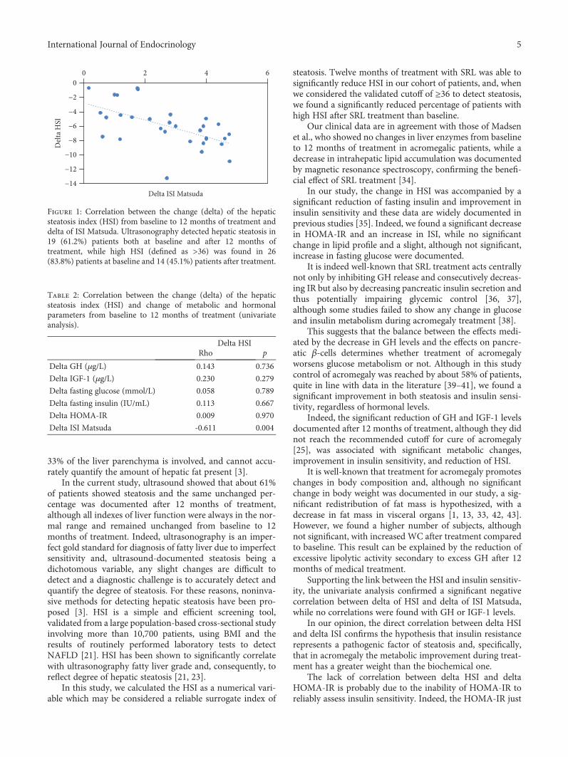
In the whole cohort of patients, the change (delta) of HSIfrom baseline to 12 months was found to be directly corre-lated with the delta of ISI (Rho -0.611; p = 0 004) (Figure 1),while no correlation was found with the delta of GH orIGF-1 levels and other metabolic parameters (Table 2).
When we analyzed patients separately according to dis-ease control, we found no significant difference betweencontrolled and uncontrolled patients in HSI values at bothbaseline (42 5 ± 5 3 vs. 40 3 ± 4 5; p = 0 297) and 12 monthsof treatment (34 9 ± 5 vs. 35 8 ± 3 4; p = 0 613). No signifi-cant difference in the number of subject with HSI > 36 (9/18 controlled patients, 50% vs. 5/13 uncontrolled patients,38.4%; p = 0 473) was found between the 2 groups at 12months of treatment. As in the whole population, bothgroups of patients showed a significant correlation betweendelta HSI and delta ISI (p = 0 026 and p = 0 029 in controlledand uncontrolled patients, respectively).
3International Journal of Endocrinology
4. Discussion
In this study, we showed that active acromegaly is associatedwith high HSI, which significantly decreases after treatmentwith SRL in concomitance with an improvement in insulinsensitivity and regardless of disease control.
It is well-known that active acromegaly is strongly associ-ated with a condition of lipotoxicity and visceral adipositydysfunction and that IR is one of the main metabolic alter-ations that characterize it [12, 29, 30].
Both lipotoxicity and IR have a significant impact onthe liver. Indeed, in active acromegaly the lipolytic effectof GH excess leads to adipose tissue redistribution [12,13]. The increased lipolysis secondary to GH excess maylead to a reduction in visceral and subcutaneous adiposetissue in active acromegalic patients [12, 13, 31–33].
However, while body fat depots are diminished, bothhepatic and extra-hepatic IR is increased [11]. Indeed, stea-tosis is a common metabolic feature of acromegaly and fatdeposition in the liver and muscle may represent one of themechanisms involved in the pathogenesis of IR in acromeg-aly [12, 14–16]although there is also evidence againsthepatic lipid accumulation in active acromegaly [20, 34].Studies have shown that GH signaling limits hepatic lipidaccumulation in mice [18, 19] and liver lipid contentproved significantly lower in acromegalic patients versushealthy subjects [20].
To date, reliable and accurate data on the prevalence ofhepatic steatosis in acromegaly are not available. Hepaticsteatosis is commonly detected by imaging such as ultra-sound or computed tomography. However, these examina-tions have poor sensitivity, detect fat only when 20% to
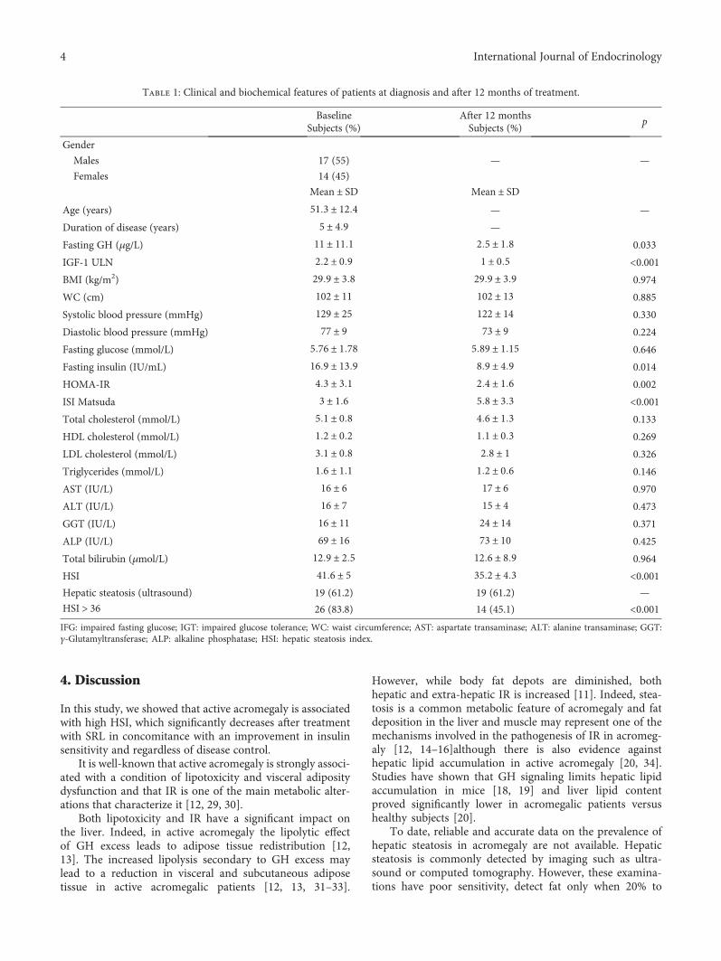
Table 1: Clinical and biochemical features of patients at diagnosis and after 12 months of treatment.
Baseline After 12 monthsp
Subjects (%) Subjects (%)
Gender
Males 17 (55) — —
Females 14 (45)
Mean ± SD Mean ± SD
Age (years) 51 3 ± 12 4 — —
Duration of disease (years) 5 ± 4 9 —
Fasting GH (μg/L) 11 ± 11 1 2 5 ± 1 8 0.033
IGF-1 ULN 2 2 ± 0 9 1 ± 0 5 <0.001BMI (kg/m2) 29 9 ± 3 8 29 9 ± 3 9 0.974
WC (cm) 102 ± 11 102 ± 13 0.885
Systolic blood pressure (mmHg) 129 ± 25 122 ± 14 0.330
Diastolic blood pressure (mmHg) 77 ± 9 73 ± 9 0.224
Fasting glucose (mmol/L) 5 76 ± 1 78 5 89 ± 1 15 0.646
Fasting insulin (IU/mL) 16 9 ± 13 9 8 9 ± 4 9 0.014
HOMA-IR 4 3 ± 3 1 2 4 ± 1 6 0.002
ISI Matsuda 3 ± 1 6 5 8 ± 3 3 <0.001Total cholesterol (mmol/L) 5 1 ± 0 8 4 6 ± 1 3 0.133
HDL cholesterol (mmol/L) 1 2 ± 0 2 1 1 ± 0 3 0.269
LDL cholesterol (mmol/L) 3 1 ± 0 8 2 8 ± 1 0.326
Triglycerides (mmol/L) 1 6 ± 1 1 1 2 ± 0 6 0.146
AST (IU/L) 16 ± 6 17 ± 6 0.970
ALT (IU/L) 16 ± 7 15 ± 4 0.473
GGT (IU/L) 16 ± 11 24 ± 14 0.371
ALP (IU/L) 69 ± 16 73 ± 10 0.425
Total bilirubin (μmol/L) 12 9 ± 2 5 12 6 ± 8 9 0.964
HSI 41 6 ± 5 35 2 ± 4 3 <0.001Hepatic steatosis (ultrasound) 19 (61.2) 19 (61.2) —
HSI > 36 26 (83.8) 14 (45.1) <0.001IFG: impaired fasting glucose; IGT: impaired glucose tolerance; WC: waist circumference; AST: aspartate transaminase; ALT: alanine transaminase; GGT:γ-Glutamyltransferase; ALP: alkaline phosphatase; HSI: hepatic steatosis index.
4 International Journal of Endocrinology
33% of the liver parenchyma is involved, and cannot accu-rately quantify the amount of hepatic fat present [3].
In the current study, ultrasound showed that about 61%of patients showed steatosis and the same unchanged per-centage was documented after 12 months of treatment,although all indexes of liver function were always in the nor-mal range and remained unchanged from baseline to 12months of treatment. Indeed, ultrasonography is an imper-fect gold standard for diagnosis of fatty liver due to imperfectsensitivity and, ultrasound-documented steatosis being adichotomous variable, any slight changes are difficult todetect and a diagnostic challenge is to accurately detect andquantify the degree of steatosis. For these reasons, noninva-sive methods for detecting hepatic steatosis have been pro-posed [3]. HSI is a simple and efficient screening tool,validated from a large population-based cross-sectional studyinvolving more than 10,700 patients, using BMI and theresults of routinely performed laboratory tests to detectNAFLD [21]. HSI has been shown to significantly correlatewith ultrasonography fatty liver grade and, consequently, toreflect degree of hepatic steatosis [21, 23].
In this study, we calculated the HSI as a numerical vari-able which may be considered a reliable surrogate index of
steatosis. Twelve months of treatment with SRL was able tosignificantly reduce HSI in our cohort of patients, and, whenwe considered the validated cutoff of ≥36 to detect steatosis,we found a significantly reduced percentage of patients withhigh HSI after SRL treatment than baseline.
Our clinical data are in agreement with those of Madsenet al., who showed no changes in liver enzymes from baselineto 12 months of treatment in acromegalic patients, while adecrease in intrahepatic lipid accumulation was documentedby magnetic resonance spectroscopy, confirming the benefi-cial effect of SRL treatment [34].
In our study, the change in HSI was accompanied by asignificant reduction of fasting insulin and improvement ininsulin sensitivity and these data are widely documented inprevious studies [35]. Indeed, we found a significant decreasein HOMA-IR and an increase in ISI, while no significantchange in lipid profile and a slight, although not significant,increase in fasting glucose were documented.
It is indeed well-known that SRL treatment acts centrallynot only by inhibiting GH release and consecutively decreas-ing IR but also by decreasing pancreatic insulin secretion andthus potentially impairing glycemic control [36, 37],although some studies failed to show any change in glucoseand insulin metabolism during acromegaly treatment [38].
This suggests that the balance between the effects medi-ated by the decrease in GH levels and the effects on pancre-atic β-cells determines whether treatment of acromegalyworsens glucose metabolism or not. Although in this studycontrol of acromegaly was reached by about 58% of patients,quite in line with data in the literature [39–41], we found asignificant improvement in both steatosis and insulin sensi-tivity, regardless of hormonal levels.
Indeed, the significant reduction of GH and IGF-1 levelsdocumented after 12 months of treatment, although they didnot reach the recommended cutoff for cure of acromegaly[25], was associated with significant metabolic changes,improvement in insulin sensitivity, and reduction of HSI.
It is well-known that treatment for acromegaly promoteschanges in body composition and, although no significantchange in body weight was documented in our study, a sig-nificant redistribution of fat mass is hypothesized, with adecrease in fat mass in visceral organs [1, 13, 33, 42, 43].However, we found a higher number of subjects, althoughnot significant, with increased WC after treatment comparedto baseline. This result can be explained by the reduction ofexcessive lipolytic activity secondary to excess GH after 12months of medical treatment.
Supporting the link between the HSI and insulin sensitiv-ity, the univariate analysis confirmed a significant negativecorrelation between delta of HSI and delta of ISI Matsuda,while no correlations were found with GH or IGF-1 levels.
In our opinion, the direct correlation between delta HSIand delta ISI confirms the hypothesis that insulin resistancerepresents a pathogenic factor of steatosis and, specifically,that in acromegaly the metabolic improvement during treat-ment has a greater weight than the biochemical one.
The lack of correlation between delta HSI and deltaHOMA-IR is probably due to the inability of HOMA-IR toreliably assess insulin sensitivity. Indeed, the HOMA-IR just
Table 2: Correlation between the change (delta) of the hepaticsteatosis index (HSI) and change of metabolic and hormonalparameters from baseline to 12 months of treatment (univariateanalysis).
Delta HSIRho p
Delta GH (μg/L) 0.143 0.736
Delta IGF-1 (μg/L) 0.230 0.279
Delta fasting glucose (mmol/L) 0.058 0.789
Delta fasting insulin (IU/mL) 0.113 0.667
Delta HOMA-IR 0.009 0.970
Delta ISI Matsuda -0.611 0.004
−14
−12
−10
−8
−6
−4
−2
00 2 4 6
Delt
a HSI
Delta ISI Matsuda
Figure 1: Correlation between the change (delta) of the hepaticsteatosis index (HSI) from baseline to 12 months of treatment anddelta of ISI Matsuda. Ultrasonography detected hepatic steatosis in19 (61.2%) patients both at baseline and after 12 months oftreatment, while high HSI (defined as >36) was found in 26(83.8%) patients at baseline and 14 (45.1%) patients after treatment.
5International Journal of Endocrinology
represents a basal index of insulin resistance closely related tobasal glucose and insulin levels, while ISI Matsuda, whichderives from glucose and insulin levels during OGTT, showsa different behavior and certainly represents a more reliableindex of insulin sensitivity than the basal HOMA-IR.
A weakness of our study could be the limited number ofpatients enrolled, as we enrolled only those who refused sur-gery as first-line treatment. In addition, in our cohort we didnot have overt diabetic patients, to avoid interference withmetabolic evaluation. Another limitation of the study isprobably the lack of direct hepatic data based on histologicexamination of liver biopsy specimens, to date consideredthe gold standard for diagnosing NAFLD, because its wide-spread use is limited by the risk associated with an invasiveprocedure, cost, and sampling error [1, 44].
In conclusion, in acromegaly HSI is mainly relatedwith IR and the reduction of GH and IGF-1 levels, andabove all the improvement in insulin sensitivity leads toan improvement of this surrogate index of hepatic steato-sis, which should always be evaluated in the follow-up ofacromegalic patients.
Data Availability
The data used to support the findings of this study are avail-able from the corresponding author upon request.
Ethical Approval
All procedures performed in studies involving human partic-ipants were in accordance with the ethical standards of theinstitutional and/or national research committee and withthe 1964 Helsinki Declaration and its later amendments orcomparable ethical standards.
Conflicts of Interest
There is no conflict of interest that could be perceived asprejudicing the impartiality of the research reported.
References
[1] M. E. Rinella, “Nonalcoholic fatty liver disease: a systematicreview,” JAMA, vol. 313, no. 22, pp. 2263–2273, 2015.
[2] J. M. Pappachan, S. Babu, B. Krishnan, and N. C. Ravindran,“Non-alcoholic fatty liver disease: a clinical update,” Journalof Clinical and Translational Hepatology, vol. 5, no. 4,pp. 384–393, 2017.
[3] N. Chalasani, Z. Younossi, J. E. Lavine et al., “The diagnosisand management of nonalcoholic fatty liver disease: practiceguidance from the American Association for the Study of LiverDiseases,” Hepatology, vol. 67, no. 1, pp. 328–357, 2018.
[4] Y. Takahashi, “The role of growth hormone and insulin-likegrowth factor-I in the liver,” International Journal of Molecu-lar Sciences, vol. 18, no. 7, 2017.
[5] N. Møller and J. O. L. Jørgensen, “Effects of growth hormoneon glucose, lipid, and protein metabolism in human subjects,”Endocrine Reviews, vol. 30, no. 2, pp. 152–177, 2009.
[6] R. A. Rizza, L. J. Mandarino, and J. E. Gerich, “Effects ofgrowth hormone on insulin action in man. Mechanisms of
insulin resistance, impaired suppression of glucose produc-tion, and impaired stimulation of glucose utilization,” Diabe-tes, vol. 31, no. 8, pp. 663–669, 1982.
[7] T. Ichikawa, K. Nakao, K. Hamasaki et al., “Role of growthhormone, insulin-like growth factor 1 and insulin-like growthfactor-binding protein 3 in development of non-alcoholic fattyliver disease,” Hepatology International, vol. 1, no. 2, pp. 287–294, 2007.
[8] T. Ichikawa, K. Hamasaki, H. Ishikawa, E. Ejima, K. Eguchi,and K. Nakao, “Non-alcoholic steatohepatitis and hepatic stea-tosis in patients with adult onset growth hormone deficiency,”Gut, vol. 52, no. 6, p. 914, 2003.
[9] Y. Takahashi, K. Iida, K. Takahashi et al., “Growth hormonereverses nonalcoholic steatohepatitis in a patient with adultgrowth hormone deficiency,” Gastroenterology, vol. 132, no. 3,pp. 938–943, 2007.
[10] D. R. Clemmons, “Roles of insulin-like growth factor-I andgrowth hormone in mediating insulin resistance in acromeg-aly,” Pituitary, vol. 5, no. 3, pp. 181–183, 2002.
[11] N. C. Olarescu and J. Bollerslev, “The impact of adipose tissueon insulin resistance in acromegaly,” Trends in Endocrinologyand Metabolism, vol. 27, no. 4, pp. 226–237, 2016.
[12] P. U. Freda, W. Shen, S. B. Heymsfield et al., “Lower visceraland subcutaneous but higher intermuscular adipose tissuedepots in patients with growth hormone and insulin-likegrowth factor I excess due to acromegaly,” The Journal of Clin-ical Endocrinology & Metabolism, vol. 93, no. 6, pp. 2334–2343, 2008.
[13] C. M. Reyes-Vidal, H. Mojahed, W. Shen et al., “Adipose tissueredistribution and ectopic lipid deposition in active acromeg-aly and effects of surgical treatment,” The Journal of ClinicalEndocrinology and Metabolism, vol. 100, no. 8, pp. 2946–2955, 2015.
[14] V. T. Samuel and G. I. Shulman, “Mechanisms for insulinresistance: common threads and missing links,” Cell, vol. 148,no. 5, pp. 852–871, 2012.
[15] A. Lettner and M. Roden, “Ectopic fat and insulin resistance,”Current Diabetes Reports, vol. 8, no. 3, pp. 185–191, 2008.
[16] M. Krssak and M. Roden, “The role of lipid accumulation inliver and muscle for insulin resistance and type 2 diabetes mel-litus in humans,” Reviews in Endocrine & Metabolic Disorders,vol. 5, no. 2, pp. 127–134, 2004.
[17] M. B. Krag, L. C. Gormsen, Z. Guo et al., “Growth hormone-induced insulin resistance is associated with increased intramyo-cellular triglyceride content but unaltered VLDL-triglyceridekinetics,” American Journal of Physiology. Endocrinology andMetabolism, vol. 292, no. 3, pp. E920–E927, 2007.
[18] J. L. Barclay, C. N. Nelson, M. Ishikawa et al., “GH-dependentSTAT5 signaling plays an important role in hepatic lipidmetabolism,” Endocrinology, vol. 152, no. 1, pp. 181–192, 2011.
[19] J. Cordoba-Chacon, N. Majumdar, E. O. List et al., “Growthhormone inhibits hepatic de novo lipogenesis in adult mice,”Diabetes, vol. 64, no. 9, pp. 3093–3103, 2015.
[20] Y. Winhofer, P. Wolf, M. Krššák et al., “No evidence of ectopiclipid accumulation in the pathophysiology of the acromegaliccardiomyopathy,” The Journal of Clinical Endocrinology andMetabolism, vol. 99, no. 11, pp. 4299–4306, 2014.
[21] J. H. Lee, D. Kim, H. J. Kim et al., “Hepatic steatosis index: asimple screening tool reflecting nonalcoholic fatty liver dis-ease,” Digestive and Liver Disease, vol. 42, no. 7, pp. 503–508, 2010.
6 International Journal of Endocrinology
[22] P. J. Meffert, S. E. Baumeister, M. M. Lerch, J. Mayerle,W. Kratzer, and H. Völzke, “Development, external validation,and comparative assessment of a new diagnostic score forhepatic steatosis,” The American Journal of Gastroenterology,vol. 109, no. 9, pp. 1404–1414, 2014.
[23] M. Papagianni, A. Sofogianni, and K. Tziomalos, “Non-invasive methods for the diagnosis of nonalcoholic fatty liverdisease,” World Journal of Hepatology, vol. 7, no. 4, pp. 638–648, 2015.
[24] L. Katznelson, Laws ER Jr, S. Melmed et al., “Acromegaly: anendocrine society clinical practice guideline,” The Journal ofClinical Endocrinology and Metabolism, vol. 99, no. 11,pp. 3933–3951, 2014.
[25] A. Giustina, P. Chanson, M. D. Bronstein et al., “A consensuson criteria for cure of acromegaly,” The Journal of ClinicalEndocrinology and Metabolism, vol. 95, no. 7, pp. 3141–3148,2010.
[26] D. R. Matthews, J. P. Hosker, A. S. Rudenski, B. A. Naylor,D. F. Treacher, and R. C. Turner, “Homeostasis model assess-ment: insulin resistance and ?-cell function from fastingplasma glucose and insulin concentrations in man,”Diabetolo-gia, vol. 28, no. 7, pp. 412–419, 1985.
[27] M. Matsuda and R. A. DeFronzo, “Insulin sensitivity indicesobtained from oral glucose tolerance testing: comparison withthe euglycemic insulin clamp,” Diabetes Care, vol. 22, no. 9,pp. 1462–1470, 1999.
[28] Expert Panel on Detection, Evaluation, and Treatment of HighBlood Cholesterol in Adults, “Executive summary of the thirdreport of the National Cholesterol Education Program (NCEP)expert panel on detection, evaluation, and treatment of highblood cholesterol in adults (adult treatment panel III),” JAMA,vol. 285, no. 19, pp. 2486–2497, 2001.
[29] A. Ciresi, M. C. Amato, G. Pizzolanti, and C. GiordanoGalluzzo, “Visceral adiposity index is associated with insu-lin sensitivity and adipocytokine levels in newly diagnosedacromegalic patients,” The Journal of Clinical Endocrinologyand Metabolism, vol. 97, no. 8, pp. 2907–2915, 2012.
[30] A. Ciresi, M. C. Amato, G. Pizzolanti, and C. Giordano,“Serum visfatin levels in acromegaly: correlation with diseaseactivity and metabolic alterations,” Growth Hormone & IGFResearch, vol. 25, no. 5, pp. 240–246, 2015.
[31] T. Ueland, S. L. Fougner, K. Godang et al., “Associationsbetween body composition, circulating interleukin-1 receptorantagonist, osteocalcin, and insulin metabolism in active acro-megaly,” The Journal of Clinical Endocrinology and Metabo-lism, vol. 95, no. 1, pp. 361–368, 2010.
[32] N. C. Olarescu, T. Ueland, K. Godang, R. Lindberg-Larsen,J. O. L. Jørgensen, and J. Bollerslev, “Inflammatory adipo-kines contribute to insulin resistance in active acromegalyand respond differently to different treatment modalities,”European Journal of Endocrinology, vol. 170, no. 1, pp. 39–48, 2014.
[33] N. C. Olarescu, A. Heck, K. Godang, T. Ueland, andJ. Bollerslev, “The metabolic risk in patients newly diagnosedwith acromegaly is related to fat distribution and circulatingadipokines and improves after treatment,” Neuroendocrinol-ogy, vol. 103, no. 3-4, pp. 197–206, 2016.
[34] M. Madsen, T. Krusenstjerna-Hafstrøm, L. Møller et al., “Fatcontent in liver and skeletal muscle changes in a reciprocalmanner in patients with acromegaly during combination ther-apy with a somatostatin analog and a GH receptor antagonist:
a randomized clinical trial,” The Journal of Clinical Endocri-nology and Metabolism, vol. 97, no. 4, pp. 1227–1235, 2012.
[35] C. Giordano, A. Ciresi, M. C. Amato et al., “Clinical and meta-bolic effects of first-line treatment with somatostatin analoguesor surgery in acromegaly: a retrospective and comparativestudy,” Pituitary, vol. 15, no. 4, pp. 539–551, 2012.
[36] M. Tzanela, D. A. Vassiliadi, N. Gavalas et al., “Glucosehomeostasis in patients with acromegaly treated with surgeryor somatostatin analogues,” Clinical Endocrinology, vol. 75,no. 1, pp. 96–102, 2011.
[37] A. Cozzolino, T. Feola, I. Simonelli et al., “Somatostatin ana-logs and glucose metabolism in acromegaly: a meta-analysisof prospective interventional studies,” The Journal of ClinicalEndocrinology and Metabolism, vol. 103, no. 6, pp. 2089–2099, 2018.
[38] B. Steffin, B. Gutt, M. Bidlingmaier, C. Dieterle, F. Oltmann,and J. Schopohl, “Effects of the long-acting somatostatin ana-logue Lanreotide autogel on glucose tolerance and insulinresistance in acromegaly,” European Journal of Endocrinology,vol. 155, no. 1, pp. 73–78, 2006.
[39] A. Colao, R. S. Auriemma, R. Pivonello, L. Kasuki, and M. R.Gadelha, “Interpreting biochemical control response rateswith first-generation somatostatin analogues in acromegaly,”Pituitary, vol. 19, no. 3, pp. 235–247, 2016.
[40] R.M. Paragliola, S.M. Corsello, and R. Salvatori, “Somatostatinreceptor ligands in acromegaly: clinical response and factorspredicting resistance,” Pituitary, vol. 20, no. 1, pp. 109–115,2017.
[41] M. R. Gadelha, L. E. Wildemberg, M. D. Bronstein, F. Gatto,and D. Ferone, “Somatostatin receptor ligands in the treat-ment of acromegaly,” Pituitary, vol. 20, no. 1, pp. 100–108,2017.
[42] C. Reyes-Vidal, J. C. Fernandez, J. N. Bruce et al., “Prospectivestudy of surgical treatment of acromegaly: effects on ghrelin,weight, adiposity, and markers of CV risk,” The Journalof Clinical Endocrinology & Metabolism, vol. 99, no. 11,pp. 4124–4132, 2014.
[43] R.-J. M. Brummer, L. Lönns, H. Kvist, U. Grangård, B. A.Bengtsson, and L. SJÖSTRÖM, “Adipose tissue and musclevolume determination by computed tomography in acromeg-aly, before and 1 year after adenomectomy,” European Journalof Clinical Investigation, vol. 23, no. 4, pp. 199–205, 1993.
[44] V. Ratziu, F. Charlotte, A. Heurtier et al., “Sampling variabilityof liver biopsy in nonalcoholic fatty liver disease,” Gastroenter-ology, vol. 128, no. 7, pp. 1898–1906, 2005.
7International Journal of Endocrinology

Stem Cells International
Hindawiwww.hindawi.com Volume 2018
Hindawiwww.hindawi.com Volume 2018
MEDIATORSINFLAMMATION
of
EndocrinologyInternational Journal of
Hindawiwww.hindawi.com Volume 2018
Hindawiwww.hindawi.com Volume 2018
Disease Markers
Hindawiwww.hindawi.com Volume 2018
BioMed Research International
OncologyJournal of
Hindawiwww.hindawi.com Volume 2013
Hindawiwww.hindawi.com Volume 2018
Oxidative Medicine and Cellular Longevity
Hindawiwww.hindawi.com Volume 2018
PPAR Research
Hindawi Publishing Corporation http://www.hindawi.com Volume 2013Hindawiwww.hindawi.com
The Scientific World Journal
Volume 2018
Immunology ResearchHindawiwww.hindawi.com Volume 2018
Journal of
ObesityJournal of
Hindawiwww.hindawi.com Volume 2018
Hindawiwww.hindawi.com Volume 2018
Computational and Mathematical Methods in Medicine
Hindawiwww.hindawi.com Volume 2018
Behavioural Neurology
OphthalmologyJournal of
Hindawiwww.hindawi.com Volume 2018
Diabetes ResearchJournal of
Hindawiwww.hindawi.com Volume 2018
Hindawiwww.hindawi.com Volume 2018
Research and TreatmentAIDS
Hindawiwww.hindawi.com Volume 2018
Gastroenterology Research and Practice
Hindawiwww.hindawi.com Volume 2018
Parkinson’s Disease
Evidence-Based Complementary andAlternative Medicine
Volume 2018Hindawiwww.hindawi.com
Submit your manuscripts atwww.hindawi.com


