Heparan sulfate is an important mediator of Ebola virus ...
12
RESEARCH Open Access Heparan sulfate is an important mediator of Ebola virus infection in polarized epithelial cells Manasi Tamhankar 1,2 , Dawn M. Gerhardt 3 , Richard S. Bennett 3 , Nicole Murphy 3 , Peter B. Jahrling 3,4 and Jean L. Patterson 1* Abstract Background: Currently, no FDA-approved vaccines or treatments are available for Ebola virus disease (EVD), and therapy remains largely supportive. Ebola virus (EBOV) has broad tissue tropism and can infect a variety of cells including epithelial cells. Epithelial cells differ from most other cell types by their polarized phenotype and barrier function. In polarized cells, the apical and basolateral membrane domains are demarcated by tight junctions, and specialized sorting machinery, which results in a difference in composition between the two membrane domains. These specialized sorting functions can have important consequences for viral infections. Differential localization of a viral receptor can restrict virus entry to a particular membrane while polarized sorting can lead to a vectorial virus release. The present study investigated the impact of cell polarity on EBOV infection. Methods: Characteristics of EBOV infection in polarized cells were evaluated in the polarized Caco-2 model grown on semipermeable transwells. Transepithelial resistance (TEER), which is a function of tight junctions, was used to assess epithelial cell polarization. EBOV infection was assessed with immunofluorescence microscopy and qPCR. Statistical significance was calculated using one-way ANOVA and significance was set at p < 0.05. Results: Our data indicate that EBOV preferentially infects cells from the basolateral route, and this preference may be influenced by the resistance across the Caco-2 monolayer. Infection occurs without changes in cellular permeability. Further, our data show that basolateral infection bias may be dependent on polarized distribution of heparan sulfate, a known viral attachment factor. Treatment with iota-carrageenan, or heparin lyase, which interrupts viral interaction with cellular heparan sulfate, significantly reduced cell susceptibility to basolateral infection, likely by inhibiting virus attachment. Conclusions: Our results show cell polarity has an impact on EBOV infection. EBOV preferentially infects polarized cells through the basolateral route. Access to heparan sulfate is an important factor during basolateral infection and blocking interaction of cellular heparan sulfate with virus leads to significant inhibition of basolateral infection in the polarized Caco-2 cell model. Keywords: Ebola virus, Caco-2, Polarized cells, Heparan sulfate * Correspondence: [email protected] 1 Department of Virology and Immunology, Texas Biomedical Research Institute, San Antonio, TX, USA Full list of author information is available at the end of the article © The Author(s). 2018 Open Access This article is distributed under the terms of the Creative Commons Attribution 4.0 International License (http://creativecommons.org/licenses/by/4.0/), which permits unrestricted use, distribution, and reproduction in any medium, provided you give appropriate credit to the original author(s) and the source, provide a link to the Creative Commons license, and indicate if changes were made. The Creative Commons Public Domain Dedication waiver (http://creativecommons.org/publicdomain/zero/1.0/) applies to the data made available in this article, unless otherwise stated. Tamhankar et al. Virology Journal (2018) 15:135 https://doi.org/10.1186/s12985-018-1045-0
Transcript of Heparan sulfate is an important mediator of Ebola virus ...
RESEARCH Open Access
Heparan sulfate is an important mediatorof Ebola virus infection in polarizedepithelial cellsManasi Tamhankar1,2, Dawn M. Gerhardt3, Richard S. Bennett3, Nicole Murphy3, Peter B. Jahrling3,4
and Jean L. Patterson1*
Abstract
Background: Currently, no FDA-approved vaccines or treatments are available for Ebola virus disease (EVD), andtherapy remains largely supportive. Ebola virus (EBOV) has broad tissue tropism and can infect a variety of cellsincluding epithelial cells. Epithelial cells differ from most other cell types by their polarized phenotype and barrierfunction. In polarized cells, the apical and basolateral membrane domains are demarcated by tight junctions, andspecialized sorting machinery, which results in a difference in composition between the two membrane domains.These specialized sorting functions can have important consequences for viral infections. Differential localization ofa viral receptor can restrict virus entry to a particular membrane while polarized sorting can lead to a vectorial virusrelease. The present study investigated the impact of cell polarity on EBOV infection.
Methods: Characteristics of EBOV infection in polarized cells were evaluated in the polarized Caco-2 model grownon semipermeable transwells. Transepithelial resistance (TEER), which is a function of tight junctions, was used toassess epithelial cell polarization. EBOV infection was assessed with immunofluorescence microscopy and qPCR.Statistical significance was calculated using one-way ANOVA and significance was set at p < 0.05.
Results: Our data indicate that EBOV preferentially infects cells from the basolateral route, and this preference maybe influenced by the resistance across the Caco-2 monolayer. Infection occurs without changes in cellularpermeability. Further, our data show that basolateral infection bias may be dependent on polarized distribution ofheparan sulfate, a known viral attachment factor. Treatment with iota-carrageenan, or heparin lyase, whichinterrupts viral interaction with cellular heparan sulfate, significantly reduced cell susceptibility to basolateralinfection, likely by inhibiting virus attachment.
Conclusions: Our results show cell polarity has an impact on EBOV infection. EBOV preferentially infects polarizedcells through the basolateral route. Access to heparan sulfate is an important factor during basolateral infection andblocking interaction of cellular heparan sulfate with virus leads to significant inhibition of basolateral infection inthe polarized Caco-2 cell model.
Keywords: Ebola virus, Caco-2, Polarized cells, Heparan sulfate
* Correspondence: [email protected] of Virology and Immunology, Texas Biomedical ResearchInstitute, San Antonio, TX, USAFull list of author information is available at the end of the article
© The Author(s). 2018 Open Access This article is distributed under the terms of the Creative Commons Attribution 4.0International License (http://creativecommons.org/licenses/by/4.0/), which permits unrestricted use, distribution, andreproduction in any medium, provided you give appropriate credit to the original author(s) and the source, provide a link tothe Creative Commons license, and indicate if changes were made. The Creative Commons Public Domain Dedication waiver(http://creativecommons.org/publicdomain/zero/1.0/) applies to the data made available in this article, unless otherwise stated.
Tamhankar et al. Virology Journal (2018) 15:135 https://doi.org/10.1186/s12985-018-1045-0
BackgroundPolarized cells often act as barriers between the externalenvironment and the underlying tissue. Due to theirasymmetric plasma membranes, these cells contain dis-tinct apical or basolateral membranes and can imposean obstacle for virus infection and spread. Viruses sub-vert this in a variety of ways, including disruption of thetight junctional barrier or transcytosis to gain access tothe basal tissue [1–5].The outbreak of Ebola virus disease (EVD) that occurred
from 2013 to 2016 in the West African countries ofGuinea, Liberia, and Sierra Leone constituted a majorhumanitarian disaster. The outbreak numbered over28,500 cases and 11,000 deaths [6]. Two more outbreakshave since occurred in the Democratic Republic of Congoin 2017, and 2018. As of August 25 2018, the latest out-break has caused 72 deaths with a total 111 cases [7]. Thishighlights the fact that EBOV will remain a health threatin the near future, and development of therapeutics is ur-gently needed to effectively combat the virus.Ebola virus infects a variety of polarized cells in vivo,
and has been isolated from a number of tissues includingthe liver and gastrointestinal tract, both of which compriseof polarized cells [8]. Gastrointestinal symptoms areamong the earliest, most common, and life-threateningclinical manifestations of EVD in humans [9]. In the 2014outbreak in Western Africa, results of a study found thatamong patients admitted to the hospital with confirmedEVD, the most common clinical syndrome was one ofgastrointestinal illness, intravascular volume depletion,and related complications [10]. Owing to the difficulties inhandling EBOV, knowledge of virus pathogenesis in polar-ized cells remains to be elucidated.Differential availability of proteins on the cell surface can
be a limiting step during the virus replication cycle. Indeed,a number of viruses induce downregulation of receptors toprevent superinfection [11, 12]. In polarized cells, proteinscan be selectively expressed on the apical or basolateralsurface through specialized sorting mechanisms [13]. Ebolavirus entry is a complex and multifactorial process, and re-striction of important entry factor(s) because of selectiveprotein localization can potentially impact the efficiency ofvirus entry. The present study investigates the impact ofpolarity on EBOV infection using the colorectal adenocar-cinoma (Caco-2) cell polarized model.
MethodsCells and virusCaco-2 cells (human epithelial adenocarcinoma cells,ATCC) were maintained in minimal essential medium(MEM; Invitrogen) supplemented with 2 or 10% fetalbovine serum (FBS) (Invitrogen). Only low passageCaco-2 cells (between passage 3 and 30) were used forseeding on transwells, and a single cell suspension was
made each time to encourage formation of a monolayer.All experiments used EBOV isolate Kikwit (Ebola virusH.sapiens-rec/COD/1995/Kikwit), a widely used strain ofEBOV, and were carried out at the biosafety level-4 facil-ities at Texas Biomedical Research Institute, San Antonio,TX or the Integrated Research Facility (IRF), NationalInstitute of Allergy and Infectious Diseases (NIAID)/National Institutes of Health, Fort Detrick, MD.
RNA extraction and qPCRTRIzol or TRIzol LS was added to the cell monolayer orsupernatant samples in the appropriate amount and ho-mogenized. RNA was extracted as per the manufacturer’sprotocol. Primers targeting EBOV nucleoprotein (NP; F5′- CATGCGTACCAGGGAGATTAC-3′, R 5′- ACTCCATCACGCTTCTTGAC -3′; amplicon length 80) wereused to quantify EBOV vRNA in the infected cells usingVerso™ 1 step RT PCR (Thermo Fisher Scientific Inc.)GAPDH was used as a reference (F 5′- CAACTCACCTCTTGGGATGAAG-3′, R 5′- CCTGGTTCAGTTTGGAGTCTATG-3′; amplicon length 90). The fold changevalues were calculated as described previously [14].
SDS-PAGE and western blottingInfected cells were harvested in RIPA lysis buffer supple-mented with LDS buffer (Invitrogen) and boiled in redu-cing sample buffer for 10 min at 100 °C. The sampleswere subjected to reducing Novex 4–12% Bis-Tris gelelectrophoresis. Separated proteins were electroblottedto PVDF membranes using the NOVEX Xcell Blot IImodule and probed using Rabbit anti-EBOV NP anti-body (IBT Bioservices, Inc).
Transepithelial electrical resistance (TEER) assayCaco-2 cells (4 × 10⁴ cells/ well) were seeded onto 6.5-mmdiameter, 1-mm pore size polycarbonate membrane trans-wells (Costar), and fresh medium was added at 2-day inter-vals. Resistance measurements were taken every other dayand expressed in ohm (Ω). At day 6 post-seeding, the cellswere verified to have around 100 (± 10%) Ω resistancebefore being used for infection. The EBOV suspension(50 μl) at a concentration of 3 pfu/cell was added eitherapically or basolaterally, incubated for 1 h at 37 °C, thenwashed three times with phosphate-buffered saline (PBS).MEM with 2% FBS medium was added, and cells wereincubated at 37 °C for the required time. For infectionstudies, TEER measurements were taken 24 and 48 hpi.
Polarized infectionCaco-2 cells were seeded onto transwells (Costar), andfresh medium was added at 2-day intervals. At day 6post-seeding, the cells were verified to have around 100(± 10%) Ω resistance before being used for infection.Cell monolayers which did not have the required
Tamhankar et al. Virology Journal (2018) 15:135 Page 2 of 12
resistance were discarded and were not used for infec-tion studies. EBOV suspension (50 μl) at a concentrationof 3 pfu/cell was added either apically or basolaterally,incubated for 1 h at 37 °C, following which were washedthree times with PBS. MEM supplemented with 2% FBSmedium was added, and cells were incubated at 37 °C.Cells were harvested in TRIzol reagent and radioimmu-noprecipitation assay (RIPA) buffer for RNA and proteinanalysis, respectively, at indicated time points, andEBOV NP vRNA was detected by quantitative reversetranscriptase (qPCR), or by western blot analysis.
Indirect immunofluorescenceCaco-2 cells were seeded into transwell inserts andinfected with EBOV After infection, cells were fixed with10% buffered formalin and processed for immunofluor-escence as described with some modifications (http://www.zonapse.net/protocols/id6.html). Cells fixed over-night were washed with PBS and incubated with im-munofluorescence buffer (20 mM of HEPES, pH 7.5,0.1% Triton-X-100, 150 mM of sodium chloride, 5 mMof EDTA, and 0.02% sodium azide as a preservative) for5 min at room temperature (RT) and all further washeswere performed with immunofluorescence buffer. Cellswere then incubated with either Rabbit anti-E-cadherinantibody (Cell Signaling Technology, Inc) to visualizeadherens junctions, or Mouse anti-EBOV GP antibody(IBT Bioservices, Inc) for visualizing EBOV infectionovernight at 4 °C. For visualization tight junctions, thecells were fixed in methanol, and processed similarly asabove. The cell monolayers were incubated with Rabbitanti-ZO-1 antibody (Cell Signaling Technology, Inc).Alexa fluor-conjugated secondary antibodies were addedfor 1 h at RT. Membranes were cut out using a scalpelblade, mounted on glass slides with Prolong anti-fademounting reagent and stained with 4′,6-diamidino-2--phenylindole (DAPI; Invitrogen). The glass slides werecovered with cover-slips and left to dry overnight in thedark at RT. The membranes were visualized using anEclipse Ti confocal microscope (Nikon) and NIS ElementsImaging Software.
Differential polarity assayCaco-2 cells (4 × 10⁴) were seeded onto 6.5-mm diameter,1-mm pore size polycarbonate membrane transwells (Co-star), and fresh medium was added at 2-day intervals. Atday 4 (average resistance 36.63 Ω), day 6 (average resistance107.32 Ω), and day 8 (average resistance 223.7 Ω)post-seeding, the cells were infected with EBOV (3 pfu/cell)either apically or basolaterally, incubated for 1 h at 37 °C,and washed three times with PBS. Then 2% FBS mediumwas added, and cells were incubated at 37 °C. Cells wereharvested 6 hpi in TRIzol reagent for qPCR analysis.
Monolayer scratch assayMonolayers of Caco-2 cells were gently scratched onceon the apical side with a 10-μl pipette tip, followedimmediately by apical addition of EBOV supernatant.Following an incubation of 1 h, the supernatant wasremoved, replaced with 2% FBS medium, and furtherincubated at 37 °C for 48 hpi. The cells were then fixedwith 10% buffered formalin and analyzed using immuno-fluorescence assay [15].
Ι-carrageenan assayFor the carrageenan assay, EBOV virus was pretreatedwith ι-carrageenan diluted in MEM without FBS supple-mentation for 30 min at 4 °C. Following incubation, cellswere infected either apically or basolaterally withEBOV-carrageenan solution (50 μl) at a final virus con-centration of 3 pfu/cell and further incubated at 37 °Cfor 1 h. The cells were then washed, the inoculum wasreplaced with MEM with 2% FBS medium, and cellswere further incubated at 37 °C. At 24 hpi, the cells wereharvested in TRIzol reagent. Quantification of the infec-tion was measured by qPCR. For the binding assay,following addition of the ι-carrageenan pretreated virus,the cells were incubated for a further 30 min at 4 °C toallow attachment but not infection. Following incuba-tion, the cells were washed with ice-cold PBS, and thecells were harvested immediately in TRIzol reagent forqPCR analysis as described earlier.
Heparin lyase assayA stock solution of (1.0 U/μl) of HL Blend fromFlavobacterium heparinum (Sigma) was prepared insterile PBS. One hour before infection, 50 μl of 0.5 U/well of HL in MEM without FBS was added to the cul-ture medium (MEM with 2% FBS) and incubated atroom temperature. Following treatment, cells were in-fected apically or basolaterally with EBOV (50 μl) at aconcentration of 3 pfu/cell and incubated at 37 °C for1 h. The cells were then washed, the inoculum was re-placed with MEM with 2% FBS medium, and cells werefurther incubated at 37 °C. At 24 hpi, the cells were har-vested in TRIzol reagent. Quantification of the infectionwas measured by qPCR. For the binding assay, followingHL pre-treatment of Caco-2 cells, was added and incu-bated for 30 min at 4 °C. Following incubation, the cellswere washed with ice-cold PBS and harvested in TRIzolreagent for analysis.
Statistical analysisGraphPad Prism (version 5.0, GraphPad) software wasused for statistical analysis. All data are shown as mean± SD calculated from three independent experiments.
Tamhankar et al. Virology Journal (2018) 15:135 Page 3 of 12
Statistical significance was calculated using one-wayANOVA and significance was set at p < 0.05.
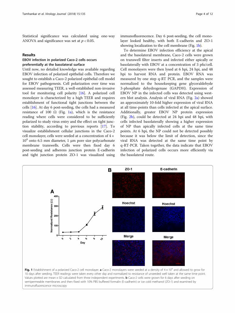
ResultsEBOV infection in polarized Caco-2 cells occurspreferentially at the basolateral surfaceUntil now, no detailed knowledge was available regardingEBOV infection of polarized epithelial cells. Therefore wesought to establish a Caco-2 polarized epithelial cell modelfor EBOV pathogenesis. Cell polarization over time wasassessed measuring TEER, a well-established non-invasivetool for monitoring cell polarity [16]. A polarized cellmonolayer is characterized by a high TEER and requiresestablishment of functional tight junctions between thecells [16]. At day 6 post-seeding, the cells had a measuredresistance of 100 Ω (Fig. 1a), which is the resistancereading where cells were considered to be sufficientlypolarized to study virus entry and the effect on tight junc-tion stability, according to previous reports [17]. Tovisualize establishment cellular junctions in the Caco-2cell monolayer, cells were seeded at a concentration of 4 ×10⁴ onto 6.5 mm diameter, 1 μm pore size polycarbonatemembrane transwells. Cells were then fixed day 6post-seeding and adherens junction protein E-cadherinand tight junction protein ZO-1 was visualized using
immunofluorescence. Day 6 post-seeding, the cell mono-layer looked healthy, with both E-cadherin and ZO-1showing localization to the cell membrane (Fig. 1b).To determine EBOV infection efficiency at the apical
and the basolateral membrane, Caco-2 cells were grownon transwell filter inserts and infected either apically orbasolaterally with EBOV at a concentration of 3 pfu/cell.Cell monolayers were then lysed at 6 hpi, 24 hpi, and 48hpi to harvest RNA and protein. EBOV RNA wasmeasured by one step q-RT PCR, and the samples werenormalized to the housekeeping gene glyceraldehyde3-phosphate dehydrogenase (GAPDH). Expression ofEBOV NP in the infected cells was detected using west-ern blot analysis. Analysis of viral RNA (Fig. 2a) showedan approximately 10-fold higher expression of viral RNAat all time-points than cells infected at the apical surface.Additionally, greater EBOV NP protein expression(Fig. 2b), could be detected at 24 hpi and 48 hpi, withcells infected basolaterally showing a higher expressionof NP than apically infected cells at the same timepoints. At 6 hpi, the NP could not be detected possiblybecause it was below the limit of detection, since theviral RNA was detected at the same time point byq-RT-PCR. Taken together, the data indicate that EBOVinfection of polarized cells occurs more efficiently viathe basolateral route.
Fig. 1 Establishment of a polarized Caco-2 cell monolayer. a Caco-2 monolayers were seeded at a density of 4 × 10⁴ and allowed to grow for10 days after seeding. TEER readings were taken every other day and normalized to resistance of unseeded well taken at the same time point.Values plotted are mean ± SD calculated from three independent experiments. b Caco-2 cells were grown for 6 days after seeding onsemipermeable membranes and then fixed with 10% PBS buffered formalin (E-cadherin) or ice cold methanol (ZO-1) and examined byimmunofluorescence microscopy
Tamhankar et al. Virology Journal (2018) 15:135 Page 4 of 12

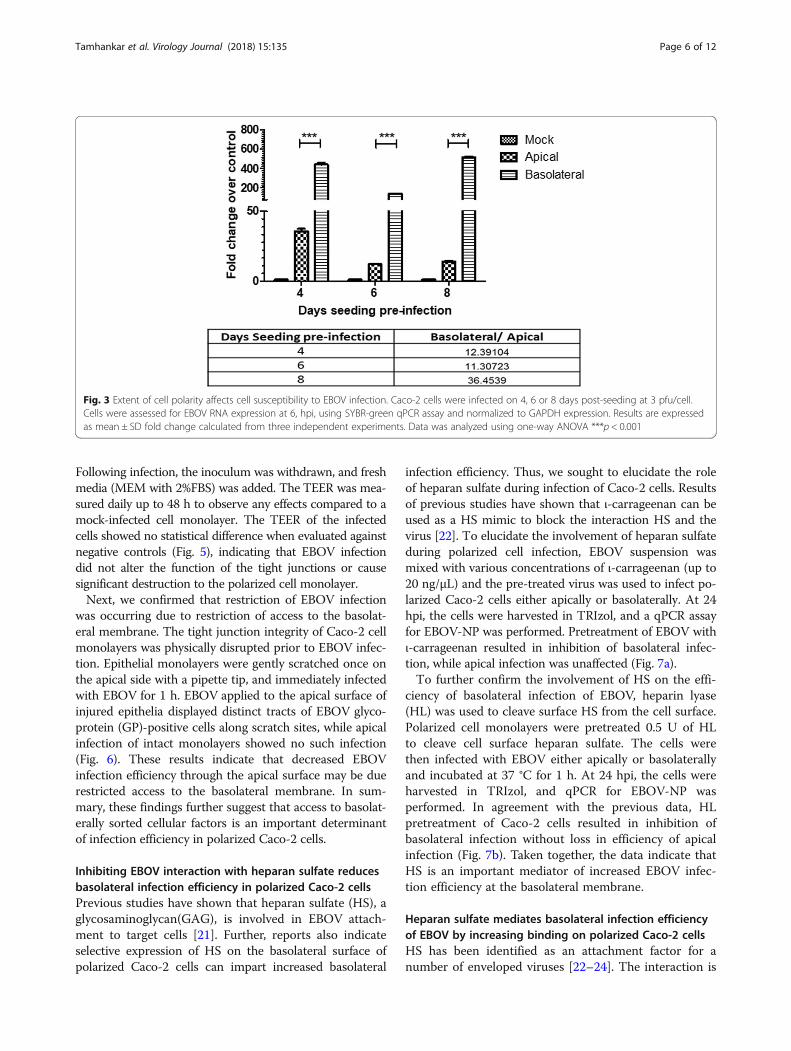
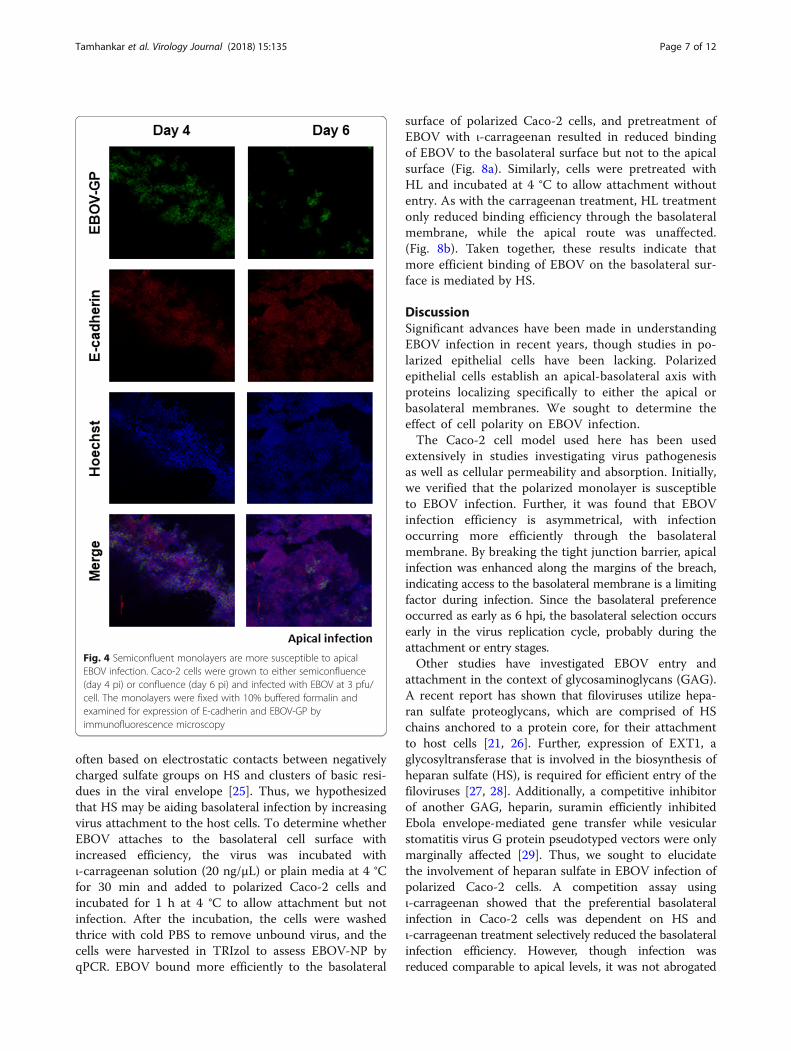
Establishment of cell polarity selectively affects apicalinfectionTo investigate the effect of increasing cell polarity onthe ability of EBOV to infect Caco-2 cells, cells wereallowed to polarize (as measured by TEER) to a lesser orgreater extent than the standard day 6 conditions andinfected apically or basolaterally with EBOV and har-vested by lysis at 6 hpi. By examining the ratio of the NPdetected in basolateral infection versus apical infectionat the same time point, an increase in relative infectionefficiency at the basolateral surface was observedbetween day 6 (11.3) and day 8 (36.45) pi. Interestingly,a higher NP expression was detected in the apicallyinfected cells at day 4 compared to day 6 pi (Fig. 3).However, no difference was observed between apicalinfection at day 6 and day 8 pi. To further confirm thisobservation, 4 or 6 day old Caco-2 monolayers were ei-ther mock-infected or EBOV-infected apically and thenfixed at 24 hpi. Day 6 monolayers showed few EBOV-GPpositive cells, in contrast to the less-polarized Day 4monolayers that showed that a majority (approximately80%) of the cell monolayer was infected, supporting the
qPCR results (Fig. 4). Thus, we theorized that cellularevents during establishment of polarity were restrictingapical infection in Caco-2 cells.
EBOV infection does not affect epithelial integrity inCaco-2 cells, restricting paracellular access to thebasolateral membraneCell polarity involves selective expression of proteins onthe apical or basolateral surface based on specific signals[18]. These two distinct membrane domains are sepa-rated by tight junctions, which also restrict paracellulartransport [19]. Thus, we hypothesized that a combinationof restricted access and a selective expression of proteinswas affecting infection efficiency and may be mediatingthe increased efficiency of basolateral infection. Tightjunctions are the major mediators of paracellular perme-ability and also play a major role in determining TEER[20]. Thus, we first sought to determine whether EBOVinfection had an impact on the tight junction integrity ofthe polarized Caco-2 monolayer. Confluent Caco-2 cellsseeded on semipermeable transwell filters were infectedeither apically or basolaterally as described before.
Fig. 2 Basolateral infection of EBOV is more efficient in Caco-2 cells a Caco-2 cells infected with EBOV at 3 pfu/cell were assessed for EBOV RNAexpression at 6, 24, and 48 hpi, using SYBR-green qPCR assay and normalized to GAPDH expression. Results are expressed in mean ± SDcalculated from three independent experiments. Data was analyzed using one-way ANOVA ***p < 0.001. b Caco-2 cells infected with EBOV at 3pfu/cell were assessed for EBOV-NP protein expression at 48 hpi by Western Blot analysis. GAPDH was used as a loading reference
Tamhankar et al. Virology Journal (2018) 15:135 Page 5 of 12
Following infection, the inoculum was withdrawn, and freshmedia (MEM with 2%FBS) was added. The TEER was mea-sured daily up to 48 h to observe any effects compared to amock-infected cell monolayer. The TEER of the infectedcells showed no statistical difference when evaluated againstnegative controls (Fig. 5), indicating that EBOV infectiondid not alter the function of the tight junctions or causesignificant destruction to the polarized cell monolayer.Next, we confirmed that restriction of EBOV infection
was occurring due to restriction of access to the basolat-eral membrane. The tight junction integrity of Caco-2 cellmonolayers was physically disrupted prior to EBOV infec-tion. Epithelial monolayers were gently scratched once onthe apical side with a pipette tip, and immediately infectedwith EBOV for 1 h. EBOV applied to the apical surface ofinjured epithelia displayed distinct tracts of EBOV glyco-protein (GP)-positive cells along scratch sites, while apicalinfection of intact monolayers showed no such infection(Fig. 6). These results indicate that decreased EBOVinfection efficiency through the apical surface may be duerestricted access to the basolateral membrane. In sum-mary, these findings further suggest that access to basolat-erally sorted cellular factors is an important determinantof infection efficiency in polarized Caco-2 cells.
Inhibiting EBOV interaction with heparan sulfate reducesbasolateral infection efficiency in polarized Caco-2 cellsPrevious studies have shown that heparan sulfate (HS), aglycosaminoglycan(GAG), is involved in EBOV attach-ment to target cells [21]. Further, reports also indicateselective expression of HS on the basolateral surface ofpolarized Caco-2 cells can impart increased basolateral
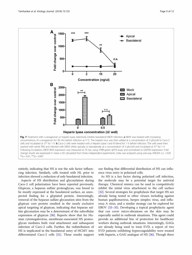
infection efficiency. Thus, we sought to elucidate the roleof heparan sulfate during infection of Caco-2 cells. Resultsof previous studies have shown that ι-carrageenan can beused as a HS mimic to block the interaction HS and thevirus [22]. To elucidate the involvement of heparan sulfateduring polarized cell infection, EBOV suspension wasmixed with various concentrations of ι-carrageenan (up to20 ng/μL) and the pre-treated virus was used to infect po-larized Caco-2 cells either apically or basolaterally. At 24hpi, the cells were harvested in TRIzol, and a qPCR assayfor EBOV-NP was performed. Pretreatment of EBOV withι-carrageenan resulted in inhibition of basolateral infec-tion, while apical infection was unaffected (Fig. 7a).To further confirm the involvement of HS on the effi-
ciency of basolateral infection of EBOV, heparin lyase(HL) was used to cleave surface HS from the cell surface.Polarized cell monolayers were pretreated 0.5 U of HLto cleave cell surface heparan sulfate. The cells werethen infected with EBOV either apically or basolaterallyand incubated at 37 °C for 1 h. At 24 hpi, the cells wereharvested in TRIzol, and qPCR for EBOV-NP wasperformed. In agreement with the previous data, HLpretreatment of Caco-2 cells resulted in inhibition ofbasolateral infection without loss in efficiency of apicalinfection (Fig. 7b). Taken together, the data indicate thatHS is an important mediator of increased EBOV infec-tion efficiency at the basolateral membrane.
Heparan sulfate mediates basolateral infection efficiencyof EBOV by increasing binding on polarized Caco-2 cellsHS has been identified as an attachment factor for anumber of enveloped viruses [22–24]. The interaction is
Fig. 3 Extent of cell polarity affects cell susceptibility to EBOV infection. Caco-2 cells were infected on 4, 6 or 8 days post-seeding at 3 pfu/cell.Cells were assessed for EBOV RNA expression at 6, hpi, using SYBR-green qPCR assay and normalized to GAPDH expression. Results are expressedas mean ± SD fold change calculated from three independent experiments. Data was analyzed using one-way ANOVA ***p < 0.001
Tamhankar et al. Virology Journal (2018) 15:135 Page 6 of 12
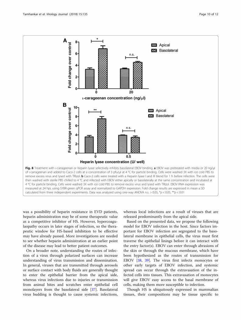
often based on electrostatic contacts between negativelycharged sulfate groups on HS and clusters of basic resi-dues in the viral envelope [25]. Thus, we hypothesizedthat HS may be aiding basolateral infection by increasingvirus attachment to the host cells. To determine whetherEBOV attaches to the basolateral cell surface withincreased efficiency, the virus was incubated withι-carrageenan solution (20 ng/μL) or plain media at 4 °Cfor 30 min and added to polarized Caco-2 cells andincubated for 1 h at 4 °C to allow attachment but notinfection. After the incubation, the cells were washedthrice with cold PBS to remove unbound virus, and thecells were harvested in TRIzol to assess EBOV-NP byqPCR. EBOV bound more efficiently to the basolateral
surface of polarized Caco-2 cells, and pretreatment ofEBOV with ι-carrageenan resulted in reduced bindingof EBOV to the basolateral surface but not to the apicalsurface (Fig. 8a). Similarly, cells were pretreated withHL and incubated at 4 °C to allow attachment withoutentry. As with the carrageenan treatment, HL treatmentonly reduced binding efficiency through the basolateralmembrane, while the apical route was unaffected.(Fig. 8b). Taken together, these results indicate thatmore efficient binding of EBOV on the basolateral sur-face is mediated by HS.
DiscussionSignificant advances have been made in understandingEBOV infection in recent years, though studies in po-larized epithelial cells have been lacking. Polarizedepithelial cells establish an apical-basolateral axis withproteins localizing specifically to either the apical orbasolateral membranes. We sought to determine theeffect of cell polarity on EBOV infection.The Caco-2 cell model used here has been used
extensively in studies investigating virus pathogenesisas well as cellular permeability and absorption. Initially,we verified that the polarized monolayer is susceptibleto EBOV infection. Further, it was found that EBOVinfection efficiency is asymmetrical, with infectionoccurring more efficiently through the basolateralmembrane. By breaking the tight junction barrier, apicalinfection was enhanced along the margins of the breach,indicating access to the basolateral membrane is a limitingfactor during infection. Since the basolateral preferenceoccurred as early as 6 hpi, the basolateral selection occursearly in the virus replication cycle, probably during theattachment or entry stages.Other studies have investigated EBOV entry and
attachment in the context of glycosaminoglycans (GAG).A recent report has shown that filoviruses utilize hepa-ran sulfate proteoglycans, which are comprised of HSchains anchored to a protein core, for their attachmentto host cells [21, 26]. Further, expression of EXT1, aglycosyltransferase that is involved in the biosynthesis ofheparan sulfate (HS), is required for efficient entry of thefiloviruses [27, 28]. Additionally, a competitive inhibitorof another GAG, heparin, suramin efficiently inhibitedEbola envelope-mediated gene transfer while vesicularstomatitis virus G protein pseudotyped vectors were onlymarginally affected [29]. Thus, we sought to elucidatethe involvement of heparan sulfate in EBOV infection ofpolarized Caco-2 cells. A competition assay usingι-carrageenan showed that the preferential basolateralinfection in Caco-2 cells was dependent on HS andι-carrageenan treatment selectively reduced the basolateralinfection efficiency. However, though infection wasreduced comparable to apical levels, it was not abrogated
Fig. 4 Semiconfluent monolayers are more susceptible to apicalEBOV infection. Caco-2 cells were grown to either semiconfluence(day 4 pi) or confluence (day 6 pi) and infected with EBOV at 3 pfu/cell. The monolayers were fixed with 10% buffered formalin andexamined for expression of E-cadherin and EBOV-GP byimmunofluorescence microscopy
Tamhankar et al. Virology Journal (2018) 15:135 Page 7 of 12
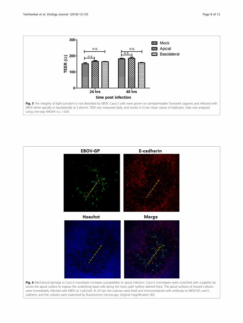
Fig. 6 Mechanical damage to Caco-2 monolayer increases susceptibility to apical infection. Caco-2 monolayers were scratched with a pipette tipacross the apical surface to expose the underlying basal cells along the injury path (yellow dashed lines). The apical surfaces of injured cultureswere immediately infected with EBOV at 3 pfu/cell. At 24 hpi, the cultures were fixed and immunostained with antibody to EBOV-GP, and E-cadherin, and the cultures were examined by fluorescence microscopy. Original magnification 40X
Fig. 5 The integrity of tight junctions is not disturbed by EBOV. Caco-2 cells were grown on semipermeable Transwell supports and infected withEBOV either apically or basolaterally at 3 pfu/ml. TEER was measured daily, and results in Ω are mean values of triplicates. Data was analyzedusing one-way ANOVA n.s. > 0.05
Tamhankar et al. Virology Journal (2018) 15:135 Page 8 of 12
entirely, indicating that HS is not the sole factor influen-cing infection. Similarly, cells treated with HL prior toinfection showed a reduction of only basolateral infection.Aspects of HS distribution and glycosylation during
Caco-2 cell polarization have been reported previously.Glypican, a heparan sulfate proteoglycan, was found tobe mostly expressed at the basolateral surface, an unex-pected finding for a glypiated protein. Interestingly,removal of the heparan sulfate glycanation sites from theglypican core protein resulted in the nearly exclusiveapical targeting of glypican, indicating that heparan sul-fate glycanation may be a determinant of the subcellularexpression of glypican [30]. Reports show that for Hu-man cytomegalovirus, membrane-associated HS proteo-glycan mediates both viral attachment and subsequentinfection of Caco-2 cells. Further, the redistribution ofHS is implicated in the basolateral entry of HCMV intodifferentiated Caco-2 cells [31]. These results support
our finding that differential distribution of HS can influ-ence virus entry in polarized cells.As HS is a key factor during polarized cell infection,
the molecule may be a potential target for antiviraltherapy. Chemical mimics can be used to competitivelyinhibit the initial virus attachment to the cell surface[32]. Several strategies for prophylaxis that target HS arealready being tested in other viruses including againsthuman papillomavirus, herpes simplex virus, and influ-enza A virus, and a similar strategy can be explored forEBOV [33–35]. Developing a topical prophylactic agentthat can cover micro-abrasions on the skin may beespecially useful in outbreak situations. This agent couldprovide an additional line of protection for healthcareworkers during outbreak situations. Interestingly, GAGsare already being used to treat EVD, a report of twoEVD patients exhibiting hypercoagulability were treatedwith heparin, a GAG analogue of HS [36]. Though there
Fig. 7 Treatment with ι-carrageenan or heparin lyase selectively inhibits basolateral EBOV infection. a EBOV was treated with increasingconcentrations of ι-carrageenan for 30 min before infection at 4 °C. The treated virus was then added at a concentration of 3 pfu/cell to Caco-2cells and incubated at 37° for 1 h. b Caco-2 cells were treated with a Heparin lyase I and III blend for 1 h before infection. The cells were thenwashed with sterile PBS and infected with EBOV either apically or basolaterally at a concentration of 3 pfu/cell and incubated at 37° for 1 h.Following incubation, EBOV RNA expression was measured at 24 hpi, using SYBR-green qPCR assay and normalized to GAPDH expression. Foldchange results are expressed in mean ± SD calculated from three independent experiments. Data was analyzed using one-way ANOVA n.s. > 0.05**p < 0.01, ***p < 0.001
Tamhankar et al. Virology Journal (2018) 15:135 Page 9 of 12
was a possibility of heparin resistance in EVD patients,heparin administration may be of some therapeutic valueas a competitive inhibitor of HS. However, hypercoagu-laopathy occurs in later stages of infection, so the thera-peutic window for HS-based inhibition to be effectivemay have already passed. More investigations are neededto see whether heparin administration at an earlier pointof the disease may lead to better patient outcomes.On a broader note, understanding the routes of infec-
tion of a virus through polarized surfaces can increaseunderstanding of virus transmission and dissemination.In general, viruses that are transmitted through aerosolsor surface contact with body fluids are generally thoughtto enter the epithelial barrier from the apical side,whereas virus infections due to injuries or transmissionfrom animal bites and scratches enter epithelial cellmonolayers from the basolateral side [37]. Basolateralvirus budding is thought to cause systemic infections,
whereas local infections are a result of viruses that arereleased predominantly from the apical side.Based on the presented data, we propose the following
model for EBOV infection in the host. Since factors im-portant for EBOV infection are segregated to the baso-lateral membrane in epithelial cells, the virus must firsttraverse the epithelial linings before it can interact withthe entry factor(s). EBOV can enter through abrasions ofthe skin or through the mucous membrane, which havebeen hypothesized as the routes of transmission forEBOV [38, 39]. The virus first infects monocytes orother early targets of EBOV infection, and systemicspread can occur through the extravasation of the in-fected cells into tissues. This extravasation of monocyteswill give EBOV easy access to the basal membrane ofcells, making them more susceptible to infection.Though HS is ubiquitously expressed in mammalian
tissues, their compositions may be tissue specific to
Fig. 8 Treatment with ι-carrageenan or heparin lyase selectively inhibits basolateral EBOV binding. a EBOV was pretreated with media or 20 ng/μlof ι-carrageenan and added to Caco-2 cells at a concentration of 3 pfu/μl at 4 °C for particle binding. Cells were washed 3X with ice cold PBS toremove excess virus and lysed with TRIzol. b Caco-2 cells were treated with a Heparin lyase I and III blend for 1 h before infection. The cells werethen washed with sterile PBS chilled to 4 °C and infected with EBOV either apically or basolaterally at the same concentration and incubated at4 °C for particle binding. Cells were washed 3X with ice cold PBS to remove excess virus and lysed with TRIzol. EBOV RNA expression wasmeasured at 24 hpi, using SYBR-green qPCR assay and normalized to GAPDH expression. Fold change results are expressed in mean ± SDcalculated from three independent experiments. Data was analyzed using one-way ANOVA n.s. > 0.05, *p < 0.05, **p < 0.01
Tamhankar et al. Virology Journal (2018) 15:135 Page 10 of 12
carry out highly diverse yet specialized roles in mamma-lian physiology [40, 41]. These HS mediated interactionsare generally electrostatic in nature, and generally showa considerable specificity with regard to the HS structureinvolved [42]. Varying distribution of HS can potentiallyhave an impact on the cell susceptibility to the virus.Thus, different polarized cells may have a slightly differ-ent susceptibility and bias depending upon the HS distri-bution and thus have different outcomes of infection.Further studies are thus needed to elucidate the specifi-city of EBOV-HS interactions regards to glycosylation aswell as structure and localization. Nevertheless, thisstudy provides a good foundation to explore EBOVpathogenesis in polarized cells.
ConclusionsOur data shows that EBOV infection in polarizedCaco-2 cells proceeds preferentially from the basolateralmembrane, Further, blocking virus access to cellularheparan sulfate leads to significant reduction of basolat-eral infection. This indicates that heparan an importantmediator for EBOV infection of polarized cells and raisesthe possibility of HS being used as a therapeutic targetduring EBOV infection.
AbbreviationsEBOV: Ebola virus; EVD: Ebola virus Disease; FBS: Fetal bovine serum;GAPDH: Glyceraldehyde 3-phosphate dehydrogenase; GP: Glycoprotein;HL: Heparin lyase; hpi: Hours post infection; HS: Heparan sulfate;MEM: Minimum Essential Medium; NP: Nucleoprotein; qPCR: Quantitativepolymerase chain reaction; RT: Room temperature; TEER: Transepithelialelectrical resistance
AcknowledgmentsWe thank Julia Michelotti for cell culture assistance at the IRF and Eric Rojofor his laboratory assistance at TBRI. We thank Laura Bollinger for technicalwriting services.
FundingWork completed at TBRI was supported by internal TBRI funding.Work completed at the IRF was supported by NIAID Division of IntramuralResearch and Division of Clinical Research and was performed under BattelleMemorial Institute Contract (No. HHSN272200700016I). The content is thispaper does not necessarily reflect the views or polices of the US Departmentof Health and Human Services (DHHS) or the institutions and companyaffiliate with the authors.
Availability of data and materialsAll data generated or analyzed during this study are included in thispublished article.
Authors’ contributionsMT participated in the conception and design of the study, carried out mostof the experiments, analyzed the data and drafted the manuscript. DMG andNM performed the experiments at IRF. RSB and PBJ oversaw the design ofthe project and supervised its execution at IRF. DMG, RSB revised themanuscript. JLP oversaw the conception and design of the study, supervisedits execution and drafted the manuscript. All the authors read and approvedthe final manuscript.
Ethics approval and consent to participateNot applicable.
Consent for publicationNot applicable.
Competing interestsThe authors declare that they have no competing interests.
Publisher’s NoteSpringer Nature remains neutral with regard to jurisdictional claims inpublished maps and institutional affiliations.
Author details1Department of Virology and Immunology, Texas Biomedical ResearchInstitute, San Antonio, TX, USA. 2University of Texas Health Science Center atSan Antonio, San Antonio, TX, USA. 3Integrated Research Facility, Division ofClinical Research, National Institute of Allergy and Infectious Diseases,National Institutes of Health, Frederick, MD, USA. 4Emerging Viral PathogensSection, National Institute of Allergy and Infectious Diseases, NationalInstitutes of Health, Frederick, MD, USA.
Received: 1 July 2018 Accepted: 20 August 2018
References1. Bomsel M. Transcytosis of infectious human immunodeficiency virus across
a tight human epithelial cell line barrier. Nat Med. 1997;3(1):42–7.2. Short KR, Kasper J, van der Aa S, Andeweg AC, Zaaraoui-Boutahar F,
Goeijenbier M, Richard M, Herold S, Becker C, Scott DP, et al. Influenza virusdamages the alveolar barrier by disrupting epithelial cell tight junctions. EurRespir J. 2016;47(3):954–66.
3. Obert G, Peiffer I, Servin AL. Rotavirus-induced structural and functionalalterations in tight junctions of polarized intestinal Caco-2 cell monolayers. JVirol. 2000;74(10):4645–51.
4. Sajjan U, Wang Q, Zhao Y, Gruenert DC, Hershenson MB. Rhinovirus disruptsthe barrier function of polarized airway epithelial cells. Am J Respir Crit CareMed. 2008;178(12):1271–81.
5. Tugizov SM, Herrera R, Palefsky JM. Epstein-Barr virus Transcytosis throughpolarized oral epithelial cells. J Virol. 2013;87(14):8179–94.
6. Outbreaks Chronology: Ebola virus Disease. 2016. https://www.cdc.gov/vhf/ebola/history/2014-2016-outbreak/index.html. Accessed 26 Aug 2018.
7. Ebola situation reports: Democratic Republic of the Congo. http://www.who.int/ebola/situation-reports/drc-2018/en. Accessed 26 Aug 2018.
8. Balda MS, Fallon MB, Van Itallie CM, Anderson JM. Structure, regulation, andpathophysiology of tight junctions in the gastrointestinal tract. Yale J BiolMed. 1992;65(6):725–40.
9. Sharma N, Cappell MS. Gastrointestinal and hepatic manifestations of Ebolavirus infection. Dig Dis Sci. 2015;60(9):2590–603.
10. Bah EI, Lamah MC, Fletcher T, Jacob ST, Brett-Major DM, Sall AA, Shindo N,Fischer WA 2nd, Lamontagne F, Saliou SM, et al. Clinical presentation ofpatients with Ebola virus disease in Conakry, Guinea. N Engl J Med. 2015;372(1):40–7.
11. Liu S, Yang W, Shen L, Turner JR, Coyne CB, Wang T. Tight junction proteinsClaudin-1 and Occludin control hepatitis C virus entry and areDownregulated during infection to prevent Superinfection. J Virol. 2009;83(4):2011–4.
12. Michel N, Allespach I, Venzke S, Fackler OT, Keppler OT. The Nef protein ofhuman immunodeficiency virus establishes Superinfection immunity by adual strategy to Downregulate cell-surface CCR5 and CD4. Curr Biol. 2005;15(8):714–23.
13. Cao X, Surma MA, Simons K. Polarized sorting and trafficking in epithelialcells. Cell Res. 2012;22(5):793–805.
14. Livak KJ, Schmittgen TD. Analysis of relative gene expression data usingreal-time quantitative PCR and the 2(-Delta Delta C(T)) method. Methods(San Diego, Calif). 2001;25(4):402–8.
15. Dylla DE, Michele DE, Campbell KP, McCray PB. Basolateral entry and releaseof new and Old World Arenaviruses from human airway epithelia. J Virol.2008;82(12):6034–8.
16. Chen S, Einspanier R, Schoen J. Transepithelial electrical resistance (TEER): afunctional parameter to monitor the quality of oviduct epithelial cellscultured on filter supports. Histochem Cell Biol. 2015;144(5):509–15.
Tamhankar et al. Virology Journal (2018) 15:135 Page 11 of 12
17. Agrawal T, Sharvani V, Nair D, Medigeshi GR. Japanese encephalitis virusdisrupts cell-cell junctions and affects the epithelial permeability barrierfunctions. PLoS One. 2013;8(7):e69465.
18. McCaffrey LM, Macara IG. Signaling pathways in cell polarity. Cold SpringHarb Perspect Biol. 2012;4(6):a009654.
19. Tang VW, Goodenough DA. Paracellular Ion Channel at the tight junction.Biophys J. 2003;84(3):1660–73.
20. Anderson JM. Molecular structure of tight junctions and their role inepithelial transport. Physiology. 2001;16(3):126.
21. Salvador B, Sexton NR, Carrion R Jr, Nunneley J, Patterson JL, Steffen I,Lu K, Muench MO, Lembo D, Simmons G. Filoviruses utilizeglycosaminoglycans for their attachment to target cells. J Virol.2013;87(6):3295–304.
22. Klimyte EM, Smith SE, Oreste P, Lembo D, Dutch RE. Inhibition of humanMetapneumovirus binding to Heparan sulfate blocks infection in humanlung cells and airway tissues. J Virol. 2016;90(20):9237–50.
23. Chen Y, Maguire T, Hileman RE, Fromm JR, Esko JD, Linhardt RJ, Marks RM.Dengue virus infectivity depends on envelope protein binding to target cellheparan sulfate. Nat Med. 1997;3(8):866–71.
24. Kroschewski H, Allison SL, Heinz FX, Mandl CW. Role of heparan sulfate forattachment and entry of tick-borne encephalitis virus. Virology. 2003;308(1):92–100.
25. de Boer SM, Kortekaas J, de Haan CAM, Rottier PJM, Moormann RJM, BoschBJ. Heparan sulfate facilitates Rift Valley fever virus entry into the cell. J Virol.2012;86(24):13767–71.
26. Bernfield M, Gotte M, Park PW, Reizes O, Fitzgerald ML, Lincecum J, Zako M.Functions of cell surface heparan sulfate proteoglycans. Annu Rev Biochem.1999;68:729–77.
27. McCormick C, Leduc Y, Martindale D, Mattison K, Esford LE, Dyer AP, TufaroF. The putative tumour suppressor EXT1 alters the expression of cell-surfaceheparan sulfate. Nat Genet. 1998;19(2):158–61.
28. O'Hearn A, Wang M, Cheng H, Lear-Rooney CM, Koning K, Rumschlag-BoomsE, Varhegyi E, Olinger G, Rong L. Role of EXT1 and Glycosaminoglycans in theearly stage of Filovirus entry. J Virol. 2015;89(10):5441–9.
29. Henss L, Beck S, Weidner T, Biedenkopf N, Sliva K, Weber C, Becker S,Schnierle BS. Suramin is a potent inhibitor of Chikungunya and Ebola viruscell entry. Virol J. 2016;13:149.
30. Mertens G, VanderSchueren B, vandenBerghe H, David G. Heparan sulfateexpression in polarized epithelial cells: the apical sorting of glypican (GPI-anchored proteoglycan) is inversely related to its heparan sulfate content. JCell Biol. 1996;132(3):487–97.
31. Esclatine A, Lemullois M, Servin AL, Quero AM, Geniteau-Legendre M.Human cytomegalovirus infects Caco-2 intestinal epithelial cellsbasolaterally regardless of the differentiation state. J Virol. 2000;74(1):513–7.
32. Buck CB, Thompson CD, Roberts JN, Muller M, Lowy DR, Schiller JT.Carrageenan is a potent inhibitor of papillomavirus infection. PLoS Pathog.2006;2(7):e69.
33. Lindahl U. Heparan sulfate-protein interactions--a concept for drug design?Thromb Haemost. 2007;98(1):109–15.
34. Cagno V, Donalisio M, Bugatti A, Civra A, Cavalli R, Ranucci E, Ferruti P,Rusnati M, Lembo D. The Agmatine-containing poly(Amidoamine) polymerAGMA1 binds cell surface Heparan sulfates and prevents attachment ofmucosal human papillomaviruses. Antimicrob Agents Chemother. 2015;59(9):5250–9.
35. Carlucci MJ, Scolaro LA, Noseda MD, Cerezo AS, Damonte EB. Protectiveeffect of a natural carrageenan on genital herpes simplex virus infection inmice. Antivir Res. 2004;64(2):137–41.
36. Wilson AJ, Martin DS, Maddox V, Rattenbury S, Bland D, Bhagani S, Cropley I,Hopkins S, Mepham S, Rodger A, et al. Thromboelastography in themanagement of coagulopathy associated with Ebola virus disease. ClinInfect Dis. 2016;62(5):610–2.
37. Schlie K, Maisa A, Freiberg F, Groseth A, Strecker T, Garten W. Viral proteindeterminants of Lassa virus entry and release from polarized epithelial cells.J Virol. 2010;84(7):3178–88.
38. Rewar S, Mirdha D. Transmission of Ebola virus disease: an overview. AnnGlob Health. 2014;80(6):444–51.
39. Vetter P, Fischer IIWA, Schibler M, Jacobs M, Bausch DG, Kaiser L. Ebola virusshedding and transmission: review of current evidence. J Infect Dis. 2016;214(suppl_3):S177–84.
40. Simon Davis DA, Parish CR. Heparan sulfate: a ubiquitous glycosaminoglycanwith multiple roles in immunity. Front Immunol. 2013;4:470.
41. Sarrazin S, Lamanna WC, Esko JD. Heparan sulfate proteoglycans. ColdSpring Harb Perspect Biol. 2011;3(7):a004952.
42. Salmivirta M, Safaiyan F, Prydz K, Andresen MS, Aryan M, Kolset SO.Differentiation-associated modulation of heparan sulfate structure andfunction in CaCo-2 colon carcinoma cells. Glycobiology. 1998;8(10):1029–36.
Tamhankar et al. Virology Journal (2018) 15:135 Page 12 of 12