
Hemostasis is the process of blood clot formation and ... · The major components of the hemostatic...
63
31/03/57 1 ทศพล มีน่วม พบ., วว. พยาธิวิทยาคลินิก ภาควิชาพยาธิวิทยา คณะแพทยศาสตร์ มหาวิทยาลัยนเรศวร Hemostasis is the process of blood clot formation and represents a coordinated response to vessel injury. The major components of the hemostatic system are • The vascular endothelium • Platelets • The coagulation and fibrinolytic systems
-
Upload
truongphuc -
Category
Documents
-
view
224 -
download
0
Transcript of Hemostasis is the process of blood clot formation and ... · The major components of the hemostatic...

31/03/57
1
ทศพล มน่ีวม พบ., วว. พยาธวิทิยาคลนิิก
ภาควชิาพยาธวิทิยา คณะแพทยศาสตร ์มหาวทิยาลยันเรศวร
Hemostasis is the process of blood clot formation and represents a coordinated response to vessel injury.
The major components of the hemostatic system are • The vascular endothelium
• Platelets
• The coagulation and fibrinolytic systems

31/03/57
2
This dynamic process is often viewed in phases • (Vascular spasm)
• Formation of a platelet plug
• Propagation of the coagulation cascade
• Formation of a clot
• Fibrinolysis of the clot.
(Vascular Platelet Coagulation phase)
The healthy endothelium is a dynamic organ • Maintaining a barrier to macromolecules.
• When injured, in contributing to the metabolic response and local vasoconstriction.
• Inhibiting platelets, suppressing coagulation and promoting fibrinolysis

31/03/57
3
Platelet Inhibition • Prostacyclin and nitric oxide
Potent vasodilators
Platelet activation and aggregation inhibition.
Anticoagulation Activity • Produce heparan sulfate proteoglycans, which bind
antithrombin and accelerate the rate at which it inhibits thrombin and coagulation enzymes.
Fibrinolytic activity • Synthesizing and releasing tissue-type and urokinase-
type plasminogen activator (t-PA and u-PA)
• Also produce type 1 plasminogen activator inhibitor1 (PAI-1), the regulator of t-PA and u-PA

31/03/57
4
Vascular tone and Permeability • synthesize prostacyclin and nitric oxide: vasodilators
• Endothelins induce vasoconstriction
They are complex cytoplasm fragment from megakaryocyte
Platelets have a life span of 7 to 10 days The platelets’s role is termed ‘primary
hemostasis’

31/03/57
5
1. Platelet adhesion : adhesion to subendothelial connective tissue 2. Platelet aggreation : links platelets to each other to form clumps.
Granules are an important component of hemostasis and contain • Platelet factor 4
• Adhesive and aggregation glycoproteins
• Coagulation factors
• Fibrinolytic inhibitors

31/03/57
6
Adhesion to subendothelial connective tissue: collagen, basement membrane, and noncollagenous microfibrils • adhesion creates the initial bleeding arrest plug
Adhesion to collagen and vWF result in platelets activation.
Release of adenosine diphosphate (ADP) • the primary mediator and amplifier of aggregation
Platelets adhesion:
Platelets adhere to exposed collage and von Willebrand factor
(vWF) and form a monolayer that supports and promote thrombin generation and subsequent fibrin formation

31/03/57
7
Induce cyclooxygenase-1 (COX-1)-dependent synthesis and release of thromboxane A, another aggregator and potent vasoconstrictor
Release of calcium, serotonin, epinephrine, and trace thrombin

31/03/57
8
• Platelet aggregation links platelets to each other to form clumps. (GPIIb/IIIa and Its ligands; Fribrinogen and vWF)

31/03/57
9
Coagulation results in the generation of thrombin, which converts soluble fibrinogen to fibrin. • To form clot
• ‘Secondary hemostasis’

31/03/57
10
Coagulation occurs through the action of discrete enzyme complexes, which are composed of • A vitamin K-dependent enzyme (II, VII, IX, X)
• A non-enzyme cofactor
• And assemble on anionic phospholipid membranes in a calcium-dependent fashion.

31/03/57
11
Fibrin, which anchors the hemostatic platelet plug, is formed from soluble plasma fibrinogen by the action of the potent protease enzyme thrombin (factor IIa).
Thrombin is formed from its inactive (zymogen) plasma precursor ‘prothrombin’ (factor II) by the action of activated factor X (Xa) and its cofactor (factor Va)
This sequence of reactions has classically been referred to as the common pathway of coagulation.
Coagulation system based on
laboratory assays
(Based on waterfall hypothesis)

31/03/57
12
Factor X can be activated by either the tissue factor (extrinsic) pathway or the contact activation (intrinsic) pathway of coagulation.
The tissue factor pathway is now considered to be the major physiologic initiator of coagulation activation.
The fibrin mesh is stabilized by covalent cross-linking mediated by factor XIII.
Coagulation system
based on
laboratory assays
(Based on waterfall
hypothesis)
major physiologic initiator

31/03/57
13
Reality..

31/03/57
14
The cell-based model of coagulation
Schematic diagram of hemostasis

31/03/57
15
The fibrinolytic protein system consists of the zymogen plasminogen and its naturally occurring activators.
Plasminogen is activated to the main clot-lysing enzyme, plasmin, by endogenous tissue plasminogen activator (tPA), single-chain urokinase plasminogen activator (ScuPA), and two-chain urokinase plasminogen activator (TcuPA).
These activators are found in the endothelium as well as in granulocytes and monocytes
SCHEMATIC REPRESENTATION OF THE FIBRINOLYTIC SYSTEM.
α2-AP, α2-Antiplasmin; PAI, plasminogen activator inhibitor; tPA, tissue plasminogen activator; uPA, urokinase plasminogen

31/03/57
16

31/03/57
17
Natural inhibitors of coagulation
• Protein C
• Protein S
Bleeding can occur if there is(are)… • abnormal platelet plug formation
• reduced thrombin generation and subsequent fibrin clot formation at the site of vascular injury
Bleeding also can occur if the platelet or fibrin clot is prematurely degraded because of excessive fibrinolysis.

31/03/57
18
Features Primary Secondary Tertiary
Components involved
Platelets, vWF and vessel wall
Coagulation Fibrinolytic factors
Site of bleeding Skin and mucocutaneous and soft tissue
Muscles, joints and deep tissues
Wounds and genitourinary tract
Physical findings Petechiae and ecchymoses
Hematomas and hemathroses
Hematuria and menorrhagia
Timing of bleeding
Immediate Delayed Delayed
Inheritance Autosomal dominant
Autosomal or X-linked recessive
Autosomal recessive
vWF: von Willebrand factor
Primary, secondary or tertiary Hemostasis
Compoenets Affected Causes
Platelets Quantitative or qualitative platelet disorders
vWF Inherited or acquired deficiency or dysfunction of vWF
Vessel wall Vasculitis or abnormalities of connective tissue supporting the vasculature

31/03/57
19
Component Affected Causes
Coagulation factors Congenital deficiency, autoantibodies, increased consumption or drugs that attenuated thrombin generation or thrombin activity
Fibrinogen Decreased production; increased consumption or synthesis of an abnormal protein
Impaired fibrin polymerization because of fibrin(ogen) degradation products or paraproteins
Fibrin cross-linking Congenital or acquired factor XIII deficiency
Component Affected Causes
Plasminogen activators Increased t-PA or u-PA release in the genitourinary tract or other tissues
Plasmin Deficiency of PAI-1 or α2-antiplasmin, resulting in an increased plasmin concentration
Plasminogen activation Enhanced plasminogen activation secondary to activation of coagulation by procoagulants, such as cancer cells, artificial surfaces or snake venoms

31/03/57
20
History taking Physical examination
• The history alone may be useful in differentiating between platelet and coagulation factor abnormalities.
• Platelet disorders are usually manifested as acquired petechiae, purpura, or mucosal bleeding.
• Coagulation problems are commonly congenital, are characterized by delayed deep muscle or joint bleeding, and are seen more often in men.
A bleeding tendency may be suspected if a patient previously experienced excessive hemorrhage after surgery or trauma, including common events such as circumcision, tonsillectomy, labor and delivery, menses, dental procedures, vaccinations, and injections.
In a patient with a history of excessive or unexplained bleeding, the initial goal is to determine whether the cause is a systemic coagulopathy or an anatomic or mechanical problem with a blood vessel.

31/03/57
21
Renal failure and the myeloproliferative disorders are associated with impaired platelet–vessel wall interactions and qualitative platelet abnormalities, connective tissue diseases and lymphomas are associated with thrombocytopenia, and liver disease causes a complex coagulopathy
Ingestion of aspirin and other nonsteroidal anti-inflammatory drugs (NSAIDs) that cause nonselective inhibition of cyclooxygenase leads to platelet dysfunction.
These drugs are often contained in over-the-counter preparations that patients may neglect to report without specific questioning.

31/03/57
22
In general, patients with thrombocytopenia or qualitative platelet or vascular disorders present with bleeding from superficial sites in the skin and mucous membranes and tends to occur spontaneously or immediately after trauma. These may involve
• Petechiae, which are pinpoint cutaneous hemorrhages that appear particularly over dependent extremities (characteristic of severe thrombocytopenia)
• ecchymoses (common bruises) • Purpura • gastrointestinal and genitourinary tract bleeding • Epistaxis and hemoptysis.
Patient with inherited or acquired coagulation factor deficiencies, such as hemophilia, or those on anticoagulant therapy tend to bleed from deeper tissue sites and in a delayed manner after trauma. These may involve
• Hemarthroses
• deep hematomas
• retroperitoneal hemorrhage

31/03/57
23
Bleeding disorders VS local bleeding? Hemostasis defects? Acquired VS Hereditary? Multiple bleeding sites? Onset? Pattern of bleeding? Familial history? Prolonged bleeding, Frequency? Previous medical illness and medication? Etc.
Vital signs Skin: nature of bleeding, signs of liver disease Mucosa: oral or nasal Lymphadenopathy Abdomen: liver size and shape, splenomegaly Joints: signs of previous bleeding Other sites of blood loss: pelvic, rectal, urinary
tract

31/03/57
24
เพิ่มรปู skin manifes
Extensive ecchymosis Collins et al. BMC Research Notes 2010, 3:161
Petechiae in newborn http://newborns.stanford.edu/PhotoGallery
Henoch-Schonlein purpura http://dermatlas.med.jhmi.edu
Complete blood count and blood smear • Assess the degree of disease associated with the
bleeding episode
Platelets count • the normal range being 150,000 to 400,000/mm3
• Platelet counts less than 100,000/mm3 define thrombocytopenia.

31/03/57
25
• Bleeding time is for determining both vascular integrity and platelet function
• The test (Ivy method) Making two standard incisions 1 mm deep and 1 cm long on the volar
aspect of the forearm under 40 mm Hg pressure via a blood pressure cuff.
The time is measured from the incision to the moment when the blood oozing from the wound is no longer absorbed by filter paper.
• Normal time is 8 minutes (Duke method; 1-3 min) 8 - 10 min : borderline
> 10 min : abnormal
• A paper conclude that the bleeding time was not effective as a screening test, and that a normal bleeding time does not exclude a bleeding disorder.
รปูจาก www.mclno.org

31/03/57
26
The activated partial thromboplastin time (aPTT) tests the components of the intrinsic and common pathways, that is, essentially all factors but VII and XIII in the entire clotting cascade.
• A phospholipid source and a contact-activating agent (kaolin) are added to anticoagulated citrate plasma.
• After an incubation period that allows factor XII to become activated, calcium is added and the clotting time is recorded.
• Factor levels are often less than 40% before the PTT is prolonged.
Reference value 22-44 seconds (รพ. มน. มีนาคม 2557) • A classic approach used by many clinicians for monitoring
unfractionated heparin therapy is to aim for a 1.5- to 2.5-fold increase in APTT.

31/03/57
27
Coagulation system
based on
laboratory assays
(Based on waterfall
hypothesis)
The prothrombin time (PT) tests the factors of the extrinsic and common pathways.
The patient’s anticoagulated plasma is combined with calcium and tissue factor.
The PT detects deficiencies in fibrinogen(factor I), prothrombin (factor II), factor V, factor VII, and factor X.
It is used to test the extrinsic pathway. • Or monitoring warfarin therapy

31/03/57
28
Coagulation system based on
laboratory assays
(Based on waterfall hypothesis)

31/03/57
29
PT > 2 seconds or more over the control time can be considered significant.
Results are usually reported as the international normalized ratio (INR), which compensates for differences in sensitivity of various thromboplastin reagents to the effects of warfarin.
INR = (patient PT/mean normal PT)ISI
• Normal value; 0.75-1.3
• Therapeutic level; 2-4.5 ISI = International Sensitivity Index
Thrombin clotting time or thrombin time • Purified thrombin is added to plasma, and the time to
clot formation is measured.
• It is a direct measure of the conversion of fibrinogen to fibrin
• It is a useful screening test for both qualitative and quantitative abnormalities of fibrinogen and inhibitors such as heparin and fibrin split products.
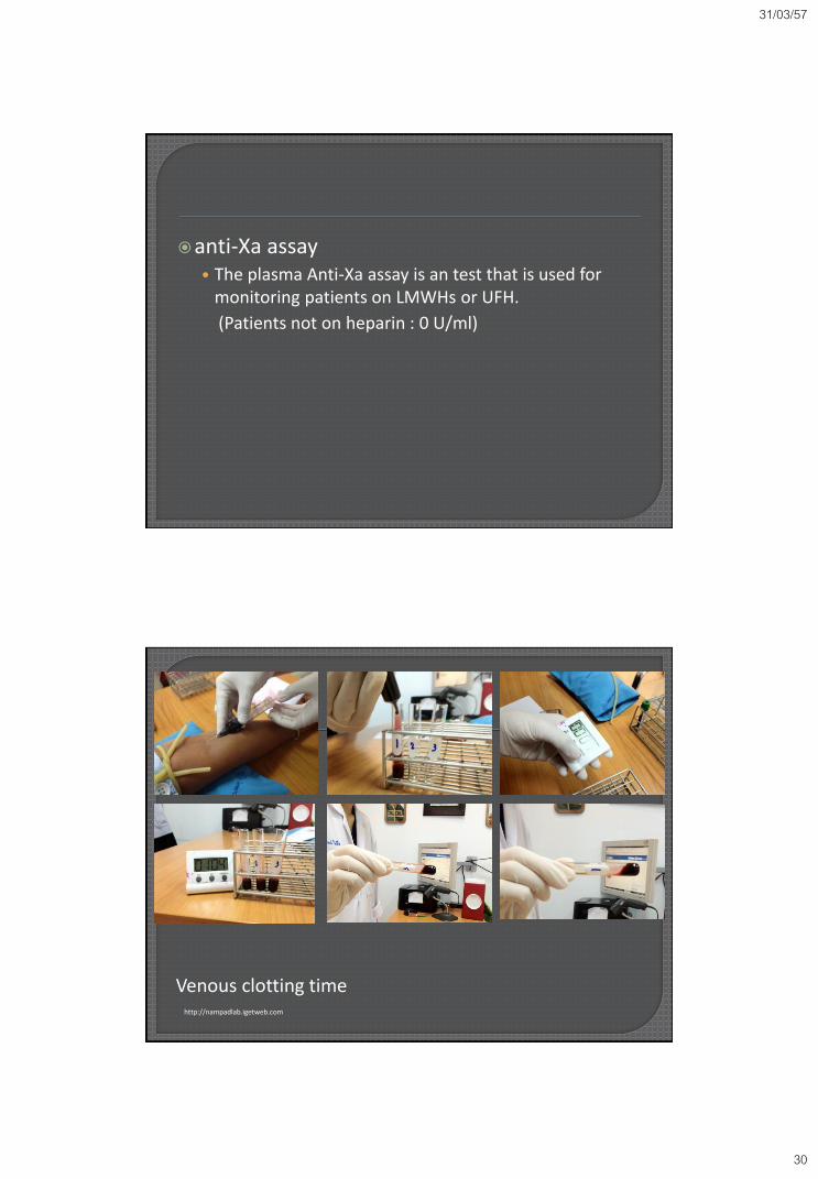
Venous clotting time • Tests the extrinsic and normal pathways.
31/03/57
30
anti-Xa assay • The plasma Anti-Xa assay is an test that is used for
monitoring patients on LMWHs or UFH.
(Patients not on heparin : 0 U/ml)
http://nampadlab.igetweb.com
Venous clotting time
31/03/57
31
Clot Solubility • The result of clot solubility testing may be the only
abnormality in disorders involving factor XIII deficiency and some abnormal fibrinogen. A washed clot is incubated in urea or acetic acid.
• If the clot is not properly cross-linked, it dissolves.
Mixing study (often 1:1) • Mixing studies based on the PT or aPTT are interpreted
based on the fact that a 50% level of any coagulation factor alone gives normal PT and aPTT values.
• Abnormal PT or aPTT
Normal value after normal plasma mixing (immediate or 2hr): Coagulation factor(s) deficit
Abnormal value after normal plasma mixing (immediate or 2hr): Coagulation factor inhibitor

31/03/57
32
Factor Level Assays • Factor levels are determined either by bioassay, in
which the ability of the sample of plasma to normalize controlled substrate-deficient plasma is evaluated, or by immunologic assay.

31/03/57
33
Long aPTT Normal PT Long PT Normal aPTT
Long aPTT Long PT
Associated with bleeding : Factor VII,IX
Anticoagulants DIC Liver disease Vitamin K deficiency Massive transfusion Dysfibrinogenemia
Not associated with bleeding : Lupus anticoagulant (LA)
Long aPTT Normal PT Long PT Normal aPTT
Long aPTT Long PT
Associated with bleeding : Factor VII,IX Acquired inhibitor of FVIII Amyloid adsorb of factor IX FXI
Slight defects in factors XI, X, II, V and dysfibrinogenemias Factor VII (rare)
Anticoagulants DIC Liver disease Vitamin K deficiency Massive transfusion Antibodies to factor V
Not associated with bleeding : Lupus anticoagulant (LA) FXII, PK, HMWK
Common pathway deficiency (rare) Dysfibrinogenemia
Single factor deficiency Perform specific inhibitor assay
Single factor deficiency Perform specific inhibitor assay
Single factor deficiency Perform specific inhibitor assay
Multiple factor deficiency : Pan-inhibitor: LA, Immunoglobulin
Multiple factor deficiency : Pan-inhibitor: LA, Immunoglobulin

31/03/57
34
• CBC and smear (EDTA—purple top)
• Platelet count (EDTA—purple top)
• Bleeding time
• Prothrombin time (citrate—blue top)
• Partial thromboplastin time (citrate—blue top)
• Other coagulation studies: fibrinogen level, thrombin time, clot
solubility, factor levels, inhibitor screens
• As necessary: electrolytes, glucose, BUN, creatinine, type and crossmatch
Bleeding disorders
Hemorrhagic diathesis may be cause by
Increased blood vessel fragility
Platelet disorder
Coagulation defect

31/03/57
35
Bleeding by vessel wall
Increased vascular fragility
Bleeding can occur with
inflammation or malformations of the blood vessels
abnormalities of the connective tissue supporting the blood vessels.
Henoch-Schonlein purpura
vasculitis that occurs with paraproteins or cryoglobulins or in patients with systemic lupus erythematosus or other immune disorders.
Hereditary hemorrhagic telangiectasia is an inherited disorder associated with malformations of the capillaries.
Can also occur with infection
Meningococcus and rickettsia
Vasculitis or DIC
Platelet disorder
Thrombocytopenia
Thrombocytopenia
is defined as a platelet count less than the normal
range, typically below 150,000/µL.

31/03/57
36
Thrombocytopenia
Platelets/mm3
100,000 : can surgery
<100,000 : prolonged bleeding time
<50,000 : bleeding after trauma/surgery
(depends on operation/procedure)
<10,000-20,000: spontaneous bleeding
<5,000 : increase risk of intracranial hemorrhage
Thrombocytopenia
There is no absolute threshold for spontaneous
bleeding due to thrombocytopenia.
It may occur at higher counts when fever, sepsis,
severe anemia, and other hemostatic defects are
present or when platelet function is impaired by
medication

31/03/57
37
Thrombocytopenia
General mechanisms of thrombocytopenia
Increased Platelet Destruction
Platelets Sequestration
Decreased Platelet Production
other e.g. dilution: massive transfusion
Mechanisms of Platelet Destruction
Type of thrombocytopenia Specific example(s)
Immune mediated
Autoantibody-mediated platelet
destruction by reticuloendothelial system (RES)
Primary immune thrombocytopenic
purpura
Secondary immune thrombocytopenic
purpura such as collagen vascular disease; Infectious mononucleosis; HIV
Alloantibody-mediated platelet destruction by RES
Neonatal alloimmune thrombocytopenia
posttransfusion purpura
passive alloimmune thrombocytopenia
alloimmune platelet transfusion refractoriness
Drug-dependent, antibody-mediated platelet destruction by RES
Drug-induced immune thrombocytopenic purpura (e.g., quinine)
Platelet activation by binding of IgG Fc of
drug-dependent IgG to platelet Fc receptor
Heparin-induced thrombocytopenia

31/03/57
38
Mechanisms of Platelet Destruction
Type of thrombocytopenia Specific example(s)
Non-immune mediated
Platelet activation by thrombin or proinflammatory cytokines
Disseminated intravascular Coagulation
septicemia/systemic inflammatory response syndromes
Platelet destruction via ingestion by macrophages (hemophagocytosis)
Infections; certain malignant lymphoproliferative disorders
Platelet destruction through platelet
interactions with altered von Willebrand factor
Thrombotic thrombocytopenic purpura hemolytic-uremic syndrome
Platelet losses on artificial surfaces Cardiopulmonary bypass surgery use of intravascular catheters
Decreased platelet survival associated with cardiovascular disease
Congenital and acquired heart disease
cardiomyopathy pulmonary embolism
Problems that lead to platelets
destruction
Cardiopulmonary bypass surgery
Use of intravascular catheters
Platelets adhere to extracorporeal synthetic surface
Fibrinogen and other plasma proteins adhere to the
artificial surfaces that provide a substrate for platelets
adhesion
Hemodilution

31/03/57
39
It is an autoimmune disease characterized by immune-
mediated platelet destruction.
Acute ITP
Platelet autoantibody
Destruction occurs in the spleen
patients with acute ITP usually have a rapid onset of bleeding
symptoms
Often in children with a history of a recent upper respiratory
infection, febrile illness or immunization
Rubella, CMV, viral hepatitis, infectious mononucleosis
Chronic ITP
In more than 70% of adults, ITP persists beyond the 6-month period
and becomes chronic.
Immune thrombocytopenic purpura (ITP)
Clincal features of ITP
Adult, female, easy bruising or nosebleeding, petechial hemorrhage, internal hemorrhage (melena, hematuria)
The most serious complication of acute ITP is intracranial hemorrhage (ICH)
Diagnosis
Clinical
Blood smear: thrombocytopenia
Bone marrow biopsy:
Increased megakaryocyte
Bleeding time : prolongd
PT and aPTT : normal
ITP is a diagnosis of exclusion.

31/03/57
40
Immune thrombocytopenic purpura (ITP)
the most common and most effective pharmacologic
therapies for acute ITP include intravenous
immunoglobulin (IVIG), Rh immune globulin (RHIG) and
corticosteroids
Drug-induced thrombocytopenia
Immune-mediated platelet destruction
Drug acting as hapten
Drug withdrawal leads to clinical improvement
e.g.
Acetaminophen, Ampicillin, Simvastatin , Ibuprofen Ranitidine,
Naproxen, Phenytoin, Rifampin, Ethambutol Trimethoprim–
sulfamethoxazole,Valproic acid, Carbamazepine, Haloperidol,
Oxaliplatin
Heparin-induced thrombocytopenia

31/03/57
41
• HIT is caused by platelet-activating immunoglobulin G
(IgG) antibodies that bind to multimolecular
complexes of platelet factor 4 (PF4) bound to heparin.
• Whereas DITP is strongly associated with petechiae
and purpura, HIT is strongly associated with thrombosis.

31/03/57
42
• D-ITP : Drug-induced thrombocytopenia
• HIT : Heparin-induced thrombocytopenia
Thrombotic thrombocytopenic purpura (TTP)
A syndrome consisting of
microangiopathic hemolytic anemia (MAHA)
Thrombocytopenia
End-organ damage secondary to microvascular thrombi.
The pathophysiology of TTP is an acute deficiency of von Willebrand factor (VWF)-cleaving metalloprotease ADAMTS13 [a disintegrin and metalloprotease with thrombospondin type 1 motif, 13]) due to
ADAMTS13 inhibitor
Genetic dysfunction
ADAMTS13 deficiency results in the accumulation of ultra-large VWF multimers, which bind platelets, and leads to both thrombi in the microvasculature an thrombocytopenia.

31/03/57
43
• Autopsy : from patient with ITP
• Microthrombi : arrows
med.md.kku.ac.th
microangiopathic hemolytic anemia (MAHA)
Schistocyte : arrow

31/03/57
44
Thrombotic thrombocytopenic purpura (TTP)
The classic pentad of anemia, thrombocytopenia,
fever, neurological signs and renal failure is
infrequently present.
Therapeutic plasma exchange (TPE) is the primary
therapy for TTP

31/03/57
45
Hemolytic uremic syndrome (HUS)
Hemolytic uremic syndrome (HUS) is a thrombotic microangiopathy (TMA) characterized by thrombocytopenia, acute renal failure and microangiopathic hemolytic anemia.
TMA is defined by endothelial cell damage, resulting in platelet-associated thrombosis and vessel obstruction.
Red blood cell (RBC) destruction occurs in the vessels narrowed by these thrombi, leading to schistocyte formation
HUS is most commonly associated with bloody diarrhea caused by shiga-like toxin-producing bacteria (~90% of HUS in children)

31/03/57
46
Platelet sequestration
Platelets may be sequestered when the spleen
enlarges owing to portal hypertension or infiltrative
diseases.
This in turn can result in moderate thrombocytopenia.
Because hypersplenism never causes a platelet count
less than 40,000 to 50,000/µL, bleeding due to
thrombocytopenia from hypersplenism alone is
unusual.

31/03/57
47
Decreased platelet production
Occurs in
Primary diseases of the bone marrow, such as acute
leukemia and aplastic anemia
Myelophthisic processes in which marrow is affected by
metastatic carcinoma, fibrosis, or other clonal
hematopoietic disorders.
Following chemotherapy and/or radiation therapy;
Decreased platelet production
Ethanol toxicity
Infections with viruses such as Dengue virus, HIV,
cytomegalovirus (CMV), Epstein-Barr virus (EBV),
and varicella.
Thrombocytopenia also occurs when normal
megakaryocyte proliferation is impaired by
myelodysplasia.

31/03/57
48
Marked reduction in all three cell lines
(erythroid, myeloid, and megakaryocytic-), with
replacement by normal fat, characterize the
classic picture of aplastic anemia.
tulane.edu
Defective platelet function
Congenital disorder
Defective platelet adhesion
Bernard-Soulier Syndrome: an abnormality in the platelet
GPIb/V/IX complex
Defective platelet aggregration
Glanzmann Thrombasthenia: a quantitative
or qualitative defect in the GPIIb/IIIa complex
Disorders of platelets secretion

31/03/57
49
Defective platelet function
Acquired disorders
Many drugs may affect platelet function.
Aspirin irreversibly acetylates and inactivates the platelet cyclooxygenase, leading to inhibition of synthesis of Thromboxane A2 (TxA2) and endoperoxides (PGG2 and PGH2).
Several other nonsteroidal antiinflammatory drugs (NSAIDs) also impair platelet function by inhibiting the cyclooxygenase enzyme and may prolong bleeding time.
Compared with aspirin, inhibition of cyclooxygenase by these agents is generally short-lived and reversible.

31/03/57
50
Defective platelet function
Ticlopidine, Clopidogrel, and Prasugel are orally administered
thienopyridine derivatives that inhibit platelet function by inhibiting the
binding of ADP to the platelet P2Y12 receptor
Uremia
The pathogenesis remains unclear.
But platelet dysfunction and impaired platelet–vessel wall interaction are
considered the major causes.
Bleeding time is prolonged in uremia.

31/03/57
51
Hemorrhagic diathesis related to
abnormalities in clotting factors
Clinical features
Large ecchymosis or hematoma after injury
Prolonged bleeding after a laceration or surgical procedure
Bleeding of GI, Urinary tract, Weight bearing joints.
Hereditary abnormalities : Hemophilia, von Willebrand disease, etc.
Acquired abnormalities : Liver disease, Dissiminated intravascular coagulation, vitamin K deficiency
von Willebrand disease
vWF serves as a carrier protein for factor VIII
And an adhesive platelet ligand that tethers the
platelet to exposed collagen at sites of vascular
injury.
von Willebrand disease is a group of inherited
bleeding disorders related to
qualitative or quantitative
defects of vWF.

31/03/57
52
Classification of von Willebrand disease
Type Description Inheritance Frequency
1 Partial quantitative deficiency of vWF. Mild abnormalities in multimer structure or distribution may occur.
AD 70-80%
2 Qualitative vWF defects. AD, AR 15-30%
2A Decreased vWF-dependent platelet adhesion and deficiency of HMW vWF multimers.
10%
2B Increased affinity for platelet GPIbα. <5%
2M Decreased vWF-dependent platelet adhesion with a normal multimer distribution.
<2%
2N Decreased affinity for FVIII. <1%
3 Almost complete deficiency of vWF. AR rare

31/03/57
53
von Willebrand disease
Mild type I vWD may not be diagnosed until adulthood
Individuals with vWD primarily complain of excessive mucocutaneous bleeding, such as Bruising without recognized trauma
Prolonged, recurrent nose bleeds
Oral cavity bleeding, including bleeding from the gums after brushing or flossing the teeth or prolonged bleeding after dental cleaning or extractions.
Prolonged excessive bleeding after surgery or trauma
Affected females frequently experience menorrhagia, usually since menarche, and have prolonged or excessive bleeding after childbirth.
von Willebrand disease
In general, the management of vWD can be
divided into three main categories:
localized measures to stop or minimize bleeding;
pharmacologic agents that provide indirect hemostatic
benefit; e.g. Tranexamic acid
treatments that directly increase plasma vWF and FVIII
levels; e.g. Desmopressin (DDAVP), vWF : FVIII
concentrates.

31/03/57
54
Hemophilia
The hemophilias include hemophilia A and hemophilia B, caused
by deficiencies or defects in clotting factor VIII (antihemophilic
factor) and factor IX (antihemophilic factor B, or Christmas
factor), respectively.
Hemophilia A and B are X-linked recessive disorders.
The prevalence in Thailand is between 1 of 13,000 to 20,000 people.
Factor XI deficiency (Hemophilia C) is an autosomal dominant
disorder (severe form is an AR disorder)
The prevalence is 1:1,000,000 in general population.
A deficiency of either of these intrinsic coagulation pathway
proteins results in inadequate formation of thrombin at sites of
vascular injury.
• Acquired hemophilia A (AHA) is a very rare disease,
caused by the development of autoantibodies, directed against circulating factor VIII of coagulation.

31/03/57
55

31/03/57
56
Hemophilia
The severity of bleeding depends upon the
percentage of circulating clotting factor activity.
5–40% are classified as having mild hemophilia
1–5% : moderate
less than 1% : severe
Approximately 60% of all cases of hemophilia A
are clinically severe, whereas only 20 to 45% of
cases of hemophilia B are severe.
Hemophilia
Severe disease, characterized by frequent
spontaneous bleeding events in joints (hemarthrosis)
and soft tissues and by profuse hemorrhage with
trauma or surgery.
Although spontaneous bleeding is uncommon in mild
deficiencies (>5% normal activity), excess bleeding
typically occurs with trauma or surgery.

31/03/57
57
Hemophilia
Diagnosis of hemophilia
A family and personal bleeding history
laboratory detection of prolongation of the aPTT (with normal PT)
Factor assay
If mild hemophilia is clinically suspected in an individual with a normal aPTT, a specific factor should be measured.
Factor antibody
History of factor replacement
Differential diagnosis : Mixing study, Factor antibody
Hemophilia
Treatment and prevention of acute bleeding
are based on replacement of the missing factor.
Recombinant factor VIII or factor IX concentrate
Cryoprecipitate (there is no FIX)
Fresh frozen plasma
Recombinant factor VIIa (For use in patients with
inhibitors to factor VIII, IX or XI)
Etc.

31/03/57
58
Disseminated Intravascular coagulation
DIC is an acquired syndrome characterized by the intravascular activation of coagulation without a specific localization and arising from different causes.
It can originate from and cause damage to the microvasculature, which if sufficiently severe, can produce organ dysfunction.
At the microcirculatory level, both thromboses and hemorrhage into the organ result in ischemia, tissue damage, and progressive organ failure.

31/03/57
59
Cause of DIC
Infectious
Meningococcemia
Bacterial infection (staphylococcal, streptococcal,
Escherichia coli, Salmonella)
Rickettsia, fungus, Malaria, virus
Tissue injury: Massive head injury, massive burn,
profound shock, etc.
Cause of DIC
Venom or toxin e.g. Russell's viper (งูแมวเซา)
Microangiopathic disorder: Severe TTP/HUS, Giant
hemangioma
Gastrointestinal: Pancreatitis, Fulminant hepatitis,
etc.
Misellaneous : Placental abruption, Acute hemolytic
transfusion reaction, ATIII or homozygous Protein C
deficiency etc.

31/03/57
60
Treatment of DIC
Identify and eliminate the underlying cause No treatment if mild, asymptomatic, and self-limited
Hemodynamic support, as indicated, in severe cases
Blood component therapy Indications: active bleeding or high risk for bleeding
Fresh-frozen plasma
Platelets
In some cases, consider cryoprecipitate, antithrombin III
Drug therapy Indications: heparin for DIC manifested by thrombosis or
acrocyanosis;
antifibrinolytic agents generally contraindicated except with life-threatening bleeding and failure of blood component therapy
Coagulation defects in liver disease
Abnormalities in coagulation
Decreased synthesis of coagulation factors
Impaired vitamin K–dependent γ-carboxylation
Dysfibrinogenemia
Disseminated intravascular coagulation
Increased fibrinolytic activity
Abnormalities in platelets
Thrombocytopenia (hypersplenism)
Abnormal platelet function

31/03/57
61
Vitamin K deficiency
Vitamin K is required for γ-carboxylation of glutamic acid residues of the
procoagulant factors II (prothrombin), VII, IX, and X and the anticoagulant
factors protein C and protein S.
Anticoagulant
Heparin
Action Inhibit thrombin (IIa), Inhibit Factor Xa, Inhibit
Factor IX and XI
Heparin binds to the enzyme inhibitor antithrombin III
(AT)
results in its activation
Low molecular weight heparin (LMWH)
Inhibit FXa >> FIIa

31/03/57
62
Monitoring UFH e.g. aPTT, anti-Xa assay
Monitoring LMWH e.g. anti-Xa assay
• Warfarin
– Action
• inhibit vitamin K epoxidase, Vitamin K dependent factor depletion
(II, VII, IX and X)
– Dose adjustment by INR (adjusted PT ratio to ISI)

31/03/57
63
Goldman L, Schafer AI. Goldman’s Cecil medicine. 24th ed. Philadelphia:Elsevier,2011. Hillyer CD, Shaz BH, Zimring JC. Transfusion medicine and hemostasis. 1st ed. Burlington, Massachusetts: Elsevier; 2009. Hoffman R, Benz Jr. EJ, Silberstein LE, Heslop H, Weitz J, Anastasi J. Hematology : Basic principle and practice. 6th ed. Philadelphia: Elsevier; 2012 Kliegman RM, Stanton BMD, Gene JS, Schon NF, Behrman RE. Nelson textbook of pediatric. 15th ed. Philadelphia: Elsevier; 2011. Marx J, Hockerger R, Walls R. Rosen’s Emergency medicine. 7th ed. Philadelphia:Elsevier; 2010. Mcpherson RA, Pincus MR, editors. Henry’s clinical diagnosis and management by laboratory methods. 22th ed. Philadelphia: Elsevier; 2011.


![COVER. CLOT. CONTROL. · [kahy-tuh-sam] 5. PERFORMANCE GAUZE ChitoSAM ™ 100 has a hemostatic performance equivalent to products impregnated or coated with chitosan hemostatic agents.](https://static.fdocuments.net/doc/165x107/5f93dd899f64d646683cc5c7/cover-clot-kahy-tuh-sam-5-performance-gauze-chitosam-a-100-has-a-hemostatic.jpg)


