Hemostasis & Coagulation Ahmad Sh. Silmi Unit 1 : Primary Hemostasis.

1
Hemostasis and Women’s HealthDorothy M. Adcock, MDTitle: HEMOSTASIS AND WOMEN’S HEALTH
I’m Dr. Dorothy Adcock. I’m happy to have this opportunity to present a lecture on Hemostasis and Women’s Health. Why is normal hemostasis, that is no abnormal bleeding tendency and no abnormal clotting tendency, especially important to women’s health? Why do we focus on hemostasis in women’s health?
Title: Why is Normal Hemostasis...

2
Title: Why Women and Hemostasis?
The reason is that physiologically women are predisposed to suffering complications if they have an abnormal hemostatic system. In particular, because of menstruation women bleed every month and those with an underlying bleeding abnormality can suffer severe consequences. Also pregnancy, there are increased risks of bleeding associated with childbirth and as we’ll discuss thrombotic risk that can impart both maternal morbidity and mortality as well as fetus. And, then finally, the use of female sex hormones, in particular progestagen and estrogen agents that are commonly administered as contraceptive agents, specifically birth control pills, infertility therapy, used in post menopausal hormone replacement therapy, in the treatment of hormone responsive cancers, and in women for breast cancer reduction risk. For these reasons a normal hemostatic system is very important to women in particular. Menorrhagia is the first topic that we will discuss. Menorrhagia refers to regular cycles but excessive amount or duration of blood flow.
Title: Menorrhagia
Normally, women bleed for about four days per month, plus or minus two days, and lose about 40 ccs of blood. Menorrhagia is a very common gynecologic symptom. And, in fact, about 10% of women that present to the gynecologist’s office complain of menorrhagia.
3
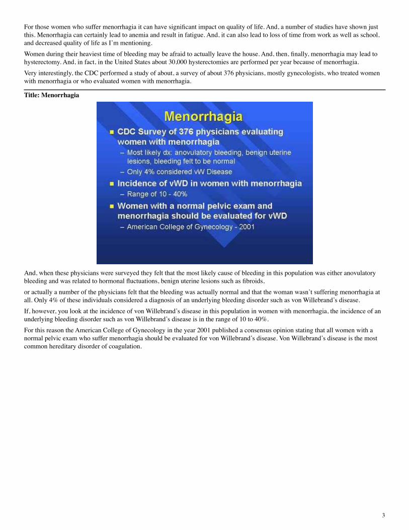
For those women who suffer menorrhagia it can have significant impact on quality of life. And, a number of studies have shown just this. Menorrhagia can certainly lead to anemia and result in fatigue. And, it can also lead to loss of time from work as well as school, and decreased quality of life as I’m mentioning. Women during their heaviest time of bleeding may be afraid to actually leave the house. And, then, finally, menorrhagia may lead to hysterectomy. And, in fact, in the United States about 30,000 hysterectomies are performed per year because of menorrhagia. Very interestingly, the CDC performed a study of about, a survey of about 376 physicians, mostly gynecologists, who treated women with menorrhagia or who evaluated women with menorrhagia.
Title: Menorrhagia
And, when these physicians were surveyed they felt that the most likely cause of bleeding in this population was either anovulatory bleeding and was related to hormonal fluctuations, benign uterine lesions such as fibroids, or actually a number of the physicians felt that the bleeding was actually normal and that the woman wasn’t suffering menorrhagia at all. Only 4% of these individuals considered a diagnosis of an underlying bleeding disorder such as von Willebrand’s disease. If, however, you look at the incidence of von Willebrand’s disease in this population in women with menorrhagia, the incidence of an underlying bleeding disorder such as von Willebrand’s disease is in the range of 10 to 40%. For this reason the American College of Gynecology in the year 2001 published a consensus opinion stating that all women with a normal pelvic exam who suffer menorrhagia should be evaluated for von Willebrand’s disease. Von Willebrand’s disease is the most common hereditary disorder of coagulation.
4
Title: Von Willebrand Disease
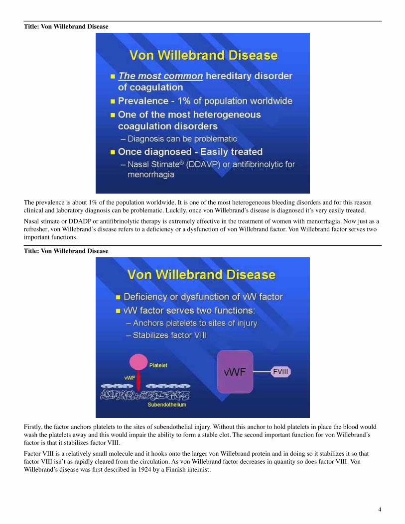
The prevalence is about 1% of the population worldwide. It is one of the most heterogeneous bleeding disorders and for this reason clinical and laboratory diagnosis can be problematic. Luckily, once von Willebrand’s disease is diagnosed it’s very easily treated. Nasal stimate or DDADP or antifibrinolytic therapy is extremely effective in the treatment of women with menorrhagia. Now just as a refresher, von Willebrand’s disease refers to a deficiency or a dysfunction of von Willebrand factor. Von Willebrand factor serves two important functions.
Title: Von Willebrand Disease
Firstly, the factor anchors platelets to the sites of subendothelial injury. Without this anchor to hold platelets in place the blood would wash the platelets away and this would impair the ability to form a stable clot. The second important function for von Willebrand’s factor is that it stabilizes factor VIII. Factor VIII is a relatively small molecule and it hooks onto the larger von Willebrand protein and in doing so it stabilizes it so that factor VIII isn’t as rapidly cleared from the circulation. As von Willebrand factor decreases in quantity so does factor VIII. Von Willebrand’s disease was first described in 1924 by a Finnish internist.
5
Title: Von Willebrand Disease
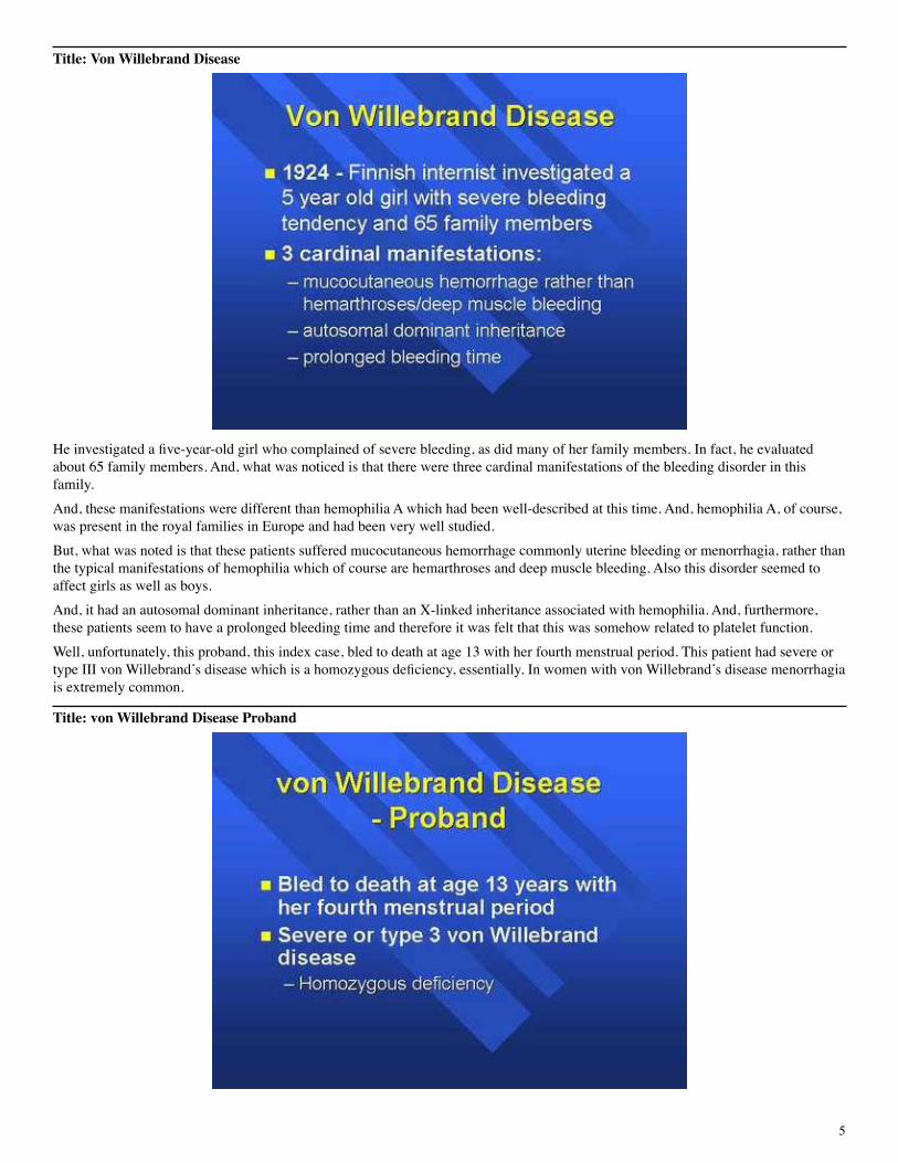
He investigated a five-year-old girl who complained of severe bleeding, as did many of her family members. In fact, he evaluated about 65 family members. And, what was noticed is that there were three cardinal manifestations of the bleeding disorder in this family. And, these manifestations were different than hemophilia A which had been well-described at this time. And, hemophilia A, of course, was present in the royal families in Europe and had been very well studied. But, what was noted is that these patients suffered mucocutaneous hemorrhage commonly uterine bleeding or menorrhagia, rather than the typical manifestations of hemophilia which of course are hemarthroses and deep muscle bleeding. Also this disorder seemed to affect girls as well as boys. And, it had an autosomal dominant inheritance, rather than an X-linked inheritance associated with hemophilia. And, furthermore, these patients seem to have a prolonged bleeding time and therefore it was felt that this was somehow related to platelet function. Well, unfortunately, this proband, this index case, bled to death at age 13 with her fourth menstrual period. This patient had severe or type III von Willebrand’s disease which is a homozygous deficiency, essentially. In women with von Willebrand’s disease menorrhagia is extremely common.
Title: von Willebrand Disease Proband
6
Title: vWD Clinical Manifestations
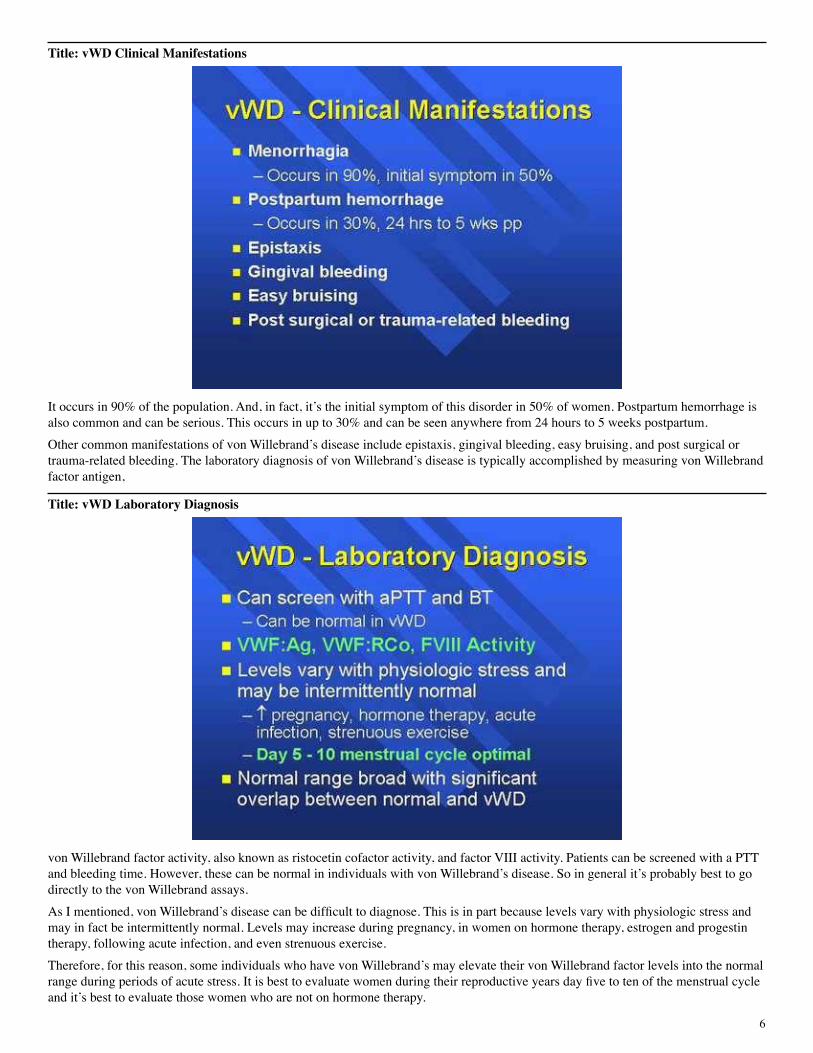
It occurs in 90% of the population. And, in fact, it’s the initial symptom of this disorder in 50% of women. Postpartum hemorrhage is also common and can be serious. This occurs in up to 30% and can be seen anywhere from 24 hours to 5 weeks postpartum. Other common manifestations of von Willebrand’s disease include epistaxis, gingival bleeding, easy bruising, and post surgical or trauma-related bleeding. The laboratory diagnosis of von Willebrand’s disease is typically accomplished by measuring von Willebrand factor antigen,
Title: vWD Laboratory Diagnosis
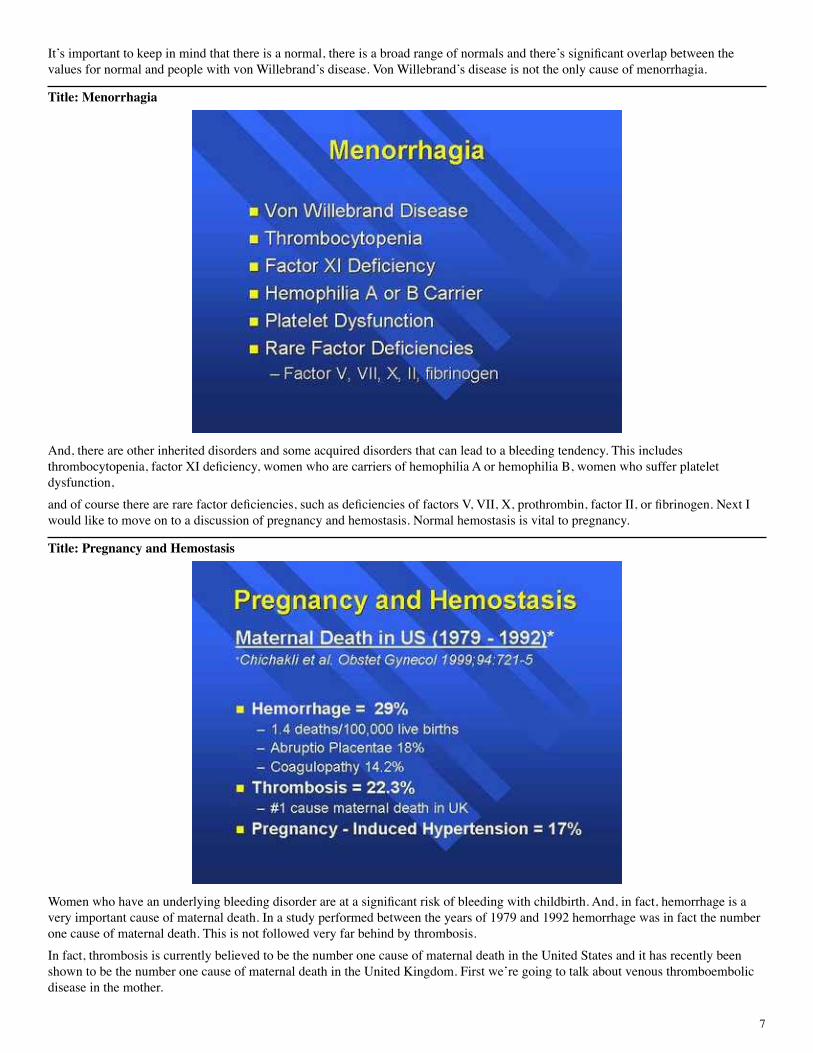
von Willebrand factor activity, also known as ristocetin cofactor activity, and factor VIII activity. Patients can be screened with a PTT and bleeding time. However, these can be normal in individuals with von Willebrand’s disease. So in general it’s probably best to go directly to the von Willebrand assays. As I mentioned, von Willebrand’s disease can be difficult to diagnose. This is in part because levels vary with physiologic stress and may in fact be intermittently normal. Levels may increase during pregnancy, in women on hormone therapy, estrogen and progestin therapy, following acute infection, and even strenuous exercise. Therefore, for this reason, some individuals who have von Willebrand’s may elevate their von Willebrand factor levels into the normal range during periods of acute stress. It is best to evaluate women during their reproductive years day five to ten of the menstrual cycle and it’s best to evaluate those women who are not on hormone therapy.
7
It’s important to keep in mind that there is a normal, there is a broad range of normals and there’s significant overlap between the values for normal and people with von Willebrand’s disease. Von Willebrand’s disease is not the only cause of menorrhagia.
Title: Menorrhagia
And, there are other inherited disorders and some acquired disorders that can lead to a bleeding tendency. This includes thrombocytopenia, factor XI deficiency, women who are carriers of hemophilia A or hemophilia B, women who suffer platelet dysfunction, and of course there are rare factor deficiencies, such as deficiencies of factors V, VII, X, prothrombin, factor II, or fibrinogen. Next I would like to move on to a discussion of pregnancy and hemostasis. Normal hemostasis is vital to pregnancy.
Title: Pregnancy and Hemostasis
Women who have an underlying bleeding disorder are at a significant risk of bleeding with childbirth. And, in fact, hemorrhage is a very important cause of maternal death. In a study performed between the years of 1979 and 1992 hemorrhage was in fact the number one cause of maternal death. This is not followed very far behind by thrombosis. In fact, thrombosis is currently believed to be the number one cause of maternal death in the United States and it has recently been shown to be the number one cause of maternal death in the United Kingdom. First we’re going to talk about venous thromboembolic disease in the mother.

8
Title: Pregnancy and Hemostasis Venous Thromboembolic Disease (VTE)
And, we’ll talk about the development of blood clots in maternal veins and the risk factors that cause these blood clots to develop, as well as the complications associated with these blood clots. Venous thromboembolic disease, or VTE as I’ve abbreviated it here, complicates about 1 in 1,000 to 1 in 2,000 pregnancies. This is actually an extraordinarily frequent complication, and as I mentioned, the number one cause of maternal death. Pregnancy is a prothrombotic state. And, the reason for this physiologically is probably to protect the mother against hemorrhage associated with childbirth. The risk of developing a blood clot during pregnancy is about six times greater than it is in the non-gravid state or the non-pregnant state. It’s very important to keep in mind that the risk is present in all three trimesters and the risk persists into the postpartum period. This is very important for treatment considerations.
Title: Pregnancy and Hemostasis Venous Thromboembolic Disease (VTE)
When blood clots develop in a pregnant female they typically develop more in the iliofemoral region than in the calf region. And, the iliofemoral region pertains to the groin region. In fact, about 72% of clots are in the groin region versus the calf region which only accounts for about 9% of clots during pregnancy. Because clots tend to form in these more proximal veins, which are larger caliber vessels, they are frequently associated with more significant complications.

9
If a blood clot from a large caliber vessel were to break loose and result in pulmonary embolus this pulmonary embolus may very well be more clinically significant because of the larger caliber of the clot itself. There’s also a greater incidence of recurrence with clots that occur in the iliofemoral versus the calf region. There’s also a greater incidence of significant post-phlebitic syndrome which can cause significant morbidity and mortality. Also, clots that develop in the pelvic region or the groin region may present with pelvic pain and this can make diagnosis in a pregnant woman very difficult, remembering that most deep venous thrombosis presents with leg swelling and leg pain. Now as I mentioned, there is a significant risk of pulmonary embolus. And, pulmonary embolus may be seen in 16% of women who are untreated. And, in fact, this is a very important cause of death in this population.
Title: Venous Thromboembolism A Multi-factorial Disease
We’re going to talk a little bit about the development of venous thrombosis. And, I’d like to explain to you that venous thrombosis is a multifactorial disease. It’s really the culmination of a number of risk factors. These risk factors can be genetic, environmental, or physiological. And, these risk factors interact to determine thrombotic risk and, ultimately, thrombus development. A thrombotic risk factor is any factor, stimulus, or condition that increases the chance to develop thrombosis. So this is much like the coronary or the atherosclerotic risk factors that we’re all very familiar with,

10
Title: Thrombotic Risk Factor
hypertension, diabetes, smoking. These thrombotic risk factors that increase the risk for venous thrombosis can be physiological, acquired, or genetic. Many of these risk factors are still unknown and we actually have a lot to learn.
Title: Concept of Risk Potential
What’s interesting about risk factors is that each risk factor differs in its amount or capacity to contribute to the thrombotic etiology. The risk varies, as I mentioned, depending on the individual factor and also the interaction of this risk factor with other risk factors. Some risk factors may interact in an additive fashion, while others interact in a synergistic fashion to greatly increase risk of thrombosis. Another important concept is the concept of thrombotic threshold. The genetic, acquired, and physiologic factors interact to increase the risk for developing thrombosis until a point when a patient’s individual thrombotic threshold is reached.

11
Title: Concept of Thrombotic Threshold
Each of us has our own thrombotic threshold. Once this threshold is approached it may be only a small or a minor provocation that induces thrombosis. This is a diagram that has been presented by Dr. Rosendaal in Lancet in 1999. And, this is a very simplistic yet illustrative model of thrombotic risk.
Title: Model Of Thrombosis Risk
As you can see on the vertical axis, thrombotic risk is listed and it increases until thrombotic threshold is achieved. On the horizontal axis is age. For all of us, our baseline risk increases with increasing age, typically after 40 years of age. If we also have an underlying genetic abnormality which would be persistent for our lifetime that would increase our baseline risk. And, as you can see here factor V Leiden is present in this particular patient and so baseline risk is increased. If there is in addition a physiological risk factor such as pregnancy you can see that transiently that patient’s risk is increased during the state of pregnancy and then if that woman, let’s say she’s 40 years old, had underlying factor V Leiden, she’s pregnant, and she’s put on bedrest for the last part of her pregnancy the immobilization may be all that is required to have that patient surpass thrombotic threshold and develop thrombosis. Ideally, what physicians and patients and need to do is be very aware of underlying risk factors so that they know what underlying risk is and ultimately understand when thrombosis may develop so that it can be prevented or if necessary anticoagulant therapy can be administered.

12
Title: Pregnancy Physiological Risk Factor
As I mentioned, pregnancy is a prothrombotic state. It is a physiological risk factor for the development of thrombosis. And, this is because there are increases in a variety of procoagulant factors as well as decreases in anticoagulant factors and in anticoagulant potential. And, ultimately this tips the balance towards thrombosis. In addition, there are physical changes associated with pregnancy that will predispose a woman to develop a blood clot. There are a number of acquired risk factors, and I’d like to point out some of the more common and more important acquired risk factors.
Title: Pregnancy Acquired Risk Factors
Cesarean delivery is an important acquired risk factor for venous thrombosis. And, in fact, an emergent cesarean delivery increases a woman’s risk tenfold. Maternal age is a very important risk factor. And, as we know many women are having children at an older age. Age greater than 35 years is associated with a three times increased risk over the baseline risk of being pregnant alone. While age greater than 40 years increases risk six times. Obesity is an important risk factor as well as a surgical procedure performed during pregnancy, prolonged immobility, and preeclampsia.

13
Title: Thrombophilia
Next we’re going to talk about thrombophilia and how this pertains to the pregnant state. Thrombophilia is defined as a predisposition to develop venous thrombosis, whether inherited or acquired. Thrombophilia may be the result of a deficiency or dysfunction of a naturally occurring anticoagulant protein such as antithrombin, protein C or protein S. More recently, it’s been shown that thrombophilia may actually be due to an increase or an excess in the procoagulant system. These abnormalities are called gain in function abnormalities. They may reflect such things as increased levels of factor VIII, factor IX, factor XI, and we’ll talk about increased factor II and factor V. If we evaluate all women who develop venous thromboembolic disease during pregnancy or the postpartum period an underlying thrombophilic condition will be identified in up to 50%. Antiphospholipid antibodies are the only acquired form of thrombophilia that we will discuss.
Title: Pregnancy and Hemostasis Venous Thromboembolic Disease

14
Title: Antiphospholipid Antibodies
They are the most common form of acquired thrombophilia. Antiphospholipid antibodies represent a heterogeneous family of antibodies that includes both lupus anticoagulants and anticardiolipin antibodies. These antibodies are associated with both venous and arterial thrombosis, and we’ll find out a little bit later also associated with fetal demise. It’s very important to keep in mind that these antibodies must be demonstrated on two or more occasions at least six to eight weeks apart because we must be able to demonstrate persistence of antiphospholipid antibodies, because only the persistent antibodies are clinically significant. Now as I mentioned, this family of antibodies includes both lupus anticoagulant and anticardiolipin antibodies. It’s therefore very important when screening for the presence of these antibodies that both of these entities be screened for,
Title: Antiphospholipid Antibodies
plasma based assays to look for the lupus anticoagulant, and ELISA based assays to look for anticardiolipin antibodies. And, most of the literature suggests there is only 60% concordance between these two families of antibodies. As I mentioned, antiphospholipid antibodies are very common, seen in about 2% of the population.
15
They can be transient. And, in fact, many of us have had antiphospholipid antibodies at one time or another. However, the transient antibodies are not thought to be clinically significant which is why we must repeat testing and confirm persistence before labeling a patient with clinically significant antiphospholipid antibodies.
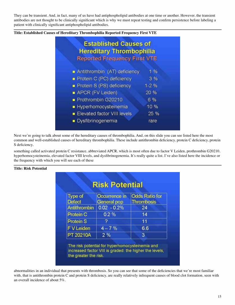
Title: Established Causes of Hereditary Thrombophilia Reported Frequency First VTE
Next we’re going to talk about some of the hereditary causes of thrombophilia. And, on this slide you can see listed here the most common and well-established causes of hereditary thrombophilia. These include antithrombin deficiency, protein C deficiency, protein S deficiency, something called activated protein C resistance, abbreviated APCR, which is most often due to factor V Leiden, prothrombin G20210, hyperhomocysteinemia, elevated factor VIII levels, and dysfibrinogenemia. It’s really quite a list. I’ve also listed here the incidence or the frequency with which you will see each of these
Title: Risk Potential
abnormalities in an individual that presents with thrombosis. So you can see that some of the deficiencies that we’re most familiar with, that is antithrombin protein C and protein S deficiency, are really relatively infrequent causes of blood clot formation, seen with an overall incidence of about 5%.

16
Activated protein C resistance is by far the most common hereditary cause of thrombophilia, accounting for about 20%. Prothrombin 20210 is also very common as is hyperhomocysteinemia and elevated factor VIII levels. Each of these different deficiency states or abnormality states, I should say, is associated with its own risk potential. Each of these factors carries with it an odds ratio for thrombosis. And, as you can see, while antithrombin deficiency is not very common in the general population, if a patient suffers antithrombin deficiency they’re at a significant risk for developing thrombosis, about 24 times that of the general population. Now if you look at factor V Leiden, it’s actually extraordinarily common with an incidence in the Caucasian population of about 5%. The odds ratio for thrombosis is not as high as the plasma based deficiencies, antithrombin, protein C, and protein S. It increases risk of thrombosis about five to seven times. Prothrombin 20210 has an incidence of about 2 to 3% in the Caucasian population and increases thrombotic risk slightly. I’ve listed here that the risk potential for hyperhomocysteinemia and factor VIII are graded, meaning the higher the levels of factor VIII or the higher the levels of homocysteine, the greater is that patient’s risk for developing thrombosis. Now I’ve listed here a cartoon of the clotting cascade. Each of the factors listed in green are the procoagulant factors that ultimately lead to fibrin clot formation. The coagulation cascade is a very, very tightly controlled system.
Title: Naturally Occuring Anticoagulants
For each procoagulant factor there is a naturally occurring anticoagulant. And, I’ve listed some of the more common anticoagulants here. Antithrombin, which is abbreviated AT, is a very important naturally occurring anticoagulant that, as you can see, inhibits a number of the activated factors or serine proteases. Individuals who are born deficient in antithrombin, those that have a heterozygous deficiency, as I mentioned, are at an increased risk of clotting which is actually quite significant, about 24 times normal. Individual, well they’re actually, homozygous deficiency is generally lethal in utero, and this suggests that this is a very important protein. Likewise, the protein C system is another critical pathway. And, protein C functions as an anticoagulant with its cofactor protein S. Together, these proteins inactivate the two cofactors in the system in the clotting cascade, factors V and factors VIII. Heterozygous deficiency of either protein C or protein S carries with it a significant risk of clotting. Homozygous deficiency is lethal in the neonatal state unless it is treated. It results in purpura fulminans. As I mentioned, one of the newer concepts in thrombophilia is that thrombophilia reflects,
17
Title: Thrombophilia Increased Gain in Function
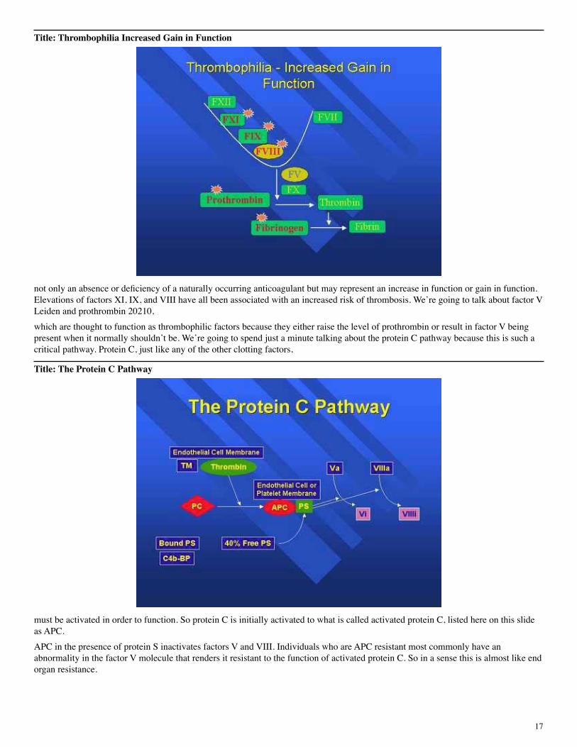
not only an absence or deficiency of a naturally occurring anticoagulant but may represent an increase in function or gain in function. Elevations of factors XI, IX, and VIII have all been associated with an increased risk of thrombosis. We’re going to talk about factor V Leiden and prothrombin 20210, which are thought to function as thrombophilic factors because they either raise the level of prothrombin or result in factor V being present when it normally shouldn’t be. We’re going to spend just a minute talking about the protein C pathway because this is such a critical pathway. Protein C, just like any of the other clotting factors,
Title: The Protein C Pathway
must be activated in order to function. So protein C is initially activated to what is called activated protein C, listed here on this slide as APC. APC in the presence of protein S inactivates factors V and VIII. Individuals who are APC resistant most commonly have an abnormality in the factor V molecule that renders it resistant to the function of activated protein C. So in a sense this is almost like end organ resistance.

18
Title: Activated Protein C Resistance
Activated protein C, as I mentioned, is most commonly due to factor V Leiden which is responsible for more than 90% of all cases of inherited APC resistance. Factor V Leiden is seen in about 5% of all Caucasians. It increases thrombotic risk five to seven times in the heterozygous state, 80 to 100 times if homozygous. The factor V molecule remains active and can promote clot formation. Abnormalities of factor V, specifically factor V Leiden, can be evaluated in the clinical laboratory by using a plasma based screening test. And, this is called the activated protein C resistance assay. These assays should always be confirmed with a molecular test to look specifically for the factor V Leiden mutation. Prothrombin G 20210A is also a very common cause of thrombophilia. It is seen in about 2.3% of the Caucasian population.
Title: Prothrombin G20210A
This is a single amino acid mutation which involves the untranslated portion of the factor II or prothrombin molecule. And, it’s believed to cause an increased risk of thrombosis by increasing factor II levels or prothrombin levels. Overall, prothrombin 20210 increases thrombotic risk about three to six times. There is no plasma based screening assay available for this mutation and a DNA based assay must be performed. Another common cause of thrombophilia reflects elevated homocysteine levels. Homocysteine is an amino acid precursor involved in the metabolic conversion of methionine.

19
Title: Homocysteine
There is very strong evidence that hyperhomocysteinemia or elevated plasma levels of homocysteine are a risk factor for premature atherosclerosis and recurrent thromboembolic disease. Elevations of homocysteine can be seen with B12, B6, and folate deficiency. And, they can also be related to underlying genetic abnormalities. These reflect abnormalities in two important enzymes in this system, specifically CBS or cystathionine beta synthase and MTHFR, also known as methylene tetrahydrofolate reductase.
Title: MTHFR C677T Polymorphism Homozygote
The MTHFR C677T polymorphism is actually a very common abnormality in the homozygous state. This abnormality is seen in about 5% of the population. It’s thought to increase, to have the potential to increase levels of homocysteine when the patient has an underlying vitamin deficiency such as decreased plasma folate levels. It’s really not known if this abnormality by itself increases risk, but it’s thought that it may interact with other risk factors that we’ve discussed increased risk, and may interact with these other risk factors in a synergistic manner. As I’ve mentioned, pregnancy is a prothrombotic state.
20
Title: Thrombophilia and Pregnancy
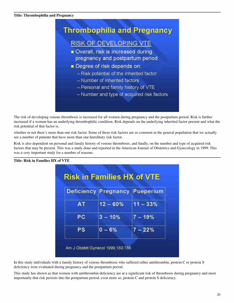
The risk of developing venous thrombosis is increased for all women during pregnancy and the postpartum period. Risk is further increased if a woman has an underlying thrombophilic condition. Risk depends on the underlying inherited factor present and what the risk potential of that factor is, whether or not there’s more than one risk factor. Some of these risk factors are so common in the general population that we actually see a number of patients that have more than one hereditary risk factor. Risk is also dependent on personal and family history of venous thrombosis, and finally, on the number and type of acquired risk factors that may be present. This was a study done and reported in the American Journal of Obstetrics and Gynecology in 1999. This was a very important study for a number of reasons.
Title: Risk in Families HX of VTE
In this study individuals with a family history of venous thrombosis who suffered either antithrombin, protein C or protein S deficiency were evaluated during pregnancy and the postpartum period. This study has shown us that women with antithrombin deficiency are at a significant risk of thrombosis during pregnancy and most importantly that risk persists into the postpartum period, even more so, protein C and protein S deficiency.

21
As you can see from this slide, while there is some risk during pregnancy these women suffer their greatest risk of developing thrombosis in the postpartum period. Another study that was reported in 1997 looked at factor V Leiden as well as antithrombin protein C and protein S deficiency.
Title: Risk of Pregnancy Associated VTE
This was a huge study performed by Dr. McColl and he evaluated 72,000 pregnancies. His numbers corroborate very well the previous study that we have just discussed. Women with antithrombin deficiency have about a 30% chance of developing thrombosis in pregnancy or the postpartum period, while those with protein C deficiency it’s about 1 in 100, factor V Leiden about 1 in 400. Based on these studies it’s now recommended that women with documented antithrombin deficiency should be treated with heparin therapy throughout pregnancy and receive anticoagulant therapy for at least eight weeks postpartum, whether or not the patient has had a personal history of thrombosis.
Title: Risk of Pregnancy Associated VTE
And, what this speaks to is the importance of identifying families with thrombophilic risk factors because women therefore can be treated and thrombosis prevented even if they don’t have a personal history of thrombosis. Women with either protein C or protein S deficiency and a family history of thrombosis, although having no personal history of thrombosis, should be anticoagulated in the postpartum period over an eight to twelve week time frame.

22
Title: Risk of Pregnancy Associated VTE
Another study that was done a little bit later than the one I mentioned previously also evaluated prothrombin G20210A. And, in this study 119 women with venous thrombosis were evaluated. And, as you can see, the risk of developing a blood clot was about 1 in 500 for factor V Leiden, surprisingly, about 1 in 200 for prothrombin 20210. But, if a woman is unfortunate to have both abnormalities her risk is really quite significant, 5 per 100. This study also evaluated homozygous MTHFR and found no increased risk in the homozygous state. Another study looked at heterozygous factor V Leiden but also looked at homozygous factor V Leiden patients.
Title: Factor V Leiden and VTE
And, in this study it was shown that about 4% of women who are homozygous for factor V Leiden develop venous thrombosis during pregnancy with a 5% incidence in the postpartum period.
23
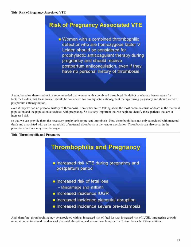
Title: Risk of Pregnancy Associated VTE
Again, based on these studies it is recommended that women with a combined thrombophilic defect or who are homozygous for factor V Leiden, that these women should be considered for prophylactic anticoagulant therapy during pregnancy and should receive postpartum anticoagulation, even if they’ve had no personal history of thrombosis. Remember we’re talking about the most common cause of death in the maternal population and the population associated with pregnancy. So it’s very important that we begin to identify these patients that are at increased risk, so that we can provide them the necessary prophylaxis to prevent thrombosis. Now thrombophilia is not only associated with maternal death and associated with an increased risk of maternal thrombosis in the venous circulation. Thrombosis can also occur in the placenta which is a very vascular organ.
Title: Thrombophilia and Pregnancy
And, therefore, thrombophilia may be associated with an increased risk of fetal loss, an increased risk of IUGR, intrauterine growth retardation, an increased incidence of placental abruption, and severe preeclampsia. I will describe each of these entities.

24
Title: Pregnancy and Thrombophilia
Each of the disorders I’ve just mentioned may be due to an abnormality of the placenta, either an abnormality within the circulation or a thrombosis. Development and sustained function of the placenta is dependent on adequate uteroplacental circulation. And, this is necessary for normal fetal development.
Title: Pregnancy and Thrombophilia
Fetal loss can be described as miscarriage or stillbirth. Miscarriage, also known as abortion, reflects delivery or loss of products before 20 weeks. About 80% of spontaneous abortions occur in the first trimester. These abortions tend to have a fetal origin, rather than maternal origin. So perhaps a chromosomal abnormality of the fetus. Fetal loss that occurs in the second trimester or beyond tends to have a maternal cause. Stillbirth is the term used for loss after about 22 weeks gestation.
25
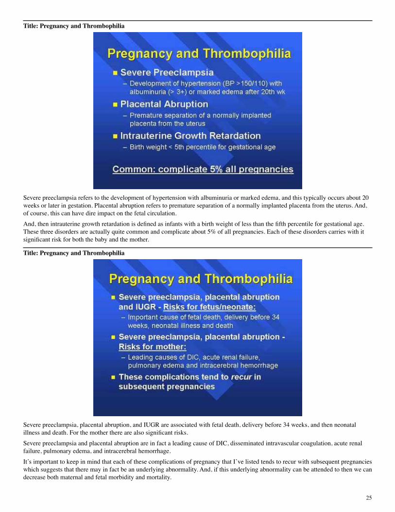
Title: Pregnancy and Thrombophilia
Severe preeclampsia refers to the development of hypertension with albuminuria or marked edema, and this typically occurs about 20 weeks or later in gestation. Placental abruption refers to premature separation of a normally implanted placenta from the uterus. And, of course, this can have dire impact on the fetal circulation. And, then intrauterine growth retardation is defined as infants with a birth weight of less than the fifth percentile for gestational age. These three disorders are actually quite common and complicate about 5% of all pregnancies. Each of these disorders carries with it significant risk for both the baby and the mother.
Title: Pregnancy and Thrombophilia
Severe preeclampsia, placental abruption, and IUGR are associated with fetal death, delivery before 34 weeks, and then neonatal illness and death. For the mother there are also significant risks. Severe preeclampsia and placental abruption are in fact a leading cause of DIC, disseminated intravascular coagulation, acute renal failure, pulmonary edema, and intracerebral hemorrhage. It’s important to keep in mind that each of these complications of pregnancy that I’ve listed tends to recur with subsequent pregnancies which suggests that there may in fact be an underlying abnormality. And, if this underlying abnormality can be attended to then we can decrease both maternal and fetal morbidity and mortality.

26
Title: Antiphospholipid Antibodies
Fetal demise is a hallmark of antiphospholipid antibody, of patients with antiphospholipid antibodies. Fetal loss may occur at any time during pregnancy. And, in fact, in women with clinically significant antiphospholipid antibodies the incidence of recurrent fetal loss is extraordinary. As you can see here, the incidence is 30 to 75%. Antiphospholipid antibodies in fetal loss have been studied for a number of decades. And, in fact, it’s been shown that women who are treated with aspirin and heparin prophylaxis may have increased fetal survival. And, as you can see here from the slide, survival may increase from 50 to 80% which is really an extraordinary accomplishment. In all women with recurrent fetal loss about 15% have antiphospholipid antibodies.
Title: Odds Ratio for Fetal Loss
Once it was realized that antiphospholipid antibodies could be associated with fetal loss, and in fact I should mention the mechanism is frequently placental thrombosis, it was realized that perhaps other causes of thrombophilia should be investigated. This was a study published in the Lancet in 1996. And, as you can see, women who suffer antithrombin, protein C protein S deficiency or have factor V Leiden have an increased risk for both miscarriage, but a greater risk for stillbirth. Women with a combined deficiency are at a markedly increased risk for stillbirth which, as you can see here, is over 14 times increased.

27
Title: Obstetric Complications and Thrombophilia
The obstetric complications that I’ve listed as well, that is preeclampsia, placental abruption, and IUGR, are also associated with placental thrombosis and hence thrombophilia. In this study that was reported in the New England Journal in 1999 110 women with the obstetric complications that I have listed without a history of venous thrombosis were evaluated for these common thrombophilic abnormalities. Remarkably, 50% of women had one or more of the abnormalities listed, specifically factor V Leiden, prothrombin 20210, or MTHFR C677T. Thirteen percent had anticardiolipin antibodies, antithrombin, protein C or protein S deficiency compared to 1% of controls.
Title: Obstetric Complications
And, in fact if you look at the odds ratio for development of the complications that we’ve listed, you can see here that the odds ratio is significant and actually correlates very well with the risk potential of the various factors that we’ve been discussing.

28
Title: Obstetric Complications Hyperhomocysteinemia
The European community has evaluated hyperhomocysteinemia probably more so than we have here in the states. And, as you can see, hyperhomocysteinemia is also associated with the complications we’ve listed as well as stillbirth.
Title: Who Should Be Screened?
What this suggests, therefore, is that there is a certain population of women that should be screened for these thrombophilic defects. Certainly, women with a family history of thrombophilia or a family history of clinically significant venous thrombosis, women who themselves have suffered a previous venous thrombosis or thrombosis during pregnancy. Something new is that women who have suffered unexplained second trimester fetal loss as well as women who suffer recurrent first trimester fetal loss should also be screened. This typically refers to three or more pregnancy losses in the first trimester. Women who have severe or recurrent preeclampsia, intrauterine growth retardation, or placental abruption should also be screened. Which assays should be included in the screening is a bit controversial and varies between experts and between institutions.
29
Title: Screening for Thrombophilia
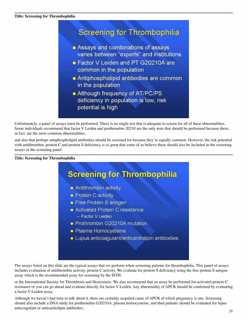
Unfortunately, a panel of assays must be performed. There is no single test that is adequate to screen for all of these abnormalities. Some individuals recommend that factor V Leiden and prothrombin 20210 are the only tests that should be performed because these, in fact, are the most common abnormalities, and also that perhaps antiphospholipid antibodies should be screened for because they’re equally common. However, the risk potential with antithrombin, protein C and protein S deficiency is so great that some of us believe these should also be included in the screening assays or the screening panel.
Title: Screening for Thrombophilia
The assays listed on this slide are the typical assays that we perform when screening patients for thrombophilia. This panel of assays includes evaluation of antithrombin activity, protein C activity. We evaluate for protein S deficiency using the free protein S antigen assay which is the recommended assay for screening by the ISTH, or the International Society for Thrombosis and Hemostasis. We also recommend that an assay be performed for activated protein C resistance or you can go ahead and evaluate directly for factor V Leiden. Any abnormality of APCR should be confirmed by evaluating a factor V Leiden assay. Although we haven’t had time to talk about it, there are certainly acquired cause of APCR of which pregnancy is one. Screening should also include a DNA study for prothrombin G20210A, plasma homocysteine, and then patients should be evaluated for lupus anticoagulant or anticardiolipin antibodies.
30
Title: Screening for Thrombophilia Effect of Pregnancy
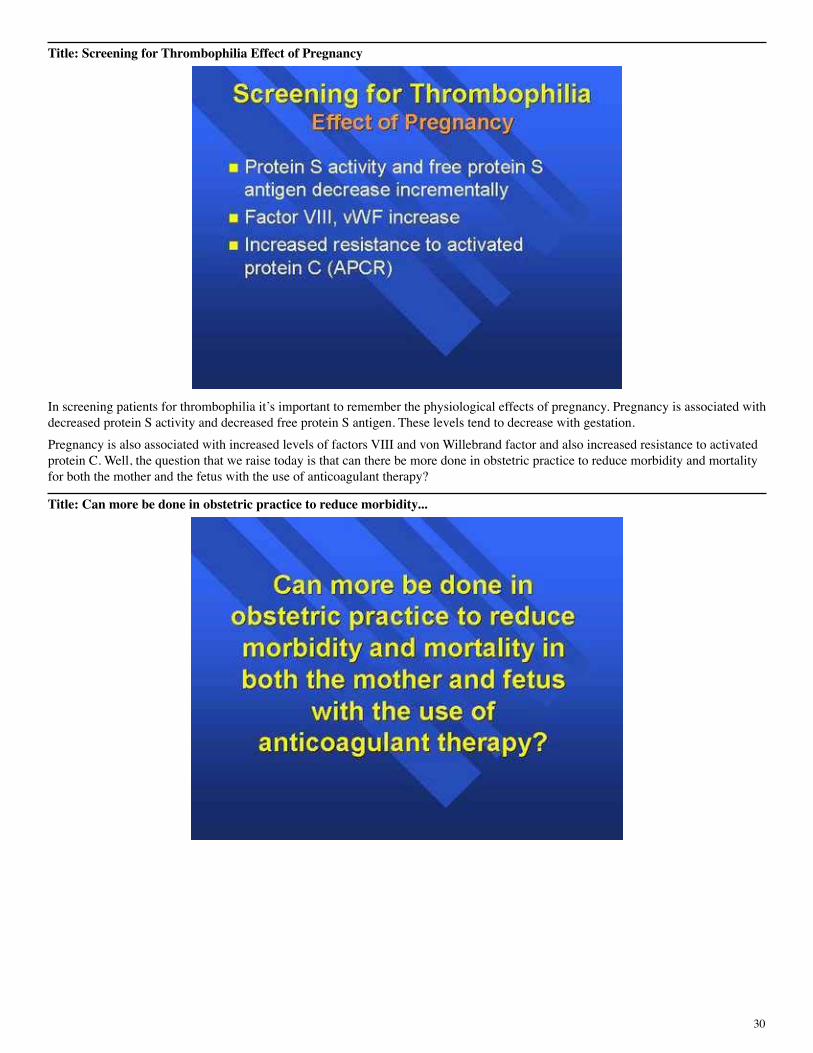
In screening patients for thrombophilia it’s important to remember the physiological effects of pregnancy. Pregnancy is associated with decreased protein S activity and decreased free protein S antigen. These levels tend to decrease with gestation. Pregnancy is also associated with increased levels of factors VIII and von Willebrand factor and also increased resistance to activated protein C. Well, the question that we raise today is that can there be more done in obstetric practice to reduce morbidity and mortality for both the mother and the fetus with the use of anticoagulant therapy?
Title: Can more be done in obstetric practice to reduce morbidity...
31
Title: Antithrombotic Therapy in Pregnancy
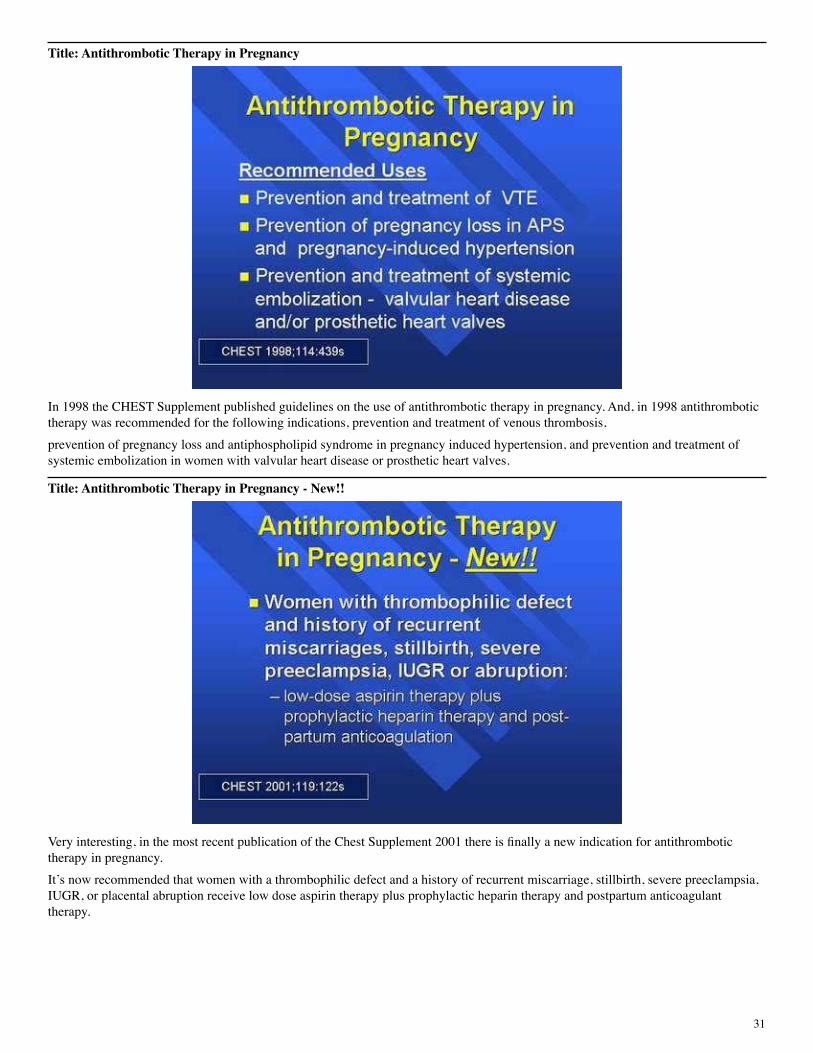
In 1998 the CHEST Supplement published guidelines on the use of antithrombotic therapy in pregnancy. And, in 1998 antithrombotic therapy was recommended for the following indications, prevention and treatment of venous thrombosis, prevention of pregnancy loss and antiphospholipid syndrome in pregnancy induced hypertension, and prevention and treatment of systemic embolization in women with valvular heart disease or prosthetic heart valves.
Title: Antithrombotic Therapy in Pregnancy - New!!
Very interesting, in the most recent publication of the Chest Supplement 2001 there is finally a new indication for antithrombotic therapy in pregnancy. It’s now recommended that women with a thrombophilic defect and a history of recurrent miscarriage, stillbirth, severe preeclampsia, IUGR, or placental abruption receive low dose aspirin therapy plus prophylactic heparin therapy and postpartum anticoagulant therapy.

32
Title: Pregnancy related complications & thrombophilia
A small number, excuse me, a number of small studies have been reported and have shown decreased complication rates and higher percentages of successful pregnancies in women who have received antithrombotic therapy during pregnancy. And, this is really very exciting. It is, however, always important to consider the risk benefit ratio for both the mother and the fetus. Which antithrombotic agents can be used during pregnancy? We’ve got three available agents, heparin and heparin-like compounds, coumarin derivatives, or warfarin, also known as warfarin, and anti-platelet agents.
Title: Antithrombotic Therapy in Pregnancy
I’ll just mention that I won’t say much about anti-platelet agents and in fact there are very few if any studies which have looked at the use of these compounds during pregnancy. Heparin therapy is the recommended anticoagulant of choice during pregnancy.
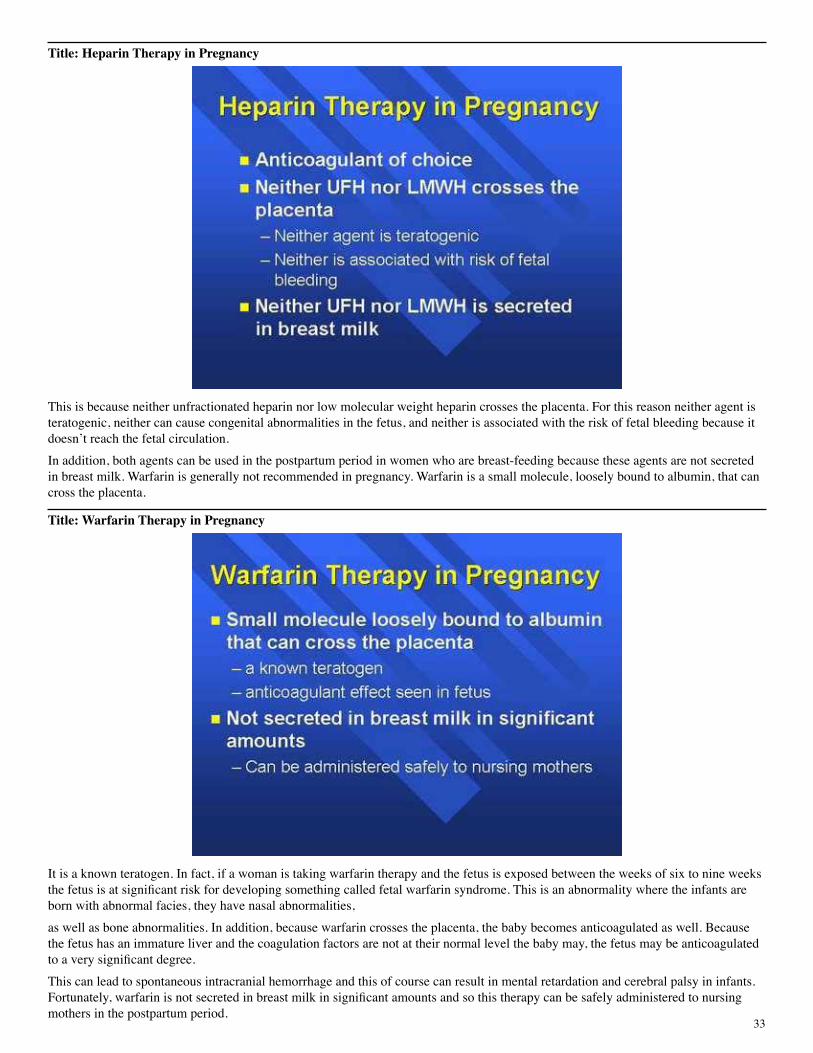
33
Title: Heparin Therapy in Pregnancy
This is because neither unfractionated heparin nor low molecular weight heparin crosses the placenta. For this reason neither agent is teratogenic, neither can cause congenital abnormalities in the fetus, and neither is associated with the risk of fetal bleeding because it doesn’t reach the fetal circulation. In addition, both agents can be used in the postpartum period in women who are breast-feeding because these agents are not secreted in breast milk. Warfarin is generally not recommended in pregnancy. Warfarin is a small molecule, loosely bound to albumin, that can cross the placenta.
Title: Warfarin Therapy in Pregnancy
It is a known teratogen. In fact, if a woman is taking warfarin therapy and the fetus is exposed between the weeks of six to nine weeks the fetus is at significant risk for developing something called fetal warfarin syndrome. This is an abnormality where the infants are born with abnormal facies, they have nasal abnormalities, as well as bone abnormalities. In addition, because warfarin crosses the placenta, the baby becomes anticoagulated as well. Because the fetus has an immature liver and the coagulation factors are not at their normal level the baby may, the fetus may be anticoagulated to a very significant degree. This can lead to spontaneous intracranial hemorrhage and this of course can result in mental retardation and cerebral palsy in infants. Fortunately, warfarin is not secreted in breast milk in significant amounts and so this therapy can be safely administered to nursing mothers in the postpartum period.

34
Keep in mind that heparin therapy must be injected and warfarin therapy can be taken orally. Danaparoid is also an anticoagulant agent that is typically the recommended agent in women with HIT, or heparin induced thrombocytopenia. Dextran as well as hirudin should be avoided.
Title: Other Antithrombotic Agents
Title: APTT to Monitor Heparin Therapy
When patients are placed on heparin therapy we typically monitor the heparin therapy and this is done most commonly with the activated partial thromboplastin time. The aPTT, or activated partial thromboplastin time, is actually a surrogate measure of heparinization and is a poor predictor of heparin anticoagulation. One of the reasons for this is that patients may have an abnormal baseline aPTT, either elevated or shortened. And, this can interfere with the interpretation of aPTT ratios in the evaluation of degree of heparinization. It’s been shown that in non-gravid or non-pregnant females or males, in the non-gravid population, you can see up to 50% discordance between aPTT levels and heparin assay levels, that is anti-Xa levels. And, I’d like to just describe the heparin assay. This is an assay that can be put on to most coagulation analyzers.

35
Title: Heparin Anti Xa Assay
It’s a chromogenic assay. It’s a very simple assay to perform and it’s been around for a number of years. This assay can typically be available on a routine or stat basis. In this assay patients’ antithrombin (or antithrombin is added from reagent) is combined with heparin that is being measured in the patient sample. As you may remember, heparin functions by potentiating antithrombin’s anticoagulant nature. Antithrombin and heparin form a complex then factor Xa is added in excess. The antithrombin heparin complex binds the Xa and the residual Xa remaining is inversely proportional to the heparin concentration. And, this residual Xa can be very easily evaluated by adding a substrate and looking for some sort of a signal. This assay, called the heparin anti-Xa assay, can be used to monitor either unfractionated heparin or low molecular weight heparin therapy.
Title: Heparin Anti Xa Assay
It’s very important that when you’re establishing this in your laboratory that you use the appropriate curve in order to evaluate each of these agents. If you’re monitoring patients on unfractionated heparin therapy you must use a standard curve that is based on unfractionated heparin and likewise for low molecular weight heparin. And, I’ve listed here the recommended therapeutic ranges for each of these agents, and it’s also important to keep in mind that the timing of these assays should be four hours post injection for low molecular weight heparin, whereas with unfractionated heparin therapy levels are typically monitored six hours post injection.
36
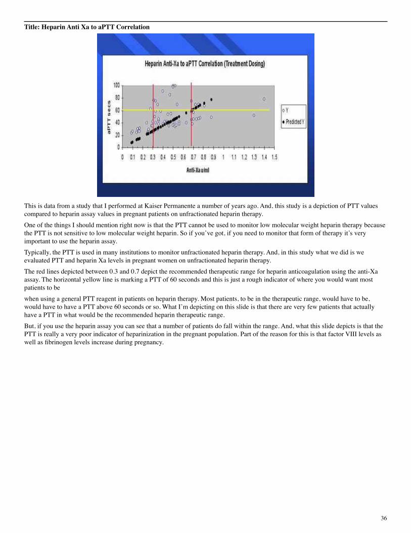
Title: Heparin Anti Xa to aPTT Correlation
This is data from a study that I performed at Kaiser Permanente a number of years ago. And, this study is a depiction of PTT values compared to heparin assay values in pregnant patients on unfractionated heparin therapy. One of the things I should mention right now is that the PTT cannot be used to monitor low molecular weight heparin therapy because the PTT is not sensitive to low molecular weight heparin. So if you’ve got, if you need to monitor that form of therapy it’s very important to use the heparin assay. Typically, the PTT is used in many institutions to monitor unfractionated heparin therapy. And, in this study what we did is we evaluated PTT and heparin Xa levels in pregnant women on unfractionated heparin therapy. The red lines depicted between 0.3 and 0.7 depict the recommended therapeutic range for heparin anticoagulation using the anti-Xa assay. The horizontal yellow line is marking a PTT of 60 seconds and this is just a rough indicator of where you would want most patients to be when using a general PTT reagent in patients on heparin therapy. Most patients, to be in the therapeutic range, would have to be, would have to have a PTT above 60 seconds or so. What I’m depicting on this slide is that there are very few patients that actually have a PTT in what would be the recommended heparin therapeutic range. But, if you use the heparin assay you can see that a number of patients do fall within the range. And, what this slide depicts is that the PTT is really a very poor indicator of heparinization in the pregnant population. Part of the reason for this is that factor VIII levels as well as fibrinogen levels increase during pregnancy.

37
Title: Effect of FVIII Levels on APTT
This was another study that I performed with Dr. Marlar at the Denver VA. And, what we showed here was that as plasma levels of factor VIII increase from about 150 to 300 or 350%, which is a very common range of elevation in an acute phase, the PTT in patients on unfractionated heparin therapy drops up to 10% and it drops significantly. So the elevated factor VIII levels and elevated fibrinogen levels decrease the PTT and therefore cause underestimation of the heparinization of the patient. This is called in vitro heparin resistance.
Title: In Vitro Heparin Resistance
Increased levels of factor VIII and fibrinogen will result in a discordance between the aPTT and anti-Xa levels resulting in attenuation of the aPTT which can be significant, particularly in the third trimester. In addition, pregnancy is not a normal physiologic state. During pregnancy heparin requirements are increased,
38
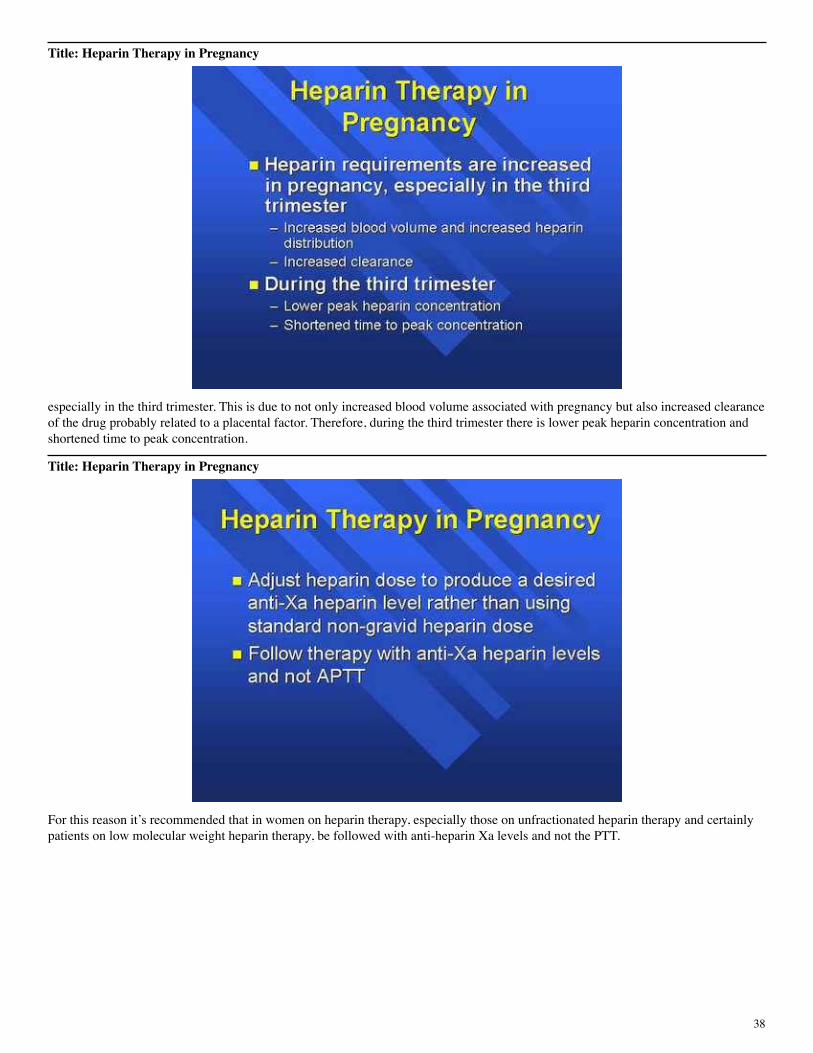
Title: Heparin Therapy in Pregnancy
especially in the third trimester. This is due to not only increased blood volume associated with pregnancy but also increased clearance of the drug probably related to a placental factor. Therefore, during the third trimester there is lower peak heparin concentration and shortened time to peak concentration.
Title: Heparin Therapy in Pregnancy
For this reason it’s recommended that in women on heparin therapy, especially those on unfractionated heparin therapy and certainly patients on low molecular weight heparin therapy, be followed with anti-heparin Xa levels and not the PTT.

39
Title: Pregnancy and Thrombophilia
It’s very important to realize that thrombophilia may be associated with serious consequences in the pregnant female, and that there may be potential benefit to both mother and baby with appropriate anticoagulant therapy as long as we administer that therapy and monitor it carefully.
Title: Oral Contraceptives and Hormone Replacement Therapy
Next, I’d like to discuss oral contraceptives and hormone replacement therapy. It’s important to realize that these therapies are used by hundreds of millions of women worldwide. It’s also important to realize that female sex hormones, particularly estrogen and progestin agents, increase the risk of venous thrombosis and to a lesser extent heart attack and stroke. When we talk about the use of female sex hormones these can be used in the form of contraception, birth control pills, post menopausal hormone replacement therapy. They can be used in the treatment of hormone responsive cancers, and also be given to women for breast cancer reduction. Let’s first discuss the risk of venous thrombosis with oral contraceptives. Oral contraceptive agents are typically classified based on generation.

40
Title: Oral Contraceptives and VTE
First generation oral contraceptives were introduced about 30 years ago and these pills had a very high estrogen content. The venous thromboembolic risk associated with these agents was quite high, greater than tenfold. Based on this risk second generation pills were developed that had a significantly lower estrogen content. With these pills the risk for venous thrombosis was about three to fourfold. In the 1990s in Europe, and later in the United States, a third generation pill was introduced which as you can see has a lower estrogen content and a varied progestin content based on the second generation pill. However, this third generation pill was associated with a six to eightfold increased risk of thrombosis. It’s important to keep in mind that the use of oral contraceptives is the major cause of thrombotic disease in young women. It’s also important that this risk of thrombosis persists even with low dose oral contraceptive pills.
Title: Oral Contraceptives and VTE
Also, what the third generation pills taught us was that it’s not only the estrogen level but also the progestagen in the combination that increases thrombotic risk. Studies have shown that the risk of venous thrombosis is greatest the first year of use and probably diminishes somewhat after that. Overall, the use of oral contraceptives increases a woman’s risk from 0.8 per 10,000 per year to 3 per 10,000 per year, remembering that in the population that takes oral contraceptive pills the underlying incidence of thromboembolic disease is low because it’s a young population.
41
So really, overall, risk is not that significant. What are some of the reasons that risk is increased? Well, oral contraceptives are associated with elevations of a variety of procoagulant factors, decreases in protein S, antithrombin and decreased fibrinolysis.
Title: Biological Mechanism
It’s also been shown that the hemostatic system of some women are apparent hyper-responders. So there are some women that show increased responsiveness of these biological effects of hormone therapy compared to others. Now what about women with underlying thrombophilia who are put on oral contraceptive pills?
Title: Oral Contraceptives and VTE
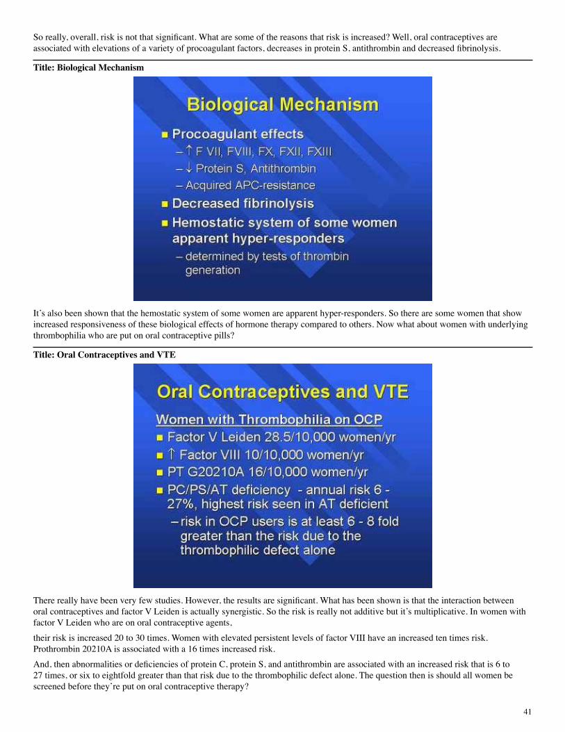
There really have been very few studies. However, the results are significant. What has been shown is that the interaction between oral contraceptives and factor V Leiden is actually synergistic. So the risk is really not additive but it’s multiplicative. In women with factor V Leiden who are on oral contraceptive agents, their risk is increased 20 to 30 times. Women with elevated persistent levels of factor VIII have an increased ten times risk. Prothrombin 20210A is associated with a 16 times increased risk. And, then abnormalities or deficiencies of protein C, protein S, and antithrombin are associated with an increased risk that is 6 to 27 times, or six to eightfold greater than that risk due to the thrombophilic defect alone. The question then is should all women be screened before they’re put on oral contraceptive therapy?
42
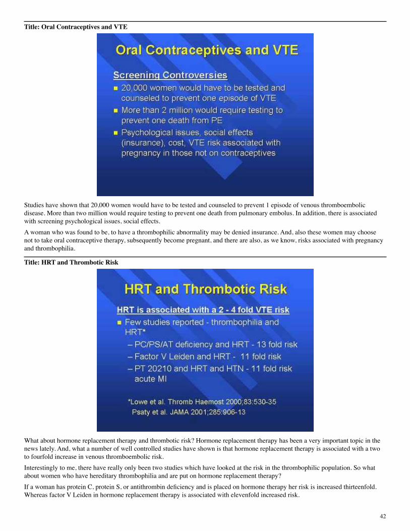
Title: Oral Contraceptives and VTE
Studies have shown that 20,000 women would have to be tested and counseled to prevent 1 episode of venous thromboembolic disease. More than two million would require testing to prevent one death from pulmonary embolus. In addition, there is associated with screening psychological issues, social effects. A woman who was found to be, to have a thrombophilic abnormality may be denied insurance. And, also these women may choose not to take oral contraceptive therapy, subsequently become pregnant, and there are also, as we know, risks associated with pregnancy and thrombophilia.
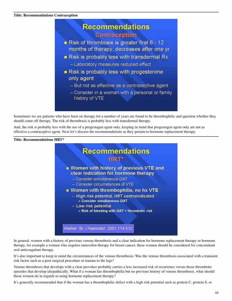
Title: HRT and Thrombotic Risk
What about hormone replacement therapy and thrombotic risk? Hormone replacement therapy has been a very important topic in the news lately. And, what a number of well controlled studies have shown is that hormone replacement therapy is associated with a two to fourfold increase in venous thromboembolic risk. Interestingly to me, there have really only been two studies which have looked at the risk in the thrombophilic population. So what about women who have hereditary thrombophilia and are put on hormone replacement therapy? If a woman has protein C, protein S, or antithrombin deficiency and is placed on hormone therapy her risk is increased thirteenfold. Whereas factor V Leiden in hormone replacement therapy is associated with elevenfold increased risk.

43
Prothrombin 20210 interestingly has been associated with an increased risk of acute myocardial infarction, especially in those women who have hypertension, and particularly in those women who smoke. Therefore, should we screen a certain population of women before they’re put on hormone therapy?
Title: Screening and Hormone Therapy
Screening may be indicated in women who have a personal or family history of venous thrombosis. Screening may also be indicated in a woman who simply does not choose to tolerate the unknown risk associated with therapy.
Title: Recommendations Contraception
In general, there are a few recommendations that we can discuss in regards to the use of oral contraceptive therapy and the risks that we’ve just discussed about venous thromboembolic disease. Because third generation oral contraceptive agents have a risk that is greater than the second generation agents these agents are generally not recommended unless second generation agents are not well tolerated. Women who have a personal or first degree relative with venous thrombosis should avoid oral contraceptive agents. Women who are on long term oral anticoagulant therapy, that I’ve listed here as OAT, are protected against the thrombotic risk associated with hormones as long as their INR is stable. It’s important to keep in mind that risk is greatest the first six to twelve months of therapy and generally decreases after one year.
44
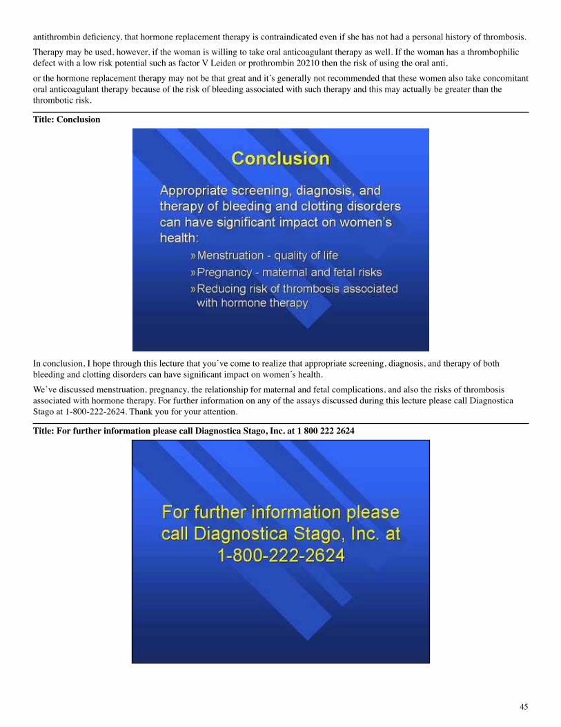
Title: Recommendations Contraception
Sometimes we see patients who have been on therapy for a number of years are found to be thrombophilic and question whether they should come off therapy. The risk of thrombosis is probably less with transdermal therapy. And, the risk is probably less with the use of a progestagen agent only, keeping in mind that progestagen agent only are not as effective a contraceptive agent. Next let’s discuss the recommendations as they pertain to hormone replacement therapy.
Title: Recommendations HRT*
In general, women with a history of previous venous thrombosis and a clear indication for hormone replacement therapy or hormone therapy, for example a women who requires tamoxifen therapy for breast cancer, these women should be considered for concomitant oral anticoagulant therapy. It’s also important to keep in mind the circumstances of the venous thrombosis: Was the venous thrombosis associated with a transient risk factor such as a post surgical procedure or trauma to the legs? Venous thrombosis that develops with a clear provoker probably carries a less increased risk of recurrence versus those thrombotic episodes that develop idiopathically. What if a woman has thrombophilia but no previous history of venous thrombosis, what should those women do in regards to using hormone replacement therapy? It’s generally recommended that if the woman has a thrombophilic defect with a high risk potential such as protein C, protein S, or
45
antithrombin deficiency, that hormone replacement therapy is contraindicated even if she has not had a personal history of thrombosis. Therapy may be used, however, if the woman is willing to take oral anticoagulant therapy as well. If the woman has a thrombophilic defect with a low risk potential such as factor V Leiden or prothrombin 20210 then the risk of using the oral anti, or the hormone replacement therapy may not be that great and it’s generally not recommended that these women also take concomitant oral anticoagulant therapy because of the risk of bleeding associated with such therapy and this may actually be greater than the thrombotic risk.
Title: Conclusion
In conclusion, I hope through this lecture that you’ve come to realize that appropriate screening, diagnosis, and therapy of both bleeding and clotting disorders can have significant impact on women’s health. We’ve discussed menstruation, pregnancy, the relationship for maternal and fetal complications, and also the risks of thrombosis associated with hormone therapy. For further information on any of the assays discussed during this lecture please call Diagnostica Stago at 1-800-222-2624. Thank you for your attention.
Title: For further information please call Diagnostica Stago, Inc. at 1 800 222 2624