Hemostasis 2 - Fiziologiefiziologie.ro/en/2014-2015/Hemostasis 2_2015.pdf · HEPARIN AND HEPARAN...
71
HEMOSTASIS 2
Transcript of Hemostasis 2 - Fiziologiefiziologie.ro/en/2014-2015/Hemostasis 2_2015.pdf · HEPARIN AND HEPARAN...

HEMOSTASIS 2

REVIEW- CLASSIC VS MODERN
ANTICOAGULANT THERAPY
Injectable- heparin and heparin derivatives
Unfractioned heparin
Low molecular weight heparins
Oral- antivitamin K inhibitors
Coumadins- most used= WARFARIN
HEPARIN AND HEPARAN SULFATE
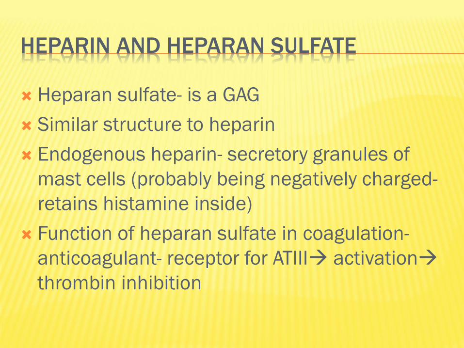
Heparan sulfate- is a GAG
Similar structure to heparin
Endogenous heparin- secretory granules of
mast cells (probably being negatively charged-
retains histamine inside)
Function of heparan sulfate in coagulation-
anticoagulant- receptor for ATIII activation
thrombin inhibition


TYPES OF HEPARIN
Unfractioned- high molecular weight; injectable
iv, hospitalised patient usually, APTT monitoring
needed (administred using a heparin pump)
Low molecular weight heparin- subcutaneously,
home therapy, no APTT monitoring, less
bleeding as side effect

UNFRACTIONED HEPARIN AS A DRUG
Needs ATIII as a cofactor
Inhibits Xa and thrombin
Clinical uses
Venous thrombosis disorders
Pulmonary embolism
Prophylaxis and treatment- thrombosis in major surgeries
Extracorporeal circulation
Blood samples drowned for lab purpuses- AC
Blood transfusions- in vitro AC

LOW MOLECULAR WEIGHT HEPARIN USE
No APTT monitoring needed
Lower molecular weight
May be administrated subcutaneously- may be
used for unhospitalized patients (once/twice a
day)
Less bleeding
Tend to replace heparin in venous thrombosis,
pulm embolism, acute coronary syndomes

ORAL ANTICOAGULANT THERAPY
Vitamin K competitors- coumadin (Warfarin)
Warfarin inhibits the vitamin K-dependent
synthesis of biologically active forms of the
calcium-dependent clotting factors II, VII, IX and
X, as well as the regulatory factors protein C,
protein S.

VITAMIN K DEPENDENT FACTORS


In the liver- vitamin K helps the carboxylation of glutamic acid residues on immature coag factors; in this process it gets oxidized
Antivitamin K drugs inhibit reductase by inhibition, vitamin K stays in an inactive form

ANTI VITAMIN K
Anticoagulant effect starts in 24-36 h from
initiation – stops in 36-72 h after it is stopped
Oral treatment
At first- injectable treatment overlaps the oral
one

ANTIVITAMIN K CLINICAL USES
Venous thrombosis
Stroke
Thrombembolism
Cardiac valve replacement
Post myocardial infarction

MONITORING HEMOSTASIS
Primary
Bleeding time
Rumpel Leede
PLT count
Secondary
Clotting time
Howell Gram time
APTT
PT (Quick time)

HOWELL GRAM INTRINSIC AND COMMON
test explores the intrinsic and common pathways; for this we add CaCl2 to citrate plasma and start monitoring the time needed to clot.
Normal values= 60 -120s
High:
TR disfunction / thrombocytopenia
intrinsic pathway factor deficit (XII,XI,IX,VIII- hemophilia)
common pathway deficit- X, V, II, I (hypofibrinogenemia/ afibrinogenemia)
-anti- clotting therapy- heparin

APTT ACTIVATED PARTIAL THROMBOPLASTIN
TIME INTRINSIC AND COMMON PATHWAYS
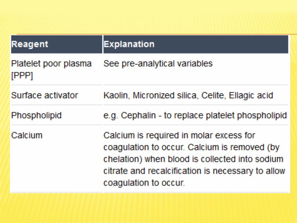
To citrate plasma (with low amount of platelets- centrifuge for 15 min- 5000 rpm) we add Ca Cl2, kaolin, cephalin and start monitoring the time needed to clot.
Cephalin is a partial tromboplastin (only phospholipid)
Kaolin or silica is a negatively charged molecule activates factor XII
Normal values = 30- 40 s
Causes of abnormal high values- the same as Howell- except for the platelet- derived causes
It is affected by unfractioned heparin- used to monitor therapy

APTT

APTT INTERPRETATION- PROLONGATION
Deficiencies of XII,XI,IX,VIII (but in mild def is normal- the def factor needs to be <20-40% for aPTT to be low)
Antibodies against fVIII (aquired hemofilia) or lupus anticoagulant present
Liver disease
Unfractioned heparin use

PROTHROMBIN TIME (QUICK METHOD-PT/QT)
EXTRINSIC AND COMMON
To citrate plasma (with high amount of
platelets) we add Ca Cl2, thromboplastin and
start monitoring the time needed to clot.
Elevated in
Deficit of I, II, V, VII, X factors
Liver failure
K vitamin deficiency/ anti vit K anticoag therapy
Normal values = 12-15 s

PROTHROMBIN TIME
Used to monitor therapy with oral anticoagulant drugs- vitamin K blocking agents- coumarins (warfarin)!!!
By INR fraction which should be measured constantly in these patients
The therapy is used in patients with high thrombotic risk

INR
International normalized ratio
ISI- depends on the tissue factor, it is established
by the manufacturer (usually between 1- 2)
Normal range for a healthy person= 0.9- 1.3
Warfarin therapy monitoring- depends on
pathology


FIBRINOLYSIS
As the clots forms, it incorporates plasmin molecules.
Plasmin is an enzyme formed from plasminogen by tissue plasminogen activator (t-PA) and urokinase type plasminogen activator (u-PA)
Plasmin breaks down fibrin polymers into fibrin fragments = fibrinolysis
Fibrinolysis helps removing the clot as the repair processes occur.
FIBRINOLYSIS
Damage to the tissues releases TPA, which together with activated components from the coagulation pathways and protein C, activates plasminogen to plasmin.
Plasmin acts on the insoluble fibrin to form a series of soluble products: FDPs.
Fibrin that has been stabilized (crosslinked) by factor XIII gives rise to crosslinked FDPs (XDPs), as well as X Y D and E fragments.
The XDPs (D-dimer, D-dimer-E fragments), and oligomers of fragments X and Y, can be detected using antibody coated latex beads.


PLASMINOGEN
Zymogen plasmin
GP
Syntesized in liver
Lyses fbrin into fibrin degradation products (FDP) and D- dimer

PHYSIOLOGICAL ACTIVATORS OF PLASMIN
tPA- released from entoth cells responsable
for intravascular fibrinolysis- mainly activates
plasminogen bound to fibrin
uPA- high conc in urine responsible for
extravascular fibrinolysis

PHYSIOLOGICAL INHIBITORS OF PLASMIN
PAI-1 is the physiological plasminogen
activatior inhibitor 1- (tPA)- from platelets
Alpha 2 antitripsin- inhibits plasmin directly


FIBRINOLYTIC THERAPY= THROMBOLYTIC
Used to dissolve blood clots- act by
plasminogen activation
3 major classes:
tPA used in miocardial infarction therapy
uPA
sPA (streptokinase activator)

CLINICAL CASE 1
23 year old male.
Over the past week noted increasing fatigue,
sore throat, earaches, headaches, and episodic
fever and chills. Unable to run his customary 25
miles per week.
Erythematous throat and tonsils.
Swollen cervical lymph nodes.
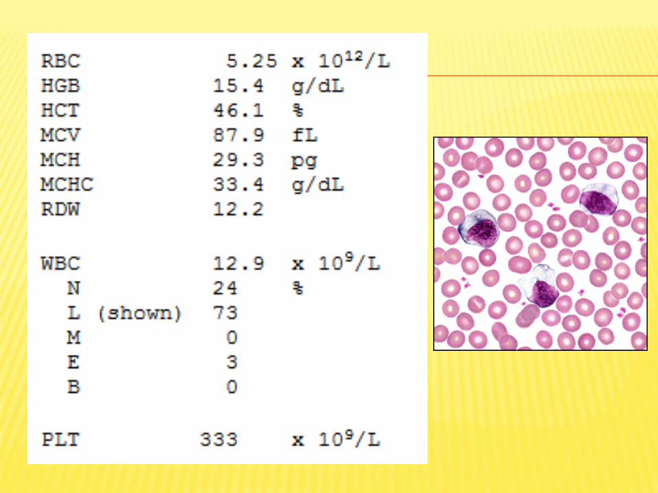
DIAGNOSIS INFECTIOUS MONONUCLEOSIS
“kissing disease”
Viral- Ebstein Barr virus
90% asymptomatic
Pharyngitis, fatigue, malaise, fever
CASE STUDY 2
70 year old female.
Symptoms of dyspnea on exertion, easy
fatigability for past 2 to 3 months.
Physical exam- palor


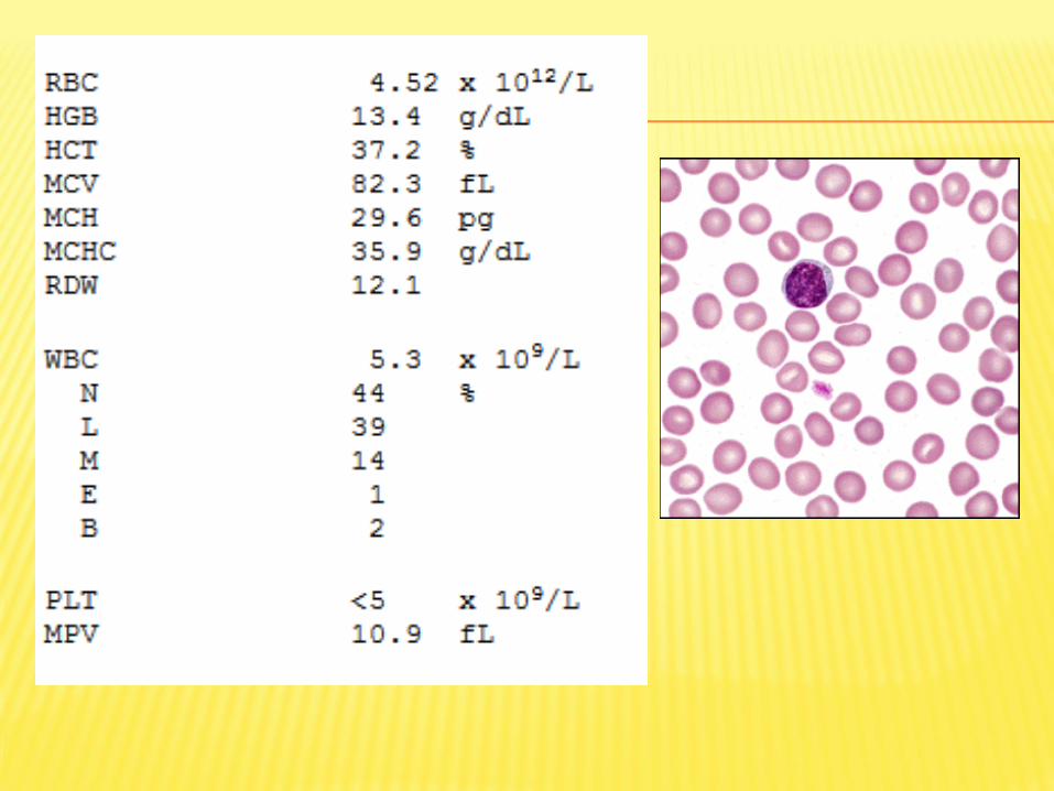
DIAGNOSIS- IRON DEFICIENCY ANEMIA

CASE STUDY 3
25 year old male.
Recurrent upper respiratory infections with
fever, nausea, and submandibular swelling for
several months prior to admission.
Noted that cuts on his hands did not heal well.
Physical Exam
Submandibular adenopathy.
No other organomegaly.


DIAGNOSIS ACUTE MYELOBLASTIC LEUKEMIA
CASE STUDY 4
40 year old female.
Brought to Emergency Room with symptoms of severe frontal headache and associated confusion. Noted to have decreased energy level and a 15 pound weight loss over the previous three months.
Physical Exam
Pale appearing, but otherwise within normal limits. No organomegaly.

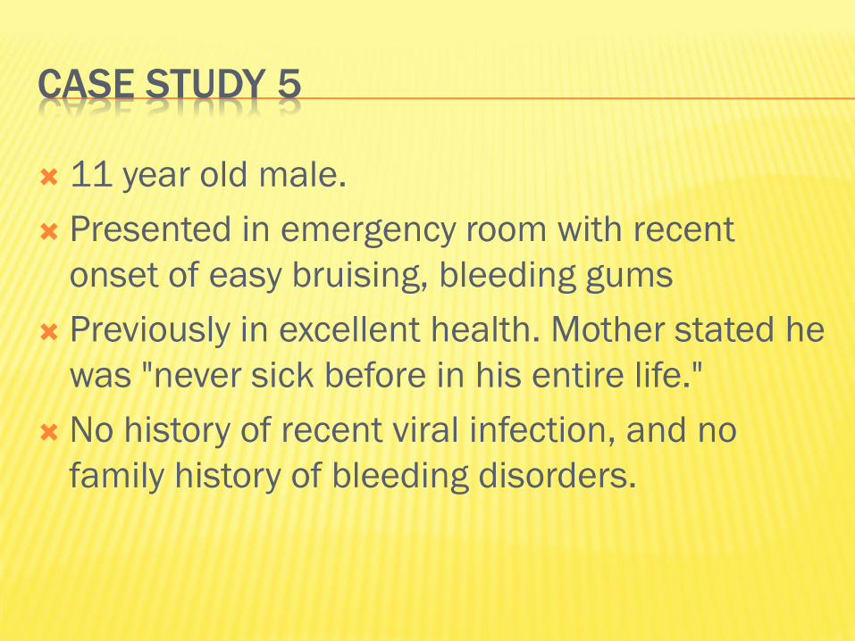
CASE STUDY 5
11 year old male.
Presented in emergency room with recent
onset of easy bruising, bleeding gums
Previously in excellent health. Mother stated he
was "never sick before in his entire life."
No history of recent viral infection, and no
family history of bleeding disorders.

HEMOSTASIS:
Bleeding time= 10 min
INR 0.91 (RI 0.85-1.15)
Howel Gram 2 min
PTT 24.8 sec (RI 23-34)
TT 15.8 sec (RI 13-18)

IMMUNE THROMBOCYTOPENIC PURPURA

CASE STUDY 6
History
54 year old female.
One year history of fatigue, weight loss, and
increasingly severe back pain.
Physical Exam
She appeared pale, but otherwise her physical
exam was within normal limits.

PROTEIN ELECTROPHORESIS
Total protein 11.0 g/dL (RI 5.2-8.3)
Serum protein electrophoresis:
Albumin 3.2 g/dL (RI 3.0-5.0)
Globulins:
Alpha1 0.4 (RI 0.1-0.5)
Alpha2 1.0 (RI 0.5-1.2)
Beta 0.8 (RI 0.5-1.1)
Gamma 5.6 (RI 0.6-1.7)

Immunoglobulins, quantitative:
IgA 9 mg/dL (RI 85-450)
IgG 5800 mg/dL (RI 800-1700)
IgM 25 mg/dL (RI 60-370)
Fibrinogen 650 mg/dl
ESR 70 mm/h

MULTIPLE MYELOMA

CASE STUDY 7
History
75 year old male.
Symptoms of severe headache and generalized pruritis.
Physical Exam
Spleen palpable 10 cm. below left costal margin. Liver palpable 3 cm. below right costal margin. The rest of the exam was within normal limits.
PULMONARY FUNCTION:
Oxygen saturation: 97% (RI 94-100)

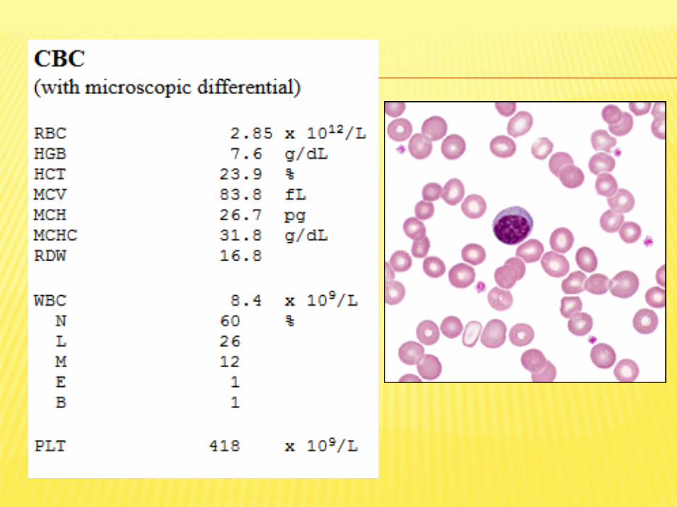
POLYCYTHEMIA VERA

PRACTICE QUESTIONS

BLOOD
1. is a type of connective tissue
2. is involved in the process of homeothermy
3. bicarbonate buffer system is the most
important extracellular system
4. ADH helps at the reabsorption of Na+ in the
distal nephron

HEMATOCRIT
1. somatic hematocrit is higher than the venous
one
2. represents the % of formed elements in the
blood
3. blood viscosity is inversely proportional to the
hematocrit value
4. splenic venous blood has the highest value

PLASMA PROTEINS
1. albumin is the main contributor to oncotic
pressure
2. the normal concentration of albumin is 7 g/dl
of blood
3. ceruloplasmin is a copper carring protein
4. albumin is a positive acute phase protein

IMMUNOGLOBULINS
1. IgM is released during primary humoral
response
2. IgG may cross the placenta
3. IgA is important in local mucosal immunity
4. IgE trigger alergic reactions

ESR
1. 1. is directly proportional with blood viscosity
2. 2. low ESR can be encountered in anemia
3. 3. it’s measured after centrifuging the blood at
3000 RPM
4. 4. detects non-specific inflammation

RED BLOOD CELLS
1. 1 g of Hb can transport 1.34 mL of oxygen
2. MCV =120fL showes that there are
macrocytes present in the blood
3. 70% of the iron in the organism is found in
hemoglobin
4. when pCO2 is low, red blood cells release O2
more easely to the tissues

WHICH OF THE FOLLOWING ARE PART OF THE
SPECIFIC IMMUNE ANSWERS
1. first line of defense mechanisms
2. second line of defense
3. all three lines of defense
4. third line of defense

CHOOSE THE RIGHT MACROPHAGES FUNCTIONS
1. antigen presenting cells
2. initiation of humoral immune answer
3. initiation of celular immune answer
4. they are the first cells to respond to tissue
infection

ANTIBODIES WORK AS
1. opsonins
2. antitoxins
3. agglutinate bacteria
4. stimulate perforin- pores formation in
antigenic cell membrane

FORMS OF CO2 TRANSPORT IN THE BLOOD ARE
carbaminoHB
bound to albumin
bicarbonates
carboxyHB

CHOOSE THE RIGHT CONDITIONS WHICH SHIFT
THE OXYHB DISSOCIATION CURVE TO THE RIGHT
Hb has low affinity for oxigen
high 2,3 DPG
low pH
low temperature

WHAT WOULD BE THE ERYTHROCYTE INDICES
PROFILE FOR A PERSON WITH NORMOCHROMIC
MACROCYTIC ANEMIA
1. MCHC= 28 g/100 ml
2. MCHC= 35 g/100 ml
3. MCV= 80 fL
4. MCV= 120 fL