Hemorrhagic enteritis virus induced changes in the lymphocyte subpopulations in turkeys and the...
12
ELSEVIER Veterinary Immunology and Immunopathology 45 (1995)139-150 Veterinary immunology and immunopathology Hemorrhagic enteritis virus induced changes in the lymphocyte subpopulations in turkeys and the effect of experimental immunodeficiency on viral pathogenesis M. Suresh, J.M.Sharma* Dept. of Veterinary PathoBiology, College of‘ Veterinary Medicine. I:niwrsity qf .Ifinnesota. St. Paul, MN 55 108. USA Accepted 3 March 1994 Abstract We examined the changes in the lymphocyte subpopulations in the spleen and peripheral blood of turkeys and the effects of experimental immunodeficiency in the B and T cell compartments on the pathogenesis of hemorrhagic enteritis (HE) in turkeys. Inoculation of turkeys with hemorrhagic enteritis virus (HEV) induced a drop in the relative propor- tions of IgM bearing cells on Day 2, 3, and 9 post-infection and an elevation in the relative proportions of CD4+ cells on Day 4 and 6 post-infection. Elevated levels of CD8 + cells were observed in the infected turkeys only on Day 16 after infection. Marked depletion of IgM+ cells may play a role in immunodepression caused by HEV. Cyclophosphamide (CY ) treatment induced B cell deficiency in turkeys severely impaired HEV replication in the spleen suggesting that B lymphocytes are important for viral replication. Cyclosporin A (CsA) selectively impaired T cell mitogenesis and protected the turkeys against HEV- induced intestinal hemorrhages. CsA did not prevent viral replication in the spleen or the associated splenomegaly This result suggested that T cell immunity may be important for intestinal hemorrhaging induced by HEV. 1. Abbreviations Con A, concanavalin A; CPM, counts per minute; CsA, cyclosporin A; ELISA, enzyme linked immunosorbent assay; HEV, hemorrhagic enteritis virus; IBDV, infectious bursal disease virus; SPF, specific-pathogen free. *Correspondingauthor. Tel. (612) 625-5276; fax (612) 625-5203. 0165-2427/95/$09.50 0 1995 Elsevier Science B.V. All rights reserved SSDI0165-2427(94)05323-K
Transcript of Hemorrhagic enteritis virus induced changes in the lymphocyte subpopulations in turkeys and the...

ELSEVIER Veterinary Immunology and Immunopathology
45 (1995)139-150
Veterinary immunology
and immunopathology
Hemorrhagic enteritis virus induced changes in the lymphocyte subpopulations in turkeys and the effect
of experimental immunodeficiency on viral pathogenesis
M. Suresh, J.M.Sharma* Dept. of Veterinary PathoBiology, College of‘ Veterinary Medicine. I:niwrsity qf .Ifinnesota.
St. Paul, MN 55 108. USA
Accepted 3 March 1994
Abstract
We examined the changes in the lymphocyte subpopulations in the spleen and peripheral blood of turkeys and the effects of experimental immunodeficiency in the B and T cell compartments on the pathogenesis of hemorrhagic enteritis (HE) in turkeys. Inoculation of turkeys with hemorrhagic enteritis virus (HEV) induced a drop in the relative propor- tions of IgM bearing cells on Day 2, 3, and 9 post-infection and an elevation in the relative proportions of CD4+ cells on Day 4 and 6 post-infection. Elevated levels of CD8 + cells were observed in the infected turkeys only on Day 16 after infection. Marked depletion of IgM+ cells may play a role in immunodepression caused by HEV. Cyclophosphamide (CY ) treatment induced B cell deficiency in turkeys severely impaired HEV replication in the spleen suggesting that B lymphocytes are important for viral replication. Cyclosporin A (CsA) selectively impaired T cell mitogenesis and protected the turkeys against HEV- induced intestinal hemorrhages. CsA did not prevent viral replication in the spleen or the associated splenomegaly This result suggested that T cell immunity may be important for intestinal hemorrhaging induced by HEV.
1. Abbreviations
Con A, concanavalin A; CPM, counts per minute; CsA, cyclosporin A; ELISA, enzyme linked immunosorbent assay; HEV, hemorrhagic enteritis virus; IBDV, infectious bursal disease virus; SPF, specific-pathogen free.
*Correspondingauthor. Tel. (612) 625-5276; fax (612) 625-5203.
0165-2427/95/$09.50 0 1995 Elsevier Science B.V. All rights reserved SSDI0165-2427(94)05323-K

140 hf. Suresh. J.M. Sharma / Lkt. I~wnunol. Iwmunopathol. 45 (1995) 139-150
2. Introduction
Hemorrhagic enteritis (HE) is an acute, highly contagious disease of turkeys caused by a type II avian adenovirus, hemorrhagic enteritis virus (HEV). The disease is characterized by splenomegaly, intestinal hemorrhage, and sudden death (Domermuth and Gross, 1991). Although HE has been recognized since 1937 (P,omeroy, 1937), the pathogenesis of the disease is poorly understood. In addi- tion to the characteristic intestinal and splenic lesions, HEV has been shown to suppress both humoral and cellular immunity in turkeys (Nagaraja et al., 1982a,b). Viruses can induce immunosuppression by various mechanisms and direct lysis of lymphoid cells is one of them (Nash, 1985; Zinkernagel and Hen- gartner, 1992). Splenic lesions in HE include lymphoid necrosis and a massive disintegration of the white pulp (Gross and Dormermuth, 1975). Whether the lymphoid necrosis is restricted to a particular subpopulation of lymphocytes is not known.
Studies on the pathogenesis of HE using immunofluorescence (Fasina and Fabricant, 1982) and immunohistochemistry (Fitzgerald et al., 1992 ) have shown that although HEV causes extensive hemorrhaging in the gut, the intestinal epi- thelial cells are not the target cells for viral replication. The immune system ap- pears to play a role in the development of HEV-induced disease. Experimentally induced immunodeficiency modulates HE (Opengart et al., 1990). Surgical sple- nectomy has been shown to abrogate intestinal lesion formation although virus replicated in extra-splenic locations (Ossa et al., 1983). Chemical bursectomy protected turkeys against HEV-induced lesions and mortality (Fadly and Nazer- ian, 1982 ) suggesting that B lymphocytes may be involved in the pathogenesis of HE. However, the mechanism of B cell involvement is not known. Also, the pos- sible contribution of T lymphocytes in the pathogenesis of HE has not been examined.
In this study, we have further examined the role of immunity in the pathogen- esis of HE. Specific objectives were to examine (a) the effect of HEV infection on lymphocyte subpopulations in the spleen and peripheral blood of turkeys, and (b) the response to HEV of turkeys experimentally immunocompromised in the T and the B cell compartments.
3. Materials and methods
3.1. Turkeys
White Nicholas turkey poults were obtained at the day of hatch from a com- mercial hatchery (Wilmar Poultry Company, Wilmar, MN) and held in isola- tion. Turkeys were exposed to HEV after 4 weeks of age when maternal antibod- ies to HEV had reached undetectable levels. Specific-pathogen free (SPF) poults, lacking anti-HEV antibodies were obtained from the National Animal Disease Center, Ames, IA. All experimental groups were reared separately for the dura-

M. Suresh. J.M. Sharma / 6’et. Imrnunol. Itnrmnopathol. 45 (1995) 139-150 141
tion of the study in Horfall-Bauer type isolation units supplied with air passed through HEPA filters.
3.2. Virus
Cell culture propagated virulent HEV (Nazerian and Fadly, 1982) kindly pro- vided by Dr. K. Nazerian, Avian Disease and Oncology Laboratory, East Lan- sing, MI was used. Each turkey was inoculated per OS with 1000 turkey infectious doses0 (TID,,) of HEV.
3.3. Immunosuppressive treatment with cyclophosphamide (CY) and cyclosporin A (CsA)
Turkeys were treated with CY (Neosar, Adria Laboratories, Columbus, OH) intraperitoneally at a daily dose of 4 mg per bird for the first 3 days post-hatching as previously described (Elmubarak et al., 198 1). The CY treated turkeys were infected with HEV 5 weeks post-treatment.
Cyclosporin A (CsA), kindly provided by Sandoz Pharmaceuticals, E. Hano- ver, NJ, was dissolved in mineral oil and administered intramuscularly every 3 days as previously described (Nowak et al., 1982) at a dosage of 100 mg kg-’ body weight per injection for 13 days.
3.4. Mitogenic assay of whole blood
The mitogenic response of whole blood cells to concanavalin A (Con A ) was determined as previously described (Sharma and Belzer, 1992). Briefly, heparin- ized blood obtained by venipuncture was diluted 1:20 with RPM1 1640 medium (Sigma Chemical Co., St Louis, MO) containing 7.5% normal turkey serum. Tri- plicate cultures of each sample were stimulated with Con A (32 lug well- ’ ). After 48 hours incubation at 39.5”C, cells in each well were pulsed with 1 &i of 3H thymidine (Amersham, 1.15 TBq mMol- ’ ) . Five hours after labeling, the cells were harvested onto glass filters. Counts per minute (CPM) values in individual cell samples were measured in a scintillation counter (Packard scintillation counter model 1900, Downers Grove, IL).
3.5. Antibody assays
The antibody responses of turkeys treated with CY or CsA were tested as pre- viously described (Suresh et al., 1993) by injecting turkeys with sheep erythro- cytes and killed Brucella abortus (National Veterinary Services Laboratory, Ames, IA). The titers were expressed as the log, of the highest dilution giving visible agglutination.
Turkeys were examined for antibodies to HEV by an enzyme linked immuno- sorbent assay (ELISA) using monoclonal antibodies to HEV kindly provided by

142 M. Suresh, J.M. Sharma / Vet. Immunol. Immunopathol. 45 (1995) I39-IS0
Dr. L. Lee, Avian Disease and Oncology Laboratory, East Lansing, MI (Nazerian et al., 1990).
3.6. ELBA for viral antigen quantitation
HEV antigen in the spleens was quantitated by an antigen capture ELISA using monoclonal antibodies to HEV as previously described (Nazerian et al., 1990).
3.7. Immunojluorescent staining of lymphocyte subsets for surface markers
Mouse monoclonal antibodies CT4 and CT8 that recognize the chicken hom- ologues of mammalian CD4 and CD8 respectively were kindly provided by Dr. C.L.H. Chen (University of Alabama). Monoclonal antibody to the p chain of the chicken IgM molecule (Erdei et al., 1983) was a gift from Drs. I. Nemeth and N. Dren (Hungarian Academy of Sciences, Budapest, Hungary). The cross reac- tivity of anti-chicken CD4, CD8, and IgM monoclonal antibodies with turkey lymphocytes and the staining procedures have been described in detail (Suresh et al., 1993). Fluorescence intensities were measured by flow cytometry (FAC- Scan, Becton Dickinson, Mountain View, CA). Gates were set to analyse only lymphocytes based on forward and 90” scatter; 10 000 cells per sample were examined.
3.8. Statistical analysis
Statistical analyses were conducted by the Statistix 3.1 program (Analytical Software, St. Paul, MN). Statistical evaluation of all results was performed by analysis of variance (ANOVA) and significant differences between group means were tested by the Tukey’s test (5% level of probability).
3.9. Experimental design
3.9. I. Experiment I To examine the effect of HEV on lymphocyte subsets, 4-week-old SPF turkeys
were inoculated with HEV. At intervals, splenic and peripheral blood mononu- clear cells were examined for the reIative proportions of different lymphocyte subsets by flow cytometry. Virus-free turkeys were tested as controls.
3.9.2. Experiment 2 A group of l-day-old commercial turkeys was treated with CY. A group of age-
matched untreated poults was maintained separately. Five weeks after CY treat- ment, CY-treated and untreated turkeys were examined for the relative bursal weights and the mitogenic responses of the whole blood cells to Con A. To assess the humoral immune competency, CY-treated and untreated turkeys were ex- amined for antibody responses 14 days after immunization with sheep erythro- cytes and B. abortus. Groups of CY-treated and untreated turkeys were inocu-
M. Suresh, J.M. Sharma / Vet. Immunol. Immunopathol. 45 (1995) 139-150 143
lated with HEV. Additional groups of virus-free CY-treated and untreated turkeys were maintained as controls. Six days post-HEV inoculation, turkeys were sacri- ficed, weighed and examined for gross lesions. Also, spleens were weighed and spleen homogenates were tested for viral antigen by ELISA.
3.9.3. Experiment 3 Four-week-old SPF-turkeys were treated with CsA. Seven days post-initiation
of CsA treatment, turkeys from CsA-treated and untreated groups were examined for the mitogenic responses of whole blood cells to Con A. CsA-treated and un- treated turkeys were inoculated with sheep erythrocytes and B. abortus 7 days post-initiation of CsA treatment to test humoral immune competence. Antibod- ies to these antigens were tested 14 days post-inoculation. Groups of CsA-treated and untreated turkeys were inoculated orally with HEV 7 days after initiation of CsA treatment. Virus-free CsA-treated and untreated turkeys were maintained as controls. Other procedures employed were similar to those in Experiment 2.
4. Results
4.1. Effect of HE V on lymphocyte subpopulations in turkeys
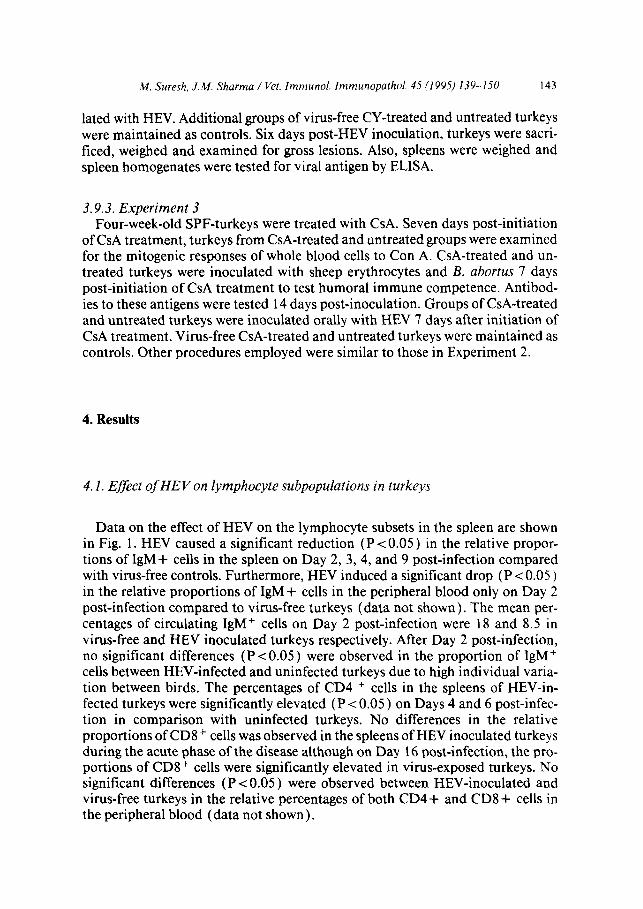
Data on the effect of HEV on the lymphocyte subsets in the spleen are shown in Fig. 1. HEV caused a significant reduction (P < 0.05 ) in the relative propor- tions of IgM+ cells in the spleen on Day 2, 3, 4, and 9 post-infection compared with virus-free controls. Furthermore, HEV induced a significant drop (P < 0.05) in the relative proportions of IgM+ cells in the peripheral blood only on Day 2 post-infection compared to virus-free turkeys (data not shown). The mean per- centages of circulating IgM+ cells on Day 2 post-infection were 18 and 8.5 in virus-free and HEV inoculated turkeys respectively. After Day 2 post-infection, no significant differences (P < 0.05 ) were observed in the proportion of IgM+ cells between HEV-infected and uninfected turkeys due to high individual varia- tion between birds. The percentages of CD4 + cells in the spleens of HEV-in- fected turkeys were significantly elevated (P < 0.05 ) on Days 4 and 6 post-infec- tion in comparison with uninfected turkeys. No differences in the relative proportions of CDS+ cells was observed in the spleens of HEV inoculated turkeys during the acute phase of the disease although on Day 16 post-infection, the pro- portions of CD8+ cells were significantly elevated in virus-exposed turkeys. No significant differences (P < 0.05 ) were observed between HEV-inoculated and virus-free turkeys in the relative percentages of both CD4 + and CD8 + cells in the peripheral blood (data not shown).

144 M. Suresh, J.M. Sharma / Vet. Immunol. Immunopathol. 45 (1995) 139-150
50 a
1
=” 40- p:
i s 30-
r .3 .= p zo- es
10-
,
2 4 6 8 10 12 14 16
Daya Postjnfution
C .
a 2 4 6 .9 10 12 14 16
Fig. 1. Flow cytometric analysis of the effect of HEV on the relative proportions of IgM+ (a), CD4+ (b), and CD8+ (c) cells in the spleens of turkeys. * Significant difference (PcO.05) from control values.

M. Suresh, J.M. Sharma / Vet. Immunol. Immunopathol. 45 (I 995) 139- 150 145
4.2. Effect of CY and CsA on immune responses of turkeys
Data in Table 1 indicate that turkeys treated with CY did not produce detect- able antibody to B. abortus and had significantly lower (P < 0.05 ) antibody titers against sheep erythrocytes than untreated controls. The relative bursal weights of CY-treated turkeys were significantly lower (PcO.05) than those of untreated turkeys. No differences (P < 0.05 ) in the mitogenic responses of the whole blood cells to con A stimulation were observed between CY-treated and untreated con- trols (Table 1) indicating that this cellular immune function was not affected by CY treatment. Taken together these data suggest that CY treatment induced a selective B cell deficiency.
Data in Table 2 show the effect of CsA on the humoral and cell-mediated re- sponses of turkeys. On Day 7 post-initiation of CsA treatment, the mitogenic responses of whole blood cells to Con A in CsA treated turkeys were significantly lower (P < 0.05 ) than those of untreated controls. CsA treatment did not com-
Table 1 Effect of CY on antibody response, bursal weight, and mitogenic responses of peripheral blood icu- kocytes to Con A (Experiment 2 )
Treatmentb Mean log2 antibody titer (n = 5 ) Mean bursal index’ Mitogenic response (II = 5 ) to (mean CPM f SD)”
Sheep erythrocytes B. abortlts Unstimulated Con A
None 5.00t l.OOd 9.20?0.88d 1.45+0.30“ 1118+361” 16839+7518d CY 0.502 1.00 0’ 0.44 & 0.04’ l782+536* 24355i 1 1758d
‘Counts per minute i standard deviation. ‘Turkeys were treated with CY, 4 mg per bird per day on the first 3 days post-hatching and immune responses were measured 5 weeks after CY treatmenmt. ‘( Bursal weight (g)/body weight (g) ) x 1000. d,‘Values within each column with different superscripts are significantly different (PC 0.05 ).
Table 2 Effect of Cyclosporin A (CsA) on humoral and cell mediated immune responses of turkeys (Experi- ment 3 )
Treatment’ Mean log, antibody titers (n= 5) to Mitogenic response” (mean CPM+SD)b
Sheep erythrocytes B. abortus Unstimulated
None 7.4F2.1d 6.6? 1.5d 1750f 1202d CsA 6.0-t 2.2d 8.6kO.5’ 2631 i 1525d
“Mitogenic responses were measured 7 days post-initiation of CsA treatment. bCounts per minute i standard deviation.
Con A
8397? 2730d 29172 1606’
‘Antigens were inoculated 7 days after initiation of CsA treatment ( 100 mg KG-’ body weight every 3 days) and antibody titers were measured 14 days after inoculation of the antigens. d.‘Values within each column with different superscripts are significantly different (PC 0.05).

146 h4. Suresh. J.M. Sharma / Vet. Immunol. Immunopathol. 45 (1995) 139-150
promise the antibody responses of turkeys to sheep erythrocytes. However, CsA- treated turkeys had significantly higher (P c 0.05 ) antibody responses against B. abortus compared with untreated turkeys. Data from Table 2 indicate that CsA induced selective T cell deficiency.
4.3. EfSect of CY and CsA induced immunodejkiency on the pathogenesis of HE
The responses of CY treated turkeys to HEV are summarized in Table 3. The CY treated turkeys did not develop detectable splenomegaly upon infection with HEV in contrast to a pronounced splenic enlargement in CY-free HEV-exposed turkeys. At Day 6 post-infection, viral antigen in the spleen was detected in one of four CY treated turkeys as opposed to seven of seven turkeys in the CY-free group. These data indicated that HEV replication was restricted in turkeys that were deficient in B cells and that B lymphocytes may be important for HEV replication.
The effects of CsA treatment on HEV-induced splenomegaly, intestinal hem- orrhaging, and viral antigen in the spleen are summarized in Table 4. None of the ten CsA-treated turkeys developed intestinal hemorrhages following exposure to HEV. On the other hand, five of ten CsA-free turkeys developed HEV-induced hemorrhagic lesions. The magnitude of splenic enlargement in CsA-treated tur- keys was comparable with that of CsA-free infected turkeys (P < 0.05 ) . Further- more, CsA treatment did not affect viral replication in the spleen because the mean viral antigen titers in the spleen of CsA-treated and CsA-free turkeys were not significantly different (P < 0.05 ). These data suggested that CsA treatment abrogated clinical HE without affecting viral replication in the spleen. Taken to- gether, these data indicate that T lymphocytes or a CsA-sensitive cell may play an important role in the development of HEV-induced intestinal lesions.
Table 3 Effect of cyclophosphamide (CY) on HEV-induced splenomegaly, and viral antigen in the spleen at Day 6 post-infection (Experiment 2)
Treatment n HEV Mean splenic indexb HEV antigen
No. positive/tested Mean log, ELBA titer
None 7 + 2.59 & 0.20’ 717 6.2+2.16’ 5 - l.37f0.27d O/5 0
CY 4 + l.49f0.76d l/4 7d 5 - 1.05? 0.25d O/5 0
“Viral antigen in the spleen was quantitated by an antigen capture ELBA using monoclonal antibodies to HEV. Sample OD values higher than the average OD of a known negative spleen sample plus 3 standard deviations were considered positive. b(Spleen weight (g)/body weight (g)) x 1000. c.dFigures within each column with different (superscripts are significantly different (PC 0.05).

M. Suresh, J.M. Sharma / Vet. Immunol. Immunopathol. 45 (1995) 139-150 147
Table 4 Effect of Cyclosporin A (CsA) treatment on HEV-induced splenomegaly, hemorrhagic enteritis and viral antigen in the spleen (Experiment 3)
Treatment HEV n Hemorrhagic enteritis Mean splenic Viral antigenb (No. positive/tested) index”
No. positive/tested Mean log, ELBA titer
-
None + 10 5/10 2.13 * 0.90’ IO/IO 7.33* 3.08’ - 5 o/5 l.OOfO.12d Of5 0
CsA t IO O/IO 1.95f0.62’ IO/IO 8.00? 2.06’ 5 o/5 1.44 t 0.25d o/5 0
“(Spleenwt. (g)/body wt. (g))xlOOO. bViral antigen in the spleen was quantitated by an antigen capture ELBA using monoclonal antibod- ies to HEV. Sample OD values higher than the average OD of a known negative spleen sample plus 3 standard deviations were considered positive. c.dFigures within each column with different superscripts are significantly different (PC 0.05 )
5. Discussion
In this study, we have shown for the first time that infection of turkeys with HEV results in a dramatic alteration in the normal population of peripheral lymphoid cells. Within the first week of exposure to HEV, we noted a significant drop in the IgM bearing cells, and an increase in the relative proportions of CD4+ cells resulting in alteration of normal CD4:CDS ratios (Fig. 1). The significance and mechanisms of these alterations in the lymphocyte subpopulations is not known. We also observed that the proportion of CD8+ cells was increased on Day 16 post-infection which may be associated with viral clearance.
The reduction in the IgM+ cells is of special interest. It is possible that this may be associated with virus-induced immunosuppression as a consequence of cyto- lytic viral infection. Additional studies are needed to establish that IgM+ cells are one of the target cells for HEV. However, the ability of cells from a B-lymphob- Iastoid cell line (Nazerian and Fadly, 1982 ) and normal non-adherent mononu- clear cells from turkey blood (Van den Hurk, 1990) to support viral replication in vitro provides circumstantial evidence that B lymphocytes are susceptible to HEV replication. Infectious bursal disease virus (IBDV), another immunosup- pressive virus of poultry, is a B cell tropic virus and causes similar IgM+ cell depletion in the spleens of infected chickens (Rodenberg et al., 1993 ) . However, it is also possible that lysis of IgM+ cells may not be a result of viral replication in these cells but may be due to the possible cytotoxic effect of unassembled pen- ton proteins released from HEV infected reticula-endothelial cells. The penton protein of mastadenoviruses has been shown to have cytotoxic activity in vitro (Valentine and Pereira, 1965; Boudin et al., 1979).
CY, an alkylating agent, has been shown to induce an irreversible suppression of the humoral immunity with only a transient effect on cellular immune re- sponses (Elmubarak et al., 1981). Results from our study also showed that CY

148 M. Suresh, J.&f. Sharma / Vet. Immunol. Immunopathol. 45 (1995) 139-150
induced a selective B cell deficiency in the turkeys. Consistent with previous find- ings (Fadly and Nazerian, 1982), we noted that CY treatment prevented HEV induced lesions. Our data extended the previous observation and showed that the absence of lesions in CY-treated birds may be due to lack of viral replication. This conclusion is based on two findings: ( 1) HEV was detected in the spleen of only one of four CY-treated turkeys in comparison with seven of seven CY-free turkeys, and (2) HEV did not induce splenomegaly in CY-treated turkeys. Sur- gical bursectomy at 1 day of age did not prevent formation of inclusion bodies in cells 3 days after intramuscular injection of HEV (Beasly and Wisdom, 1978 ) thus suggesting that unlike CY, bursectomy did not prevent viral replication. One plausible explanation for the lack of effect of bursectomy on viral replication may be that surgical bursectomy at hatch did not cause total B cell depletion. Studies on avian B cell ontogeny have revealed that B cell emigration from the bursa to the periphery begins well before hatch (Weill and Reynaud, 1987). The effect of CY on the other hand is more systemic and bursal as well as extra bursal B cells are destroyed (Elmubarak et al., 198 1).
To date, ours is the first attempt to study the effect of T cell deficiency on HE in turkeys. We used CsA to induce selective T cell deficiency. As expected, treat- ment of turkeys with CsA resulted in inhibition of mitogenic responses of T lym- phocytes without detectable effect on humoral immunity. The most noticeable finding was that turkeys deficient in a T cell function were resistant to HEV- induced intestinal hemorrhages. HEV clearly replicated normally in CsA treated turkeys. Splenomegaly and viral antigen in the spleen were equally apparent in CsA-treated and CsA-free turkeys. The major therapeutic effect of CsA in mam- mals is to inhibit T lymphocyte activation (Schreiber and Crabtree, 1992). If a similar effect occurs in turkeys, our data suggests that T cell immunity may play a pivotal role in the causation of intestinal hemorrhage in HE. The ability of CsA treatment to induce remission of symptoms in cases of intractable diarrhea of infants (Sanderson et al., 199 1) and Crohn’s disease (Brynskov et al., 1989) has provided evidence of a primary role of activated T cells in the pathogenesis of these conditions. The spleen, which is the primary site of HEV replication (Fa- sina and Fabricant, 1982; Silim and Thorsen, 198 1 ), has been found to be a pre- requisite for lesion development in the intestine (Ossa et al., 1983). In contrast, we show that in HE, the presence of viral antigen in the spleen did not immi- nently lead to intestinal hemorrhages. CsA treatment prevented intestinal lesions despite extensive viral replication in the spleen. An earlier study had indicated that infection of turkeys with HEV resulted in an increase in the numbers of duo- denal mast cells (Opengart et al., 1992). CsA treatment may have abrogated in- testinal hemorrhaging by its inhibitory effect on T lymphocytes to produce IL-3, a mast cell growth factor (Razin et al., 1984). If T lymphocytes are directly in- volved in the genesis of intestinal lesions, the possible roles of inflammatory cy- tokines need to be evaluated.

M. Suresh, J.M. Sharma / Vet. Immunol. Immunopathol. 45 (1995) 139-150 149
Acknowledgments
Many thanks are due to Dr. Jamil Ahmad, Terence Pertile, Dr. Kemal Karaca and Susan Belzer for their suggestions and assistance in this study. This work was supported by a grant from the Minnesota Turkey Research and Promotion Coun- cil and the Minnesota Turkey Growers Association.
References
Beasly, J.N. and Wisdom, J., 1978. Studies on the pathogenesis of hemorrhagic enteritis of turkeys. .Avian Dis., 22: 3 13-3 19.
Boudin, M.L., Moncamy, M., D’Halluin, J.C. and Boulanger, P., 1979. Isolation and characterization of adenovirus type 2 vertex capsomere (penton base). Virology, 92: 125-I 38.
Brynskov, J., Freund, L., Rasmussen, S.N. et al., 1989. A placebo controlled, double-blind. random- ized trial of cyclosporine therapy in active chronic Crohn’s disease. N. Engl. J. Med., 32 I: 84% 850.
Domermuth, C.H. and Gross, W.B., 1991. Hemorrhagic enteritis and related infections. In: B.W. Calnek, H.J. Barnes, C.W. Beard, W.M. Reid, and H.W. Yoder, Jr. (Editors), Diseases of poultry. Iowa University Press, Ames, IA, pp. 567-572.
Erdei, J., Katona, A., Nemeth, I. and Fachet, J., 1983. Isolation and characterization of murine mon- oclonal antibodies against chicken immunoglobulin heavy chain determinants. Seminar to Open- gart (abstract). In: Proc. Jt. Congr. Eur. Tissue Cult. Sot. Eur. Reticuloendothel. Budapest, Hun- gary, 9-13 May 1983.
Elmubarak, AK., Sharma, J.M., Lee, L.F. and Sanger, V.L., 198 1. Suppression of immunologic func- tion and degeneration of lymphoid organs in cyclophosphamide-treated turkeys. Am. J. Vet. Res., 42 (13): 2122-2128.
Fadly, A.M. and Nazerian, K., 1982. Evidence of bursal involvement in the pathogenesis of hemor- rhagic enteritis of turkeys. Avian Dis., 26: 525-533.
Fasina. SO. and Fabricant, J., 1982. In vitro studies of hemorrhagic enteritis virus with immunoflu- orescent antibody technique. Avian Dis., 26 ( 1 ): 150- 157.
Fitzgerald, SD.. Reed, W.M. and Burnstein, T., 1992. Detection of type II avian adenoviral antigen in tissue sections using immunohistochemical staining. Avian Dis., 36: 341-347.
Gross, W.B. and Domermuth, C.H.. 1975. Spleen lesions of hemorrhagic enteritis of turkeys. 4vian Dis.. 20: 455-466.
Nagaraja, K.V.. Emery, D.J., Patel, B.L., Pomeroy, B.S. and Newman, J.A., 1982a. In vitro evaluation of B-lymphocyte function in turkeys infected with hemorrhagic enteritis virus. Am. J. Vet. Rcs.. 43: 502-504.
Nagaraja, K.V., Patel, B.L., Emery, D.A., Pomeroy, B.S. and Newman. J..4., 1982b. In vitro depres- sion of the mitogenic response of lymphocytes from turkeys infected with hemorrhagic enteritis virus. Am. J. Vet. Res.. 43: 134-136.
Nash. A.A.. 1985. Tolerance and suppression m virus diseases. Brit. Med. Bull., 4 1 ( 1 ): 4 l-45. Nazerian, K. and Fadly, A.M., 1982. Propagation of virulent and avirulent turkey hemorrhagic enter-
itis virus in cell culture. Avian Dis., 26: 816-827. Nazerian, K., Lee. L.F. and Payne, W.S., 1990. A double-antibody enzyme-linked immunosorbent
assay for the detection of turkey hemorrhagic enteritis virus antibody and antigen. Avian Dis.. 34: 4’5-432.
Nowak, J.S.. Kai. O., Peck, R. and Franklin, R.M., 1982. The effects of cyclosporin .4 on the chicken immune system. Eur. J. Immunol.. 12: 867-876.

150 M. Suresh, J.M. Sharma / Vet. Immunol. Immunopathol. 45 (1995) 139-150
Opengart, K.N., Eyre, P. and Domermuth, C.H., 1990. The effects of steroidal and nonsteroidal anti- inflammatory agents on the manifestations of hemorrhagic enteritis in turkeys. In Proc. 127th AVMA Conv., San Antonio, TX, 2 1-25 July 1990 (abstract),
Opengart, K., Eyre, P. and Domermuth, C.H., 1992. Increased numbers of duodenal mucosal mast cells in turkeys inoculated with hemorrhagic enteritis virus. Am. J. Vet. Res., 53 (5 ): 8 14-8 19.
Ossa, J.E., Alexander, J. and Schurig, G.G., 1983. Role of splenectomy in prevention of hemorrhagic enteritis and death from hemorrhagic enteritis virus in turkeys. Avian Dis., 27: 1106-I 11 I.
Pomeroy, B.S. and Fenstermarcher, R., 1937. Hemorrhagic enteritis in turkeys. Poult. Sci.. 16: 378- 382.
Razin, E., Ihle, J.N., Seldin, D. et al., 1984. Interleukin 3: a differentiation and growth factor for the mouse mast cell that contain chondroitin sulfate E proteoglycan. J. Immunol., 132: 1479-1486.
Rodenberg, J., Sharma, J.M., Belzer, SW., Nordgren, R.M. and Naqi, S., 1993. Flow cytometric anal- ysis of B-cell and T-cell subpopulations in specific-pathogen-free chickens infected with infectious bursal disease virus. Avian Dis., in press.
Sanderson, I.R., Phillips, A.D., Spencer, J. and Walker-Smith, J.A., 1991. Response of autoimmune enteropathy tocyclosporin A therapy. Gut., 32: 1421-1425.
Schreiber, S.L. and Crabtree, G.R., 1992. The mechanism of action of cyclosporin A and FK506. Immunol. Today, 13: 136-142.
Sharma, J.M. and Belzer, SW., 1992. Blastogenic response of whole blood cells of turkeys to a T cell mitogen. Dev. Comp. Immunol., 16: 77-84.
Silim, A. and Thorsen, J., 1981. Hemorrhagic enteritis: Virus distribution and sequential develop- ment of antibody in turkeys. Avian Dis., 25: 444-453.
Suresh, M., Sharma, J.M. and Belzer, S.W., 1993. Studies on lymphocyte subpopulations and the effect of age on immune competence in turkeys. Dev. Comp. Immunol., in press.
Valentine, R.C. and Pereira, H.G., 1965. Antigens and the structure of the adenovirus. J. Mol. Biol., 13: 13-20.
Van den Hurk, J.V., 1990. Propagation of group II avian adenoviruses in turkey and chicken leuko- cytes. Avian Dis., 34: 12-25.
Weill, J.C. and Reynaud, C.-A., 1987. The chicken B cell compartment. Science, 238: 1094-1098. Zinkernagel, R.F. and Hengartner, H., 1992. Virally induced immunosuppression. Curr. Opin. Im-
munol., 4: 408-412.