Hemophilic psuedotumor- is there a role of radiotherapy ... Report/w22JYlr_ijal.pdfHemophilic...
5
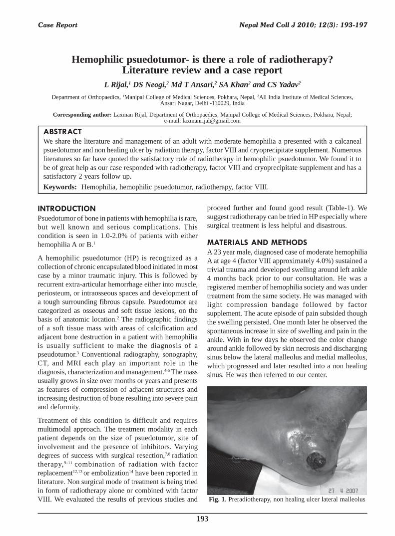
193 Hemophilic psuedotumor- is there a role of radiotherapy? Literature review and a case report L Rijal, 1 DS Neogi, 2 Md T Ansari, 2 SA Khan 2 and CS Yadav 2 Department of Orthopaedics, 1 Manipal College of Medical Sciences, Pokhara, Nepal, 2 All India Institute of Medical Sciences, Ansari Nagar, Delhi -110029, India Corresponding author: Laxman Rijal, Department of Orthopaedics, Manipal College of Medical Sciences, Pokhara, Nepal; e-mail: [email protected] ABSTRACT We share the literature and management of an adult with moderate hemophilia a presented with a calcaneal psuedotumor and non healing ulcer by radiation therapy, factor VIII and cryoprecipitate supplement. Numerous literatures so far have quoted the satisfactory role of radiotherapy in hemophilic psuedotumor. We found it to be of great help as our case responded with radiotherapy, factor VIII and cryoprecipitate supplement and has a satisfactory 2 years follow up. Keywords: Hemophilia, hemophilic psuedotumor, radiotherapy, factor VIII. INTRODUCTION Psuedotumor of bone in patients with hemophilia is rare, but well known and serious complications. This condition is seen in 1.0-2.0% of patients with either hemophilia A or B. 1 A hemophilic psuedotumor (HP) is recognized as a collection of chronic encapsulated blood initiated in most case by a minor traumatic injury. This is followed by recurrent extra-articular hemorrhage either into muscle, periosteum, or intraosseous spaces and development of a tough surrounding fibrous capsule. Psuedotumor are categorized as osseous and soft tissue lesions, on the basis of anatomic location. 2 The radiographic findings of a soft tissue mass with areas of calcification and adjacent bone destruction in a patient with hemophilia is usually sufficient to make the diagnosis of a pseudotumor. 3 Conventional radiography, sonography, CT, and MRI each play an important role in the diagnosis, characterization and management. 4-6 The mass usually grows in size over months or years and presents as features of compression of adjacent structures and increasing destruction of bone resulting into severe pain and deformity. Treatment of this condition is difficult and requires multimodal approach. The treatment modality in each patient depends on the size of psuedotumor, site of involvement and the presence of inhibitors. Varying degrees of success with surgical resection, 7,8 radiation therapy, 9-11 combination of radiation with factor replacement 12,13 or embolization 14 have been reported in literature. Non surgical mode of treatment is being tried in form of radiotherapy alone or combined with factor VIII. We evaluated the results of previous studies and proceed further and found good result (Table-1). We suggest radiotherapy can be tried in HP especially where surgical treatment is less helpful and disastrous. MATERIALS AND METHODS A 23 year male, diagnosed case of moderate hemophilia A at age 4 (factor VIII approximately 4.0%) sustained a trivial trauma and developed swelling around left ankle 4 months back prior to our consultation. He was a registered member of hemophilia society and was under treatment from the same society. He was managed with light compression bandage followed by factor supplement. The acute episode of pain subsided though the swelling persisted. One month later he observed the spontaneous increase in size of swelling and pain in the ankle. With in few days he observed the color change around ankle followed by skin necrosis and discharging sinus below the lateral malleolus and medial malleolus, which progressed and later resulted into a non healing sinus. He was then referred to our center. Case Report Nepal Med Coll J 2010; 12(3): 193-197 Fig. 1. Preradiotherapy, non healing ulcer lateral malleolus
Transcript of Hemophilic psuedotumor- is there a role of radiotherapy ... Report/w22JYlr_ijal.pdfHemophilic...
193
Hemophilic psuedotumor- is there a role of radiotherapy?Literature review and a case report
L Rijal,1 DS Neogi,2 Md T Ansari,2 SA Khan2 and CS Yadav2
Department of Orthopaedics, 1Manipal College of Medical Sciences, Pokhara, Nepal, 2All India Institute of Medical Sciences,Ansari Nagar, Delhi -110029, India
Corresponding author: Laxman Rijal, Department of Orthopaedics, Manipal College of Medical Sciences, Pokhara, Nepal;e-mail: [email protected]
ABSTRACT
We share the literature and management of an adult with moderate hemophilia a presented with a calcanealpsuedotumor and non healing ulcer by radiation therapy, factor VIII and cryoprecipitate supplement. Numerousliteratures so far have quoted the satisfactory role of radiotherapy in hemophilic psuedotumor. We found it tobe of great help as our case responded with radiotherapy, factor VIII and cryoprecipitate supplement and has asatisfactory 2 years follow up.Keywords: Hemophilia, hemophilic psuedotumor, radiotherapy, factor VIII.
INTRODUCTION
Psuedotumor of bone in patients with hemophilia is rare,but well known and serious complications. Thiscondition is seen in 1.0-2.0% of patients with eitherhemophilia A or B.1
A hemophilic psuedotumor (HP) is recognized as acollection of chronic encapsulated blood initiated in mostcase by a minor traumatic injury. This is followed byrecurrent extra-articular hemorrhage either into muscle,periosteum, or intraosseous spaces and development ofa tough surrounding fibrous capsule. Psuedotumor arecategorized as osseous and soft tissue lesions, on thebasis of anatomic location.2 The radiographic findingsof a soft tissue mass with areas of calcification andadjacent bone destruction in a patient with hemophiliais usually sufficient to make the diagnosis of apseudotumor.3 Conventional radiography, sonography,CT, and MRI each play an important role in thediagnosis, characterization and management.4-6 The massusually grows in size over months or years and presentsas features of compression of adjacent structures andincreasing destruction of bone resulting into severe painand deformity.
Treatment of this condition is difficult and requiresmultimodal approach. The treatment modality in eachpatient depends on the size of psuedotumor, site ofinvolvement and the presence of inhibitors. Varyingdegrees of success with surgical resection,7,8 radiationtherapy,9-11 combination of radiation with factorreplacement12,13 or embolization14 have been reported inliterature. Non surgical mode of treatment is being triedin form of radiotherapy alone or combined with factorVIII. We evaluated the results of previous studies and
proceed further and found good result (Table-1). Wesuggest radiotherapy can be tried in HP especially wheresurgical treatment is less helpful and disastrous.
MATERIALS AND METHODS
A 23 year male, diagnosed case of moderate hemophiliaA at age 4 (factor VIII approximately 4.0%) sustained atrivial trauma and developed swelling around left ankle4 months back prior to our consultation. He was aregistered member of hemophilia society and was undertreatment from the same society. He was managed withlight compression bandage followed by factorsupplement. The acute episode of pain subsided thoughthe swelling persisted. One month later he observed thespontaneous increase in size of swelling and pain in theankle. With in few days he observed the color changearound ankle followed by skin necrosis and dischargingsinus below the lateral malleolus and medial malleolus,which progressed and later resulted into a non healingsinus. He was then referred to our center.
Case Report Nepal Med Coll J 2010; 12(3): 193-197
Fig. 1. Preradiotherapy, non healing ulcer lateral malleolus
194
On examination, there was a non healing ulcer of 3x3cm just below the medial and lateral malleolus (Fig.1).Peri-malleolar swelling and change of skin color to duskyred. Slough and necrotic tissue with sero-sanguinushdischarge was seen in the bed of ulcer but there was nofrank pus. Hematological and radiological evaluationwas done immediately after admitting the patient.Hematology confirmed the diagnosis of moderate
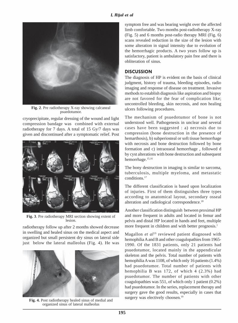
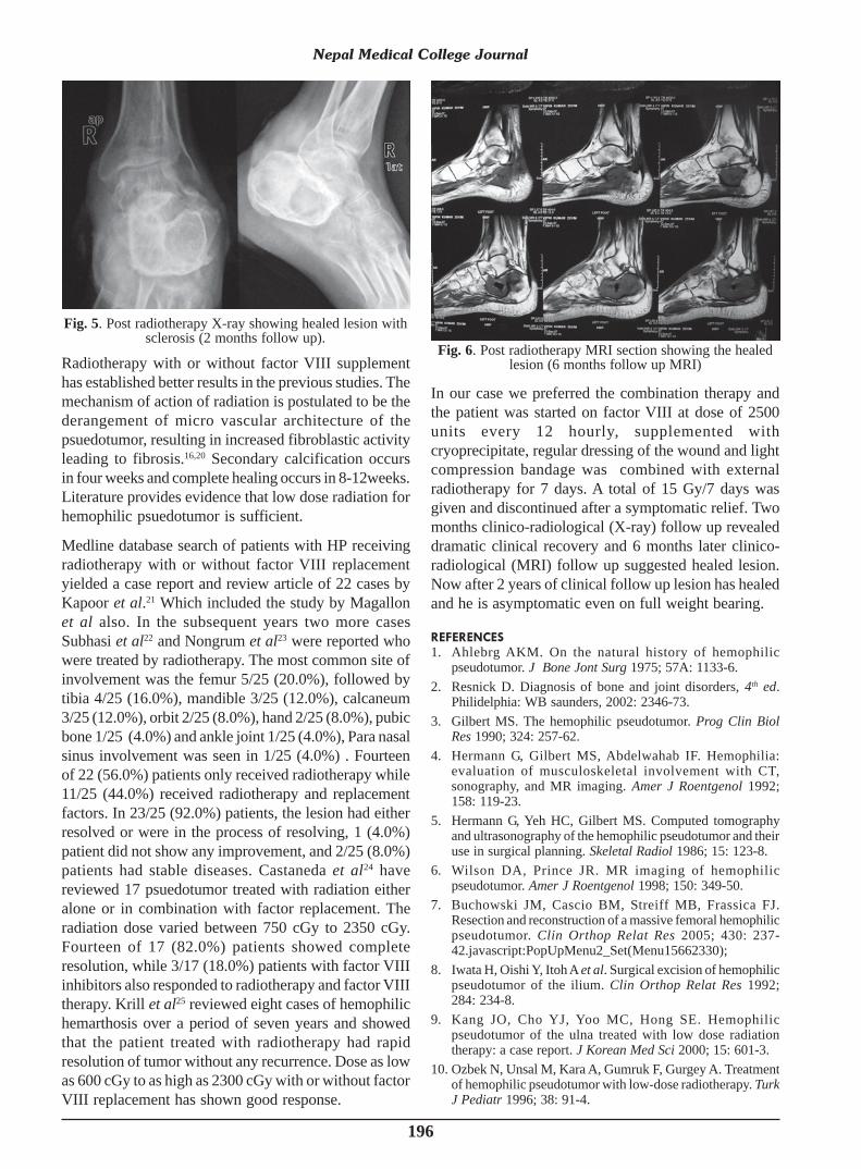
hemophilia A and imaging with X ray (Fig. 2) and MRI(Fig. 3) suggested large lytic lesion with heterogeneousinternal contents (? Hemorrhage) in the calcaneum withcutaneous sinus on the medial and posterolateral aspect.The findings were consistent with HP with edema oftalus and fluid in the ankle and subtalar joint.
The patient was started on factor VIII at dose of 2500units every 12 hourly, supplemented with
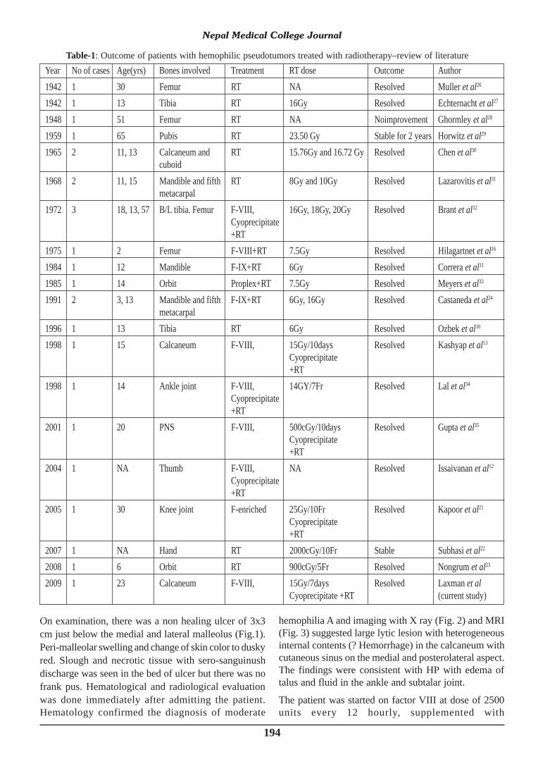
Table-1: Outcome of patients with hemophilic pseudotumors treated with radiotherapy–review of literatureYear No of cases Age(yrs) Bones involved Treatment RT dose Outcome Author1942 1 30 Femur RT NA Resolved Muller et al26
1942 1 13 Tibia RT 16Gy Resolved Echternacht et al27
1948 1 51 Femur RT NA Noimprovement Ghormley et al28
1959 1 65 Pubis RT 23.50 Gy Stable for 2 years Horwitz et al29
1965 2 11, 13 Calcaneum and RT 15.76Gy and 16.72 Gy Resolved Chen et al30
cuboid1968 2 11, 15 Mandible and fifth RT 8Gy and 10Gy Resolved Lazarovitis et al31
metacarpal1972 3 18, 13, 57 B/L tibia. Femur F-VIII, 16Gy, 18Gy, 20Gy Resolved Brant et al32
Cyoprecipitate+RT
1975 1 2 Femur F-VIII+RT 7.5Gy Resolved Hilagartnet et al16
1984 1 12 Mandible F-IX+RT 6Gy Resolved Correra et al11
1985 1 14 Orbit Proplex+RT 7.5Gy Resolved Meyers et al33
1991 2 3, 13 Mandible and fifth F-IX+RT 6Gy, 16Gy Resolved Castaneda et al24
metacarpal1996 1 13 Tibia RT 6Gy Resolved Ozbek et al10
1998 1 15 Calcaneum F-VIII, 15Gy/10days Resolved Kashyap et al13
Cyoprecipitate+RT
1998 1 14 Ankle joint F-VIII, 14GY/7Fr Resolved Lal et al34
Cyoprecipitate+RT
2001 1 20 PNS F-VIII, 500cGy/10days Resolved Gupta et al35
Cyoprecipitate+RT
2004 1 NA Thumb F-VIII, NA Resolved Issaivanan et al12
Cyoprecipitate+RT
2005 1 30 Knee joint F-enriched 25Gy/10Fr Resolved Kapoor et al21
Cyoprecipitate+RT
2007 1 NA Hand RT 2000cGy/10Fr Stable Subhasi et al22
2008 1 6 Orbit RT 900cGy/5Fr Resolved Nongrum et al23
2009 1 23 Calcaneum F-VIII, 15Gy/7days Resolved Laxman et alCyoprecipitate +RT (current study)
Nepal Medical College Journal
195
cryoprecipitate, regular dressing of the wound and lightcompression bandage was combined with externalradiotherapy for 7 days. A total of 15 Gy/7 days wasgiven and discontinued after a symptomatic relief. Post
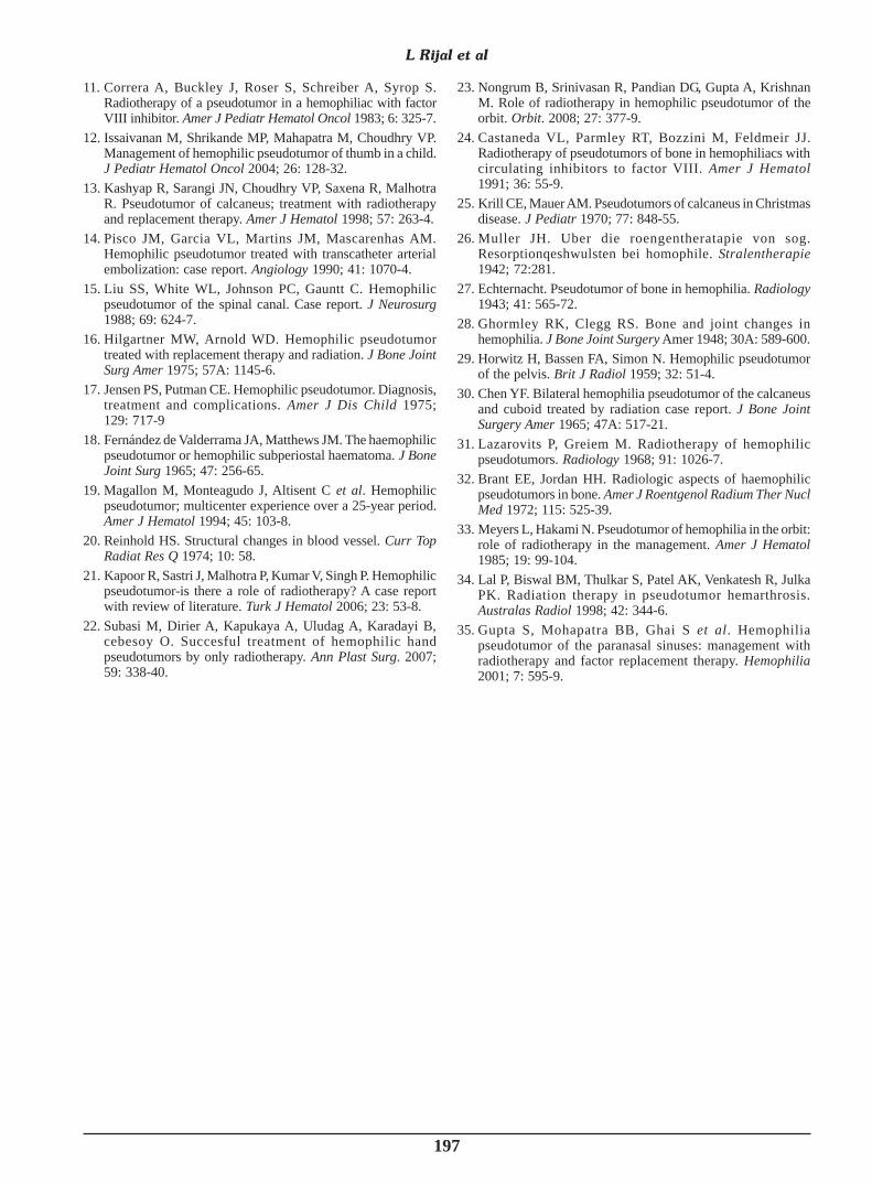
symptom free and was bearing weight over the affectedlimb comfortable. Two months post-radiotherapy X-ray(Fig. 5) and 6 months post-radio therapy MRI (Fig. 6)scans revealed reduction in the size of the lesion withsome alteration in signal intensity due to evolution ofthe hemorrhagic products. A two years follow up issatisfactory, patient is ambulatory pain free and there isobliteration of sinus.
DISCUSSION
The diagnosis of HP is evident on the basis of clinicaljudgment, history of trauma, bleeding episodes, radioimaging and response of disease on treatment. Invasivemethods to establish diagnosis like aspiration and biopsyare not favored for the fear of complication like;uncontrolled bleeding, skin necrosis, and non healingulcers following procedures.
The mechanism of psuedotumor of bone is notunderstood well. Pathogenesis in unclear and severalcases have been suggested : a) necrosis due tocompression (bone destruction in the presence ofhemarthosis), b) subperiosteal or soft tissue hemorrhagewith necrosis and bone destruction followed by boneformation and c) intraosseal hemorrhage , followed dby cyst alterations with bone destruction and subsequenthemorrhage.15,16
The bony destruction in imaging is similar to sarcoma,tuberculosis, multiple myeloma, and metastaticconditions.17
The different classification is based upon localizationof injuries. First of them distinguishes three typesaccording to anatomical layout, secondary ossealalteration and radiological correspondence.18
Another classification distinguish between proximal HPand more frequent in adults and located in femur andpelvis and distal HP located in hands and feet, multiplemore frequent in children and with better prognosis.1
Magallon et al19 reviewed patient diagnosed withhemophilia A and B and other coagulopathies from 1965-1990. Of the 1831 patients, only 21 patients hadpsuedotumor, located mainly in the appendicularskeleton and the pelvis. Total number of patients withhemophilia A was 1108, of which only 16 patients (1.4%)had psuedotumor. Total number of patients withhemophilia B was 172, of which 4 (2.3%) hadpsuedotumor. The number of patients with othercoagulopathies was 551, of which only 1 patient (0.2%)had psuedotumor. In the series, replacement therapy andsurgery gave the good results, especially in cases thatsurgery was electively choosen.19
L Rijal et al
Fig. 2. Pre radiotherapy X-ray showing calcanealpsuedotumor.
radiotherapy follow up after 2 months showed decreasein swelling and healed sinus on the medical aspect andorganized but small persistent dry sinus on lateral sidejust below the lateral malleolus (Fig. 4). He was
Fig. 3. Pre radiotherapy MRI section showing extent oflesion.
Fig. 4. Post radiotherapy healed sinus of medial andorganized sinus of lateral malleolus
196
Radiotherapy with or without factor VIII supplementhas established better results in the previous studies. Themechanism of action of radiation is postulated to be thederangement of micro vascular architecture of thepsuedotumor, resulting in increased fibroblastic activityleading to fibrosis.16,20 Secondary calcification occursin four weeks and complete healing occurs in 8-12weeks.Literature provides evidence that low dose radiation forhemophilic psuedotumor is sufficient.
Medline database search of patients with HP receivingradiotherapy with or without factor VIII replacementyielded a case report and review article of 22 cases byKapoor et al.21 Which included the study by Magallonet al also. In the subsequent years two more casesSubhasi et al22 and Nongrum et al23 were reported whowere treated by radiotherapy. The most common site ofinvolvement was the femur 5/25 (20.0%), followed bytibia 4/25 (16.0%), mandible 3/25 (12.0%), calcaneum3/25 (12.0%), orbit 2/25 (8.0%), hand 2/25 (8.0%), pubicbone 1/25 (4.0%) and ankle joint 1/25 (4.0%), Para nasalsinus involvement was seen in 1/25 (4.0%) . Fourteenof 22 (56.0%) patients only received radiotherapy while11/25 (44.0%) received radiotherapy and replacementfactors. In 23/25 (92.0%) patients, the lesion had eitherresolved or were in the process of resolving, 1 (4.0%)patient did not show any improvement, and 2/25 (8.0%)patients had stable diseases. Castaneda et al24 havereviewed 17 psuedotumor treated with radiation eitheralone or in combination with factor replacement. Theradiation dose varied between 750 cGy to 2350 cGy.Fourteen of 17 (82.0%) patients showed completeresolution, while 3/17 (18.0%) patients with factor VIIIinhibitors also responded to radiotherapy and factor VIIItherapy. Krill et al25 reviewed eight cases of hemophilichemarthosis over a period of seven years and showedthat the patient treated with radiotherapy had rapidresolution of tumor without any recurrence. Dose as lowas 600 cGy to as high as 2300 cGy with or without factorVIII replacement has shown good response.
In our case we preferred the combination therapy andthe patient was started on factor VIII at dose of 2500units every 12 hourly, supplemented withcryoprecipitate, regular dressing of the wound and lightcompression bandage was combined with externalradiotherapy for 7 days. A total of 15 Gy/7 days wasgiven and discontinued after a symptomatic relief. Twomonths clinico-radiological (X-ray) follow up revealeddramatic clinical recovery and 6 months later clinico-radiological (MRI) follow up suggested healed lesion.Now after 2 years of clinical follow up lesion has healedand he is asymptomatic even on full weight bearing.
REFERENCES
1. Ahlebrg AKM. On the natural history of hemophilicpseudotumor. J Bone Jont Surg 1975; 57A: 1133-6.
2. Resnick D. Diagnosis of bone and joint disorders, 4th ed.Philidelphia: WB saunders, 2002: 2346-73.
3. Gilbert MS. The hemophilic pseudotumor. Prog Clin BiolRes 1990; 324: 257-62.
4. Hermann G, Gilbert MS, Abdelwahab IF. Hemophilia:evaluation of musculoskeletal involvement with CT,sonography, and MR imaging. Amer J Roentgenol 1992;158: 119-23.
5. Hermann G, Yeh HC, Gilbert MS. Computed tomographyand ultrasonography of the hemophilic pseudotumor and theiruse in surgical planning. Skeletal Radiol 1986; 15: 123-8.
6. Wilson DA, Prince JR. MR imaging of hemophilicpseudotumor. Amer J Roentgenol 1998; 150: 349-50.
7. Buchowski JM, Cascio BM, Streiff MB, Frassica FJ.Resection and reconstruction of a massive femoral hemophilicpseudotumor. Clin Orthop Relat Res 2005; 430: 237-42.javascript:PopUpMenu2_Set(Menu15662330);
8. Iwata H, Oishi Y, Itoh A et al. Surgical excision of hemophilicpseudotumor of the ilium. Clin Orthop Relat Res 1992;284: 234-8.
9. Kang JO, Cho YJ, Yoo MC, Hong SE. Hemophilicpseudotumor of the ulna treated with low dose radiationtherapy: a case report. J Korean Med Sci 2000; 15: 601-3.
10. Ozbek N, Unsal M, Kara A, Gumruk F, Gurgey A. Treatmentof hemophilic pseudotumor with low-dose radiotherapy. TurkJ Pediatr 1996; 38: 91-4.
Nepal Medical College Journal
Fig. 5. Post radiotherapy X-ray showing healed lesion withsclerosis (2 months follow up).
Fig. 6. Post radiotherapy MRI section showing the healedlesion (6 months follow up MRI)
197
11. Correra A, Buckley J, Roser S, Schreiber A, Syrop S.Radiotherapy of a pseudotumor in a hemophiliac with factorVIII inhibitor. Amer J Pediatr Hematol Oncol 1983; 6: 325-7.
12. Issaivanan M, Shrikande MP, Mahapatra M, Choudhry VP.Management of hemophilic pseudotumor of thumb in a child.J Pediatr Hematol Oncol 2004; 26: 128-32.
13. Kashyap R, Sarangi JN, Choudhry VP, Saxena R, MalhotraR. Pseudotumor of calcaneus; treatment with radiotherapyand replacement therapy. Amer J Hematol 1998; 57: 263-4.
14. Pisco JM, Garcia VL, Martins JM, Mascarenhas AM.Hemophilic pseudotumor treated with transcatheter arterialembolization: case report. Angiology 1990; 41: 1070-4.
15. Liu SS, White WL, Johnson PC, Gauntt C. Hemophilicpseudotumor of the spinal canal. Case report. J Neurosurg1988; 69: 624-7.
16. Hilgartner MW, Arnold WD. Hemophilic pseudotumortreated with replacement therapy and radiation. J Bone JointSurg Amer 1975; 57A: 1145-6.
17. Jensen PS, Putman CE. Hemophilic pseudotumor. Diagnosis,treatment and complications. Amer J Dis Child 1975;129: 717-9
18. Fernández de Valderrama JA, Matthews JM. The haemophilicpseudotumor or hemophilic subperiostal haematoma. J BoneJoint Surg 1965; 47: 256-65.
19. Magallon M, Monteagudo J, Altisent C et al. Hemophilicpseudotumor; multicenter experience over a 25-year period.Amer J Hematol 1994; 45: 103-8.
20. Reinhold HS. Structural changes in blood vessel. Curr TopRadiat Res Q 1974; 10: 58.
21. Kapoor R, Sastri J, Malhotra P, Kumar V, Singh P. Hemophilicpseudotumor-is there a role of radiotherapy? A case reportwith review of literature. Turk J Hematol 2006; 23: 53-8.
22. Subasi M, Dirier A, Kapukaya A, Uludag A, Karadayi B,cebesoy O. Succesful treatment of hemophilic handpseudotumors by only radiotherapy. Ann Plast Surg. 2007;59: 338-40.
23. Nongrum B, Srinivasan R, Pandian DG, Gupta A, KrishnanM. Role of radiotherapy in hemophilic pseudotumor of theorbit. Orbit. 2008; 27: 377-9.
24. Castaneda VL, Parmley RT, Bozzini M, Feldmeir JJ.Radiotherapy of pseudotumors of bone in hemophiliacs withcirculating inhibitors to factor VIII. Amer J Hematol1991; 36: 55-9.
25. Krill CE, Mauer AM. Pseudotumors of calcaneus in Christmasdisease. J Pediatr 1970; 77: 848-55.
26. Muller JH. Uber die roengentheratapie von sog.Resorptionqeshwulsten bei homophile. Stralentherapie1942; 72:281.
27. Echternacht. Pseudotumor of bone in hemophilia. Radiology1943; 41: 565-72.
28. Ghormley RK, Clegg RS. Bone and joint changes inhemophilia. J Bone Joint Surgery Amer 1948; 30A: 589-600.
29. Horwitz H, Bassen FA, Simon N. Hemophilic pseudotumorof the pelvis. Brit J Radiol 1959; 32: 51-4.
30. Chen YF. Bilateral hemophilia pseudotumor of the calcaneusand cuboid treated by radiation case report. J Bone JointSurgery Amer 1965; 47A: 517-21.
31. Lazarovits P, Greiem M. Radiotherapy of hemophilicpseudotumors. Radiology 1968; 91: 1026-7.
32. Brant EE, Jordan HH. Radiologic aspects of haemophilicpseudotumors in bone. Amer J Roentgenol Radium Ther NuclMed 1972; 115: 525-39.
33. Meyers L, Hakami N. Pseudotumor of hemophilia in the orbit:role of radiotherapy in the management. Amer J Hematol1985; 19: 99-104.
34. Lal P, Biswal BM, Thulkar S, Patel AK, Venkatesh R, JulkaPK. Radiation therapy in pseudotumor hemarthrosis.Australas Radiol 1998; 42: 344-6.
35. Gupta S, Mohapatra BB, Ghai S et al. Hemophiliapseudotumor of the paranasal sinuses: management withradiotherapy and factor replacement therapy. Hemophilia2001; 7: 595-9.
L Rijal et al