Hemolytic Disease of the Fetus and Newborn: Modern ... · antigens and in predicting fetal anemia...
21
Hemolytic Disease of the Fetus and Newborn: Modern Practice and Future Investigations Jeanne E. Hendrickson, Meghan Delaney PII: S0887-7963(16)30011-6 DOI: doi: 10.1016/j.tmrv.2016.05.008 Reference: YTMRV 50466 To appear in: Transfusion Medicine Reviews Received date: 20 February 2016 Accepted date: 23 May 2016 Please cite this article as: Hendrickson Jeanne E., Delaney Meghan, Hemolytic Disease of the Fetus and Newborn: Modern Practice and Future Investigations, Transfusion Medicine Reviews (2016), doi: 10.1016/j.tmrv.2016.05.008 This is a PDF file of an unedited manuscript that has been accepted for publication. As a service to our customers we are providing this early version of the manuscript. The manuscript will undergo copyediting, typesetting, and review of the resulting proof before it is published in its final form. Please note that during the production process errors may be discovered which could affect the content, and all legal disclaimers that apply to the journal pertain.
Transcript of Hemolytic Disease of the Fetus and Newborn: Modern ... · antigens and in predicting fetal anemia...
�������� ����� ��
Hemolytic Disease of the Fetus and Newborn: Modern Practice and FutureInvestigations
Jeanne E. Hendrickson, Meghan Delaney
PII: S0887-7963(16)30011-6DOI: doi: 10.1016/j.tmrv.2016.05.008Reference: YTMRV 50466
To appear in: Transfusion Medicine Reviews
Received date: 20 February 2016Accepted date: 23 May 2016
Please cite this article as: Hendrickson Jeanne E., Delaney Meghan, Hemolytic Disease ofthe Fetus and Newborn: Modern Practice and Future Investigations, Transfusion MedicineReviews (2016), doi: 10.1016/j.tmrv.2016.05.008
This is a PDF file of an unedited manuscript that has been accepted for publication.As a service to our customers we are providing this early version of the manuscript.The manuscript will undergo copyediting, typesetting, and review of the resulting proofbefore it is published in its final form. Please note that during the production processerrors may be discovered which could affect the content, and all legal disclaimers thatapply to the journal pertain.
ACC
EPTE
D M
ANU
SCR
IPT
ACCEPTED MANUSCRIPT
Page 1 of 20
Hemolytic Disease of the Fetus and Newborn: Modern Practice and Future Investigations
Jeanne E. Hendrickson, MD and Meghan Delaney, DO, MPH1,2,3
1. Laboratory Medicine, Seattle Children’s Hospital, Seattle, WA, USA
2. Department of Laboratory Medicine & Pediatrics, University of Washington, Seattle, WA, USA
3. Bloodworks NW, Seattle, WA, USA
Keywords: hemolytic disease of the fetus and newborn, red blood cell, alloimmunization
Corresponding author:
Jeanne E. Hendrickson, MD; Yale University Department of Laboratory Medicine, 330 Cedar Street,
Clinic Building 405, PO Box 208035, New Haven, CT 06520-0835, USA
Tel.: +1-203-737-8316
E-mail: [email protected]
ACC
EPTE
D M
ANU
SCR
IPT
ACCEPTED MANUSCRIPT
Page 2 of 20
Abstract:
Red blood cell (RBC) sensitization occurs in some women in response to exposure to paternally derived
RBC antigens during pregnancy or to non-self antigens on transfused RBCs during their lifetime. Once
sensitized, future pregnancies may be at risk for hemolytic disease of the fetus and newborn (HDFN).
Although great strides have been made over the past few decades in terms of identifying blood group
antigens and in predicting fetal anemia through the use of non-invasive monitoring, many questions
remain in terms of understanding RBC alloimmunization risk factors, preventative therapies, and
treatment strategies. At the present time, there is room for improvement in these areas in both developed
and developing countries. Evidence based, universal guidelines describing recommended RBC antigen
matching transfusion strategies for girls or women, prior to pregnancy or during intrauterine transfusions
(IUTs), would be welcomed. A better understanding of the mechanism(s) of action of RhIg, first
introduced over half of a century ago and one of the most successful immunoprophylaxis therapies in
existence today, would also be a large step forward. For example, answers to questions of the role(s) that
fetal RBC clearance, antigen masking, antigen modulation, and immune suppression play in the
effectiveness of RhIg may help to guide the development of novel preventative therapies during
pregnancy for immunization to RhD and non-RhD antigens. Further, a better understanding of the
importance of anti-RhD or other alloantibody glycosylation patterns may be beneficial not only in
developing such novel immunoprophylaxis therapies, but also in predicting the clinical significance of
existing maternal alloantibodies. One other area of need includes the development of therapies beyond
IUTs to mitigate the dangers of maternal alloantibodies to developing fetuses. We challenge physicians,
scientists, and funding agencies to prioritize studies of RBC alloimmunization and HDFN, and to invest
in the children of our future.
ACC
EPTE
D M
ANU
SCR
IPT
ACCEPTED MANUSCRIPT
Page 3 of 20
Introduction
Maternal alloimmunization to blood group antigens occurs through exposure to non-self antigens on
RBCs via transfusion or prior pregnancy. RBC alloantibodies may be detrimental in transfusion or
pregnancy settings, depending on the specificity and depending on the presence or absence of the cognate
antigen(s) on transfused or fetal RBCs. In a transfusion setting, incompatible RBCs may be hemolyzed.
In a pregnancy setting, fetal RBCs expressing a paternally derived antigen against which a mother is
alloimmunized may be hemolyzed or may be altered such that erythropoiesis is suppressed, either
resulting in hemolytic disease of the fetus and newborn (HDFN).
Alloantibodies against more than 50 non-ABO blood group antigens have been implicated in
HDFN, with many blood group antigens historically first identified after the birth of a hydropic infant1. In
addition to antibodies against non-ABO blood group antigens, naturally occurring maternal
isohemagglutinins, often in group O mothers, are capable of leading to anemia in fetuses expressing the A
or B antigens. This anemia, though relatively common, is usually mild and rarely requires intervention.
The majority of clinically significant HDFN cases are due to alloantibodies against antigens in the Rh,
Kell, Duffy, Kidd, and MNS families2-4
, with 1/300-1/600 live births being affected by maternal RBC
alloimmunization5. Of note, maternal alloantibodies against Rh antigens are significantly less likely to
develop when fetal cells are ABO incompatible with maternal plasma, presumably due to the rapid
clearance of fetal RBCs by maternal isohemagglutinins. Although alloantibodies to antigens in the Rh
family remain a leading cause of severe HDFN world-wide, antibodies against the antigens in the Kell
family are emerging to be a leading cause of HDFN in parts of the world where RhIg is widely used for
immunoprophylaxis6.
The outcomes of antigen positive fetuses developing in utero of alloimmunized women vary,
depending on characteristics of the maternal antibody and the RBC antigen. Fetuses affected by maternal
anti-D alloantibodies may have anemia and such severe hyperbilirubinemia that they develop kernicterus.
ACC
EPTE
D M
ANU
SCR
IPT
ACCEPTED MANUSCRIPT
Page 4 of 20
Fetuses affected by maternal anti-Kell antibodies, however, are more likely to have anemia and
reticulocytopenia but rarely have significant hyperbilirubinemia. Because many different maternal
antibodies are capable of leading to hydrops fetalis and intrauterine fetal demise, the early diagnosis of at
risk pregnancies is critically important. Maternal antibody titers against antigens such as RhD are
commonly reported by US transfusion medicine services. Bioassays such as chemiluminescence or
monocyte monolayer assays have a higher specificity to predict fetal outcomes than maternal antibody
titers7, 8
, though they are not routinely used in the US. Recent studies suggest that other antibody
characteristics, including glycosylation patterns, may associate even more closely with antibody
dependent cellular cytotoxicity and fetal outcome than titer9, 10
.
A better understanding of characteristics of maternal antibodies and fetal RBC antigens that result
in adverse fetal outcomes would be helpful in the development of novel/targeted therapeutic
interventions, as discussed in more detail below. For example, it is unclear why maternal antibodies
against antigens on the KEL glycoprotein so efficiently lead to reticulocytopenia. Is has been
hypothesized that the expression of the Kell antigen on early fetal RBC precursors plays an important
role, with direct suppression of erythropoiesis by maternal anti-Kell reproduced in cultures in vitro11
.
However, phagocytosis of RBC precursors expressing the Kell antigen has also been observed in vitro12
,
making it plausible that the lack of hyperbilirubinemia observed in fetuses affected by maternal anti-Kell
antibodies is due in part to immune mediated clearance of very early RBC precursors which don’t contain
hemoglobin. In this review, we summarize the latest clinical approaches to HDFN and describe research
that is underway as well as the topics in need of further investigation and development to continue to
improve management of this disorder.
Diagnosis and Treatment
At the first prenatal visit, all pregnant women should be tested for RBC alloantibodies using the indirect
antibody test (IAT). After the woman is found the have RBC antibodies, the risk of clinically significant
ACC
EPTE
D M
ANU
SCR
IPT
ACCEPTED MANUSCRIPT
Page 5 of 20
HDFN is determined by several techniques. Using the maternal sample, serial (usually monthly) RBC
antibody titration is done to assess if the fetal red blood cells are acting as an immunizing stimulus. If
available for testing, the paternal blood type provides inheritance information; homozygous fathers have
100% chance of passing the implicated antigen to the fetus; heterozygous fathers have a 50% chance of
having an offspring with the offending blood group antigen. In the case of anti-D sensitization,
serological testing cannot determine zygosity and molecular testing for the RHD gene copy number are
needed 13
. Fetal DNA can also be obtained by amniocentesis for blood group genotyping to directly
determine the fetal blood type using cultured amniocytes. Due to the risk of fetal loss with the procedure,
newer techniques have been developed that isolate fetal DNA from a maternal peripheral blood sample
for RBC genotyping14, 15
.
To determine the clinical significance of HDFN, the fetus should be monitored for well-being by
ultrasound to look for evidence of ascites (hydrops), heart rate monitoring and for anemia. Fetal anemia
assessment is carried out non-invasively using ultrasonic measurement of fetal blood flow through the
large cerebral vessels, usually the middle cerebral artery. The velocity of the blood flow indicates the
degree of anemia.
Although maternal alloantibody titers are used to predict the risk to the fetus, some fetuses have
severe anemia despite low titers and others have no anemia despite high titers. Pan-IgG reagents are
typically used for measuring titers in the US, but evaluating IgG subtypes may be informative for certain
antibodies16
. In one study, for example, fetal anemia correlated positively with the amount of maternal
IgG1 anti-D, and negatively with the amount of IgG3 anti-D bound to fetal RBCs17
. In addition to
antibody titers, some countries also use in vitro antibody dependent cellular cytotoxicity (ADCC)
biological assays to predict alloantibody activity18, 19
The importance of antibody glycosylation patterns on FcγR binding avidity on clinical outcomes
is increasingly being appreciated, in multiple biologic systems20
. Recent studies have described a
significant association between such antibody glycosylation patterns and fetal outcomes. Maternal anti-D
alloantibodies with the lowest degrees of fucosylation (as measured by mass spectroscopy), for example,
ACC
EPTE
D M
ANU
SCR
IPT
ACCEPTED MANUSCRIPT
Page 6 of 20
have been reported to be associated with more severe fetal anemia9. Similarly, maternal anti-platelet
glycoprotein alloantibodies with low fucosylation patterns have been shown to be more clinically
significant than those with higher fucosylation patterns21
. One potential explanation for the increase in
clinical significance is high binding avidity to FcγRIIIa monocytes and FcγIIIb polymorphonuclear cells.
The treatment of a fetus affected by HDFN is focused on monitoring and support until delivery.
Fetuses at gestation >16-24 weeks and are found to have blood flow velocities 1.5 times the multiple of
the mean (MoM) is indicative of moderate to severe anemia. Periumbilical blood testing is indicated to
directly sample the fetal circulation 2, 22
. This is often directly followed by intrauterine red blood cell
transfusion (IUT) if the fetus is not of acceptable gestational age for delivery. These RBC transfusions
are selected as Group O, negative for the offending antigen(s), leukocyte reduced, irradiated, and
concentrated to ensure maximal delivery and to ensure the hematocrit of the fetus is ≥30%. Extended
RBC antigen matching is done at some centers. IUT is able to preserve neurologic outcome in many
children, though those with severe hydrops may develop cerebral palsy, severe developmental delay, and
deafness23
. Potential alternatives or adjunctive maternal therapies during pregnancy to decrease the
severity of fetal anemia include IVIG and plasma exchange (PE), though the evidence to support the
efficacy of these therapies is limited. PE has been reported in women with high titer RBC antibodies and
antecedent HDFN24
; it is given a Category II by the American Society for Apheresis, meaning it is
considered second line therapy25
.
After delivery, infants are closely monitored for hemoglobin and bilirubin levels as their own
physiological systems must function independently from the mother. Infants affected by HDFN must be
intensively observed with laboratory monitoring of hemoglobin and bilirubin to determine if
phototherapy, simple transfusion, or exchange transfusion are necessary26, 27
. Although exchange
transfusion is a critical therapy for prevention of kernicterus in neonates with rapidly rising bilirubin, it
does have complications28
. IVIg has been shown to reduce the need for exchange transfusion in some
studies29, 30
. However, it does not affect anemia significantly, and top off transfusions may still be needed
ACC
EPTE
D M
ANU
SCR
IPT
ACCEPTED MANUSCRIPT
Page 7 of 20
27. In addition, IVIg contains anti-A and anti-B antibodies which may potentially worsen HDFN among
group A or B newborns.
The majority of maternal alloantibody transfer into the fetal circulation ends at birth. However,
neonatal anemia may persist beyond the natural half-life of the antibodies in some circumstances, and
may resolve more quickly than predicted in other circumstances. Anemia may persist regardless of the
presence of circulating alloantibodies in neonates who undergo multiple intrauterine transfusions, due to
the suppression of erythropoiesis. Further, it is possible that maternally derived alloantibodies in
neonates who have undergone intrauterine transfusions or post-natal exchange transfusions may persist
longer than one may predict, due to a lack of antigen positive RBCs to bind the antibodies. As the infant
ages, signs of poor feeding and increased sleep can be signs of persistent hypoproliferative anemia.
Continued monitoring with regular checks of hematocrit and reticulocytes provides critical information
for marrow recovery or the need for red cell transfusion. Of note, recent studies have introduced the idea
that maternal antibodies against antigens on platelets or RBCs may be transferred to neonates via breast
milk. A study describing the transfer of anti-platelet IgA antibodies in breast milk from mother to neonate
and resulting in prolonged neonatal thrombocytopenia31
led to murine studies evaluating the possibility of
RBC alloantibody transfer in breast milk. Anti-KEL glycoprotein RBC alloantibodies (IgG and IgA)
were, in fact, shown to be transferred via breast milk from mothers to nursing pups in a murine fostering
model32
. In considering the translation of these murine findings to humans, however, one must remain
mindful that murine FcRn receptors transfer IgG significantly more efficiently from intestine to
circulation than human receptors33, 34
.
Prevention
The prevention of maternal alloimmunization falls into three different categories; primary, secondary and
tertiary (Table 1). Primary prevention focuses on preventing the possibility of maternal
alloimmunization through reduced exposure to foreign RBC antigens. In most industrialized nations, the
emergency supply of RBCs that is kept sequestered to support sudden and unexpected bleeding
ACC
EPTE
D M
ANU
SCR
IPT
ACCEPTED MANUSCRIPT
Page 8 of 20
emergencies includes Group O (universal donor) RBC products. These products may be RhD negative or
positive. In many institutions, particularly those supporting trauma programs, the RhD positive RBC
products are allowed to be given to males so that the rarer RhD negative RBC products can be
preferentially provided to females of childbearing potential, such as those <50 years 35
. These widely
accepted practices are in place to provide a measure of serological prevention for young women that
require life-saving transfusion that does not endanger future pregnancies. Although not as universally
practiced, some nations and regions attempt to prevent exposure of other RBC antigens when females of
childbearing potential are transfused with RBC products in routine medical and surgical settings. For
example, several European nations provide K antigen negative, and/or E, and c antigen matched products
to all females less than 45 – 50 years (exact practices vary by region). It is not clear if the approach of
extended RBC antigen matching is entirely successful due to other routes of potential exposure, such as
through previous pregnancy and receipt of transfusion outside of the region. However, for regions that
are able to accomplish it, there may be a measure of prevention added 36
.
The administration of Rh immunoglobulin (RhIg) is secondary prevention aimed at removing or
masking fetal RhD positive RBCs found in the maternal circulation during pregnancy or after delivery.
The product has had an enormous impact on the prevalence of HDFN due to anti-D, decreasing the
incidence from 16% to <0.1% 37
. Notably, many low resource countries do not have prenatal screening
programs and RhIg availability, putting infants born in these regions at risk.
Appropriately identifying women who need RhIg is an area of study. Partially because of the
product’s success and also because it was implemented before there was a feasible way to determine the
fetal RhD type and precise maternal RhD typing, practice has evolved to administer RhIg to all RhD
negative pregnant women prenatally, and often postnatally, even when the infant’s RBC type could also
be determined at that time. Moreover, practice often includes giving RhIg to all women with weak RhD
testing results, referred to as weak D blood type because there has not been acceptance or access to testing
to more precisely guide prevention measures. The advent of genetic testing for RHD has led to
ACC
EPTE
D M
ANU
SCR
IPT
ACCEPTED MANUSCRIPT
Page 9 of 20
improvements in the ability to guide RhIg administration decisions and to stratify risk for women who
already have made anti-D. There are some examples of forward thinking national approaches to testing
fetal DNA from a sample of peripheral maternal blood to determine which pregnant women should
receive RhIg due to pregnancy with an RhD positive fetus15
. In 2015, a practice recommendation was
made by several professional organizations (AABB, CAP, ACOG, Armed Forces Blood Program, ABC,
American Red Cross) to begin to phase in the use of RhD genotyping methods to categorize women with
initial RhD typing results that are weak instead of strongly reactive in the serological test system 38
. The
recommendation suggests that if the woman carries the RHD genetic variants weak D type 1, 2, or 3, then
she can safely be managed as RhD positive and does not need RhIg. The testing offers more precise
information than can be provided by traditional blood typing approaches and can provide a measure of
assurance for women that have been given different blood typing results at different times as well as
improve resource utilization of RhD negative blood products and RhIg. In fact, a financial modelling
study of the suggested approach found that the genotyping approach is cost neutral or incrementally cost
saving than providing RhIg to all women who are weak D by serological methods39
.
Rarely, an RhD negative female that is of childbearing potential and desiring of future
pregnancy may receive RhD positive RBC units due to emergency needs, or occasionally, due to errors in
the process of blood sample collection and preparation of the blood product(s). In these exceptional
circumstances, the use of large doses of RhIg and/or automated red blood cell exchange (RCE) using an
apheresis instrument are types of secondary prevention that have been reported. Several case reports have
been published that cite the use of approximately 20 ug of RhIg for each mL of RhD positive RBCs
transfused, although data are limited and there is no accepted standard40
. For RhD negative females that
have received larger quantities of RhD positive RBC products, an RCE procedure has been used to lower
the RhD positive RBCs in circulation 41, 42
. Then the patient is administered RhIg to cover the remaining
calculated residual RhD positive RBCs. The ASFA plans to rank this as a category III in the 2016
Seventh Special Issue to be published in the Journal of Clinical Apheresis, suggesting that apheresis
should be used on a case-by-case basis due to the paucity of published experience and efficacy.
ACC
EPTE
D M
ANU
SCR
IPT
ACCEPTED MANUSCRIPT
Page 10 of 20
The use of extended antigen matched RBC units for IUT transfusions is tertiary prevention. In
this setting, the pregnant woman already has alloimmunization to RBC antigens and has a fetus that is
affected with anemia due to the RBC antibody. Although there is not currently a thorough scientific
understanding to pinpoint the factors that make a person’s immune system respond to RBC antigens,
there is evidence that such “immune responder” women are at risk for further RBC alloimmunization to
other RBC antigens that they have not yet formed antibodies against. The broadening of her RBC
antibody specificities may impact future pregnancies. Researchers in the Netherlands have led several
studies investigating the effect of providing antigen matched RBC units to women undergoing IUT
procedures. Supporting the “immune responder” hypothesis, they found that when Rh (C, c, E, e) and K
antigen matched RBCs were used for IUT, 25% of women (53/212) formed additional RBC antibodies43
.
In an attempt to abrogate this response, the group attempted to provide IUT transfusions with the Rh, K
blood antigens as above, plus the addition of Fy, Jk and Ss blood group antigens for 159 women that
needed IUT for HDFN. They found that it was increasingly difficult to find appropriately matched units
(48% received full antigen matched, 52% received partially antigen matched), and that 4.3% of women
getting high match and 11% getting moderate match formed additional antibodies44
. Together, these data
show the difficulty of prevention of alloimmunization for patients that appear to have “immune
responder” characteristics. Further research is needed to define and understand the underlying biological
mechanisms.
Research in Focus
RhIg and alternative RhD specific therapies
RhIg, in widespread use in many countries for half of a century now, is one of the most successful
immunomodulatory therapies in existence today. Despite its efficacy, the mechanism of action remains
poorly understood45, 46
. The current practice of generating RhIg by immunizing male RhD negative
volunteers not only puts the volunteers at risk for hemolytic transfusion reactions should they receive
RhD positive RBCs in trauma/emergent situations, however, but it also leads to variability in RhIg
ACC
EPTE
D M
ANU
SCR
IPT
ACCEPTED MANUSCRIPT
Page 11 of 20
product composition. For example, differences in fucosylation patterns have recently been identified
between brands of RhIg, with these patterns theoretically impacting product efficacy by altering Fcγ
binding47
.
Investigations of alternative sources of anti-D (including in vitro production) have been hampered
due in part to a lack of animal models available for reductionist/mechanistic studies; the generation of an
animal model of RhD alloimmunization has been difficult48, 49
. It is logical that RhIg simply prevents a
mother’s immune system from “seeing” the RhD antigen on fetal RBCs, given observational studies in
the pre-RhIg era of the risk of maternal anti-D formation in ABO compatible pregnancies (16% risk of
RhD alloimmunization) versus ABO incompatible pregnancies (<2% risk of RhD alloimmunization)37, 50
.
However, some RhD antigen sites on fetal RBCs are thought to remain unbound with the low dose of
RhIg given. Further, correlations between RBC clearance and anti-D alloimmunization have not
consistently been observed, with some monoclonal anti-D preparations that increase RBC clearance
enhancing rather than preventing alloimmunization51
. A similar finding of increased alloimmunization in
situations of rapid RBC clearance has been reported in at least one murine model, suggesting that rapid
clearance of a bolus of antigen positive RBCs has the capability, in certain situations or with certain
antigens, to enhance RBC alloimmunization52, 53
. Of note, antigen presentation alone is known to be
insufficient to lead to an alloantibody response in some blood group systems54
as well as in multiple non-
RBC systems, with a simultaneous danger signal of sorts also being necessary55, 56
.
Antibody mediated immune suppression
The term “antibody mediated immune suppression” (AMIS) has been applied to models in which
passively infused antibodies such as RhIg prevent active alloimmunization. The actual degree of “immune
suppression” (versus other mechanism(s) of action) of these antibodies, however, remains unknown.
Regardless of mechanism, an antigen specific effect is seen in many systems. For example, passively
infused polyclonal antibody (“KELIg”) against the KEL glycoprotein antigen expressed on murine RBCs
prevents alloimmunization to transfused RBCs expressing the KEL glycoprotein.57
ACC
EPTE
D M
ANU
SCR
IPT
ACCEPTED MANUSCRIPT
Page 12 of 20
The phenomenon of a passively administered antibody altering active RBC alloimmunization has
been investigated not only in the KEL murine model but also in murine models with other antigens and in
murine models transfused with sheep RBCs.58
Multiple subtypes of monoclonal antibodies as well as
polyclonal antibodies have been investigated, with some but not all being capable of preventing
alloimmunization altogether or of decreasing the magnitude of an alloimmune response59-61
. In at least
one model, the efficacy of passively infused antibodies at preventing an alloimmune response to
transfused RBCs has been shown to be independent of the inhibitory FcRIIb receptor60
.
Antigen modulation
Antigen modulation, also known as antigen loss or antigen suppression, has been described to occur on
RBCs as well as on WBCs in the presence of antibodies, in humans and in animal models alike62, 63
. At
the present time, it is not known whether RhD antigen modulation occurs in the presence of RhIg and, if it
occurs, whether it is in part responsible for the immunoprophylaxis effect of RhIg. Of note, modulation
of the RhD antigen in an RhD positive patient treated with RhIg for ITP has recently been reported,
suggesting that modulation of the RhD antigen on fetal RBCs is plausible64
. If antigen modulation
correlates with immunoprophylaxis effect, then a better understanding of antigen/antibody characteristics
as well as recipient cell types that impact this modulation will be informative as novel therapies to prevent
maternal alloimmunization are developed.
Reductionist studies in murine models have allowed antigen modulation to be studied in greater
detail than human studies permit. Studies completed using transgenic murine RBCs labeled with
lipophilic dyes or those that are naturally fluorescent have found that passive administration of KELIg
modulates the KEL glycoprotein antigen to the point that it is not detectable by flow cytometric methods
within 24-48 hours after transfusion57
. Although the relationship between antigen modulation and RBC
clearance is not well understood, the accelerated clearance of antibody bound RBCs slows in some
models once the cognate antigen can no longer be detected65
.
ACC
EPTE
D M
ANU
SCR
IPT
ACCEPTED MANUSCRIPT
Page 13 of 20
Alternate strategies to prevent pregnancy associated alloimunization
Recent work in murine models, based on the success of RhIg, has focused on using antibodies to prevent
alloimmunization. However, there may be alternate approaches beyond the use of antibodies for
prevention of alloimmunization in pregnancy. One approach that has been studied in a transfusion setting
includes using RBCs themselves to induce non-responsiveness. Murine studies have shown that RBCs
expressing the human glycophorin A (hGPA) antigen are not immunogenic in a transfusion setting in the
absence of an adjuvant. Exposure to hGPA RBCs in this murine model not only leads to non-
responsiveness, but instead results in tolerance: subsequent transfusions, given even in the presence of an
adjuvant, are incapable of leading to alloimmunization54
. If antigen specific non-responsiveness extends
beyond transfusion to exposure in a pregnancy setting, then one may potentially envision a strategy of
tolerance induction using RBCs themselves.
Conclusions
Although much progress has been made in the past few decades in terms of understanding the molecular
basis of blood group antigens and in predicting fetal anemia through the use of non-invasive monitoring,
there has been no significant progress in the prevention or treatment of RBC alloimmunization in
pregnancy. Practical clinical questions and research challenges remain (Table 2); these include applying
advances in immunobiology, molecular medicine, and transgenic technology to the study of HDFN. A
better understanding of which patients are at risk of alloantibody formation may allow more personalized
transfusion or pregnancy immunoprophylaxis strategies. Though challenging to design and execute,
studies in this area may yield revolutionary improvements in preventative approaches. Beyond
preventative therapies, the development of targeted strategies to mitigate the dangers of existing maternal
antibodies to developing fetuses would also be transformative. We challenge physicians, scientists, and
ACC
EPTE
D M
ANU
SCR
IPT
ACCEPTED MANUSCRIPT
Page 14 of 20
funding agencies to dedicate resources to RBC alloimmunization and HDFN, and to invest in the children
of our future.
Conflict of interest
The authors declare no conflicts of interest related to this manuscript.
ACC
EPTE
D M
ANU
SCR
IPT
ACCEPTED MANUSCRIPT
Page 15 of 20
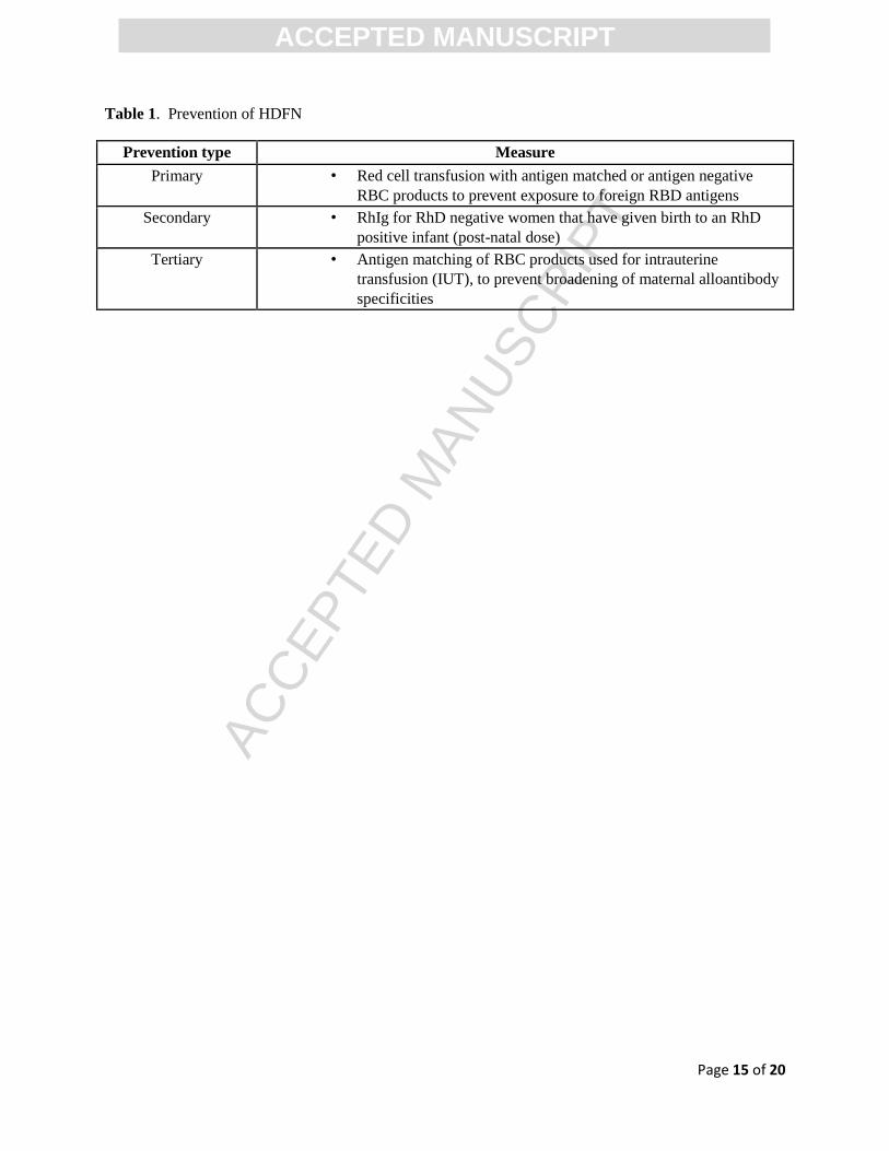
Table 1. Prevention of HDFN
Prevention type Measure
Primary • Red cell transfusion with antigen matched or antigen negative
RBC products to prevent exposure to foreign RBD antigens
Secondary • RhIg for RhD negative women that have given birth to an RhD
positive infant (post-natal dose)
Tertiary • Antigen matching of RBC products used for intrauterine
transfusion (IUT), to prevent broadening of maternal alloantibody
specificities
ACC
EPTE
D M
ANU
SCR
IPT
ACCEPTED MANUSCRIPT
Page 16 of 20
Table 2. Clinical questions and research challenges
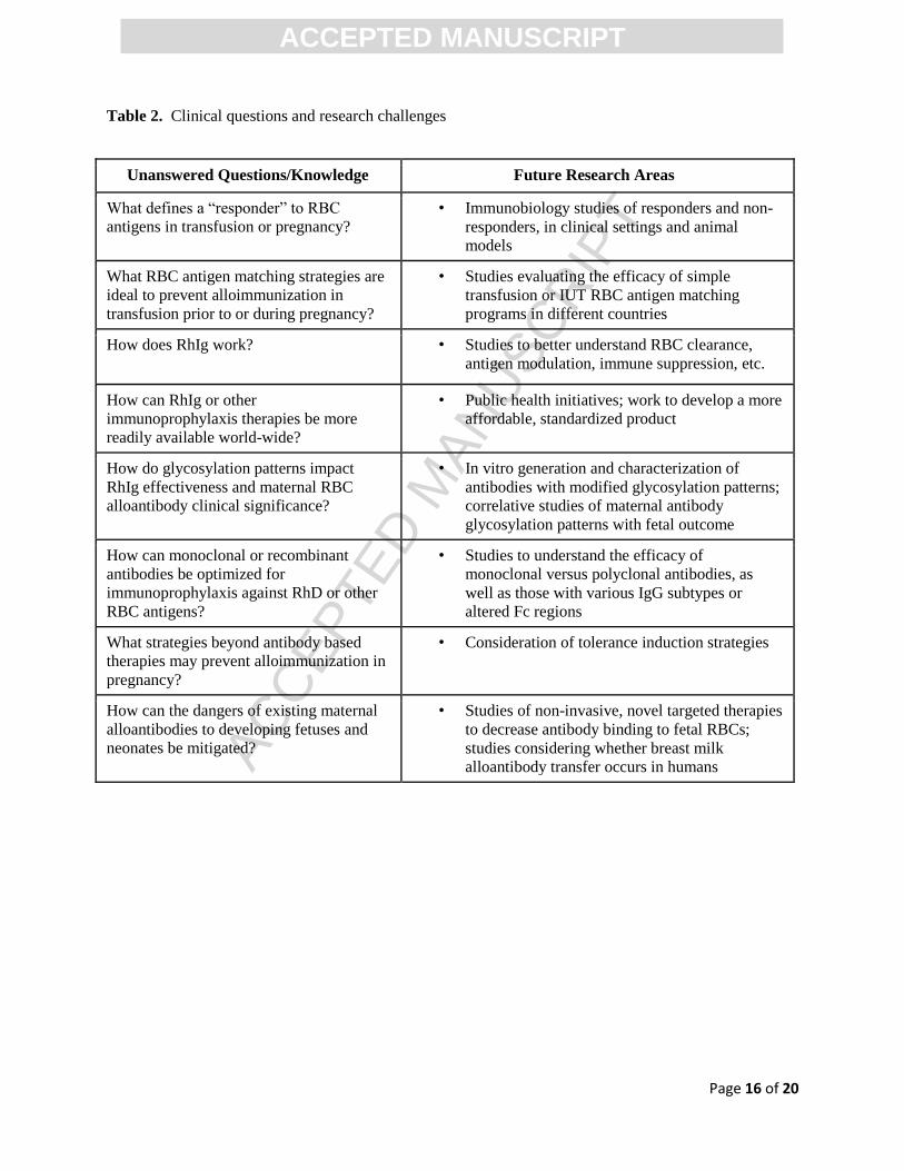
Unanswered Questions/Knowledge Future Research Areas
What defines a “responder” to RBC
antigens in transfusion or pregnancy?
• Immunobiology studies of responders and non-
responders, in clinical settings and animal
models
What RBC antigen matching strategies are
ideal to prevent alloimmunization in
transfusion prior to or during pregnancy?
• Studies evaluating the efficacy of simple
transfusion or IUT RBC antigen matching
programs in different countries
How does RhIg work? • Studies to better understand RBC clearance,
antigen modulation, immune suppression, etc.
How can RhIg or other
immunoprophylaxis therapies be more
readily available world-wide?
• Public health initiatives; work to develop a more
affordable, standardized product
How do glycosylation patterns impact
RhIg effectiveness and maternal RBC
alloantibody clinical significance?
• In vitro generation and characterization of
antibodies with modified glycosylation patterns;
correlative studies of maternal antibody
glycosylation patterns with fetal outcome
How can monoclonal or recombinant
antibodies be optimized for
immunoprophylaxis against RhD or other
RBC antigens?
• Studies to understand the efficacy of
monoclonal versus polyclonal antibodies, as
well as those with various IgG subtypes or
altered Fc regions
What strategies beyond antibody based
therapies may prevent alloimmunization in
pregnancy?
• Consideration of tolerance induction strategies
How can the dangers of existing maternal
alloantibodies to developing fetuses and
neonates be mitigated?
• Studies of non-invasive, novel targeted therapies
to decrease antibody binding to fetal RBCs;
studies considering whether breast milk
alloantibody transfer occurs in humans
ACC
EPTE
D M
ANU
SCR
IPT
ACCEPTED MANUSCRIPT
Page 17 of 20
REFERENCES
1. Reid ME, Lomas-Francis C. The Blood Group Antigen Facts Book. 2nd ed. Amsterdam: Elsevier Academic Press; 2004. 2. Moise KJ, Jr., Argoti PS. Management and prevention of red cell alloimmunization in pregnancy: a systematic review. Obstet gynecol. 2012;120:1132-9. 3. Delaney M, Matthews DC. Hemolytic disease of the fetus and newborn: managing the mother, fetus, and newborn. Hematology Am Soc Hematol Educ Program. 2015;2015:146-51. 4. Eder AF. Update on HDFN: new information on long-standing controversies. Immunohematology. 2006;22:188-95. 5. Markham KB, Rossi KQ, Nagaraja HN, O'Shaughnessy RW. Hemolytic disease of the fetus and newborn due to multiple maternal antibodies. Am J Obstet Gynecol. 2015;213:68 e1-5. 6. Moise KJ, Jr. Non-anti-D antibodies in red-cell alloimmunization. Eur J of Obstet Gynecol Reprod Biol. 2000;92:75-81. 7. Hadley AG, Wilkes A, Goodrick J, Penman D, Soothill P, Lucas G. The ability of the chemiluminescence test to predict clinical outcome and the necessity for amniocenteses in pregnancies at risk of haemolytic disease of the newborn. Br J Obstet Gynaecol. 1998;105:231-4. 8. Nance SJ, Nelson JM, Horenstein J, Arndt PA, Platt LD, Garratty G. Monocyte monolayer assay: an efficient noninvasive technique for predicting the severity of hemolytic disease of the newborn. Am J Clin Pathol. 1989;92:89-92. 9. Kapur R, Della Valle L, Sonneveld M, Hipgrave Ederveen A, Visser R, Ligthart P, et al. Low anti-RhD IgG-Fc-fucosylation in pregnancy: a new variable predicting severity in haemolytic disease of the fetus and newborn. Br J Haematol. 2014;166:936-45. 10. Sonneveld ME, Plom R, Admiraal J, Koeleman C, Hipgrave-Ederveen A, Koelewijn J, et al. IgG alloantibodies agianst RBC induced by pregnancy or transfusion have unique glycosylation patterns which correlate with clinical outcomes of hemolytic disease of the fetus or newborn. Blood ASH Annual Meeting Supplement 2015, Abstract 660. 11. Vaughan JI, Manning M, Warwick RM, Letsky EA, Murray NA, Roberts IA. Inhibition of erythroid progenitor cells by anti-Kell antibodies in fetal alloimmune anemia. N Engl J Med. 1998;338:798-803. 12. Daniels G, Hadley A, Green CA. Causes of fetal anemia in hemolytic disease due to anti-K. Transfusion. 2003;43:115-6. 13. Wagner FF, Flegel WA. RHD gene deletion occurred in the Rhesus box. Blood. 2000;95:3662. 14. Finning K, Martin P, Summers J, Daniels G. Fetal genotyping for the K (Kell) and Rh C, c, and E blood groups on cell-free fetal DNA in maternal plasma. Transfusion. 2007;47:2126. 15. Finning KM, Martin PG, Soothill PW, Avent ND. Prediction of fetal D status from maternal plasma: introduction of a new noninvasive fetal RHD genotyping service. Transfusion. 2002;42:1079. 16. Ahaded A, Brossard Y, Debbia M, Lambin P. Quantitative determination of anti-K (KEL1) IgG and IgG subclasses in the serum of severely alloimmunized pregnant women by ELISA. Transfusion. 2000;40:1239-45. 17. Lambin P, Debbia M, Puillandre P, Brossard Y. IgG1 and IgG3 anti-D in maternal serum and on the RBCs of infants suffering from HDN: relationship with the severity of the disease. Transfusion. 2002;42:1537-46. 18. Oepkes D, van Kamp IL, Simon MJ, Mesman J, Overbeeke MA, Kanhai HH. Clinical value of an antibody-dependent cell-mediated cytotoxicity assay in the management of Rh D alloimmunization. Am J Obstet Gynecol. 2001;184:1015-20. 19. de Haas M, Thurik FF, Koelewijn JM, van der Schoot CE. Haemolytic disease of the fetus and newborn. Vox Sang. 2015;109:99-113.
ACC
EPTE
D M
ANU
SCR
IPT
ACCEPTED MANUSCRIPT
Page 18 of 20
20. Higel F, Seidl A, Sorgel F, Friess W. N-glycosylation heterogeneity and the influence on structure, function and pharmacokinetics of monoclonal antibodies and Fc fusion proteins. Eur J pharmaceutics and biopharmaceutics : official journal of Arbeitsgemeinschaft fur Pharmazeutische Verfahrenstechnik eV. 2016;100:94-100. 21. Kapur R, Kustiawan I, Vestrheim A, Koeleman CA, Visser R, Einarsdottir HK, et al. A prominent lack of IgG1-Fc fucosylation of platelet alloantibodies in pregnancy. Blood. 2014;123:471-80. 22. ACOG Practice Bulletin No. 75: Management of alloimmunization during pregnancy. Obstet Gynecol. 2006;108:457-64. 23. Lindenburg IT, Smits-Wintjens VE, van Klink JM, Verduin E, van Kamp IL, Walther FJ, et al. Long-term neurodevelopmental outcome after intrauterine transfusion for hemolytic disease of the fetus/newborn: the LOTUS study. Am J Obstet Gynecol. 2012;206:141.e1-8. 24. Ruma MS, Moise KJ, Jr., Kim E, Murtha AP, Prutsman WJ, Hassan SS, et al. Combined plasmapheresis and intravenous immune globulin for the treatment of severe maternal red cell alloimmunization. Am J Obstet Gynecol. 2007;196:138.e1-6. 25. Schwartz J, Winters JL, Padmanabhan A, Balogun RA, Delaney M, Linenberger ML, et al. Guidelines on the use of therapeutic apheresis in clinical practice-evidence-based approach from the Writing Committee of the American Society for Apheresis: the sixth special issue. J Clin Apheresis. 2013;28:145-284. 26. Management of hyperbilirubinemia in the newborn infant 35 or more weeks of gestation. Pediatrics. 2004;114:297-316. 27. Rath ME, Smits-Wintjens VE, Walther FJ, Lopriore E. Hematological morbidity and management in neonates with hemolytic disease due to red cell alloimmunization. Early Hum Dev. 2011;87:583-8. 28. Smits-Wintjens VE, Rath ME, van Zwet EW, Oepkes D, Brand A, Walther FJ, et al. Neonatal morbidity after exchange transfusion for red cell alloimmune hemolytic disease. Neonatology. 2013;103:141-7. 29. Corvaglia L, Legnani E, Galletti S, Arcuri S, Aceti A, Faldella G. Intravenous immunoglobulin to treat neonatal alloimmune haemolytic disease. The journal of maternal-fetal & neonatal medicine : the official journal of the European Association of Perinatal Medicine, the Federation of Asia and Oceania Perinatal Societies, the International Society of Perinatal Obstet. 2012;25:2782-5. 30. Elalfy MS, Elbarbary NS, Abaza HW. Early intravenous immunoglobin (two-dose regimen) in the management of severe Rh hemolytic disease of newborn--a prospective randomized controlled trial. Eur J Pediatr. 2011;170:461-7. 31. Hauschner H, Rosenberg N, Seligsohn U, Mendelsohn R, Simmonds A, Shiff Y, et al. Persistent neonatal thrombocytopenia can be caused by IgA antiplatelet antibodies in breast milk of immune thrombocytopenic mothers. Blood. 2015;126:661-4. 32. Santhanakrishnan M, Tormey CA, Natarajan P, Liu J, Hendrickson JE. Clinically significant anti-KEL RBC alloantibodies are transferred by breast milk in a murine model. Vox Sang. 2016 Mar 7. doi: 10.1111/vox.12387. 33. Van de Perre P. Transfer of antibody via mother's milk. Vaccine. 2003;21:3374-6. 34. Paveglio S, Puddington L, Rafti E, Matson AP. FcRn-mediated intestinal absorption of IgG anti-IgE/IgE immune complexes in mice. Clinical and experimental allergy : journal of the British Society for Allergy and Clinical Immunology. 2012;42:1791-800. 35. Callum JL, Waters JH, Shaz BH, Sloan SR, Murphy MF. The AABB recommendations for the Choosing Wisely campaign of the American Board of Internal Medicine. Transfusion. 2014;54:2344. 36. van der Schoot CE, Tax GH, Rijnders RJ, de Haas M, Christiaens GC. Prenatal typing of Rh and Kell blood group system antigens: the edge of a watershed. Transfus Med Rev. 2003;17:31. 37. Bowman JM. Controversies in Rh prophylaxis. Who needs Rh immune globulin and when should it be given? Am J Obstet Gynecol. 1985;151:289-94.
ACC
EPTE
D M
ANU
SCR
IPT
ACCEPTED MANUSCRIPT
Page 19 of 20
38. Sandler SG, Flegel WA, Westhoff CM, Denomme GA, Delaney M, Keller MA, et al. It's time to phase in RHD genotyping for patients with a serologic weak D phenotype. Transfusion. 2015;55:680-9. 39. Kacker S, Vassallo R, Keller MA, Westhoff CM, Frick KD, Sandler SG, et al. Financial implications of RHD genotyping of pregnant women with a serologic weak D phenotype. Transfusion. 2015; 55(9):2095-103. 40. Anderson B, Shad AT, Gootenberg JE, Sandler SG. Successful prevention of post-transfusion Rh alloimmunization by intravenous Rho (D) immune globulin (WinRho SD). Am J Hematol. 1999;60:245-7. 41. Laspina S, O'Riordan J M, Lawlor E, Murphy WG. Prevention of post-transfusion RhD immunization using red cell exchange and intravenous anti-D immunoglobulin. Vox Sang. 2005;89:49-51. 42. Nester TA, Rumsey DM, Howell CC, Gilligan DM, Drachman JG, Maier RV, et al. Prevention of immunization to D+ red blood cells with red blood cell exchange and intravenous Rh immune globulin. Transfusion. 2004;44:1720-3. 43. Schonewille H, Klumper FJ, van de Watering LM, Kanhai HH, Brand A. High additional maternal red cell alloimmunization after Rhesus- and K-matched intrauterine intravascular transfusions for hemolytic disease of the fetus. Am J Obstet Gynecol. 2007;196:143.e1. 44. Schonewille H, Prinsen-Zander KJ, Reijnart M, van de Watering L, Zwaginga JJ, Meerman RH, et al. Extended matched intrauterine transfusions reduce maternal Duffy, Kidd, and S antibody formation. Transfusion. 2015;55:2912-9. 45. Brinc D, Lazarus AH. Mechanisms of anti-D action in the prevention of hemolytic disease of the fetus and newborn. Hematology Am Soc Hematol Educ Program. 2009:185-91. 46. Kumpel BM. On the mechanism of tolerance to the Rh D antigen mediated by passive anti-D (Rh D prophylaxis). Immunol Lett. 2002;82:67-73. 47. Kapur R, Della Valle L, Verhagen OJ, Hipgrave Ederveen A, Ligthart P, de Haas M, et al. Prophylactic anti-D preparations display variable decreases in Fc-fucosylation of anti-D. Transfusion. 2015;55:553-62. 48. Goossens D, da Silva N, Metral S, Cortes U, Callebaut I, Picot J, et al. Mice expressing RHAG and RHD human blood group genes. PLoS One. 2013;8:e80460. 49. Bernardo L, Denomme GA, Shah K, Lazarus AH. RhD Specific Antibodies Are Not Detectable in HLA-DRB1(*)1501 Mice Challenged with Human RhD Positive Erythrocytes. Adv Hematol. 2014;2014:470242. 50. Finn R, Clarke CA, Donohoe WT, Mc CR, Sheppard PM, Lehane D, et al. Experimental studies on the prevention of Rh haemolytic disease. Br Med J. 1961;1:1486-90. 51. Kumpel BM. Efficacy of RhD monoclonal antibodies in clinical trials as replacement therapy for prophylactic anti-D immunoglobulin: more questions than answers. Vox Sang. 2007;93:99-111. 52. Hendrickson JE, Hod EA, Spitalnik SL, Hillyer CD, Zimring JC. Storage of murine red blood cells enhances alloantibody responses to an erythroid-specific model antigen. Transfusion. 2010;50:642-8. 53. Hendrickson JE, Hod EA, Cadwell CM, Eisenbarth SC, Spiegel DA, Tormey CA, et al. Rapid clearance of transfused murine red blood cells is associated with recipient cytokine storm and enhanced alloimmunogenicity. Transfusion. 2011;51:2445-54. 54. Smith NH, Hod EA, Spitalnik SL, Zimring JC, Hendrickson JE. Transfusion in the absence of inflammation induces antigen-specific tolerance to murine RBCs. Blood. 2012;119:1566-9. 55. Hendrickson JE, Desmarets M, Deshpande SS, Chadwick TE, Hillyer CD, Roback JD, et al. Recipient inflammation affects the frequency and magnitude of immunization to transfused red blood cells. Transfusion. 2006;46:1526-36. 56. Schenten D, Medzhitov R. The control of adaptive immune responses by the innate immune system. Adv Immunol. 2011;109:87-124. 57. Stowell SR, Arthur CM, Girard-Pierce KR, Sullivan HC, Santhanakrishnan M, Natarajan P, et al. Anti-KEL sera prevents alloimmunization to transfused KEL RBCs in a murine model. Haematologica. 2015;100:e394-7.
ACC
EPTE
D M
ANU
SCR
IPT
ACCEPTED MANUSCRIPT
Page 20 of 20
58. Brinc D, Le-Tien H, Crow AR, Freedman J, Lazarus AH. IgG-mediated immunosuppression is not dependent on erythrocyte clearance or immunological evasion: implications for the mechanism of action of anti-D in the prevention of haemolytic disease of the newborn? Br J Haematol. 2007;139:275-9. 59. Brinc D, Le-Tien H, Crow AR, Siragam V, Freedman J, Lazarus AH. Immunoglobulin G-mediated regulation of the murine immune response to transfused red blood cells occurs in the absence of active immune suppression: implications for the mechanism of action of anti-D in the prevention of haemolytic disease of the fetus and newborn? Immunol. 2008;124:141-6. 60. Bernardo L, Yu H, Amash A, Zimring JC, Lazarus AH. IgG-Mediated Immune Suppression to Erythrocytes by Polyclonal Antibodies Can Occur in the Absence of Activating or Inhibitory Fcgamma Receptors in a Full Mouse Model. J Immunol. 2015;195:2224-30. 61. Stowell SR, Liepkalns JS, Hendrickson JE, Girard-Pierce KR, Smith NH, Arthur CM, et al. Antigen modulation confers protection to red blood cells from antibody through Fcgamma receptor ligation. J Immunol. 2013;191:5013-25. 62. Zimring JC, Cadwell CM, Spitalnik SL. Antigen loss from antibody-coated red blood cells. Transfus Med Rev. 2009;23:189-204. 63. Taylor RP, Lindorfer MA. Fcgamma-receptor-mediated trogocytosis impacts mAb-based therapies: historical precedence and recent developments. Blood. 2015;125:762-6. 64. Sullivan HC, Arthur CM, Patel SR, Hendrickson JE, Lazarus AH, Stowell SR. Anti-RhD mediates loss of RhD antigen following anti-RhD infusion. Blood. 2015;126:3570. 65. Girard-Pierce KR, Stowell SR, Smith NH, Arthur CM, Sullivan HC, Hendrickson JE, et al. A novel role for C3 in antibody-induced red blood cell clearance and antigen modulation. Blood. 2013;122:1793-801.
Highlights (3-5 bullet points, max 85 characters including spaces)
RBC alloimmunization and HDFN remain significant problems in all countries
A better understanding of factors influencing responsiveness to RBC antigens is needed
Optimized RBC antigen matching transfusion strategies for girls and women are necessary
Understanding how RhIg works is essential for novel immunoprophylaxis development
Targeted therapies to mitigate the dangers of alloantibodies to fetuses would be beneficial