Hematological Issues in Acute Care- Part B - etouches · PDF fileHEMATOLOGICAL ISSUES IN ACUTE...
32
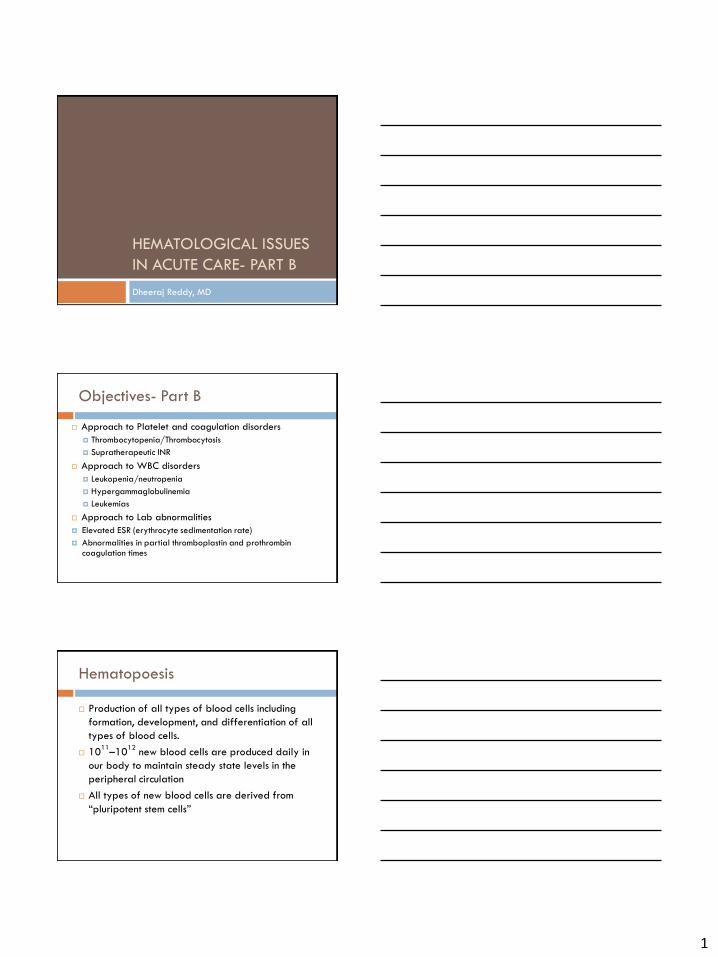
1 HEMATOLOGICAL ISSUES IN ACUTE CARE- PART B Dheeraj Reddy, MD Objectives- Part B Approach to Platelet and coagulation disorders Thrombocytopenia/Thrombocytosis Supratherapeutic INR Approach to WBC disorders Leukopenia/neutropenia Hypergammaglobulinemia Leukemias Approach to Lab abnormalities Elevated ESR (erythrocyte sedimentation rate) Abnormalities in partial thromboplastin and prothrombin coagulation times Hematopoesis Production of all types of blood cells including formation, development, and differentiation of all types of blood cells. 10 11 –10 12 new blood cells are produced daily in our body to maintain steady state levels in the peripheral circulation All types of new blood cells are derived from “pluripotent stem cells”
Transcript of Hematological Issues in Acute Care- Part B - etouches · PDF fileHEMATOLOGICAL ISSUES IN ACUTE...
1
HEMATOLOGICAL ISSUES
IN ACUTE CARE- PART B
Dheeraj Reddy, MD
Objectives- Part B
Approach to Platelet and coagulation disorders
Thrombocytopenia/Thrombocytosis
Supratherapeutic INR
Approach to WBC disorders
Leukopenia/neutropenia
Hypergammaglobulinemia
Leukemias
Approach to Lab abnormalities
Elevated ESR (erythrocyte sedimentation rate)
Abnormalities in partial thromboplastin and prothrombin coagulation times
Hematopoesis
Production of all types of blood cells including
formation, development, and differentiation of all
types of blood cells.
1011
–1012
new blood cells are produced daily in
our body to maintain steady state levels in the
peripheral circulation
All types of new blood cells are derived from
“pluripotent stem cells”
2
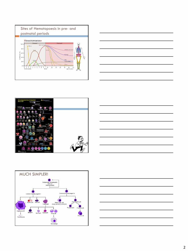
Sites of Hematopoesis in pre- and
postnatal periods
MUCH SIMPLER!
3
PLATELET AND
COAGULATION SECTION
Platelet anatomy
Shape: round and flat discs, with diameter of 1-2 m and volume of 7-9 fL
Plasma membrane
◦ Receptor glycoproteins eg GPIIb / IIIa ( IIb3 )
◦ Surface-connected canalicular system
Cytoskeleton
Organelles ◦ Mitochondria, lysosomes,
and peroxisomes ◦ Alpha granules ( ~ 80 per
platelet )
◦ Dense granules ( 3 - 8 per platelet )
4
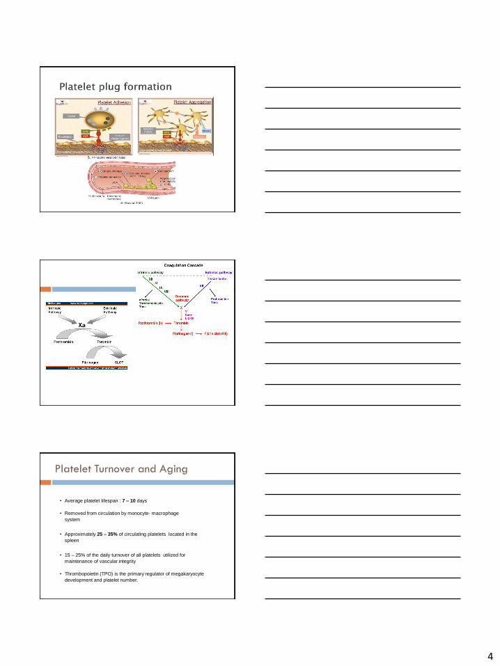
Platelet Turnover and Aging
• Average platelet lifespan : 7 – 10 days
• Removed from circulation by monocyte- macrophage
system
• Approximately 25 – 35% of circulating platelets located in the
spleen
• 15 – 25% of the daily turnover of all platelets utilized for
maintenance of vascular integrity
• Thrombopoietin (TPO) is the primary regulator of megakaryocyte
development and platelet number.
5
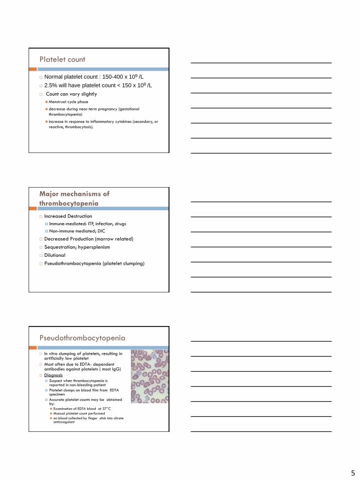
Platelet count
Normal platelet count : 150-400 x 109 /L
2.5% will have platelet count < 150 x 109 /L
Count can vary slightly
Menstrual cycle phase
decrease during near-term pregnancy (gestational
thrombocytopenia)
increase in response to inflammatory cytokines (secondary, or
reactive, thrombocytosis).
Major mechanisms of
thrombocytopenia
Increased Destruction
Immune-mediated: ITP, infection, drugs
Non-immune mediated; DIC
Decreased Production (marrow related)
Sequestration; hypersplenism
Dilutional
Pseudothrombocytopenia (platelet clumping)
Pseudothrombocytopenia
In vitro clumping of platelets, resulting in artificially low platelet
Most often due to EDTA- dependent antibodies against platelets ( most IgG)
Diagnosis
Suspect when thrombocytopenia is reported in non-bleeding patient
Platelet clumps on blood film from EDTA specimen
Accurate platelet counts may be obtained by:
Examination of EDTA blood at 37°C
Manual platelet count performed
on blood collected by finger stick into citrate anticoagulant
6
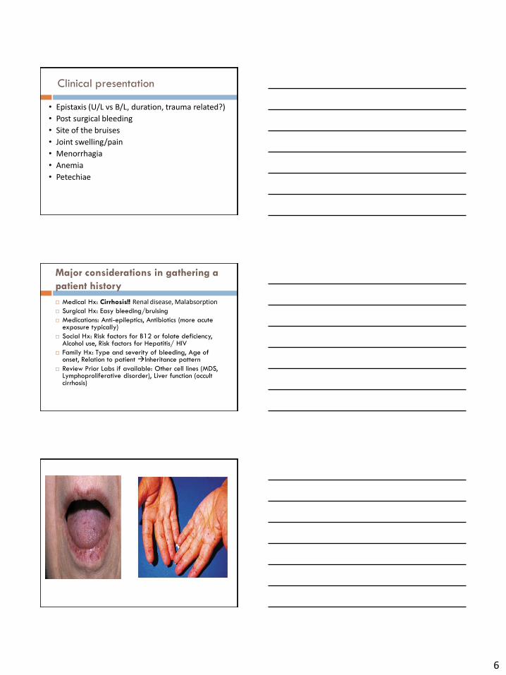
Clinical presentation
• Epistaxis (U/L vs B/L, duration, trauma related?)
• Post surgical bleeding
• Site of the bruises
• Joint swelling/pain
• Menorrhagia
• Anemia
• Petechiae
Major considerations in gathering a
patient history
Medical Hx: Cirrhosis!! Renal disease, Malabsorption
Surgical Hx: Easy bleeding/bruising
Medications: Anti-epileptics, Antibiotics (more acute exposure typically)
Social Hx: Risk factors for B12 or folate deficiency, Alcohol use, Risk factors for Hepatitis/ HIV
Family Hx: Type and severity of bleeding, Age of onset, Relation to patient Inheritance pattern
Review Prior Labs if available: Other cell lines (MDS, Lymphoproliferative disorder), Liver function (occult cirrhosis)
7
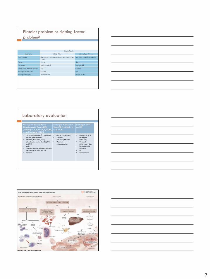
Platelet problem or clotting factor
problem?
Laboratory evaluation
Prolonged Activated Partial
Thromboplastin Time (aPTT)
(>26-35s) - I, II, V, VIII,IX, X, XI, XII, prekallikrein and HMWK
Prolonged Prothrombin
Time (PT) (>10-14s) - I, II, V, VII, X
Prolonged aPTT
and PT
• No clinical bleeding↓ factors XII,
HMWK, prekallikrein
• Variable, but usually mild,
bleeding↓ factor XI, mild↓ FVIII
and FIX
• VwD
• Frequent, severe bleedingsevere
deficiencies of FVIII and FIX
• Heparin
• Factor VII deficiency
• Vitamin K
deficiencyearly
• Warfarin
anticoagulation
• Factor II, V, X, or
fibrinogen
deficiency
• Vitamin K
deficiency Late
• Direct thrombin
inhibitors
• DIC
• Liver disease
Hunt BJ. N Engl J Med 2014;370:847-859
8
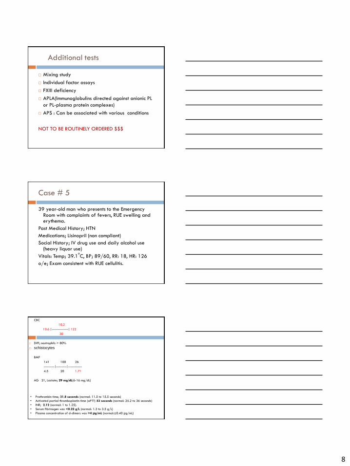
Additional tests
Mixing study
Individual factor assays
FXIII deficiency
APLA(Immunoglobulins directed against anionic PL
or PL-plasma protein complexes)
APS : Can be associated with various conditions
NOT TO BE ROUTINELY ORDERED $$$
Case # 5
39 year-old man who presents to the Emergency Room with complaints of fevers, RUE swelling and erythema.
Past Medical History; HTN
Medications; Lisinopril (non compliant)
Social History; IV drug use and daily alcohol use (heavy liquor use)
Vitals: Temp; 39.1oC, BP; 89/60, RR: 18, HR: 126
o/e; Exam consistent with RUE cellulitis.
CBC
10.2
13.6 |--------------| 122
30
Diff; neutrophils > 80%
schistocytes
BMP
141 100 26
---------|---------|------------
4.5 20 1.71
AG 21, Lactate; 29 mg/dL(6-16 mg/dL)
• Prothrombin time; 31.8 seconds (normal: 11.5 to 15.5 seconds)
• Activated partial thromboplastin time (aPTT) 53 seconds (normal: 25.2 to 36 seconds)
• INR; 2.12 (normal: 1 to 1.25).
• Serum fibrinogen was <0.22 g/L (normal: 1.3 to 3.5 g/L)
• Plasma concentration of d-dimers was >4 μg/mL (normal:≤0.40 μg/mL)
9
Disseminated Intravascular Coagulation
(DIC)
A heterogeneous group of clinicopathologic
syndromes
Characterized by dysregulated generation of
thrombin (pathologic thrombin formation)
Leading to intravascular fibrin formation, and
Secondary fibrinolysis (plasmin generation),
Often resulting in hemorrhage, thrombosis, and/or
multi-organ system failure
Some Causes of DIC
Infections
Bacteremia
Rickettsial infections
Metabolic disorders
Hypotension
Hypoxia
Hyper/hypothermia
Obstetrical complications
Placental abruption
Placenta previa
Pregnancy-induced HTN
Amniotic fluid embolism
Retained dead fetus
Tumors
Adenocarcinoma
Tumor Lysis Syndrome
AML: M3(APL), M4 or M5
Trauma
Crush injuries
Head injuries
Toxins
Viper venom bites
Drugs
L-asparaginase
Prothrombin complex concentrates
Heparin (via HIT)
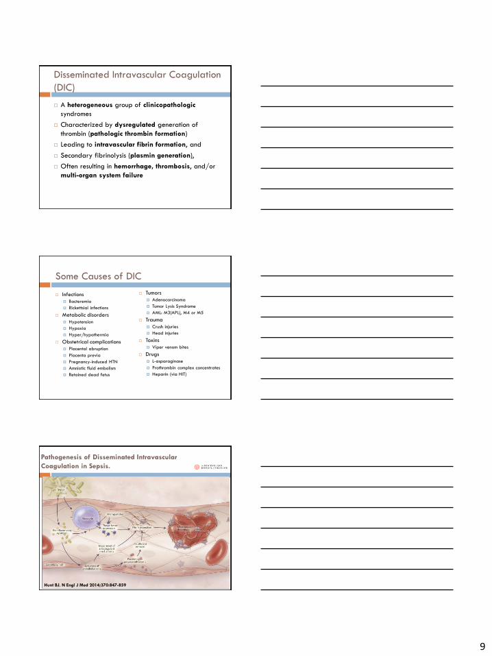
Pathogenesis of Disseminated Intravascular
Coagulation in Sepsis.
Hunt BJ. N Engl J Med 2014;370:847-859
10
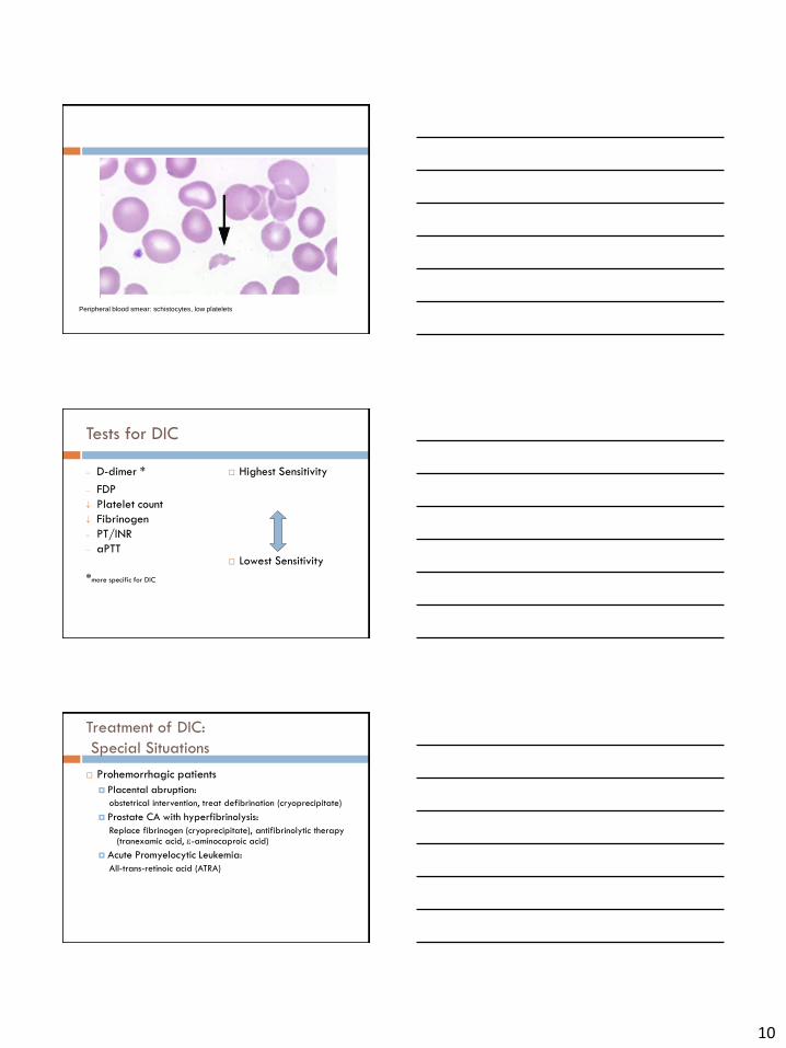
Peripheral blood smear: schistocytes, low platelets
Tests for DIC
D-dimer *
FDP
Platelet count
Fibrinogen
PT/INR
aPTT
*more specific for DIC
Highest Sensitivity
Lowest Sensitivity
Treatment of DIC:
Special Situations
Prohemorrhagic patients
Placental abruption:
obstetrical intervention, treat defibrination (cryoprecipitate)
Prostate CA with hyperfibrinolysis:
Replace fibrinogen (cryoprecipitate), antifibrinolytic therapy (tranexamic acid, -aminocaproic acid)
Acute Promyelocytic Leukemia:
All-trans-retinoic acid (ATRA)
11
Treatment of DIC:
Special Situations
Prothrombotic patients
Adenocarcinoma (Trousseau’s syndrome):
Heparin, avoid warfarin
Septicemia with acral gangrene (purpura fulminans):
Vitamin K, heparin, FFP
Heparin-induced thrombocytopenia:
Unusual DIC picture with increased thrombin generation without low
fibrinogen, PT, aPTT, THUS use agent that reduces thrombin
generation (lepirudin, argatroban, danaproid)
Laboratory Findings in Various Platelet
and Coagulation Disorders in the ICU
Hunt BJ. N Engl J Med 2014;370:847-859
AAAB guidelines on prophylactic
platelet transfusion (2015)
Prophylactic platelet transfusion, with therapy-induced hypoproliferative thrombocytopenia. <10 × 109 cells/L to reduce the risk for spontaneous bleeding. Give single apheresis unit or equivalent. Greater doses are not more effective, and lower doses equal to one half of a standard apheresis unit are equally effective.
Patients having elective central venous catheter placement with a platelet count less than 20 × 109 cells/L.
Patients having elective diagnostic lumbar puncture with a platelet count less than 50 × 109 cells/L.
Patients having major elective nonneuraxial surgery with a platelet count less than 50 × 109 cells/L.
No role routine for patients who are nonthrombocytopenic and have cardiac surgery with cardiopulmonary bypass.

12
Case # 6
47 year old man presents to ER for LLE swelling. He had a 3 day hospitalization, 4 weeks ago for a non-ST elevation myocardial infarction for which he underwent cardiac catheterization and was given low-molecular-weight heparin.
Current medications; aspirin, clopidogrel, pravastatin, and lisinopril.
o/e: Left thigh is swollen and tender.
CBC, BMP, LFTs are unremarkable except for a platelet count of 102,000/µL (150,000-350,000/μL previously)
Duplex ultrasonography + Deep venous thrombosis
Unfractionated heparin is administered. Twelve hours later, the patient’s platelet count is 27,000/µL
Heparin-induced thrombocytopenia
Complication of heparin therapy.
2 types of HIT.
Type 1 HIT: NON IMMUNE MEDIATED within the first 2
days after exposure to heparin, and the platelet count
normalizes with continued heparin therapy. Results from
the direct interaction of heparin with the platelet
membrane, resulting in enhanced platelet aggregation
Type 2 HIT : IMMUNE MDIATED disorder that typically
occurs 4-10 days after exposure to heparin and has life-
and limb-threatening thrombotic complications
13
Risk factors;
include unfractionated rather than LMW heparin (but
HIT can occur in any patient following any heparin
exposure)
higher heparin doses
female sex
possibly age
Thrombosis skin necrosis, limb gangrene, and
organ infarction
Clinical features
Patients present typically 5–14 days after starting heparin treatment, with a fall in platelet count of more than 30% from baseline NOTE count may still be in the reference range.
Can be asymptomatic, or develop venous/arterial thrombosis and skin lesions, including overt skin necrosis.
May complain of pain/itch at injection or shivering following heparin injections.
4 T score
4-T score
Pre-test probability of the diagnosis is assessed using the 4Ts
scoring system
14
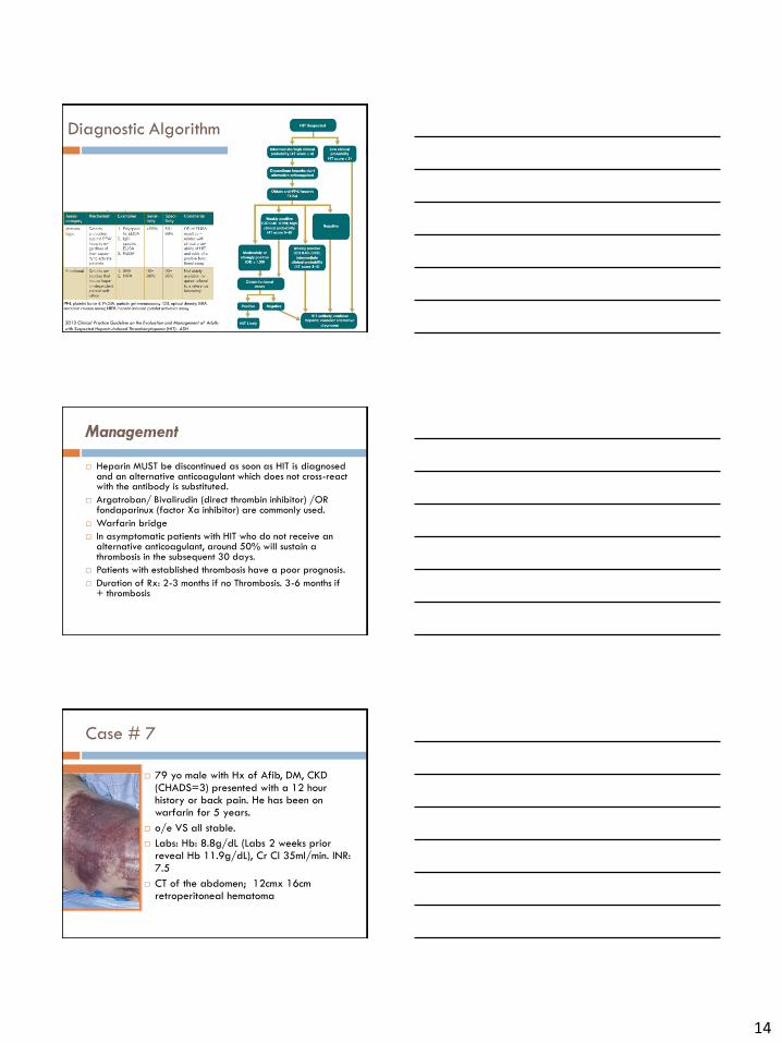
Diagnostic Algorithm
2013 Clinical Practice Guideline on the Evaluation and Management of Adults
with Suspected Heparin-Induced Thrombocytopenia (HIT)- ASH
Management
Heparin MUST be discontinued as soon as HIT is diagnosed and an alternative anticoagulant which does not cross-react with the antibody is substituted.
Argatroban/ Bivalirudin (direct thrombin inhibitor) /OR fondaparinux (factor Xa inhibitor) are commonly used.
Warfarin bridge
In asymptomatic patients with HIT who do not receive an alternative anticoagulant, around 50% will sustain a thrombosis in the subsequent 30 days.
Patients with established thrombosis have a poor prognosis.
Duration of Rx: 2-3 months if no Thrombosis. 3-6 months if + thrombosis
Case # 7
79 yo male with Hx of Afib, DM, CKD (CHADS=3) presented with a 12 hour history or back pain. He has been on warfarin for 5 years.
o/e VS all stable.
Labs: Hb: 8.8g/dL (Labs 2 weeks prior reveal Hb 11.9g/dL), Cr Cl 35ml/min. INR: 7.5
CT of the abdomen; 12cmx 16cm retroperitoneal hematoma
15
General Principles of Management of Anticoagulant
Associated Bleeding
HASHTI
1. Hold further doses of anticoagulant
2. Consider Antidote
3. Supportive treatment: volume resuscitation, inotropes as needed
4. Local or surgical Hemostatic measures: topical agents (aminocaproic acid, tranexamic acid)
5. Transfusion (red cells, platelets, FFP as indicated)
6. Investigate for bleeding source
American Society of Hematology- 2011 pocket
guide
EMERGENT TREATMENT OF BLEEDING
Only antidote for the newer oral anti-coagulants, Dabigatran; idarucizumab
Oral activated charcoal retards absorption of recently ingested drug within a couple hours of presentation
Hemodialysis removes Dabigatran (as only 35% is protein-bound), but not Apixaban/Rivaroxaban
Recombinant Factor VIIa- reverse life-threatening bleeds ( but can cause disseminated intravascular coagulation and systemic thrombosis)
Prothrombin Complex Concentrates- 3/4factor prothrombin complex concentrates (PCCs)- these contain Vitamin K dependent factors in high doses and have only been demonstrated in small trials.
16
Back to the patient
The patient received 10mg IV Vitamin K, 4 units of
FFP and Prothrombin Complex Concentrates
H/H dropped to 7.3/22. He also received 2 units
of packed red blood cells
INR returned to 1.1
Extensive conversation was held with patient
regarding benefits vs risks of anticoagulation. His
PCP was also involved, and mutually agreed that its
safer for patient to be off anti-coagulation.
Thrombocytosis
Presence of high levels of platelets
Can be primary (not common) or secondary (Reative; much more common)
Primary: Essential Thrombocytosis myeloproliferative disorder. Diagnosis of Exclusion
Platelet count > 450 × 103/µL for at least 2 months.
Acquired V617F JAK2 mutation present
bleeding/thrombosis, headache, nausea, vomiting, abdominal pain, visual disturbances, dizziness, fainting, and numbness
Secondary Thrombocytosis
Inflammation
Acute and chronic infection
Connective tissue disease
Malignancy
Kawasaki syndrome
Iron deficiency
Marrow recovery
Sickle cell disease
Post-splenectomy
17
WBC ORDERS
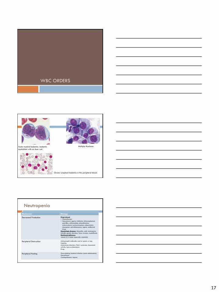
Multiple Myeloma
Chronic lymphoid leukemia in the peripheral blood.
Acute myeloid leukemia. Leukemic
myeloblast with an Auer rod.
Neutropenia
Mechanism Etiology
Decreased Production Drug-induced
• Chemotherapy
• Noncytotoxic agents; Antibiotics (chloramphenicol,
penicillins, sulfonamides), phenothiazines,
anticonvulsants (carbamazepine), antipsychotics
(clozapine), anti-inflammatory agents, antithyroid
drugs
Hematologic diseases: idiopathic, cyclic neutropenia,
infantile genetic disorders, Tumor invasion, myelofibrosis
Nutritional deficiency
vitamin B12, folate (especially alcoholics)
Peripheral Destruction Antineutrophil antibodies and/or splenic or lung
trapping
Autoimmune disorders; Felty’s syndrome, rheumatoid
arthritis, lupus erythematosus
Drugs
Peripheral Pooling Overwhelming bacterial infection (acute endotoxemia)
Hemodialysis
Cardiopulmonary bypass
18
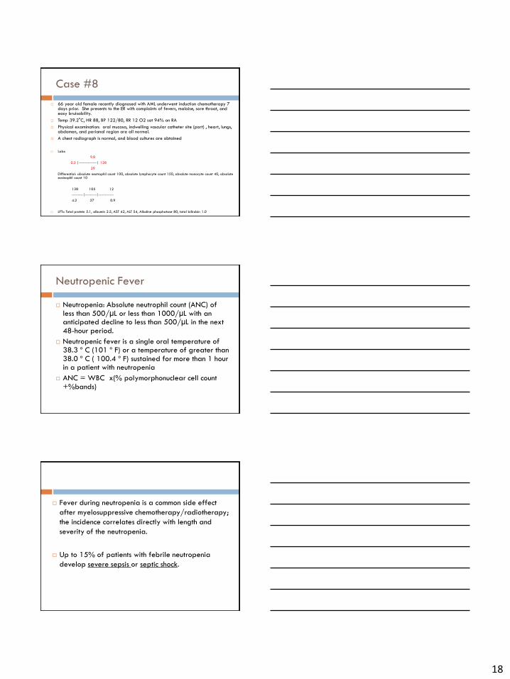
Case #8
66 year old female recently diagnosed with AML underwent induction chemotherapy 7 days prior. She presents to the ER with complaints of fevers, malaise, sore throat, and easy bruisability.
Temp 39.2oC, HR 88, BP 122/80, RR 12 O2 sat 94% on RA
Physical examination: oral mucosa, indwelling vascular catheter site (port) , heart, lungs, abdomen, and perianal region are all normal.
A chest radiograph is normal, and blood cultures are obtained
Labs:
9.8
0.3 |--------------| 128
29
Differential: absolute neutrophil count 100, absolute lymphocyte count 150, absolute monocyte count 40, absolute eosinophil count 10
138 105 12
---------|---------|------------
4.2 27 0.9
LFTs: Total protein 5.1, albumin 2.5, AST 42, ALT 54, Alkaline phosphatase 80, total bilirubin 1.0
Neutropenic Fever
Neutropenia: Absolute neutrophil count (ANC) of less than 500/µL or less than 1000/µL with an anticipated decline to less than 500/µL in the next 48-hour period.
Neutropenic fever is a single oral temperature of 38.3 º C (101 º F) or a temperature of greater than 38.0 º C ( 100.4 º F) sustained for more than 1 hour in a patient with neutropenia
ANC = WBC x(% polymorphonuclear cell count +%bands)
Fever during neutropenia is a common side effect
after myelosuppressive chemotherapy/radiotherapy;
the incidence correlates directly with length and
severity of the neutropenia.
Up to 15% of patients with febrile neutropenia
develop severe sepsis or septic shock.
19
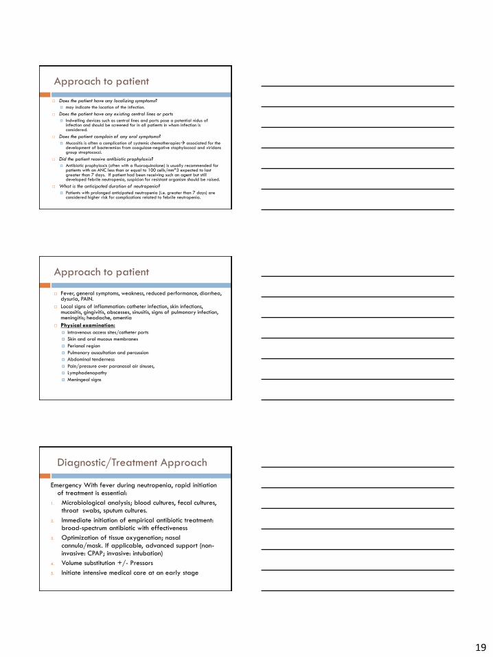
Approach to patient
Does the patient have any localizing symptoms?
may indicate the location of the infection.
Does the patient have any existing central lines or ports
Indwelling devices such as central lines and ports pose a potential nidus of infection and should be screened for in all patients in whom infection is considered.
Does the patient complain of any oral symptoms?
Mucositis is often a complication of systemic chemotherapies associated for the development of bacteremias from coagulase-negative staphylococci and viridans group streptococci.
Did the patient receive antibiotic prophylaxis?
Antibiotic prophylaxis (often with a fluoroquinolone) is usually recommended for patients with an ANC less than or equal to 100 cells/mm^3 expected to last greater than 7 days. If patient had been receiving such an agent but still developed febrile neutropenia, suspicion for resistant organism should be raised.
What is the anticipated duration of neutropenia?
Patients with prolonged anticipated neutropenia (i.e. greater than 7 days) are considered higher risk for complications related to febrile neutropenia.
Approach to patient
Fever, general symptoms, weakness, reduced performance, diarrhea, dysuria, PAIN.
Local signs of inflammation: catheter infection, skin infections, mucositis, gingivitis, abscesses, sinusitis, signs of pulmonary infection, meningitis; headache, amentia
Physical examination:
Intravenous access sites/catheter ports
Skin and oral mucous membranes
Perianal region
Pulmonary auscultation and percussion
Abdominal tenderness
Pain/pressure over paranasal air sinuses,
Lymphadenopathy
Meningeal signs
Diagnostic/Treatment Approach
Emergency With fever during neutropenia, rapid initiation of treatment is essential:
1. Microbiological analysis; blood cultures, fecal cultures, throat swabs, sputum cultures.
2. Immediate initiation of empirical antibiotic treatment: broad-spectrum antibiotic with effectiveness
3. Optimization of tissue oxygenation; nasal cannula/mask. If applicable, advanced support (non-invasive: CPAP; invasive: intubation)
4. Volume substitution +/- Pressors
5. Initiate intensive medical care at an early stage
20
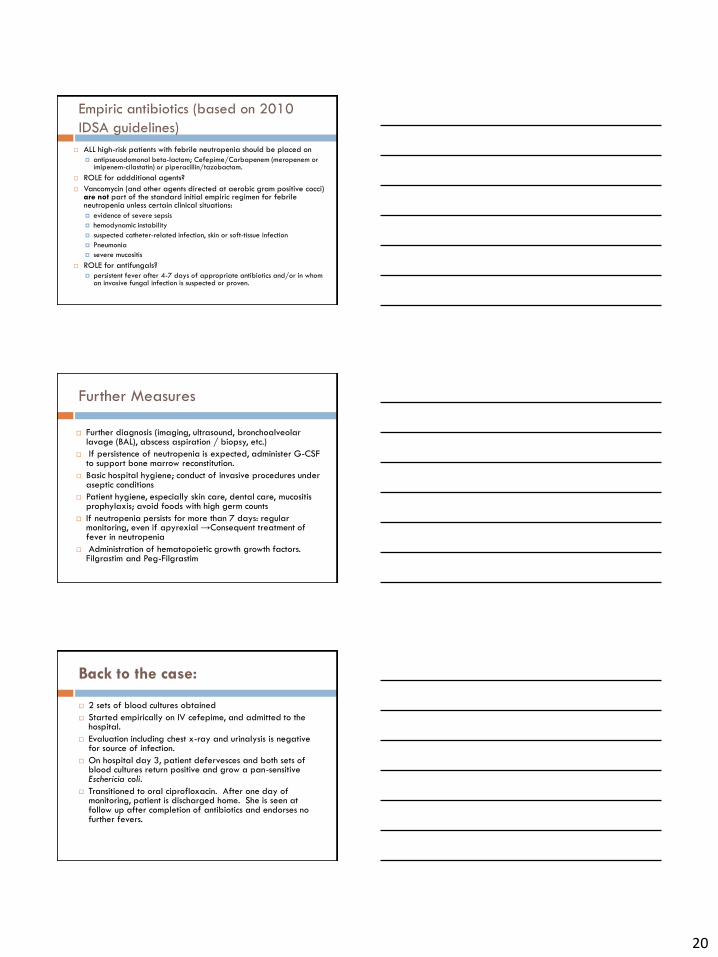
Empiric antibiotics (based on 2010
IDSA guidelines)
ALL high-risk patients with febrile neutropenia should be placed on
antipseuodomonal beta-lactam; Cefepime/Carbapenem (meropenem or imipenem-cilastatin) or piperacillin/tazobactam.
ROLE for addditional agents?
Vancomycin (and other agents directed at aerobic gram positive cocci) are not part of the standard initial empiric regimen for febrile neutropenia unless certain clinical situations:
evidence of severe sepsis
hemodynamic instability
suspected catheter-related infection, skin or soft-tissue infection
Pneumonia
severe mucositis
ROLE for antifungals?
persistent fever after 4-7 days of appropriate antibiotics and/or in whom an invasive fungal infection is suspected or proven.
Further Measures
Further diagnosis (imaging, ultrasound, bronchoalveolar lavage (BAL), abscess aspiration / biopsy, etc.)
If persistence of neutropenia is expected, administer G-CSF to support bone marrow reconstitution.
Basic hospital hygiene; conduct of invasive procedures under aseptic conditions
Patient hygiene, especially skin care, dental care, mucositis prophylaxis; avoid foods with high germ counts
If neutropenia persists for more than 7 days: regular monitoring, even if apyrexial →Consequent treatment of fever in neutropenia
Administration of hematopoietic growth growth factors. Filgrastim and Peg-Filgrastim
Back to the case:
2 sets of blood cultures obtained
Started empirically on IV cefepime, and admitted to the hospital.
Evaluation including chest x-ray and urinalysis is negative for source of infection.
On hospital day 3, patient defervesces and both sets of blood cultures return positive and grow a pan-sensitive Eschericia coli.
Transitioned to oral ciprofloxacin. After one day of monitoring, patient is discharged home. She is seen at follow up after completion of antibiotics and endorses no further fevers.
21
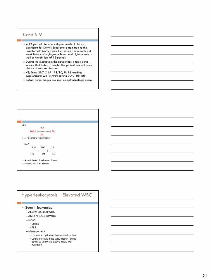
Case # 9
A 22 year old female with past medical history significant for Down’s Syndrome is admitted to the hospital with blurry vision. Her care giver reports a 3 week history of high grade fevers and night sweats as well as weight loss of 15 pounds.
During the evaluation, the patient has a tonic clonic seizure that lasted 1 minute. The patient has no known history of seizure disorder.
VS; Temp 39.7 C, BP 118/80, RR 18 needing supplemental O2 (3L/min) satting 92%. HR 108
Retinal hemorrhages are seen on opthalmologic exam.
CBC
10.6
105.6 |--------------| 88
30
Myeloblast predominance
BMP
137 100 36
---------|---------|------------
4.5 24 1.31
A peripheral blood smear is sent
PT/INR. APTT all normal.
Hyperleukocytosis: Elevated WBC
• Seen in leukemias
– ALL>/=200,000 WBC
– AML>/=100,000 WBC
– Risks
• Stroke
• TLS
– Management
• Hydration, hydration, hydration=first line
• Leukophoresis if the WBC doesn’t come
down to below the above levels with
hydration
22
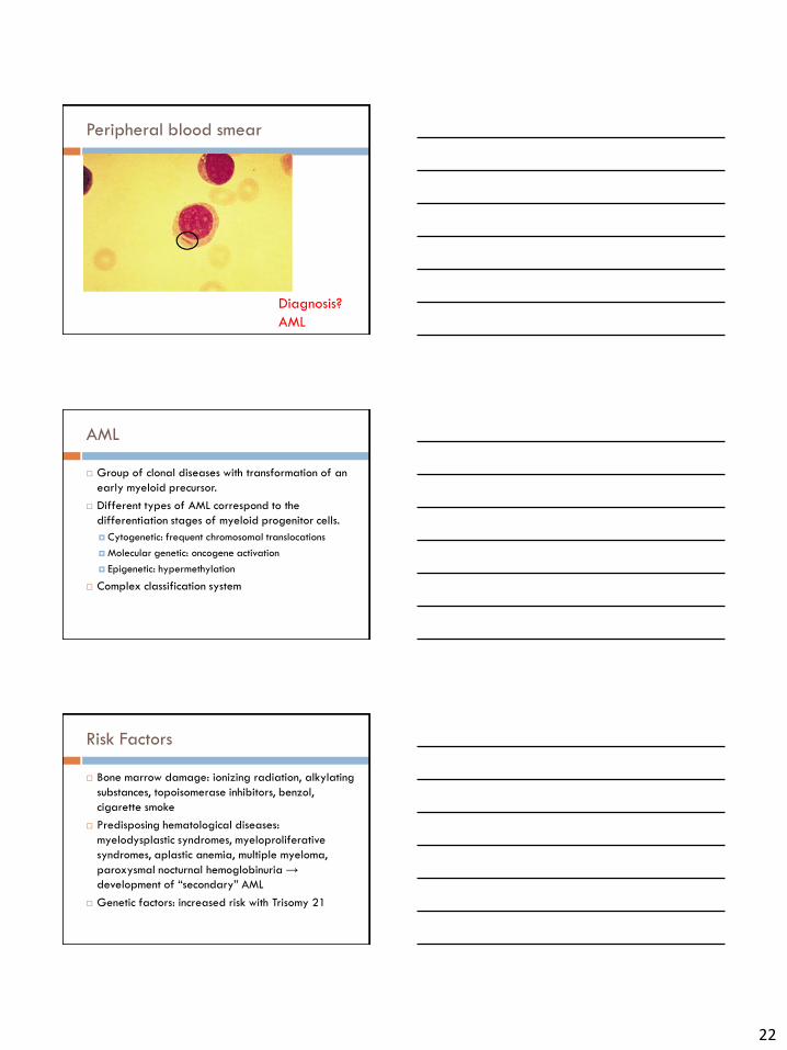
Peripheral blood smear
Diagnosis?
AML
AML
Group of clonal diseases with transformation of an
early myeloid precursor.
Different types of AML correspond to the
differentiation stages of myeloid progenitor cells.
Cytogenetic: frequent chromosomal translocations
Molecular genetic: oncogene activation
Epigenetic: hypermethylation
Complex classification system
Risk Factors
Bone marrow damage: ionizing radiation, alkylating
substances, topoisomerase inhibitors, benzol,
cigarette smoke
Predisposing hematological diseases:
myelodysplastic syndromes, myeloproliferative
syndromes, aplastic anemia, multiple myeloma,
paroxysmal nocturnal hemoglobinuria →
development of “secondary” AML
Genetic factors: increased risk with Trisomy 21
23
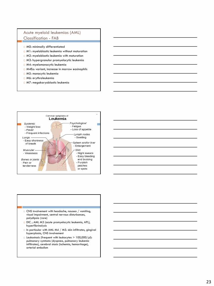
Acute myeloid leukemias (AML)
Classification - FAB
M0: minimally differentiated
M1: myeloblastic leukemia without maturation
M2: myeloblastic leukemia with maturation
M3: hypergranular promyelocytic leukemia
M4: myelomonocytic leukemia
M4Eo: variant, increase in marrow eosinophils
M5: monocytic leukemia
M6: erythroleukemia
M7: megakaryoblastic leukemia
CNS involvement with headache, nausea / vomiting, visual impairment, central nervous disturbances, polydipsia (rare)
DIC ; AML M3 (acute promyelocytic leukemia, APL), hyperfibrinolysis
In particular with AML M4 / M5: skin infiltrates, gingival hyperplasia, CNS involvement
Leukostasis (frequent with leukocytes > 100,000/μl): pulmonary symtoms (dyspnea, pulmonary leukemic infiltrates), cerebral stasis (ischemia, hemorrhage), arterial embolism
24
Medical History, Clinical Signs; History with risk factors,
family history (immediate search for possible matched
related blood stem cell donors)
Examination: skin, mucous membranes (gingival
hyperplasia), lung (infections), lymph node , abdomen
(hepatosplenomegaly), neurological findings
Laboratory Tests CBC with differential blood count, Peripheral blood smear
BMP, LFTs,
LDH (elevated with increased cell turnover)
Coagulation parameters (DIC, hyperfibrinolysis)
Microbiological diagnostics if febrile, virus serum titers Bone marrow aspirate
HLA typing of patient and all siblings (search for HLA-identical family donor for possible matched related allogeneic blood stem cell transplantation)
Leukocytosis with detection of the same blast population as in the bone marrow.
Anemia and thrombocytopenia as signs of suppression of normal hematopoiesis
Back to the patient…..
Immediate measures, will be to aggressively
hydrate, and monitor her hematologic parameters
Monitor respiratory status.
Role of Leukapharesis in controversial, but was
successfully administered.
Rapid cytoreduction can be achieved with induction
chemotherapy WBC count decreased to 35K
within 24 hours.
25
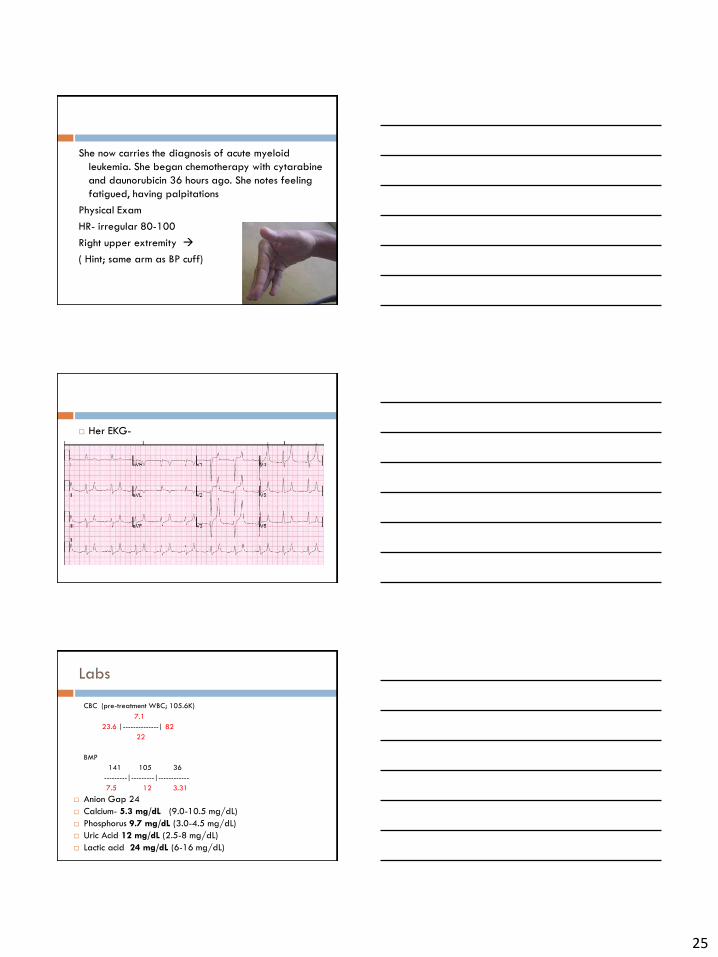
She now carries the diagnosis of acute myeloid
leukemia. She began chemotherapy with cytarabine
and daunorubicin 36 hours ago. She notes feeling
fatigued, having palpitations
Physical Exam
HR- irregular 80-100
Right upper extremity
( Hint; same arm as BP cuff)
Her EKG-
Labs
CBC (pre-treatment WBC; 105.6K)
7.1
23.6 |--------------| 82
22
BMP
141 105 36
---------|---------|------------
7.5 12 3.31
Anion Gap 24
Calcium- 5.3 mg/dL (9.0-10.5 mg/dL)
Phosphorus 9.7 mg/dL (3.0-4.5 mg/dL)
Uric Acid 12 mg/dL (2.5-8 mg/dL)
Lactic acid 24 mg/dL (6-16 mg/dL)
26
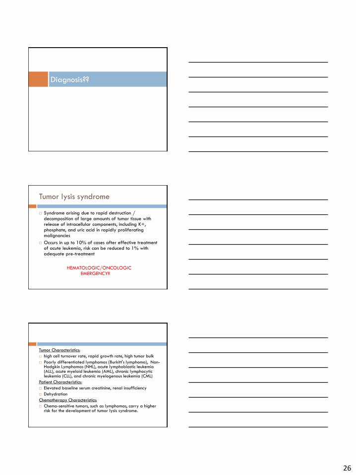
Diagnosis??
Tumor lysis syndrome
Syndrome arising due to rapid destruction / decomposition of large amounts of tumor tissue with release of intracellular components, including K+, phosphate, and uric acid in rapidly proliferating malignancies
Occurs in up to 10% of cases after effective treatment of acute leukemia, risk can be reduced to 1% with adequate pre-treatment
HEMATOLOGIC/ONCOLOGIC EMERGENCY!!
Tumor Characteristics:
high cell turnover rate, rapid growth rate, high tumor bulk
Poorly differentiated lymphomas (Burkitt's lymphoma), Non-Hodgkin Lymphomas (NHL), acute lymphoblastic leukemia (ALL), acute myeloid leukemia (AML), chronic lymphocytic leukemia (CLL), and chronic myelogenous leukemia (CML)
Patient Characteristics:
Elevated baseline serum creatinine, renal insufficiency
Dehydration
Chemotherapy Characteristics:
Chemo-sensitive tumors, such as lymphomas, carry a higher risk for the development of tumor lysis syndrome.
27
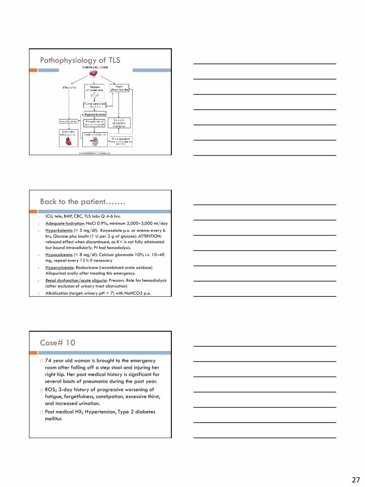
Pathophysiology of TLS
Back to the patient…….
1. ICU, tele, BMP, CBC, TLS labs Q 4-6 hrs.
2. Adequate hydration: NaCl 0.9%, minimum 2,000–3,000 ml/day
3. Hyperkalemia (> 5 mg/dl): Kayexelate p.o. or enema every 6
hrs, Glucose plus insulin (1 U per 2 g of glucose). ATTENTION:
rebound effect when discontinued, as K+ is not fully eliminated
but bound intracellularly. Pt had hemodialysis.
4. Hypocalcemia (< 8 mg/dl): Calcium gluconate 10% i.v. 10–40
mg, repeat every 12 h if necessary
5. Hyperuricemia: Rasburicase (recombinant urate oxidase)
Allopurinol orally after treating this emergency.
6. Renal dysfunction/acute oliguria: Pressors. Role for hemodialysis
(after exclusion of urinary tract obstruction)
7. Alkalization (target: urinary pH > 7) with NaHCO3 p.o.
Case# 10
74 year old woman is brought to the emergency
room after falling off a step stool and injuring her
right hip. Her past medical history is significant for
several bouts of pneumonia during the past year.
ROS; 3-day history of progressive worsening of
fatigue, forgetfulness, constipation, excessive thirst,
and increased urination.
Past medical HX; Hypertension, Type 2 diabetes
mellitus
28
On physical examination, she appears somnolent
but is easily arousable.
Temp is 37.1 °C (98.8 °F), BP 110/70 mm Hg, HR
120/min, and respiration rate is 17/min.
Oral mucosa is dry, and the conjunctivae are pale.
The lungs are clear.
No lymphadenopathy
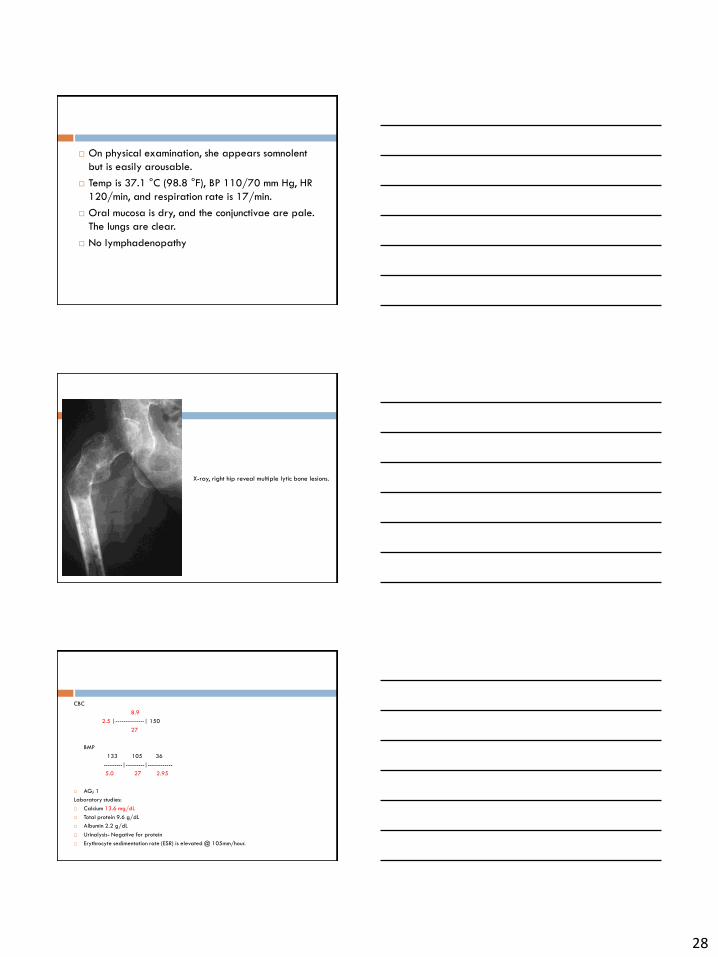
X-ray, right hip reveal multiple lytic bone lesions.
CBC
8.9
2.5 |--------------| 150
27
BMP
133 105 36
---------|---------|------------
5.0 27 2.95
AG; 1
Laboratory studies:
Calcium 13.6 mg/dL
Total protein 9.6 g/dL
Albumin 2.2 g/dL
Urinalysis- Negative for protein
Erythrocyte sedimentation rate (ESR) is elevated @ 105mm/hour.
29
Elevated Erythrocyte Sedimentation
Rate
Measures the rate at which red blood cells
sediment in a period of one hour.
Non-specific measure of inflammation
Indicator of the balance between + charged pro-
sedimentation factors (mainly fibrinogen) vs – anti-
sedimentation factors (mainly erythrocytes).
Erythrocyte Sedimentation Rate
Simple, inexpensive BUT non-specific test that is used to help detect inflammation associated with conditions such as infections, cancers, and autoimmune diseases.
ESR> 100 mm/hr
Multiple myeloma
Temporal arteritis
Polymyalgia rheumatic
Flares
Systemic lupus erythematosus
Rheumatoid arthritis
Inflammatory bowel disease
Peripheral blood
smear; Normochromic,
normocytic
erythrocytes with
rouleaux formation
Bone marrow; Plasma
Cells
30
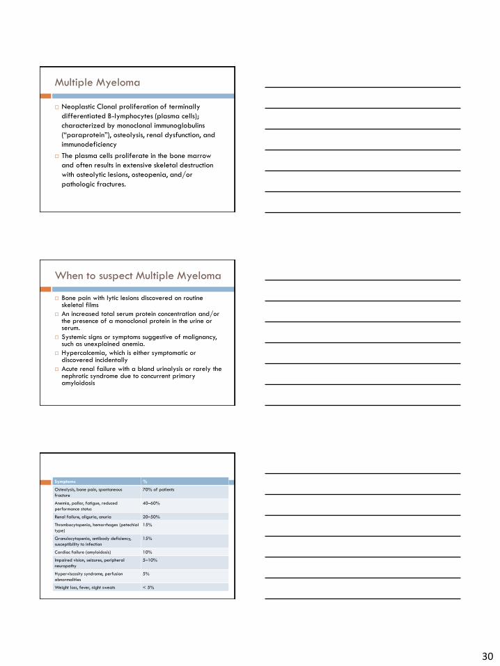
Multiple Myeloma
Neoplastic Clonal proliferation of terminally
differentiated B-lymphocytes (plasma cells);
characterized by monoclonal immunoglobulins
(“paraprotein”), osteolysis, renal dysfunction, and
immunodeficiency
The plasma cells proliferate in the bone marrow
and often results in extensive skeletal destruction
with osteolytic lesions, osteopenia, and/or
pathologic fractures.
When to suspect Multiple Myeloma
Bone pain with lytic lesions discovered on routine skeletal films
An increased total serum protein concentration and/or the presence of a monoclonal protein in the urine or serum.
Systemic signs or symptoms suggestive of malignancy, such as unexplained anemia.
Hypercalcemia, which is either symptomatic or discovered incidentally
Acute renal failure with a bland urinalysis or rarely the nephrotic syndrome due to concurrent primary amyloidosis
Symptoms %
Osteolysis, bone pain, spontaneous
fracture
70% of patients
Anemia, pallor, fatigue, reduced
performance status
40–60%
Renal failure, oliguria, anuria 20–50%
Thrombocytopenia, hemorrhages (petechial
type)
15%
Granulocytopenia, antibody deficiency,
susceptibility to infection
15%
Cardiac failure (amyloidosis) 10%
Impaired vision, seizures, peripheral
neuropathy
5–10%
Hyperviscosity syndrome, perfusion
abnormalities
5%
Weight loss, fever, night sweats < 5%
31
International Myeloma Working Group Criteria for
the Diagnosis of Multiple Myeloma
1. Monoclonal plasma cells in the bone marrow >10%
and/or presence of a biopsy-proven plasmacytoma.
2. Monoclonal protein present in the serum and/or urine.
3. Myeloma-related organ dysfunction (1 or more):
Calcium elevation in the blood (serum calcium >10.5 mg/L
[2.63 mmol/L] or upper limit of normal)
Renal insufficiency (serum creatinine >2 mg/dL [152.6
µmol/L])
Anemia (hemoglobin <10 g/dL [100 g/L] or 2 g < normal)
Lytic Bone lesions or osteoporosis
Laboratory Tests
CBC with differential
BMP, Ca2+,
serum creatinine, urea, uric acid, bilirubin,
albumin, LDH, CRP, ESR ↑, β2-microglobulin ↑
Total serum protein ↑, serum protein electrophoresis, immunofixation, detection of monoclonal paraprotein (“M-gradient”)
Urinary protein, urinary protein electrophoresis (M-gradient), detection of urinary light chains (“Bence-Jones proteinuria”) in 60% of cases
Detection of serum light chains (recently available assay), serum analysis more sensitive than urinary analysis)
Quantitative immunoglobulin level determination immunoelectrophoresis
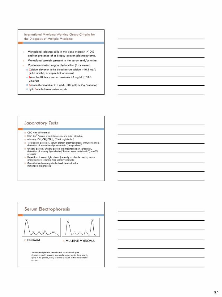
Serum Electrophoresis
NORMAL
MULTIPLE MYELOMA
Serum electrophoresis demonstrates an M-protein spike
M-protein usually presents as a single narrow peak, like a church
spire, in the gamma, beta, or alpha-2 region of the densitometer
tracing
32
Imaging
X-ray (lateral skull, lateral spine, humerus, pelvis, femur): osteolysis or diffuse osteoporosis of the axial skeleton, multiple osteolytic skull lesions (punched-out skull)
Suspected risk of fracture due to osteolysis (spinal column): CT / MRI / PET
Spinal Cord compression; from an extramedullary plasmacytoma or a bone fragment due to fracture of a vertebral body Suspected in patients presenting with severe back pain along with weakness/parasthesia of the lower extremities, or bladder or bowel dysfunction or incontinence; EMERGENCY!!
**Avoid iodine-containing contrast media due to potential nephrotoxicity**
Back to the Patient- Immediate support
High fluid intake to treat renal impairment and
hypercalcemia
Analgesia for bone pain.
Bisphosphonates Rx hypercalcemia and to delay
other skeletal related events
Allopurinolprevent urate nephropathy.
Lenalidomide–dexamethasone, bortezomib
induction therapy was initiated.
References
Harrison’s Principles of Internal Medicine, 18th Edition
UpToDate
Concise Manual of Hematology and Oncology
Hematology: Basic Principles and Practice, 6th Edition
Hematology in Clinical Practice, 5th Edition
2012 Clinical Practice Guide on Red Blood Cell Transfusion
2013 Clinical Practice Guideline on the Evaluation and Management of Adults with Suspected Heparin-Induced Thrombocytopenia (HIT)
MKSAP- 17
ACP Textbook of Medicine 2012
NEJM; Hunt BJ. N Engl J Med 2014;370:847-859