Hemangiomas and Vascular Malformations Yağmur AYDIN, M.D. University of Istanbul, Cerrahpasa...
52
Hemangiomas and Vascular Malformations Yağmur AYDIN, M.D. University of Istanbul, Cerrahpasa Medical Faculty Department of Plastic, Reconst. and Aesthetic Surgery
-
Upload
edmund-benson -
Category
Documents
-
view
221 -
download
0
Transcript of Hemangiomas and Vascular Malformations Yağmur AYDIN, M.D. University of Istanbul, Cerrahpasa...

Hemangiomas and Vascular Malformations
Yağmur AYDIN, M.D.
University of Istanbul, Cerrahpasa Medical Faculty Department of Plastic, Reconst. and Aesthetic Surgery

Vascular Anomalies
• In U.S, 40000 babies with vascular anomailes are born in a year
• 1of 10 children has vascular anomaly• A common mistake has been done with the
naming– Strawbery, cavernous, capillary hemangioma
• Mulliken and Glowacki in 1982 – classification according to the biological characteristics (endothelial properties)

Vascular Anomalies
• Tumors– Hemangioma– pyogenic granuloma– Kaposiphorm hemangio-endothelioma
• Malformations– Capillary– Lymphatic– Venous– Arteriovenous– combined


Hemangioma
• The most common tumor of infancy and childhood (4-10%)• 3-5 times more seen in girls• More seen in premature infants (<1200 grams% 23)• Not frequent in darker-skinned babies • Usually occurs in first 2 weeks after birth• Initially, a pale-colored, telangiectatic or macular red stain
or purple-colored stain• Single lesion in 80%, 20% more than one lesion• In patients with more than one lesion accompanies other
system hemangiomas ( liver etc.)

Clinical Appearance
• Lesions located in the superficial dermis– Hard, shiny dark-red coloured bulky lesion
• Lesion located in deep dermis, subcutaneous fat or muscle– Showing a slight bulking, hot, bluish
coloured lesion

The incidence of hemangiomas
• Craniofacial area (60%)• The body (25%)• Extremities (15%)

3 phases of hemangiomas
• Proliferation period (0-1 years)– Rapidly dividing endothelial cells
• İnvolution period (1-5 years)– Reduced endothelial proliferation, increased apoptosis,
fibrofatty replacement, reduced tumor volume, skin softening• Involution completion term (> 5 years)
– Thin veins and capillaries that drain current fibrofatty
islets

• Proliferation period:– Rapid growth (up to 10-12. months)
• Involution period (1-7 years)– Growth slows down,compatible with the
child's growth rate – Fading of the skin starting from the center of the
lesion,softening– The color disappears untill 5-7 years– Normal skin takes place in nearly 50% of children

Differential Diagnosis
• Lymphatic malformation –deep located hemangioma (neck, axilla)
• Capillary malformation - macular hemangioma• Vascular tumors of infancy - Fibrosarcoma etc.
– Radiological studies– Biopsy

Clinical evaluation
• Clear and open dialogue with the family• Confirm the diagnosis• Documentation with photos• The need for medical or surgical treatment• Other diagnostic studies to investigate the
extension of hemangiomas and other anomalies• Provide support groups and publications for
family

Complications
• Ulceration, bleeding• Infection• Visual impairment• Airway obstruction• Obstruction in the ear canal• Congestive heart failure
– diffuse neonatal hemangiomathosis – large visceral hemangiomas

Problems
• Hemangiomas located in head&neck (PHACES syndrome.)
• Hemangiomas covering the visual area• Perioral location• Respiratory distress (subglottic hemangioma• Ulcer

• •
hemangioma covering the visual field in 2 months old infant
Rapid reduction after oral and intra lesional corticosteroid injection
Residue mass at age 1,atrophy, telangiectasia

Treatment
• Follow up and observation• Training and convincing the family• Systemic corticosteroid• Second-generation drugs (vincristine, interferon
alpha)• Laser therapy - pulsed-dye laser
– ulcerated hemanjyom– Remaining telangiectasia after involution

Hemangiomas requiring treatment
• Covering the visual area, the air way, the ear canal
• Leading to congestive heart failure• Showing ulceration and bleeding

Treatment• Dangerous and life-threatening complications occur in 10% of
patient• The first option in medical treatment application
of corticosteroids (90% response)• Corticosteroid
– Topical / injection into the lesion• Triamcinolone 3-5 mg / kg• 3-5 times application (6-8 weeks apart)
– systemic application• Oral prednisone or prednisolone 2-3 mg / kg /day• Once every 2-4 weeks (10-12 months)

Other medical treatment agents(vicristine, interpheron alpha)
• Cases non –responsive to corticosteroid therapy• If long-term use of corticosteroids is
contraindicated• If complications develop after the use
of corticosteroids• In cases that the family does not want
to use (rare)

Systemic steroid treatment

Ulcer
local diffuse

Ulcer Treatment
• Dressings• Corticosteroid• Laser( flashlamp dye laser)• Total excision (if primary closure is possible)

Hemangioma
Ulceration

• residual athrophic • tissue and wrinkled skin after
involution• of hemangioma

Clinical appearance after involution of a large perioral and periorbital hemangioma

Vascular Malformations
• As a result of an error during embryological and fetal development
• Classification– Clinical– Radiological– Histological

Vascular Malformations
• Capillary• Lymphatic• Venous• Combined
– Arteriovenous– Lymphatico-venous– Lympathico-capillary-venous

Vascular Malformations
• Slow-Flow– Capillary– Lymphatic– venous
• Fast-flow– Arteriovenous
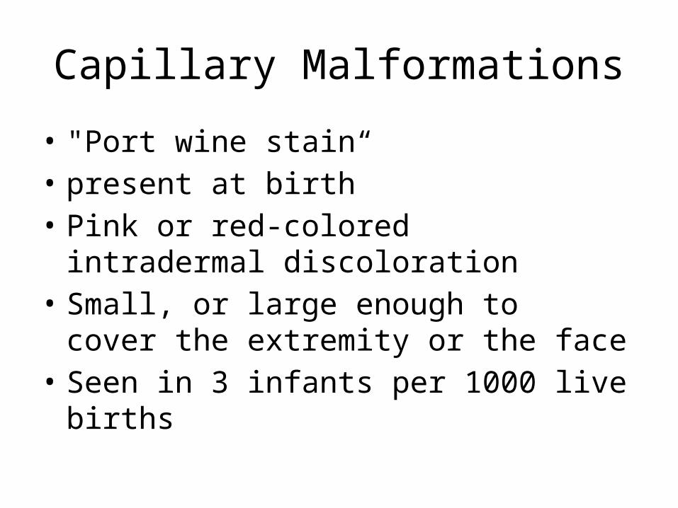
Capillary Malformations
• "Port wine stain“• present at birth• Pink or red-colored intradermal discoloration• Small, or large enough to cover the extremity
or the face• Seen in 3 infants per 1000 live births

• Real-capillary malformations– Progressive– Grow thicker over time, darken and nodule
develops inside• "Salmon patch", "Nevus simplex", "vascular stain ”
“birth stain”– Often seen in the middle part of the face, neck– Generally fade and reduce at the age of 1

• Other underlying disease or syndrome• Capillary malformation on midline lumbar or cervical region,
(spinal dysraphism, tethered spinal cord)• Sturge-Weber syndrome (capillary malformation of the
distribution area of the trigeminal nerve,lepthomeningeal vascular abnormalities,seizures)
• Klippel-Trenaunay syndrome(slow- flow capillary-lympho-venous malformation , elongation on axial plane and overgrowth of a limb

Sturge-Weber syndrome• capillary malformation of the trigeminal nerve distribution area• lepthomeningeal vascular anomaly• Seisures

Klippel-Trenaunay syndrome• capillary malformation• soft tissue and bone overgrowth• varices

Treatment
• Flashlamp-pumped pulsed dye laser– 577, 585, or 595 nm wavelength– Targets oxyhemoglobin– Provokes intravascular thrombosis
• 50- 90% of patients present discoloration• Treatment gives the best results in early childhood• Other lasers (non-responsive patients)
– Alexandrite (755 nm)– Neodymium: yttrium-aluminum-garnet (1064nm)– Intense pulsed light (IPL)

Port wine stainThe result obtained after two applications of pulsed dye laser

Lympathic Malformations• Seen as local sponge-like lesions or difuse lesions covering an
anatomical organ or area• Radiological and histological
– Microcystic– Macrocyctic– Mixed
• Usually occurs at birth or first 2 ages• most common seen in cervicofacial area• Axilla, chest, mediastinum,
retroperitoneal area,perineum, gluteal area• Overlying skin is intact, looks bluish • Small ,thin vesicles are pathognomonic for dermal involvement

Treatment
• Bleeding• Recurrent infection (cellulitis)• Body contouring• Correction of funtional deficits
– Sclerotherapy (macrocyctic lesions)– Intralesional injection of bleomycin– OK-432– Argon, neodynium: YAG, or carbon dioxide laser

Surgical treatment indications
• Lesions blocking the respiratory way• Lesions that create feeding problems• Lesions making distortion

Venous Malformation
• Soft, fading with pressure, bluish coloured masses under the skin
• Swelling with physical activity and when slouched down
• Palpable thrombi• Morning pain (stasis and microthrombi)• Frequent localization of head and neck• More expansive than it looks
(muscle,bone, oral mucosa, salivary gland)

Diagnosis
• Magnetic resonance imaging (MRI)– To diagnose– To evaluate the extent of malformation
• Bleeding-coagulation profile should be assessed– coagulopathy

Treatment
• Percutaneous sclerotherapy– absolute ethanol– hypertonic saline– sodium sulfate tetradesil
• Elastic compression stockings (extremity)• Aspirin (daily)
– painful thrombus– For prophylaxis of phlebitis
• Surgical Treatment– head and neck lesions causing cosmetic problem– severe pain and bleeding– Lesions with well-defined borders


Venous malformation spreading into the vulva and inner thigh muscles
Partial excision and injection
of sclerosing agents

• Venous malformation: 2 months after1 time the Nd: YAG laser application • complete regression

Arterio-venous malformations
• Connection exists between the arterial system and venous system
• They usually present at birth• Often incorrectly diagnosed (KM or
hemangioma)• There is a rapid flow between the two systems• Fast flow is evident in childhood

Arterio-venous malformations
• Over time the stain on the skin – Erythema– The local rise of temperature – Thrill – Murmur
• The mass may expand• Rapid growth may be seen during puberty or
trauma

Arterio-venous malformations
• Arteriovenous shunts– Ischemia and related symptoms and signs– painless ulceration
• Persistent pain, occasional bleeding• Widespread AVM may increase cardiac
output and cause congestive heart

Diagnosis
• Ultrasonography• Coloured Doppler• MRI• Angiography
– Feeder and draining vessels– Varying degrees of arterial dilatation– curved vessels– A-V shunts– Enlarged draining veins

Treatment
• Embolization• Sclerotherapy• Surgical resection and reconstruction
– Preoperative angiography• Determines feeder and draining vessels• Embolization (adhesive or with special springs)

Arterio-venous malformations
• Intracranial (most common)• Extracranial
– Head and neck– Extremity– Body– İnternal organs

• AVM in the ear1.Preoperative embolization2.Ear amputation and partial
thickness skin grafting3. ear prosthesis

Differential Diagnosis
Hemangioma• Occurs shortly
after birth• Rapid
growth, stagnation and reduction phases
• More seen in girls• Endothelial hyperplasia
vascular malformation• Development commensurate with
the growth• More noticable at puberty• Does not reduce• No gender difference• Lack of normal endothelium