HemagglutininfromAcrididae(Grasshopper ...
73
Hemagglutinin from Acrididae (Grasshopper) : preparation and properties by Mark Richard Stebbins A thesis submitted in partial fulfillment of the requirements for the degree of Master of Science in Biochemistry Montana State University © Copyright by Mark Richard Stebbins (1984) Abstract: The proteinaceous hemagglutinin (lectin) present in the hemolymph of Melanoplus sanguinipes (F.), was isolated and biochemically characterized. The protein was purified to homogeneity by affinity chromatography on a column of Sepharose-galactose. The hemagglutinin showed broad specificity and agglutinated several erythrocyte types. Gel filtration and electrophoresis showed that grasshopper hemagglutinin was a high molecular weight (600-700 K dalton) non-covalent aggregate of 70 K dalton subunits. The 70 K dalton subunits contained two disulfide-linked polypeptide chains of molecular weight 40,000 and 28,000 respectively. The purified hemagglutinin contained a preponderance of acidic and polar amino acid residues and a small amount of glucosamine. Hemagglutination activity toward human asialo erythrocytes was destroyed by treatment of the hemagglutinin with trypsin, heat or EDTA. Hemagglutination inhibition studies showed that low concentrations (<5 mM) of both galactosidic and glucosidic carbohydrates are bound by the hemagglutinin and cause inhibition of erythrocyte agglutination. The strongest inhibitors of hemagglutination were the alpha anomers of D-galactose. Hemolymphatic hemagglutinin isolated from Melanoplus differentiaIis yielded identical physicochemical results as did hemagglutinin from Melanoplus sanguinipes. It was concluded that a single hemagglutinin protein was the substance responsible for all hemagglu-tinating activity present in the hemolymph of either species. Research directed toward the elucidation of the possible roles that grasshopper hemagglutinin plays in grasshopper immune/defense mechanisms was initiated by producing antibodies to purified hemagglutinin in rabbits and mice. These antibodies were specific for grasshopper hemagglutinin as shown by gel double diffusion and immunoelectrophoresis. Individual subunits were recognized by rabbit antiserum as shown by an indirect immunochemical detection procedure where rabbit antiserum (bound to protein subunits immobilized on a nitrocellulose filter) is recognized by an enzyme-conjugated "second" antibody. Applications of this research toward future immunological localization studies of grasshopper hemagglutinin in insect hemocyte maintainance cultures are discussed.
Transcript of HemagglutininfromAcrididae(Grasshopper ...

Hemagglutinin from Acrididae (Grasshopper) : preparation and propertiesby Mark Richard Stebbins
A thesis submitted in partial fulfillment of the requirements for the degree of Master of Science inBiochemistryMontana State University© Copyright by Mark Richard Stebbins (1984)
Abstract:The proteinaceous hemagglutinin (lectin) present in the hemolymph of Melanoplus sanguinipes (F.),was isolated and biochemically characterized. The protein was purified to homogeneity by affinitychromatography on a column of Sepharose-galactose. The hemagglutinin showed broad specificity andagglutinated several erythrocyte types. Gel filtration and electrophoresis showed that grasshopperhemagglutinin was a high molecular weight (600-700 K dalton) non-covalent aggregate of 70 K daltonsubunits. The 70 K dalton subunits contained two disulfide-linked polypeptide chains of molecularweight 40,000 and 28,000 respectively. The purified hemagglutinin contained a preponderance ofacidic and polar amino acid residues and a small amount of glucosamine. Hemagglutination activitytoward human asialo erythrocytes was destroyed by treatment of the hemagglutinin with trypsin, heator EDTA. Hemagglutination inhibition studies showed that low concentrations (<5 mM) of bothgalactosidic and glucosidic carbohydrates are bound by the hemagglutinin and cause inhibition oferythrocyte agglutination. The strongest inhibitors of hemagglutination were the alpha anomers ofD-galactose. Hemolymphatic hemagglutinin isolated from Melanoplus differentiaIis yielded identicalphysicochemical results as did hemagglutinin from Melanoplus sanguinipes. It was concluded that asingle hemagglutinin protein was the substance responsible for all hemagglu-tinating activity present inthe hemolymph of either species.
Research directed toward the elucidation of the possible roles that grasshopper hemagglutinin plays ingrasshopper immune/defense mechanisms was initiated by producing antibodies to purifiedhemagglutinin in rabbits and mice. These antibodies were specific for grasshopper hemagglutinin asshown by gel double diffusion and immunoelectrophoresis. Individual subunits were recognized byrabbit antiserum as shown by an indirect immunochemical detection procedure where rabbit antiserum(bound to protein subunits immobilized on a nitrocellulose filter) is recognized by anenzyme-conjugated "second" antibody. Applications of this research toward future immunologicallocalization studies of grasshopper hemagglutinin in insect hemocyte maintainance cultures arediscussed.

HEMAGGLUTININ FROM ACRIDIDAE (GRASSHOPPER)
PREPARATION AND PROPERTIES
by
Mark Richard Stebbins
A thesis submitted in partial fulfillment
of the requirements for the degree
of
Master of Science
. inBiochemistry
MONTANA STATE UNIVERSITY Bozeman, Montana
June, 1984
APPROVAL
of a thesis submitted by
Mark Richard Stebbins
This thesis has been read by each member of the thesis committee and has been found to be satisfactory regarding content, English usage, format, citations, bibliographic style, and consistency, and is ready for submission to the College of Graduate Studies.
Dare <Graduate Committee
Approved for the Major Department
Head, Major Department
Approved for the College of Graduate Studies

iii
STATEMENT OF PERMISSION TO USE
In presenting this thesis in partial fulfillment of the requirements for a master's degree at Montana State
University, I agree that the Library shall make it
available to borrowers under rules of the Library. Brief
quotations from this thesis are allowable without special
permission, provided that accurate acknowledgement of
source is made.
Permission for extensive quotation from or reproduc
tion of this thesis may be granted by my major.professor,
or in his absence, by the Director of Libraries when, in
the opinion of either, the proposed use of the material is
for scholarly purposes. Any copying or use of the
material in this thesis for financial gain shall not be
allowed without my written permission.
Signature
Date Mu1 , JfM~~v ^
iv
ACKNOWLEDGEMENTS
I would like to thank my research advisor. Dr. Ken Hapner, for his enthusiasm and guidance during the course of this project.
I also wish to express my appreciation to the
following persons from Montana State University who
generously offered advice and assistance during my
graduate study.
Dr. John E . Robbins, Chemistry Dept.
Sharon J . Hapner, Biology Dept.
Dr. Clifford W. Bond, Microbiology Dept.
Dr. Guylyn R. Warren, Chemistry Dept.
Dr. Samuel J . Rogers, Chemistry Dept.

V
TABLE OF CONTENTS
PageLIST OF TABLES........................ . :.............. vii
LIST OF FIGURES................. viii
ABSTRACT...................... . . ........... ..... ...... I
INTRODUCTION........................................... 2
RESEARCH OBJECTIVES.... .......... ....... ;............ 9
MATERIALS AND METHODS................................. 10
Isolation of Grasshopper Hemagglutinin............ 10Collection of hemolymph......................... 10Hemagglutination assay..................... . ... 10Preparation of asialo erythrocytes............. 11Affinity chromatography............... '......... ' 11Protein assay.................................... 12
Carbohydrate Inhibiton of Hemagglutination........ 12Biochemical Characterization of GrasshopperHemagglutinin....................................... 13
Gel filtration............................... 13Polyacrylamide gel electrophoresis (PAGE)----- 13Nondenaturing discontinuous PAGE....'........ 13Sodium dodecyl sulfate (SDS) PAGE.............. 14Urea PAGE. . . ....................... .■..... ........ 15Isoelectric focusing....................■........ 15Amino acid analysis.... ............. .'......... 16Stability........................................ 16
Production of Antiserum in Rabbits................ 17Production of Antibodies in
Murine Ascitic Fluid................... ......... 17Gel Double Diffusion............. ................... 18Immunochemical Detection of GrasshopperHemagglutinin............... 18
Immunoelectrophoresis........................... 18Protein transfer from PAGE gels to
nitrocellulose filters.............'.......... ". 19Glucose oxidase conjugated _goat-anti-
(rabbit IgG) IgG............................. 2 0

TABLE OF CONTENTS--Continued
PageRESULTS.........'....................................... 21
Purification of Grasshopper Hemagglutinin......... 21Affinity chromatography......................... 21Stepwise elution................................ 22Elution with other desorbants.................. 22
Erythrocyte Agglutination.......................... 24Native Molecular Structure of GrasshopperHemagglutinin.... ...... '........................... 25
Gel filtration....... .......... ........... . . 2 5Nondenaturing electrophoresis. . . . . ... ......... 27Isoelectric focusing.................... 27
Subunit-Structure of Grasshopper Hemagglutinin.... • 30SDS electrophoresis............................. 30Electrophoresis in urea......................... 30Isoelectric focusing in urea................... 33
Amino Acid Composition............................. 3 3Carbohydrate Inhibition of GrasshopperHemagglutinin..................... 34Molecular Stability of Grasshopper Hemagglutinin.. 37Antigenicity of Grasshopper Hemagglutinin......... 40'
• Rabbit antiserum...... .................... .'..... 40Immune murine ascitic fluid.................... 40
Immunological Detection of GrasshopperHemagglutinin....................................... 43
Immunoelectrophoresis........................... 43Glucose oxidase immunoenzyme................... 43
DISCUSSION.............................................. 45
Purification........................................ 45Molecular Structure................................. 48Inhibition of Hemagglutination.................... 52Stability.-.............. ............................. 5 4Immunological Studies..........-.................... 55
CONCLUSIONS..... ................. ................ ..... 5 8
REFERENCES CITED..................... '.................. 6 0
vi
APPENDIX 63

vii
LIST OF TABLES
Table Page
1. Hemagglutination of Various Erythrocytes ■ by Whole Hemolymph, Absorbed Hemolymph
and Purified Hemagglutinin....................... 252. Amino Acid Composition of Purified Hemagglutinin
from Melanoplus sanguinipes andMeianoplus differentia I is........................ 3 4
3. Carbohydrate Inhibition of Hemagglutination by Whole Grasshopper Hemolymphand. Purified Hemagglutinin....................... 36

viii
LIST OF FIGURES
Figure Page
1. Affinity chromatography of grasshopperhemolymph.................................. 23
2. Gel filtration of grasshopper hemolymph........ 26
3. Nondenaturing polyacrylamide gel electrophoresis of purifiedgrasshopper hemagglutinin.............. 28
4. SDS polyacrylamide gel electrophoresis of whole grasshopper hemolymph andpurified hemagglutinin.......................... 29
5. Urea polyacrylamide gel electrophoresisof grasshopper hemagglutinin.... . . ......... 31
6. Isoelectric focusing of native and ureadenatured grasshopper hemagglutinin............ 32
7. Heat stability of hemagglutinating activity of whole grasshopper hemolymph andpurified hemagglutinin. .......................... 38
8. Trypsin stability of hemagglutinating activity of whole grasshopper hemolymph andpurified hemagglutinin.......................... 39
9. Gel double diffusion of whole grasshopper hemolymph, affinity adsorbed hemolymph and purified hemagglutinin vs.rabbit antiserum................................. 41
10. Immunoelectrophoresis of purifiedhemagglutinin....... 42

I
ABSTRACT
The proteinaceous hemagglutinin (lectin) present in the hemolymph of Melanoplus sanguinipes (F.) , was isolated and biochemically characterized. The protein was purified to homogeneity by affinity chromatography on a column ofI Sepharose-galactose. The hemagglutinin showed broad specificity and agglutinated several erythrocyte types. Gel filtration and electrophoresis showed that grasshopper hemagglutinin was a high molecular weight (600-700 K da Iton) non-covalent aggregate of 70 K da Iton subunits. The 70 K daIton subunits contained two disulfide-linked polypeptide chains of molecular weight 40,000 and 28,000 respectively. The purified hemagglutinin contained a preponderance of acidic and polar amino acid residues and a small amount of glucosamine. Hemagglutination activity toward human asialo erythrocytes was destroyed by treatment of the hemagglutinin with trypsin, heat or EDTA. Hemagglutination inhibition studies showed that low concentrations (<5 mM) of both galactosidic and glucosidic carbohydrates are bound by the hemagglutinin and cause inhibition of erythrocyte agglutination. The strongest inhibitors of hemagglutination were the alpha anomers of D-galactose. Hemolymphatic hemagglutinin isolated from Melanoplus d ifferentia Iis yielded identical physico- chemic.aI results' as. did hemagglutinin from Melanoplus sanguinipes. It was concluded that a single hemagglutinin protein was the substance responsible for all hemagglu- tinating activity present in the hemolymph of either species.
Research directed toward the elucidation of the possible roles that grasshopper hemagglutinin plays in grasshopper immune/defense mechanisms was initiated by producing antibodies to purified hemagglutinin in rabbits and mice. These antibodies were specific for grasshopper hemagglutinin as shown by gel double diffusion and immunoelectrophoresis. Individual subunits were recognized by rabbit antiserum as shown by an indirect immunochemical detection procedure where rabbit antiserum (bound to protein subunits immobilized bn a nitrocellulose filter) is recognized by an enzyme-conjugated "second" antibody. Applications of this research toward future immunological localization studies of grasshopper hemagglutinin in insect hemocyte maintainance cultures are discussed.

2
INTRODUCTION
An organism's survival is often dependent in part
upon endogenous "immune" protection systems that render
the host exempt from the potentialIy harmful effects of
pathogens and other foreign substances. Higher vertebrate
organisms possess an integrated cellularly and humoralIy
mediated antibody immune system, the ha I !.marks of which
are the immunoglobulins, complement proteins, and
lymphocytes [I].
What do we know about the immune systems of lower
vertebrates and invertebrates, particularly insects? It
has been speculated that the mammalian immune system may
have evolved from the hemocyte cells of invertebrates [2].
Certain Iy the success of the insects' strategy for
survival is manifest in their numbers. Over 700,000
currently living species of insects have been identified,
which amounts to over one-half of all living things found
on the earth. x Several lines of evidence are available to
clearly indicate that the immunoglobulin-complement system
is not present in the insect. Two basic events are known
to occur upon the introduction of foreign matter into the
hemocoel of an insect: phagocytosis of smaller particles
(< 10 micrometers, urn) and encapsulation of larger

3p a r t i c l e s [3]. P h a g o c y t o s i s initially requires recognition of the foreign substance. This is followed by chemotactic attraction and subsequent attachment of the foreign substance to the phagocyte. The final phase is
ingestion and neutralization o.f the foreign material.
Particles too large to be phagocytosed are encapsulated
and neutralized in a membranous capsule. Encapsulated
bacteria have been shown to aggregate into m e Ianized
nodules [4]. What, then, is the nature of nonself
recognition in insects ? Is recognition accomplished by
cells alone? Are humoral factors involved in recognition?
One clue may lie in a group of proteins called agglutinins
that occur ubiquitously in hemolymph, the blood of
insects.
Agglutinins are polyvalent lectins that can recognize
and bind to specific carbohydrate molecular structures on
cell surfaces of bacteria and vertebrate erythrocytes.
The cells are thus crosslinked and clumped or aggregated.
In the case of red cells, the term hemagglutination is
descriptive of the activity. The clumping activity can be
visually observed, and this fact forms the basis for the
convenient hemagglutination assay for detection of
hemagglutinins.
Soluble hemagglutinin activity is generally present
in the hemolymph of invertebrate organisms [5,6] and most
current research on invertebrate hemagglutinins is

4
directed toward their putative (carbohydrate). recognitory
capabilities as humoral and/or cellular immunosubstances
[7]. Hemagglutinins have been implicated as opsonins in
•some invertebrates including crayfish [8], mollusks [9]
and oysters [10,11] . Nonetheless, the in vivo function(s)
of invertebrate hemagglutinins is unknown and experimental data supportive of their involvement in immune mechanisms
is largely circumstantial and inconclusive.
The reasons for studying insect immune systems are
two-fold. First, annual losses of food crops in the
United States caused by insects have been estimated at 13%
[12]. In the western United States grasshoppers alone
destroy more than 30 million dollars worth of food crops
each year [13]. Our rapidly increasing human population
demands increased global food production capacity which is
increasingly dependent on the effective control of insect
pests. Insect control involving toxic pesticides is
facing many drawbacks including technological limitations
due to evolving genetic resistance, nonspecificity and
sociological-legal restraints [14]. These facts along
with recent advances in biotechnology have opened the door
for research and development of biological control
systems; that is, the strategic introduction of bacteria
or viruses that are pathogenic to a specific (pest)
organism [15]. The major obstacle limiting progress in
this area is the highly successful immunodefense system of

5insects, the biochemical basis of which is poorly- understood .
Another reason for studying insect hemagglutinins is
that lectins bind to cell surface polysaccharides, glycoproteins, and glycolipids in a specific fashion and-
provide a way to study the architecture of cell surfaces
[16]. Some lectins differentially agglutinate certain
mammalian cells in culture, depending on the structure of
their surface polysaccharides. Since these cell surface
c a r b o h y d r a t e s are often ant i g e n i c in a nonself
environment, lectins can provide a means by which to
indicate donor-recipient tissue computability in tissue
transplants based on a similar agglutination, pattern [17].
Also, some lectins preferentially agglutinate viralIy or
chemically transformed mammalian cells in culture as well
as cells from spontaneous tumors [18] and are therefore
useful tools for the cytogeneticist and the cancer
researcher [19].
The possible involvement of hemagglutinins in insect
defense systems is described in reviews by Whitcomb, et
al. [20] and more recently by Lackie [17] and Ratner and
Vinson [21]. Insect agglutinins may be active in both
recognitory and processing (phagocytosis, encapsulation)
phases of immunosurveillance and protection. Elucidation
of the i_n vivo function of insect agglutinins has been
hampered by the unavailability of highly purified and

6characterized agglutinins, and associated immunological detection procedures. ,
Hemagglutinating activity has been described in the hemolymph of several insect specimens including grass
hoppers [22,23], crickets [24], flesh fly [25], cockroaches [26,27-, 28,29,30,31] , beetles [32], locusts [28],
milkweed bug [33], and butterflies and moths [34,35] .
An injury induced hemagglutinin from Sarcophaga
peregrina larvae that is also detected in high amounts in
the early pupal stage was isolated and characterized by
Komano, et a I. [25]. It is a 190,000 molecular weight
(MW) protein consisting of four 32,000 MW and two 30,000
MW noncovalently associated subunits. Only the 32,000 MW
subunit is present in normal larvae. The authors suggest
that the 30,000 MW subunit may be produced from 32,000 MW
subunits by a protease that is activated commensurate with
body wall injury [36]. This idea is based on indirect
evidence that both 32,000 MW and 30,000 MW subunits show
similar tryptic peptide maps. Alternatively, the 30,000
MW subunit may be synthesized de novo. In a further study
using a radioimmunoassay Komano, et al. [37] showed that
the lectin was synthesized in the fat body and secreted
into the hemolymph both on injury and on pupation. Also,
the amount of lectin on the outer hemocyte surface
increased upon injury and on pupation. The authors
suggest that this.hemagglutinin may be involved in
7nonspecific recognition in an immune system that
culminates in phagocytosis of foreign substances or fragments of tissue undergoing metamorphosis.
Hapner and Jermyn [24] isolated a hemagglutinin from
the cricket TeleogrylIus commodus (Walker) on an affinity
matrix of Sepharose-fetuin. Cricket hemagglutinin.was
completely desorbed from the affinity column with buffer
that contained 0.1 M N-acetyl neuraminic acid, and
incompletely with buffer containing '0.1 M 2-acetamido-2-
deoxy-D-glucose. Purified cricket hemagglutinin activity
was inhibited by the two desorbants as well as by N-
acety1-D-glucosamine (10 mM), N-acety1-D-galactosamine (50
mM), and EDTA (10 mM). The unpurified cricket hemagglu
tinin was shown to be a high molecular weight glycoprotein
complex of disulfide-linked 31,000 MW and 53,000 MW
polypeptide chains.
Amirante, et a I. [38] described the presence of two
hemagglutinins in the hemolymph of the cockroach
Leucophaea m aderae L. Amirante and Mazzalai [39] used
fluorescein-labeled antiserum, to show that both hemagglu
tinins were synthesized in granular hemocytes and spherule
cells. The authors propose that the two hemagglutinins
are probably released into the hemolymph where they may be
responsible for "cellular immunological reactions".

8Grasshopper hemolymph nonspecificalIy agglutinates
all human ABO and many animal erythrocytes. The hemagglutinin activity exhibits a broad pattern of carbohydrate
inhibition of hemagglutination and shows highest sensitivity to inhibition by both galactosidic and glucosidic
structures [22]. This broad range of hemagglutination and
carbohydrate inhibition resides, in individual insects [23]
and is not the result of the pooling of hemolymph from
many insects. Individual grasshoppers are therefore
viewed as either containing complex mixtures of agglu
tinins of various specificities ■(heteroagglutinins) or a
single hemagglutinin of broad red cell and carbohydrate
binding capability.

9
RESEARCH OBJECTIVES
The specific objectives of this study are:
a. Purify and characterize the hemagglutinin from
the hemolymph of Melanoplus sanguinipes and Melanoplus
differentialis.
b. Immunize rabbits and mice using purified
hemagglutinin as the immunogen.
c. Develop methodology for indirect immunochemical
localization procedures for grasshopper hemagglutinin.

10
MATERIALS AND METHODS
Isolation'of Grasshopper Hemagglutinin
Collection of hemolymph. Adu.lt IYL_ sanguinipes and M.
differentiaIis grasshoppers were provided from permanent
colonies at the USDA Rangeland Insect Laboratory, Bozeman,
MT. Hemolymph was collected with a capillary pipette from
ether-anaesthetized insects as previously described [22].
The hemolymph was pipetted into an equal volume of ice
cold Dulbecco's phosphate buffered saline (DPBS) (1.5 mM
KH2PO4, 8 mM Na2HPO4, 0.9 mM CaCl2, 2.7 mM KCl, .0.5 mM
MgCl2, 0.135 M NaCl, pH. 7.2). which contained 0.001 M
phenyl thiourea' (PTU) to inhibit melanin formation. Hemo-
cytes and coagulum were removed by centrifugation at 3000
g and the clear yellow supernatant was stored at -20°C.
Hemagglutination assay. Human ABO erythrocytes were
a gift from Physicians Laboratory Service, Inc. (Bozeman,
MT) and animal erythrocytes were purchased from Colorado
Serum Company (Denver, CO). Erythrocytes were washed four
times by centrifugation in ice cold DPBS prior to use.
Hemagglutination activity was detected at 22°C by serial
two-fold dilution of 25 microliter (uI) hemagglutinin
sample with 25 ul DPBS using plastic V-bOttom microtiter
dishes. After dilution of the sample, 25 ul of a 2.5%

11suspension of erythrocytes in DPBS was added and
agglutination was visually determined after 30 min. The
reciprocal of the highest dilution causing agglutination
of erythrocytes was the hemagglutination titer. Controls
not containing hemagglutinin were always performed.
Preparation of asialo erythrocytes. Asialo human erythrocytes were prepared by incubating ,for one hour at
37°C 0.5 ml human O+ erythrocytes with 3 mg neuraminidase
(type 5, Sigma Chemical Cd., St. Louis, MO) in 10 ml DPBS
at pH 5.7. The asialo red cells were washed four times
with ice cold DPBS prior to use.
Affinity chromatography. D-Galactose was covalently
attached to Sepharose 4B (Pharmacia, Piscataway, NJ)' by
the divinyIsuIfone method [40]. A 0.5 x 3 cm column was
prepared and washed with 100 volumes of ice cold DPBS.
About 50 ml of grasshopper hemolymph (previously diluted
with an equal volume of DPBS, I mM PTU) was passed through
the column (10 ml/hr) at 4°C. The column was then, washed
with ice cold DPBS until the absorbance of the effluent at
280 nm returned to zero. Adsorbed hemagglutinin activity
was released from the column upon elution with DPBS that
contained 0.2 M D-galactose. After the first one ml
fraction was collected, the column flow was stopped and
the column was allowed to incubate for several hours at
22°C before the second one ml fraction was collected.
Subsequent fractions were collected after similar

12incubation periods. Hemagglutination titer was
immediately determined by hemagglutination assay using
asialo human O+ erythrocytes.
Protein assay. Protein concentrations of eluted,
fractions were determined relative to a bovine serum albumin standard by the method of Bradford [41] with the
Bio Rad protein assay kit (Bio Rad Laboratories, Richmond,
CA). Collected fractions were stored at -20°C.
Carbohydrate Inhibition of Hemagglutination
Minimal inhibitory concentrations of carbohydrates
were determined by performing the hemagglutination assay
in the presence of carbohydrates ranging downward in
concentration from 100 mM to 0.3 mM. The initial sample
of hemagglutinin was adjusted by dilution to a titer of
64-128. The serially diluted hemagglutinin and the added
carbohydrate (25 u I ) were incubated 5 min prior to
addition of 25 ul human O+ asialo erythrocytes. All
determinations were done in duplicate and titer was
visually estimated after one hour. Controls in which DPBS
was substituted for the carbohydrate solution were done
concurrently. Direct comparison of the hemagglutination
titer for the inhibited and noninhibited assays allowed
determination of the inhibitor concentration which
decreased the titer by 50% (one agglutination well).

13Biochemical Characterization of Grasshopper Hemagglutinin
GeJL filtration. Whole hemo lymph and purified
hemagglutinin were chromatographed separately on a 1.5 x
120 cm column of Sepharose 6B. The column was developed
at 22°C at a flow rate of 12 ml/hr with' a pH 7.2 buffer
consisting of 10 mM tris, 150 mM NaCl, I m-M CaC 12 and 50 mM D-galactose. Calibration standards were thyroglobulin
(669,000), ferritin (440,000), catalase (232,000) and
aldolase (151,000) (Pharmacia). Column effluent was
monitored at 280 nm and 2 ml fractions were collected.
Aliquots were assayed for hemagglutinin activity using
asialo erythrocytes.
Polyacrylam ide geI electrophoresis (PAGE). Protein
samples were electrophoresed, under various conditions, in
140 x 160 x 1.5 mm polyacrylamide slab gels using the
apparatus' and procedures from Hoefer Scientific (San
Francisco, CA). Acrylamide and sodium dodecyl sulfate
were from Sigma. N,N'-methylenebisacrylamide and
N,N,N' ,N'-tetramethylethylenediamine were from Aldrich
Chemical Company (Milwaukee, WI). Enzyme grade urea was
from Bethesda Research Laboratories (Gaithersburg, MD) and
protein molecular weight markers were from Pharmacia. Al I
other chemicals were of reagent grade.Nondenaturing discontinuous PAGE. Nondenaturing
discontinuous PAGE was, carried out using a 3.1%, pH-7.5
stacking gel and a 5%, pH 8.3 separating gel, both
14containing 0.2 M D-galactose.■ Samples contained
approximately 10 ug protein mixed with 1/10 volume 50%
sucrose. Electrophoresis was carried out at 20'mA/gel for
6 hr at 13°C. Gels were fixed for I hr in 12.5% (w/v)
trichloroacetic acid in water, stained 2 hr in 0.25% (w/v)
Coomassie Blue G- 2 5 0 in water and destained 24 hr in
methanol/acetic acid/water (5:7:88 by vol). The position
of non-denatured haemagglutinin was related to the
standard protein markers: thyrogIobuIin (669,000),
ferritin (440,000), catalase (232,000) and lactate
dehydrogenase (140,00 0).
Sodium dodecyI sulfate (SDS) PAGE. Electrophoresis
in SDS polyacrylamide gel slabs was done using 4%, pH 6.8
stacking and 12%, pH 8.8 separating gels according to
Laemmli [42]. Samples that contained 10-30 ug protein
were denatured by heating 2 min at 95 °C in an egual
volume of pH 6.8 buffer (0.1 M tris'HCl, 4% SDS and 20%
glycerol) that did or did not contain 2-mercaptoethanol.
Standard molecular weight markers were treated similarly.
Electrophoresis was continued for ,3 hr at 22°C and 30
mA/gel. Gels were stained 4 hr in 0.12% (w/v) Coomassie
Blue R-250 in methanol/acetic acid/water (5:1:4 by vol),
destained in the same solvent for I hr and then destained
24 hr in methanol/acetic' acid/water (5:7:88 by vol).
Standard curves were calculated from the migration
positions of bovine serum albumin (68,000), ovalbumin
15(43,000) , chymotrypsinogen (25,700) and lysozyme (14,300) /relative to that of the phenol red marker dye. Apparent
molecular weights for reduced and non-reduced conditions
were extrapolated from plots relating migration and log
molecular weight for the reduced and non-reduced standard
protein markers, respectively.
Electrophoresis using urea as the
denaturant was performed as in the nondenaturing s:ystern
except that both stacking and separating gels contained 6
M urea and 0.02 M EDTA. Before application to the gel,
solid urea was added to all protein samples to give a
final concentration of 6 M. Reduced protein, samples were
prepared by incubation in 5% 2-mercaptoe.thanol for 2 min
at 95°C prior to addition of urea. Samples contained 10-
20 ug protein. Electrophoresis was performed at 200 volts
for 18 hr at 22°C.
_! s oe _1 e c t r _i c f o cuj5 ing^ Isoelectric focusing of
purified hemagglutinin was done in 5'x 90 mm 6% poly
acrylamide gel rods with a Hoefer DE 102 Tube GeI Unit
according to Wrigley [43]. Carrier ampholyte in the pH
range 3-10 was from LKB Products (Bromma, Sweden). All
nondenaturing gels c o n t a i n e d 0.2 M D-galactose.
Isoelectric focusing under denaturing conditions was
performed in gels containing 6 M urea. Isoelectric
focusing was continued for 4 hr at I mA/ tube and at 13°C.
Focused gels were fixed 2 hr in methanol/water (3:7 v/v)

16that contained 3.45% (w/v) suIfosaIicyIic acid and 11.5%
(w/v) trichloroacetic acid. They were then soaked in
destain solution (ethanol/acetic acid/water, 25:8:67 by
yol) for 2 hr and stained 20 min at 50°C with 0.12% (w/v)
Coomassie Blue R-250 in destain solution. .Destaining was
continued 12-24 hr or' until bands were visible. .
Amino acid analysis. . Samples of hemagglutinin (100
ug) were refluxed i.n vacuo in 6 M HCl for 18 hr at IlO0C.
The hydrolyzates were dried in a vacuum desiccator,
dissolved in pH 2.2 citrate sample buffer .and analyzed on
a Beckman 120C amino acid analyzer according to Spackman,
et al. [44]. Performic acid oxidation of protein samples
prior to hydrolysis was performed by the method, of Hirs
[45]. Gysteic acid was assumed to have a ninhydrin color
value equal to that of aspartic acid. No. corrections were
made for incomplete hydrolysis or partial- hydrolytic
destruction of amino acid residues.
Stability. Heat stability of the hemagglutinin was
examined by periodic hemagglutination assay of 250 uI
s a m p l e s i n c u b a t e d at 3 7 °C and 5 5 ° C in D P ES.
Susceptibility to trypsin was similarly determined by.
incubating hemagglutinin (250 ul) with 25 ug active
trypsin (Sigma) at 3 7°C. The hemagglutination titer (human G+ asial-o erythrocytes) was adjusted to 1024 by
dilution with DPBS for both whole hemolymph and purified
hemagglutinin prior to each experiment.

17Production of Antiserum in Rabbits
Female New Zealand white rabbits were each immunized
with 100 ug of purified hemagglutinin according to the
multiple intradermal injection method of Vaitukaitus [46].
The immunogen was prepared by emulsifying one ml of
hemagglutinin solution (DPBS,- 0.2 M D-galactose) with one
ml of complete Freund's adjuvant that contained 5 mg/ml T.
baciI Ius. For each animal, control serum was obtained
before immunization and antiserum was collected weekly
beginning 6 weeks post immunization.
Production of Antibodies in Murine Ascitic Fluids
Two BALB/c-BYJ mice were immunized with purified
hemagglutinin according to the method of Tung [47]. Eachr
mouse was injected intraperitonealIy on days 0, 14, 21,
28, and 35 with an emulsion of complete Freund's adjuvant
and purified hemagglutinin solution (9:1 v/v) . Each 0.2
ml immunization contained approximately 40 ug purified
hemagglutinin. Mice were tapped when ascitic fluid build
up became appreciable, usually every 3 days after the 5th
injection. An 18 gauge needle (without syringe) was
inserted into the peritoneal cavity and the fluid was
collected directly into a centrifuge tube. Typically
about 5 ml of hyperimmune ascitic fluid was obtained from
each tap. Sodium azide was added to the ascitic fluid to
a final concentration of 0.025% (w/v) , and the mixture was

18allowed to incubate overnight at 22°C. Cellular debris
was removed by centrifugation at 8,000 rpm (SS-34 rotor, 5
min) and at 4°C. The supernatant was centrifuged again at 4 °C (15,000 rpm, 20 min) and the fatty layer formed in
this step was removed by aspiration. The remaining
solution was then filtered through glass wool to remove
any residual lipid. Hyperimmune ascitic fluid was stored
in I ml aliquots at -50°C.
Gel Double Diffusion
Antibody- production from rabbit antiserum and murine
ascitic fluid was monitored by double diffusion of
antiserum and hemagglutinin in 0.5% agarose gels [48].
Agarose was dissolved in DPBS and in DPBS that contained
0.1 M each D-galactose and D-glucose. Titer values of
antisera and ascites were the reciprocal of the highest,
dilution that produced a precipitin line after 48 hr
incubation in a moist environment at 22°C.
Immunochemical Detection of Grasshopper Hemagglutinin
Immunoelectrophoresis. Purified grasshopper hemag
glutinin were electrophoresed in 1.5 mm thick 0.5% agarose
gels on plain 25 x 75 mm microscope slides according to
the method of Grabar and Williams [49]. The gels were
prepared using reservoir buffer (0.025 M barbital, 0.005 M
sodium barbital, pH 8.-6) that contained 0.2 M galactose
and 0.025% (w/v) NaNg. Electrophoresis was carried out at

19I mA/gel for 2-3 hr in a cooled (13°C) horizontal electro
phoresis unit. Following, electrophoresis the center trough was filled with 0.1 ml of rabbit antiserum and the
gel was incubated at 3 7°C for 24 hr. Precipitin lines
were evaluated visually and stained with amido black to
obtain a permanent record. Gels to be stained were first
washed in 300 ml of 10 m M PBS (pH 7.2) for 24 hr (I
change) to remove non-precipitated protein. . Gels were
then rinsed in deionized water (5 min), covered with wet
filter paper, and air-dried at 37°C overnight. The slides
were stained for 5 minutes at 2 2°C in 1% amido black (w/v)
in destain solution, acetic acid/water (7:93 v/v), and
destained with 3 successive 100 ml washes. The gels were
air-dried and the blue banding patterns were evaluated
visually.
Projte^n transfer from PAGE) gels to nitrocellulose
filters. The methods of Towbin [50] were used to
electroelute proteins from various types of polyacrylamide
gel slabs onto nitrocellulose filter paper of 0.2 urn
porosity (Schleicher & Schue11, Keene, NE). FOr review,
see Gershoni and Palade [51]. The transfer apparatus was
a Hoefer TE series transphor unit, and all transfers were
performed at 13°. SDS-PAGE and nondenaturing-PAGE gels
were transferred in buffer (25mM tris, 192 mM glycine, 20%
methanol v/v> pH 8.3) with the nitrocellulose on the
anodic side of the gel for 30-60 minutes (depending on the

20percentage of acrylamide in the gel) at a current of 0.7-
0.8 amps. Urea gels were transferred in 0.7% acetic acid
with the nitrocellulose on the cathodic side of the gel
for 45 minutes. Following the transfer, the gels were
stained with Coomassie Blue in the usual fashion.
Nitrocellulose strips were stained either with amido black
or immunochemicalIy with glucose oxidase (Appendix I).G lucose oxida_se conjugated goat anti- (rabbit IgG)
IgG. This glucose oxidase immunoenzyme was purchased from
Cappel Laboratories (Malvern, PA) and was used to identify
native grasshopper hemagglutinin or subunits thereof that
had been immobilized on nitrocellulose filters. Generally
the method of Rathev, et al. [52] was followed, and the
staining procedure is outlined in the Appendix. The
glucose oxidase method was also used to stain strips of
nitrocellulose onto which had been spotted 2 ul of various
hemagglutinin and control protein solutions. Following-
disclosure, nitrocellulose strips were dried overnight
between weighted blotter paper.

21
RESULTS
These results describe the isolation, biochemical characterization, and immunological characterization of the hemagglutinin from sanguinipes. During this work,
parallel studies with hemolymphatic hemagglutinin from M.
differentialis were performed which yielded virtually
identical results.
Purification of Grasshopper Hemagglutinin
Affinity chromatography. The hemagglutinin present
in grasshopper hemolymph was purified, on a column of
Sepharose-galactose as shown in Figure I. In a typical
affinity purification experiment, about 350 ug of
hemagglutinin was isolated from a 50 ml hemolymph sample.
This represented hemo lymph collected from approximately
300-400 insects. The minimal concentration of purified
hemagglutinin capable of agglutinating human O + asialo
erythrocytes was 20 n g / m l . . Hemolymph, prior to
application to the column, typically had a hemagglutina
tion titer in the range 512-1024. The- titer value of
hemolymph emerging from the column was in the range 8-16
showing that approximately 98% of the hemagglutinin
activity in the original sample was adsorbed by the
A
22affinity matrix. When desorbing buffer (DPBS, 0.2 M D-
galactose) was applied to the affinity column (arrow in Fig. I), a small peak of 280 nm absorbancy and a
coincident peak of hemagglutinin activity emerged from the
column. The hemagglutinin activity trailed somewhat
behind the absorbancy peak indicating that release of
activity from within the Sepharose-galactose matrix was
not instantaneous.
Stepwise elution. In later experiments the adsorbed
affinity matrix was incubated in one column volume of desorbing buffer for several hours prior to elution. This
procedure resulted in a slightly higher yield and a more
highly concentrated preparation of hemagglutinin.
Elution w ith other desorbants. Adsorbed hemagglu
tinin was also released from the affinity column by
elution with either DPBS that contained 0.2 M sucrose or
by 5 mM sodium phosphate buffer (pH 7.2) that contained 1%
sodium dodecyl sulfate and 50 mM EDTA. In both cases,
molecular characteristics of the desorbed protein were
indistinguishable from those associated with hemagglutinin
released from the affinity column by elution with DPBS
that contained 0.2 M D-galactose.

AB
SOR
BA
NC
Y, 2
80 n
m0 .1 5 -
0.10 —
0.05 —
Figure I. Affinity chromatography of grasshopper hemolymph.Elution of pure grasshopper hemagglutinin is with 0.2 M galactose in DPBS (arrow).
TITE
R
24Erythrocyte agglutination '
Hemolymph that had passed through the Sepharose- galactose column was examined for possible residual
hemagglutinating activity toward other human and animal
erythrocyte types. The ability of purified hemagglutinin
and of whole (non-adsorbed) hemolymph to agglutinate these
cells was also examined. These data are summarized in
Table I and indicate that essentially all hemagglutinin
activity was removed from the original hemolymph sample by
one pass over the affinity matrix, and was regained upon
elution with desorbing buffer (DPBS, O .'2 M D-ga lactose) .
This particular sample had generally low activity and, in
contrast with previously shown data (Hapner, [23], did not
agglutinate normal human red cells. • Rabbit erythrocytes
behaved anomalously since not only did adsorbed hemolymph
show a high titer, but heat-denatured purified hemagglu
tinin showed a high amount of agglutinating capability as
well. The control experiment, however, showed that
rabbit cells are not agglutinated and settle normally in
DPBS alone. Agglutination of rabbit erythrocytes was
therefore viewed as being caused by nonspecific factors,
perhaps hydrophobic protein interactions.

Table I. Hemagglutination of Various Erythrocytes by Whole Hemolymph, Absorbed Hemolymph and Purified Hemagglutinin.
Cell type
Hemagglutination titer*
WholeHemolymph
AdsorbedHemolymph
PureHemagglutinin
A+ NA NA NAB + NA NA NAAB + NA NA NAO + NA NA NAAsialo O+ 128 T 4096Pig 128 T 4096Cat 32 NA 1024Calf 16 T 64Chick NA NA NASheep NA NA. NARabbit 2048 1024 4096
*T, Trace; NA, No Activity
Native Molecular Structure of Grasshopper Hemagglutinin
Ge I filtration. The native molecular weight of
grasshopper hemagglutinin was examined by gel filtration.
As shown in Figure 2, the hemagglutinin activity from a
sample of whole hemolymph emerged as a single peak of high
molecular weight near 700,000. This peak emerged
separately from major regions of protein absorbancy
indicating that it was not associated with principal
hemolymph proteins. When a sample of purified hemagglu
tinin was placed on the column, under identical flow
conditions, the elution position of hemagglutinin activity
was unchanged from that shown in Figure 2. The size of
the molecular aggregate was therefore independent of other

ABSO
RBAN
CY.
280
nm
ZtJ669 KI II !
20 40 60 80 100FRACTION
Figure 2. Gel filtration of grasshopper hemolymph. The arrow indicates the emerging position of a 669,000 MW standard protein.
TITE
R (
-

27hemolymph factors. If D-galactose was omitted from the elution buffer, no detectable hemagglutinin activity was
recovered from the gel filtration column, suggesting a
structural dependence of the hemagglutinin on the presence
of D-galactose.
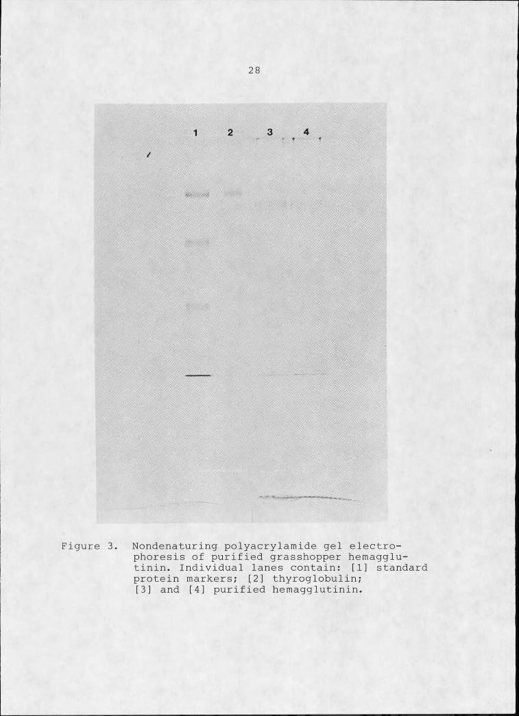
Nondenaturing e lectrophoresis. Electrophoresis of
hemagglutinin under nondenaturing conditions produced a
single protein band of molecular weight near 590,000
(Figure 3). Some diffuse lightly stained areas were
detectable in the lower molecular weight regions of the
gel slab indicating the possible presence of contaminants
or protein fragments dissociated from the high molecular
weight aggregate. Omission of D-galactose from- the
polyacrylamide slab, or prior incubation of the hemagglu
tinin sample i'n 5 mM EDTA resulted in the disappearance of
the 590,000 molecular weight protein band, a result
analogous to the above observation concerning gel filtra
tion in the absence of D-galactose.
Isoelectric focusing. A single broad band in the
slightly acidic pH range resulted upon isoelectric
focusing of purified grasshopper hemagglutinin under
nondenaturing conditions (Figure 6, gel I). Purified,
native hemagglutinin was therefore viewed as being a
homogeneous population of protein of.- nearly identical
ionic character.
28
1 2 3 4♦ f t
Z
Figure 3. Nondenaturing polyacrylamide gel electrophoresis of purified grasshopper hemagglutinin. Individual lanes contain: [1] standardprotein markers; [2] thyrogIobuIin;[3] and [4] purified hemagglutinin.

29
MIGRATION-----------►
1I I I
Figure 4. SDS polyacrylamide gel electrophoresis of whole grasshopper hemolymph and purified hemagglutinin. Lanes I, 2, 3 contain nonreduced protein, and lanes 4, 5, 6 contain reduced protein. Lanes 1,6, molecular weight markers; lanes 2,5 whole grasshopper hemolymph; lanes lanes 3,4, purified hemagglutinin.

30Subunit Structure of Grasshopper Hemagglutinin
SDS electrophoresis. The subunit structure of grass
hopper hemagglutinin was examined by electrophoresis in
SDS polyacrylamide gels. Figure 4 shows that in the
presence of SDS the agglutinin traveled as a single band
of 70,000 molecular weight (MW). Upon reduction with 2-
mercaptoethanol the 70,000 MW band disappeared and two new
bands appeared at 40,000 MW and 28,000 MW. Neither the
reduced nor the non-reduced bands corresponded to any
principal protein bands resulting from the simultaneous
electrophoresis of whole hemolymph. In the case of
nonreduced hemagglutinin some darkly staining material
remained at the top of the separating gel (Figure 4, , lane
3) and was apparently aggregated or precipitated protein
incompletely soIubiIized by the SDS. Samples of whole
hemolymph (lane 2) and the protein mole.cular weight
standards (lane I) behaved similarly.
Electrophoresis in urea. The subunit structure of
grasshopper hemagglutinin was analyzed further by urea
electrophoresis and isoelectric focusing in urea. When
purified hemagglutinin was electrophoresed in polyacryla
mide gels containing 6 M urea, the nonreduced molecule
migrated as a homogeneous ionic species (Figure 5, band
A). When the hemagglutinin was incubated in 2-mercapto-
ethanol prior to electrophoresis, the single band
disappeared and a diffuse area of staining of greater

31
Figure 5. Urea polyacrylamide gel electrophoresis ofgrasshopper hemagglutinin. Band A, nonreduced hemagglutinin; Band B , reduced hemagglutinin.

32
I
10t \
2
pH
Figure 6. Isoelectric focusing of native and urea-denatured grasshopper hemagglutinin. Gel I, native hemagglutinin; gel 2, urea-denatured hemagglutinin.

33mobility, possibly representing several bands, appeared (Figure 5, band B) .
Isoelectric focusing in urea. Isoelectric focusing
of purified hemagglutinin in gels containing 6 M urea
showed that the denatured subunits (70,000 MW by SDS electrophoresis) do exhibit some charge heterogeneity
since several distinct closely spaced bands were present
in the acidic region of the pH gradient (Figure 6, gel 2).
These bands were in a position corresponding to a slightly
more acidic pi than was the band observed for the native
molecule (Figure 6, gel I). '
Amino Acid Composition.
Results of amino acid analysis of grasshopper
h e m a g g l u t i n i n from both Mjl s^n gu j njl p e s and Mjl
differentialis are shown in Table 3. Given the limits of
experimental error in calculating integration values by
the half height method, grasshopper hemagglutinin from M.
sanguinipes and • IVL differentials appear extremely similar
in amino acid content. The protein contained relatively
high amounts of aspartic acid and glutamic acid, and a low
amount of methionine. The presence of a low amount of
cystine present in the molecule was confirmed by showing
the presence of cysteic acid after performic acid
oxidation of grasshopper hemagglutinin (data not shown).
A ninhydrin sensitive peak in the position of glucosamine

34suggested- that grasshopper hemagglutinin contained small
amounts of associated carbohydrate.
Table 2. Amino Acid Composition of Purified Hemagglutinin from Melanoplus sanguinipes and Melanoplus .. differentia I is.
Amino AcidgAA/lOOg protein' residues/70 KdaltonM . sang M. diff M . ,sang ' M. diff
Lysine 4.6 4.6 25 25Histidine 3.0 3.4 16 18Arginine 5.4 6.0 24 27Aspartic Acid 12.2 11.9 74 72Threonine 6.3 6.2 44 43Serine 4.7 4.5 3 8 36Glutamic Acid 14.1 ■ 14.0 76 76Proline 6.3 5.4 45 39Glycine 3.4 4.5 42 55Alanine 5.1 • 5.5 50 54Cystine 4.1 4.1 14 14Valine 5.3 4.9 37 35Methionine 1.8 1.3 10 7Isoleucine■ 4.6 4.4 28 2 7Leucine ' 7.4 7.6 ' 46 47Tyrosine 5.2 5.4 22 23Phenylalanine 4.8 5.0 23 24Tryptophan - - - -Glucosamine 1.7 1.2 7 5
*not determined
^®:£k£hydrate Inhibition of Grasshopper HemagglutininThe carbohydrate binding specificity of grasshopper
hemagglutinin was examined through hemagglutination
inhibition tests. Table 3 lists the minimal inhibitory
concentrations determined for several carbohydrates and
carbohydrate derivatives. Table 3 also includes minimal

35inhibitory concentrations previously determined [23] for
whole grasshopper hemolymph.
The similar broad inhibition pattern obtained for
both whole hemolymph and purified hemagglutinin showed that certain glucosidic carbohydrates and certain galacto-. sidic carbohydrates were both capable of inhibiting
hemagglutination when present at concentrations in the 1-5
mM range. These results suggested that grasshopper
hemagglutinin has broad carbohydrate specificity and was
not limited to interaction with a single structural type
of carbohydrate receptor. The best carbohydrate inhibi
tors appeared to be the alpha-anomers of simple galacto-
sides, however there was no clear preference over several
other galactose or glucose containing oligosaccharides.
E D T A , a divalent metal ion chelator, was among the
strongest inhibitors and was effective at 1.2 mM. . Inhibi
tion of hemagglutination by EDTA was apparently due to the
removal of divalent cations from the hemagglutinin that
were obligatory for the active conformation of the protein.

36
Table 2. Carbohydrate Inhibition of Hemagglutination by Whole Grasshopper Hemolymph and Purified Hemagglutinin.
Minimal Inhibitory Concentration (mM)
Inhibitor*Whole
HemolymphPure
Hemagglutinin
Galactosidic:«<-PNP-galactose 1.5. 0.6eC-Me-galactose 6.2 1.5Stachyose 3.1 2.52-deoxygalactose 6.2 . 2.5Raffinose 6.2 2.5P-Me-gal acto.se 12 . 3.1Fucose 12 , 3.1Galacturonate 6.2 6.2L-Fucose 25. 12.Melibiose 12 . 12.Galactose 2 5 . 12.Galactonic- -lactone 25. 12.Lactulose 25. 2 5 .P-PNP-galactose 12 . 2 5 .
Glucosidic:Palatinose 6.2 3.1Maltotriose ND 6.2Melizitose 12 . 6.2«-PNP-glucose 12 . 12.«-Me-glucose 25 . 25 .Maltose 25 . 25.Sucrose 2 5 . 50 .Trehalose ND >10 0.
Other:L-Rhamnose 12. 6.2L-Sorbose 25 . 12 .L-Arabinose 2 5- 2 5.EDTA 1.5 1.5
* Abbreviations: PNP', para-nitrophenyl; Me, methyl;IlD, not done; EDTA > ethylenediaminetetraacetate.
Data from Hapner, [23] .
37Molecular Stability of Grasshopper Hemagglutinin
Grasshopper hemagglutinin lost no hemagglutination
activity when stored for weeks at -20°C in DPBS that contained 0.2 M D-galactose. Activity was slowly lost
(days) when the purified hemagglutinin was warmed to room
temperature. Dialysis or concentration by membrane
ultrafiltration of solutions of hemagglutinin resulted in
irreversible loss of activity. Hemagglutinating activity
of both purified hemagglutinin and whole hemolymph was
rapidly lost upon incubation at 56°C whereas both retained
full activity after 6 hr at 3 7°C (Figure 7). Purified
hemagglutinin was less stable than that that in whole
hemolymph and lost all activity in one minute. Treatment
of purified hemagglutinin with 0.1% active trypsin at 37°C
resulted in rapid and reproducible disappearance of
activity after four hours incubation as shown in Figure 8.
There appeared to be a lag period during which the protein
retained resistance to trypsin and then suddenly, activity
was destroyed. Control experiments without trypsin
retained hemagglutinin activity throughout the incubation
period. Low concentrations (SmM) of EDTA destroyed all
hemagglutinin activity in both whole grasshopper hemolymph
and purified hemagglutinin.

TITE
R
1 0 2 4 « ^
2 5 6 '
5 6 0C
120 180 2 4 0
TIME , MINUTES
Figure 7. Heat stability of hemagglutinating activity of whole grasshopper hemolymph and purified hemagglutinin.(----, whole hemolymph; ---- , purified hemagglutinin).

TITE
R
1024.
5 1 2 .
2 5 6 -
1 2 8 -
6 4 -
3 2 .
1 6 -
8-
4 -
2 ■
IIIII
IIIIII *
OJVD
1 2 3 4 5 6
T I ME,HOURS
Figure 8. Trypsin stability of hemagglutinating activity of whole grasshopper hemolymph and purified hemagglutinin.(----, whole hemolymph; ---- , purified hemagglutinin).

40Antigenicity of Grasshopper Hemagglutinin
Rabbit antiserum. Small amounts (50-100 ug) of
purified hemagglutinin elicited antibody production in
both rabbits and- mice. Rabbit antiserum reached an immuno
double diffusion titer value of 8-16 at about 15 weeks
after immunization. The rabbit antiserum produced- a
precipitin band against either whole grasshopper hemolymph
or purified grasshopper hemagglutinin when,, subjected to
gel double diffusion (Figure 9). No precipitin band was
formed against affinity adsorbed hemolymph that contained
no hemagglutinin. When double diffusion of purified
grasshopper hemagglutinin versus antiserum was performed
in agarose that contained both 0.1 M D-glucpse and 0.1 M
D-galactose, a double line occurred, whereas when the
sugarrs were absent from the gel only a single band was
produced. Whole grasshopper hemolymph always produced a
single precipitin band regardless of the presence or
absence of sugars. The double band was viewed as possibly
resulting from aggregation anomalies of the hemagglutinin
molecule in the absence of hemolymphatic factors.
Immune murine ascitic fluid. Hyperimmune ascitic
fluid was tapped from immunized mice beginning on about
day .3 8 of the immunization schedule (3 days after the
fifth injection). Subsequent taps were performed every 3
to 5 days following the first tap until the production of
ascites fluid subsided. Typically, about 25 ml of ascites

41
Figure 9. Gel double diffusion of whole grasshopperhemolymph, affinity adsorbed hemolymph, and purified hemagglutinin vs. rabbit antiserum. Wells 2,5, whole hemolymph; wells 3,6, affinity adsorbed hemolymph; wells 1,4, purified hemagglutinin; center well, rabbit antiserum.

42
(+) "«-------- (-)
Figure 10. Immunoelectrophoresis of purified hemagglutinin.

43fluid was collected over a period of about 3 weeks. Mouse
ascitic fluid typically had an immuno double diffusion
titer value of 32.
Immunological Detection of Grasshopper Hemagglutinin
Immunoelectrophoresis. Samples of purified hemagglutinin previously electrophoresed under 'nondenaturing
conditions in agarose gels formed one major precipitin
band when diffused against rabbit antiserum as shown in
Figure 10. A minor band also occurred near the origin.
The presence of two bands indicated that at least two
species of immunoreactive protein (perhaps two aggregate
forms, as seen in gel double diffusion) were present in
the initial purified hemagglutinin sample.
Glucose oxidase immunoenzyme. Nonreduced and reduced
samples of grasshopper hemagglutinin were electrophoresed
on SDS polyacrylamide gel slabs, electroeluted onto
nitrocellulose filters and subjected to immunochemical
staining with glucose oxidase. Preliminary results
indicated that all subunits of grasshopper hemagglutinin
were specifically recognized by the primary (rabbit)
antiserum. This method was extremely sensitive as shown
by its ability to detect as little as 1-2 ng of purified
hemagglutinin that had been previously spotted onto a
strip of nitrocellulose. Controls which gave no staining

were DPBS> DPBS-O.2 M galactose, human serum, 3%
BSA-saline, and standard molecular weight proteins.
44(w/v)

45
DISCUSSION
Previous work in this laboratory established the
presence of hemagglutinating .activity in the hemolymph of
several genera of acrididae (grasshoppers) [22].
Individual grasshoppers representing four Melanoplus spp.
were subsequently shown to contain similar- broad-spectrum
hemolymphatic hemagglutinin [23]. The data presented in
this thesis are concerned with the biochemical nature of
the hemagglutinin from Me l^nop IlU s_ ^angu_in Ipes^ and
Melanoplus differentiaIis. The major conclusion reached
as a result of this research is that a single hemagglu
tinin protein is responsible for all of . the observed
hemolymphatic hemagglutinating activity.
Purification
Affinity chromatography of grasshopper hemolymph on a
matrix of Sepharose-galactose was a reliable one-step method for isolating pure, active hemagglutinin in high
yield. Grasshopper hemagglutinin was estimated to account
for only 0.1% of the total soluble protein in hemolymph
and affinity chromatography provided a means to obtain
highly purified grasshopper hemagglutinin in sufficient
concentration for direct biochemical analyses. Purified
hemagglutinin was also obtained in sufficient amounts to

46elicit specific antibody production in rabbits and mice (see below).
Elution of grasshopper hemagglutinin adsorbed to the affinity matrix was normally performed with 0.2 M galactose in DPBS (Figure I). The released protein had an identical broad-spectrum erythrocyte binding profile as
did the original sample of whole hemolymph from which it
had been prepared (Table I). Only trace amounts of
hemagglutinating activity was present in the adsorbed
hemolymph that had passed through the affinity matrix.
Elution of activity was also performed with 0.2 M sucrose
in DPBS, and the protein eluted in this fashion exhibited
an identical erythrocyte binding . profile as well as
identical physicochemical properties as did the hemagglu
tinin eluted With DPBS-galactose. Elution of adsorbed
protein from the affinity matrix was also performed using
denaturing (SDS) buffers. Although the desorbed protein
in the latter case was inactive (incapable of hemaggluti
nation) , • it showed identical electrophoretic characteris
tics as did hemagglutinin desorbed under nohdenaturIng
conditions.
These findings confirmed that all the hemaggluti-
nating activity was removed from the affinity matrix with
either glucosyIic or galactosylic carbohydrates, and
further that both carbohydrate binding capabilities reside
in the same molecule. Moreover, only hemagglutinin

47protein was retained by the affinity column because the
eluted proteins were in all cases homogeneous and identical, as discussed below.
Typically, 350 ug of hemagglutinin was purified from a 50 ml diluted hemolymph sample (25. ml actual hemolymph)
as determined by protein assay. Since individual
grasshopper specimens generally yielded about 35 ul of
hemolymph, this preparation represented about 700 insects.
Hemagglutinin was therefore a minor component (0.5
ug/insect) of the total hemoIymphatic protein content.
This conclusion was further supported by gel filtration
(Figure 2) and SDS-PAGE (Figure 4) data which showed that
grasshopper hemagglutinin did not correspond to any major
hemolymphatic proteins. It was essential therefore to
elute grasshopper hemagglutinin from the affinity column
as efficiently as possible to obtain samples in high
enough concentrations for biochemical analyses. As shown
in Figure I, some peak trailing occurred when the affinity
column was eluted with 0.2 M galactose. In fact, activity
was detectable in the effluent after 20 ml of desorbant
buffer had passed through the column. Since grasshopper
hemagglutinin was unstable toward ultrafiltration, and
could not be concentrated in that fashion, this problem
was minimized by incorporating a stepwise elution
procedure. After the first one ml fraction was collected,
the column was allowed to incubate several hours (or

48overnight)' in desorbing buffer before the second fraction
was collected. The procedure was repeated several times. Using this technique, essentially all activity was
obtained in the first 3 or 4 fractions. The cause of the
peak trailing is unclear, but it may be. due to high
affinity between the hemagglutinin and the galactose
matrix and restricted accessibility of binding sites.
Molecular Structure
The- native molecular weight of grasshopper hemagglu
tinin was estimated to be about 700,000 by gel filtration
(Figure 2), and 590,000 by electrophoresis under native
conditions (Figure 3). These values are significantly
larger than those obtained for the hemagglutinin, from the
flesh fly, Sarcophaga peregrina, which has a native MW of
190,000 [25]. On the other hand Melanoplus hemagglutinin
is smaller than the hemagglutinin from the cricket,
TeleogrylIus commodus, which has a native molecular weight
estimated to be several million [24].
Retention of the native aggregated structure of
grasshopper hemagglutinin during gel filtration and
nondenaturing PAGE was dependent on the presence of
stabilizing carbohydrate. No activity was detected upon
gel filtration unless galactose (0.1 M) was incorporated
into the filtration buffer. Similarly, no 590,000 MW band
occurred in nondenaturing polyacrylamide gels unless

49
galactose was incorporated into the acrylamide solution prior to polymerization.
The diffuse lightly stained material of lower MW that
resulted during electrophoresis in nondenaturing gels
(Figure 3) probably represented partial dissociation of
the native aggregate. The possibility exists that this material accounted for the difference in native MW values
as determined by gel filtration and electrophoresis,
however no firm conclusion was possible.
Nondenaturing PAGE and gel filtration both suggested
that purified grasshopper hemagglutinin is a homogeneous
moiety, a conclusion that is further supported by
isoelectric focusing data. Isoelectric focusing of
purified g.rasshopper hemagglutinin in native conditions
(Figure 6, gel I) indicated that it is homogeneous with
regard to isoelectric pH. When the native aggregate was
dissociated into 70,000 MW subunits by urea-denaturation,
the subunits focused at a position closer to the acidic
end of the gel (Figure 6, gel 2). These data suggested
that some acidic amino acid side chains were masked in the■
native aggregated structure and become exposed upon urea
denaturation.
Determinations of the subunit structure of grass
hopper hemagglutinin showed that the native aggregate was
composed of 70,000 MW subunits, which, in turn were
comprised of disulfide linked 40,000 MW and 28,000 MW

50polypeptide' chains. The hemagglutinin from Sarcophaga is
comprised.of 30,000 MW and 32,000 MW polypeptides and the
.lighter fragment is probably synthesized by proteolytic
modification of the heavier, perhaps in response to a
wound-related inductive stimulus [53]. Although no
evidence was sought in this research, a similar event
could occur in Melanoplus whereby 28,000 MW polypeptides
may be proteolytic products of 40,000 MW polypeptides.
Comparisons of the molecular structure of the
hemagglutinins from different types of insects reveals
few, if any, similarities. For example, TeleogrylIus
hemagglutinin [24] exclusively employs disulfide bonding
to form the high MW (several million) native structure.
Me lanoplus hemagglutinin utilizes disulfide bonding to
form. 70,000 MW subunits which are then noncovalently
associated to form the native structure. Finally, the
native structure of Sarcophaga hemagglutinin [25] involves
no d i s u l f i d e bonding, rather only n o n c o v a l e n t
associations.Ionic homogeniety of the subunits from grasshopper
hemagglutinin was investigated further by electrophoresis
of nonreduced and reduced subunits in polyacrylamide gels
containing 6 M urea, and by isoelectric focusing of
nonreduced subunits. In urea-electrophoresis, proteins
migrate as a function of their molecular charge and their
molecular size. Denatured subunits (70,000 MW by sodium

51dodecyl sulfate-PAGE) migrated in a homogeneous band when
electrophoresed in urea whereas the 28,000 MW and 40,000
MW subunits migrated as a smear of closely spaced bands of
uncertain number (Figure 5). Isoelectric focusing of
70,000 MW subunits showed that the single band observed on
urea-PAGE was actually comprised of several slightly
different isoforms of the protein (Figure 6, gel 2).
These isoforms were approximately equal in staining
intensity, and their significance with respect to the
structure.of the native hemagglutinin is unknown.
The amino acid analysis of grasshopper hemagglutinin
(Table 2) showed high amounts of acidic and polar amino
acids, and a. low amount of methionine. All of the other
common amino acids were present in moderate amounts. A
preponderance-of acidic amino acids is compatible with
isoelectric focusing data that show grasshopper hemagglu
tinin to be an acidic molecule. Grasshopper hemagglutinin
is apparently a glycoprotein as evidenced by the presence
of glucosamine which accounts for approximately 1-2% of
the total molecular weight of the molecule.
Only slight differences were observed in the amino
acid compositions between hemagglutinin prepared from M.
sanguinipes hemolymph and that from Mjl differentialis.
Experimental limitations are inherent in the analysis of
small quantities of protein, including the requirement for
hand-calculating integration values by the half-height

52method. Therefore, the numerical values in Table 2 are
probably not as significant as shown and, although the
reverse could be true, it is possible that the amino acid
composition of the two' hemagglutinins were even more
similar than Table 2 indicates.
Inhibition of Hemagglutination
As shown in Table 3, purified grasshopper hemagglu
tinin accounted for the complete carbohydrate inhibition
characteristics previously described for whole hemolymph
[23]. Table I shows that passage over the galactose
affinity column removed most hemagglutinating activity
from whole hemolymph and that both whole hemolymph and
purified hemagglutinin have the same broad erythrocyte
agglutination profile. These data, along with physico
chemical data, support the conclusion that all of the
hemagglutinating activity present in the hemolymph of both
M .• sanguinipes and Mjl differentialis is due to a single
hemagglutinin protein.
A specific feature of grasshopper hemagglutinin is
that it exhibits broad specificity by binding both D-
glucosidic and'D-galactosidic structures. This represents
a deviation from most described lectins which generally
exhibit very strict specificity for a single type of
carbohydrate structure. Sarcophaga hemagglutinin is
highly specific for D-galactose and lactose [25], and

53hemagglutinin from Te I e ogr y_l is inhibited by N-
acetylated sugars [24]. The hemagglutinins from Peri-p^aneta am er i. c ana (cockroach) and S ch_i s t oc er c a
gregaria (locust) ■ hemolymph are inhibited by both D-
glucosylic and. D-galactosylic carbohydrates [28], however,
neither of these activities can yet be attributed to a
single molecule as the hemagglutinin(s) have not been
purified.
The strongest inhibitors of grasshopper hemagglutinin
were the alpha anomers of D-galactose. Alpha-PNP-D-
galactose and aIpha-methyI-D-galactose inhibited hemagglu
tination at a concentration 20 times less than did their
glucosidic analogs. D-galactosidic dI- and oligo
saccharides inhibited hemagglutination at concentrations
approximately equal to their glucosidic counterparts.
Although most D-glucosidic disaccharides were inhibitory,
trehalose was not inhibitory at comparable concentrations.
Whether grasshopper hemagglutinin is, in vivo, more
specific for either D-glucosidic structures or D-
galactosidic structures, or whether its actual target is a
different, presently unidentified carbohydrate, remains to
be determined.
Among the strongest inhibitors of hemagglutination
was EDTA. .This was presumably not due to inhibition per
se, but rather to the removal of divalent cations upon
which the native structure of the. hemagglutinin is

54dependent.. The possible involvement of divalent cations
in the native structure of grasshopper hemagglutinin was
also suggested by the fact that a 590,000 MW band did not
occur when an EDTA-treated sample of hemagglutinin was
subjected to electrophoresis. This dissociation was
apparently . nonreversible, since EDTA-inactivated samples
of purified hemagglutinin showed no hemagglutination
activity after incubation with excess divalent cations.
Stability
Purified grasshopper hemagglutinin was stable for
months when stored at -20°C in DPBS that contained 0.2 M
D-galactose. As previously mentioned, galactose appeared
to stabilize the native structure of grasshopper hemagglu
tinin as it was necessary to incorporate galactose into
gel filtration buffers to avoid loss of activity, and into
nondenaturing polyacrylamide electrophoresis gels to
prevent dissociation of the native aggregate.
Hemagglutinating activity of both whole grasshopper
hemolymph and purified hemagglutinin was destroyed by
heating one minute at 56°C, whereas both were stable at
3 7 °C (Figure 7). Whole hemolymph was stable upon
incubation with trypsin, whereas purified hemagglutinin
was reproducibly destroyed after 4 hours (Figure 8).
Sarcophaga hemagglutinin is resistant to heat treatment at
80°C for 5 minutes and to trypsin for a duration of 30

55minutes (37°C). The fact that grasshopper hemagglutinin
was initially stable toward trypsin suggested the presence
of a shielded, trypsin-sensitive peptide bond(s), whose
integrity is obligatory for hemagglutination.
Immunological StudiesSmall amounts of purified grasshopper hemagglutinin
successfully elicited an immunological response in
rabbits. Specific antisera have also been elicited in
rabbits against Sarcophaga hemagglutinin [25] and against
the heteroagglutinins from Leucophaea [31].
The antiserum obtained in this study was specific for
grasshopper hemagglutinin as shown by gel double diffusion
(Figure 9)., Single homogeneous precipitin lines were
observed for both whole grasshopper hemolymph and purified
hemagglutinin, whereas no precipitin was formed against
hemolymph that contained no hemagglutinin. An anomalous
result was obtained when both 0.1 M glucose and 0.1 M
galactose were incorporated into the double diffusion gel
in that for purified hemagglutinin, a second,. lighter
precipitin line was also present closer to the peripheral
(hemagglutinin) well. In the latter case, only the major
line was continuous with the (single) line produced for
whole hemolymph. .A similar observation was seen upon
Immunoelectrophoresis (Figure 10) where a second minor
precipitin arc occurred near the origin when purified

56hemagglutinin was electrophoresed in agarose gels that
contained 0.2 M galactose. The significance of these
minor bands is not understood, however, they -may represent
hemagglutinin molecules that have spontaneously aggregated
into larger, slower migrating molecular forms. In the
case of Immunoelectrophoresis, however, the possibility
exists that aggregation of hemagglutinin occurs in such a
way as to result in the '.formation of aggregates that do
not migrate in the given electrophoretic conditions, but
simply diffuse away from the well during and after
electrophoresis.
Purified hemagglutinin was also antigenic in mice, as
shown by precipitin formation on gel double diffusion
between purified antigen (hemagglutinin) and mouse ascitic
fluid. Preliminary indications suggested that the murine
ascitic fluid had a higher titer value than did rabbit
antiserum. Preliminary data involving the indirect
identification of antigenic subunits with antibody-
conjugated glucose oxidase immunoenzyme suggested that all
structural components of grasshopper hemagglutinin (i.e.
30,000, 40,000 and 70,000 MW) contained antigenic
determinants that are recognized by primary (rabbit)
antiserum.The production of specific antibodies against
purified grasshopper hemagglutinin and the development of
specific associated immunochemical detection procedures is

57the first step in succeeding research, designed to
establish the possible association of grasshopper hemagglutinin with specific grasshopper hemocytes.

58
CONCLUSIONS
The major conclusion to be drawn from data derived
from this research is that one protein (lectin) is the
substance responsible for all hemagglutinating activity
present in the hemolymph of either Melanoplus sanguinipes
or Melanoplus differentiaIis. Grasshopper hemagglutinin
is an ionically homogeneous 600,000-700,000 molecular
weight aggregate of 70,000 M W ■subunits. The subunits are
comprised of 28,000 MW and 40,000 MW polypeptide chains
that are connected by disulfide bonds. The hemagglutinin
is an acidic molecule (estimated pi 5-6), and it contains
a preponderance of acidic and polar amino acids as well as
a small amount of associated carbohydrate. Grasshopper
hemagglutinin exhibits broad-spectrum carbohydrate binding
capability and it is strongly inhibited by both D-
glucosidic and D-galactosidle structures. The strongest
inhibitors are the alpha anomers of D-galactose. Purified
hemagglutinin is stable when stored at - 2 0°C in the
presence of galactose but is destroyed by heating to 56°C
or by exposure to trypsin. Divalent cations are
apparently required for activity and they may be involved
in the maintenance of the aggregated form of the molecule.

59Grasshopper hemagglutinin is antigenic in rabbits and
mice. Initial experimentation utilizing an indirect
immunoenzyme assay indicates that all components of the
hemagglutinin molecule (i.e. 70,000 MW, 40,000 MW and
28,000 MW subunits) • are immunoreactive with, primary (rabbit) antiserum.
Throughout this research, comparative experiments
were performed with hemagglutinin from two species of
Melanoplus. In all cases, essentially identical results
were obtained, and it appears likely that the same
hemagglutinin protein is present throughout M e lanoplus
Whether or not this is the case generally in the
Acr i d i d a e , must be determined through, additional
experimentation.

60
REFERENCES CITED
1. .Hildemann, W.H. (19 74) . Life Sci. 14:605-614.
2. Burnet, F .M . (1968). Nature 218:426-430.3. Salt, G. (1970). The Cellular Defense Reactions of
Insects. Cambridge Univ. Press, New. York and London.
4. Rate I if f e , N.A. and S.J. Gagen. (19 76). J. Inv. Pathol. 28:373-382.
5. Cohen, E . (1974). Ann. N . Y . Acad. Sci. 234:1-412.
6. Yeaton, R.W. (1981). Dev. Comp. Immunol. 5:391-402.
'I. Lackie, A.M. (1980). Parasitol. 80:393-412.
8. McKay, D., C.R. Jenkin and D. Rowley. (19 69). , Aust.J . Exp. Biol. Med. Sci. 47:125-134.
9. Anderson, R.S., and R.A. Good. (19 76). J. Inv. Pathol. 27:57-64.
10. Vasta, G .R . (1982). J. Inv. Pathol. 40:367-377.
11. Tripp, M .R . (1966). J. Inv. Pathol. 8:478-484.
12. Pimental, D., (ed.) (19 81). Handbook of PestManagement in Agriculture. Vol. I, CRC Press, Boca Raton, EL. pp. 3-78.
13. Berry, R.A. (1978). Insects and Mites of Economic Importance in the Northwest. Oregaon State Univ. Press, Corvallis, OR. p. 98.
14. Carter, L.J. (1976). Science 191:836-837.
15. Pimental, D., (ed.) (1981). Handbook of PestManagement in Agriculture. Vol. 2, CRC Press, Boca Raton, EL. pp. 318-356.
16. Nicolson, G .L . (1974). Int. Rev. Cytol. 48:90-190.
Lackie, A.M. (1981). Dev. Comp. Immunol. 5:191-204.17.

61REFERENCES CITED— Continued'
18. Se I a , B.A., H. Lis, N. Sharon and L. Sachs. (1970). J. Membrane Biol. 3:267.
19. Sharonh, N. and H. Lis. (19 7 2) . Science. 177:949-959 .
20. Whitcomb, R.F., M . Shapiro and R.R. Granados. (1974) Physiology of Insecta, (Rockstein, M., ed.) , Vo,I. 5, Academic Press, New York. pp. 448-537.
21. Ratner, S. and S.B'. Vinson. (1983). Amer. Zool. 23:185-194.
22. Jurehka, R., K. Manfredi and K.D. Hapner. (1982). JInsect Physiol. 28(2):177-181.
23. Hapner, K.D. (1983). J. Insect Physiol. 29 (I):101- 106.
24. Hapner, K.D. and M.A. Jermyn. (1981). Insect■ Biochem. 11(3):287-295.
25. Komano, H., D. Mizuno and S,. Natori. (19 8 0) . J.Biol. Chem. 255(7):2919-2924.
26. Scott, M .T. (19 71). Archs. Zoo I. Exp. Gen. 112:73-80 .
27. Scott, M .T . (1972). J . Insect. Pathol. 19:66-71.
28. Lackie, A .M . (1981). J. Insect Physiol. 27:139-143
29. Anderson, (1972) . Infection and Immunity. 5:55-59..
30. Don Ion, W.C. and C.T. Wemyss. (1976),. J. Inv. Pathol. 28:191-194.
31. Amirante, G.A. (1976). Experientia. 32:526-528.
32. Stynen, D., M. Peferoen and A.DeLoof. (19 8 2) . J. Insect Physiol. 28:465-470.
33. Feir7 D., and M.A. Wa I z. (1964). Ann. Entomol,. Soc. Amer. 57:388.
34. Umetsu, K.S., S. Kosaka, S. Kashimura and T. Suzuki. (1979). Tokoru J. Esp. Med. 129:161-167.
35. Yeaton, R.W. (1.9.80). Ph'.D. Diss.,, Univ. of PA., 229 p.
!

62REFERENCES CITED— Continued
37. Komano, H., R. Nozawa, D. Mizuno and S. Na tori.(1983). -J. Biol. Chem . 258 (4) : 21.43-2147 .
38. AmIrante, A.G., F.L. DeBernardi and P.C. Magnetti.(19 76.) . Boll. Zool . 43 : 63-67.
.39. Amirante, G.A. and F.G. Mazzalai. (1978). Dev. Comp Immunol. 2:735-740.
40. Fornstedt, N. and J. Porath. (1975). FEBS Lett. 57:187-191.
41. Bradford, M.M. (1976). Anal. Biochem. 72:248.
42. Laemmli, U.K. and M . Favre. (1973). J. Mol. Biol.80:575-599.
43. Wrigley, 1971. (1971). Methods Enzymol. 22:559-564
44. Spackman, D.H., W.H. Stein and S. Moore. (1958).Anal. Chem. 30:1190.
45. Hirs, C.H.W. (1967). Methods Enzymol. 11:197-199.
46. Vaitukaitus, J.L. (1981). Methods. EnzymoI. 73:46-5 2 .
47. Tung, A .S . (1983). Methods Enzymol. 93:12-23.
48. Ouchterlony, 0. (1948). Ark. Kemi Mineral. Geol.26 B :1-9 .
49. Grabar, P. and C.A. Williams. (1953) . Biochim. Biophys. Acta. 10:193.
50. Towbin, H., T. Staehelin and J. Gordon. (1979).Proc. Nat. Acad. Sci. 46(9) : 4350-4354.
51. Gershoni, J.M. and G.E. Palade. (1983). Anal. Biochem. 131:1-15.
52. Rathlev, .T, J.M. Hocko, G.F. Franks, S.C. Suffin,C.M. O'Donnell and D.D. Porter. (1981). Clin. Chem.27 (9) :1513-1515.
53. Komano, H., D. Mizuno and S. Nator i. (1981). J.Biol. Chem. 256(14):7087-7089.

63
APPENDIX
Amido Black stain procedure:
1. Stain: 0.1% (w/v) amido black in destain3-5 min
2. Destain: methanol/acetic acid/water (90:2:8 byvo I)
3 X 3 min
Indirect immunostaining with glucose oxidase iminunoenzyme:
1. Incubate strip in saline (IOmM tris, 0.9% (w/v)NaCl, pH 7.2
3 7°C I hr
2. Rinse twice with saline r.t. 5 min total
3. Incubate strip in primary antiserum that hasbeen diluted 1:50 with 3% (w/v) BSA in saline (step I)
3 7°C I hr
4. Rinse strip five times with saline r.t. 30 min total
5. Incubate strip in second antibody (i.e. anti-IgG/glucose oxidase conjugate (Img/ml that has been diluted 1:1000 with 3% BSA-saline)
' 3 7°C I hr
6. Rinse strip five times with saline r.t. 30 min total
7. Incubate strip in disclosing solution:-2.5 mM' p-nitro blue tetrazolium (Sigma) -41.7 mM D-glucose -0.326 mM phenazine methosulfate
3 7°C 1-12 hrRinse strip with water and dry between weighted blotter paper or paper towels.
8.

MONTANA STATE UNIVERSITY LIBRARIES
762 100 5551 2
T-N378 St 31cop.2