Helicobacter pylori Associated Dyspepsia in...
26
Chapter 5 Helicobacter pylori—Associated Dyspepsia in Paediatrics Mónica Roxo-Rosa, Mónica Oleastro and Ana Isabel Lopes Additional information is available at the end of the chapter http://dx.doi.org/10.5772/56551 1. Introduction Helicobacter pylori ubiquitously infects the human gastric mucosa since time immemorial, predictably before the man’s diaspora out of East Africa around 58,000 years ago [1]. Colonization may have been somehow beneficial for human carriers, allowing the co- evolution of this gram-negative bacterium and its host over the centuries. Yet, at least nowadays [2], this may not be a peaceful association, with infection almost invariably causing an acute host immune response. However, in a fully adapted manner, H. pylori avoids recognition and, thus, clearance, by the host immune system, with both infection and the consequent gastritis persisting throughout the patients’ life. The clinical outcome of this persistence is dependent on a sophisticated crosstalk between the host and the pathogen. If often asymptomatic, the H. pylori-associated non-ulcer dyspepsia is clearly the strongest aetiological factor for severe gastric diseases that will develop late in adult life in a minority of infected patients, i.e., peptic ulcer disease, both gastric and duodenal ulcers, and gastric cancer, namely, adenocarcinoma and mucosa associated lymphoid tissue (MALT) lymphoma (reviewed in [3]). Peptic ulcer disease rarely occurs soon after H. pylori infection [4-8] that generally starts in childhood; this presumably reflects marked differen‐ ces in the virulence [9-16] and/or in the susceptibility of young patients [17-19]. This chapter, focussing on the paediatric population, seeks to explore: the prevalence of H. pylori infection; the molecular mechanism used by H. pylori during colonization and infection; the role of this bacterium in the development of peptic ulcer-related organic dyspepsia; and the genetic/proteome profile of the H. pylori-strains associated with peptic ulcer disease. © 2013 Roxo-Rosa et al.; licensee InTech. This is a paper distributed under the terms of the Creative Commons Attribution License (http://creativecommons.org/licenses/by/3.0), which permits unrestricted use, distribution, and reproduction in any medium, provided the original work is properly cited.
Transcript of Helicobacter pylori Associated Dyspepsia in...
Chapter 5
Helicobacter pylori—Associated Dyspepsia in Paediatrics
Mónica Roxo-Rosa, Mónica Oleastro andAna Isabel Lopes
Additional information is available at the end of the chapter
http://dx.doi.org/10.5772/56551
1. Introduction
Helicobacter pylori ubiquitously infects the human gastric mucosa since time immemorial,predictably before the man’s diaspora out of East Africa around 58,000 years ago [1].Colonization may have been somehow beneficial for human carriers, allowing the co-evolution of this gram-negative bacterium and its host over the centuries. Yet, at leastnowadays [2], this may not be a peaceful association, with infection almost invariablycausing an acute host immune response. However, in a fully adapted manner, H. pyloriavoids recognition and, thus, clearance, by the host immune system, with both infectionand the consequent gastritis persisting throughout the patients’ life. The clinical outcomeof this persistence is dependent on a sophisticated crosstalk between the host and thepathogen. If often asymptomatic, the H. pylori-associated non-ulcer dyspepsia is clearly thestrongest aetiological factor for severe gastric diseases that will develop late in adult life ina minority of infected patients, i.e., peptic ulcer disease, both gastric and duodenal ulcers,and gastric cancer, namely, adenocarcinoma and mucosa associated lymphoid tissue(MALT) lymphoma (reviewed in [3]). Peptic ulcer disease rarely occurs soon after H. pyloriinfection [4-8] that generally starts in childhood; this presumably reflects marked differen‐ces in the virulence [9-16] and/or in the susceptibility of young patients [17-19].
This chapter, focussing on the paediatric population, seeks to explore: the prevalence of H.pylori infection; the molecular mechanism used by H. pylori during colonization and infection;the role of this bacterium in the development of peptic ulcer-related organic dyspepsia; andthe genetic/proteome profile of the H. pylori-strains associated with peptic ulcer disease.
© 2013 Roxo-Rosa et al.; licensee InTech. This is a paper distributed under the terms of the Creative CommonsAttribution License (http://creativecommons.org/licenses/by/3.0), which permits unrestricted use,distribution, and reproduction in any medium, provided the original work is properly cited.
1.1. Prevalence of infection
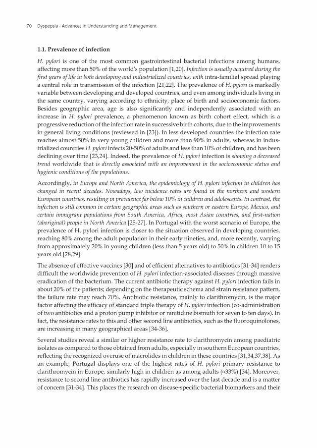
H. pylori is one of the most common gastrointestinal bacterial infections among humans,affecting more than 50% of the world's population [1,20]. Infection is usually acquired during thefirst years of life in both developing and industrialized countries, with intra-familial spread playinga central role in transmission of the infection [21,22]. The prevalence of H. pylori is markedlyvariable between developing and developed countries, and even among individuals living inthe same country, varying according to ethnicity, place of birth and socioeconomic factors.Besides geographic area, age is also significantly and independently associated with anincrease in H. pylori prevalence, a phenomenon known as birth cohort effect, which is aprogressive reduction of the infection rate in successive birth cohorts, due to the improvementsin general living conditions (reviewed in [23]). In less developed countries the infection ratereaches almost 50% in very young children and more than 90% in adults, whereas in indus‐trialized countries H. pylori infects 20-50% of adults and less than 10% of children, and has beendeclining over time [23,24]. Indeed, the prevalence of H. pylori infection is showing a decreasedtrend worldwide that is directly associated with an improvement in the socioeconomic status andhygienic conditions of the populations.
Accordingly, in Europe and North America, the epidemiology of H. pylori infection in children haschanged in recent decades. Nowadays, low incidence rates are found in the northern and westernEuropean countries, resulting in prevalence far below 10% in children and adolescents. In contrast, theinfection is still common in certain geographic areas such as southern or eastern Europe, Mexico, andcertain immigrant populations from South America, Africa, most Asian countries, and first-nation(aboriginal) people in North America [25-27]. In Portugal with the worst scenario of Europe, theprevalence of H. pylori infection is closer to the situation observed in developing countries,reaching 80% among the adult population in their early nineties, and, more recently, varyingfrom approximately 20% in young children (less than 5 years old) to 50% in children 10 to 15years old [28,29].
The absence of effective vaccines [30] and of efficient alternatives to antibiotics [31-34] rendersdifficult the worldwide prevention of H. pylori infection-associated diseases through massiveeradication of the bacterium. The current antibiotic therapy against H. pylori infection fails inabout 20% of the patients; depending on the therapeutic schema and strain resistance pattern,the failure rate may reach 70%. Antibiotic resistance, mainly to clarithromycin, is the majorfactor affecting the efficacy of standard triple therapy of H. pylori infection (co-administrationof two antibiotics and a proton pump inhibitor or ranitidine bismuth for seven to ten days). Infact, the resistance rates to this and other second line antibiotics, such as the fluoroquinolones,are increasing in many geographical areas [34-36].
Several studies reveal a similar or higher resistance rate to clarithromycin among paediatricisolates as compared to those obtained from adults, especially in southern European countries,reflecting the recognized overuse of macrolides in children in these countries [31,34,37,38]. Asan example, Portugal displays one of the highest rates of H. pylori primary resistance toclarithromycin in Europe, similarly high in children as among adults (≈33%) [34]. Moreover,resistance to second line antibiotics has rapidly increased over the last decade and is a matterof concern [31-34]. This places the research on disease-specific bacterial biomarkers and their
Dyspepsia - Advances in Understanding and Management70
associated molecular mechanisms as a top priority to define disease-risk and to target H.pylori eradication in high-risk individuals. Ultimately, it may provide novel bacterial and/orhost’s therapeutic or vaccine targets.
1.2. Molecular mechanisms of H. pylori colonization and infection
1.2.1. Acid resistance and motility
In a fully adapted manner, during colonization and persistence, this neutralophile bacteriumresists gastric acidity mainly through its urease activity. Its urease enzyme, a Ni2+-containingdodecameric protein of approximately 1100 kDa, composed of 12 small subunits, UreA (27kDa), and 12 large subunits, UreB (62 kDa), catalyzes the hydrolysis of urea into ammonia andcarbon dioxide, buffering both the bacteria cytoplasm and periplasm [39]. Accounting for5-10% of the total protein content, urease is one of the most abundant proteins in the H.pylori proteome [16,30]. Probably due to the toxicity of ammonia, urease activity is known tobe dependent on low pH and/or Ni2+ concentration conditions [39,40], being essential forbacteria survival only under acidic conditions. In the early stages of colonization, H. pyloriseeks parts of the stomach with higher pH, such as the antrum (the distal part of the stomach).Indeed, this bacterium uses the pH gradient as chemotactic signal to achieve regions of neutralpH, since its spatial orientation is lost in the absence of the mucus pH gradient [41]. Thus, theacid-producing parietal cells may protect the corpus region from initial invasion.
Efficient colonization of the gastric niche by H. pylori is also dependent on how fast it escapesfrom the lumen of the stomach and reaches the mucus layer, avoiding elimination by gastricperistalsis [42]. Its helical shape and the two to six polar, sheathed flagella provide swimmingabilities. According to a longstanding theory [43], the helical shape allows H. pylori to have acorkscrew motion which, although not being essential for motility, enhances its ability to swimthrough the viscous mucus layer. The machinery that gives rise to the spiral shape of thisbacterium remains largely unknown, but seems dependent on the coordinated action ofmultiple proteins in a shape-generating pathway that leads to the relaxation of the peptido‐glycan crosslinking [44]. Flagella are, however, essential for H. pylori motility. Indeed, aflagel‐lated strains (obtained by elimination of both flaA and flaB, genes encoding the two majorcomponents of flagellar filament, flagellins A and B) [45], as well as strains presenting non-functional flagella (in knockout motB models lacking the gene that encodes the MotB flagellarmotor protein), are non-motile [42]. Such mutants are able to establish only transient coloni‐zation in animal models [42,46]. Moreover, lower-motility strains are long known to inducein vitro reduced inflammation levels, when compared to higher motility strains [47]. Once inthe mucus layer of the stomach, H. pylori resides here thereafter, either freely swimming [43]or attached to host’s extracellular mucins [41], getting closer to the host’s gastric epithelialsurface whenever necessary. Occasionally H. pylori also can be internalized, entering the gastricepithelial cells [48]. Invasion beyond the epithelial layer is, however, is a rare event.
Helicobacter pylori—Associated Dyspepsia in Paediatricshttp://dx.doi.org/10.5772/56551
71
1.2.2. Bacterial Adherence
In the human stomach, the vast majority of H. pylori cells exist in their motile form within themucus layer lining; only a small portion (≈30%) are adherent to the surfaces of epithelial cells[41]. Nevertheless, adherence to the gastric epithelium is important for the ability of H. pylorito cause disease because this intimate attachment facilitates: 1) persistence, by preventing thebacteria from being eliminated from the stomach through mucus turnover and gastricperistalsis, and also by enabling the bacteria to replicate; 2) evasion from the human immunesystem; 3) efficient delivery of the bacterial toxic proteins; and 4) acquiring nutrients releasedfrom the damaged host cells.
H. pylori expresses a multitude of different adhesins. Best characterized is the blood groupantigen-binding adhesin (BabA), a ligand of ABO (of the blood group system) Lewis b (Leb)antigens [49]. Sequence analyses reveals the existence of two allelic variants of babA, the babA1and babA2 alleles, which are identical except for a 10 base pair insertion that results in atranslational initiation codon present in babA2 but absent in babA1, and of a highly homologousgene, babB. Of these, only babA2 allele encodes a functional Leb adhesin [49]. BabA is not likelyto be essential for the colonization; BabA-expressing strains are no different in this stepcompared to BabA-non-expressing strains [50]. BabA, being an adhesin, however likely playsan important role in the induction of host inflammatory response. Indeed, babA2 allele isclinically important, namely in a vacA/cagA-positive genetic background (two additionalimportant virulence factors discussed in section 1.2.3. of this chapter), which is associated withpeptic ulcer disease and gastric cancer [51]. BabA-expressing strains induces change in theglycosylation pattern of the gastric mucosa of humans and animal models [50].
The second best characterized H. pylori adhesin is the sialic acid binding adhesin (SabA) whichmediates attachment to the inflammation-associated (sialylated Lewisx and Lewisa) antigens[52]. In fact, gastric tissue inflammation and malignant transformation promote synthesis ofsialylated glycoconjugates, which are rare in healthy human stomachs [52, 53]. Accordingly,high levels of sialylated glycoconjugates are found in H. pylori infected persons; these decreaseafter eradication of the infection and resolution of the gastritis [54]. H. pylori can agglutinateerythrocytes and neutrophils in vitro. The SabA adhesin is the hemagglutinin of H. pylori andallows bacterial adherence to blood cells; this may result in systemic dissemination of thepathogen [55]. Moreover, the binding of SabA to sialic acid carries neutrophil receptors,essential for the nonopsonic activation of human neutrophils [56]. Neutrophils play a majorrole in the epithelium injury, since these cells have direct toxic effects on the epithelial cells,through the induction of an oxidative burst, with the release of reactive oxygen and nitrogenspecies. Thus, the neutrophil activating capacity of SabA makes this protein an additionalvirulence factor that is important in the pathogenesis of H. pylori infection.
The outer membrane inflammatory protein (OipA), a member of the Hop family of proteins,is another H. pylori membrane protein with an active role in bacterial adherence, for which nohost receptors are known. Its encoding gene (oipA) is present in all H. pylori strains but it isonly expressed in those presenting the oipA “on” (i.e., functional) genotype. This is regulatedby slipped-strand mispairing, depending on the number of CT repeats in the 5’ region of thegene [57]. OipA expression by H. pylori is associated with high bacterial densities, severe
Dyspepsia - Advances in Understanding and Management72
neutrophil infiltration and, ultimately, with peptic ulceration and gastric cancer [58,59].Supporting the later association, the inactivation of oipA results in a reduced nuclear translo‐cation of β-catenin, a known factor involved in the transcriptional up-regulation of genesimplicated in carcinogenesis [59].
1.2.3. Delivery and virulence factors
After adherence, H. pylori delivers its virulence factors into the cytoplasm of the host’s cells byusing a type IV secretion system (T4SS) and/or outer membrane vesicles. The genes encodingthe components of the T4SS, which is a syringe-like pilus protruding from the bacterial surfaceused to inject virulence factors in host target cells’ cytoplasm, are located in the cag pathoge‐nicity island (PAI) [60]. This is an approximately 40 kpb chromosomal insertion that is thoughtto have been incorporated into the H. pylori genome by horizontal transfer from an unknownsource [61]. As a result, strains are heterogenic regarding the presence of this chromosomalregion, varying between those that contain the intact cag PAI to those that completely lack it.For those lacking an intact T4SS, the delivery of their virulence factors is totally dependent onthe secretion of outer membrane vesicles with a still poorly known content; these are endocy‐tosed by the host epithelial cells (reviewed in [62]).
Encoded by cytotoxin associated gene A, one of the 32 genes of the cag PAI region, CagA isperhaps the most extensively studied translocated protein, the only known effector proteininjected by the T4SS. Once injected into cytoplasm of target cells, CagA interferes with severalhost cell signalling cascades, ultimately inducing abnormal proliferation, cytoskeletonrearrangements and inflammation through the release of cytokines, such as interleukin-1β(IL-1β), IL-8 and tumour necrosis factor-α (TNF-α) (reviewed in [63]). There are two types ofclinical isolates regarding CagA: those producing this protein (cagA-positive H. pylori strains),and the CagA-nonproducing strains (cagA-negative H. pylori strains). The cagA-positive strainsare considerably more virulent [60]. In Western countries, individuals carriers of cagA-positivestrains are thus at higher risk of peptic ulcer disease and/or gastric cancer [64,65], leading tothe classification of CagA as a bacterium-derived oncogenic protein [66]. In a not fullyundisclosed manner, the virulence of the cagA-positive strains is associated with the numberand the type of the phosphorylation motifs of the C-terminal variable region of the protein. Ofthese motifs, defined as EPIYA (Glu-Pro-Ile-Tyr-Ala) A, B, C and D according to differentflanking amino acids, CagA protein nearly always possesses EIPYA-A and B segments,followed by none, one, two or three C segments in Western-strains or a D segment in strainsof East Asian countries. The East Asian-type of CagA (ABD) is known to be more carcinogenicthan the Western-type; within the latter, the variants possessing multiple EPIYA-C motifs(ABCC or ABCCC) are more virulent compared with those with a single segment (ABC)(reviewed in [63]). Moreover, the association of CagA expression levels with polymorphismsin the cagA promoter region may further contribute to differences in virulence among cagA-positive strains and different disease-associated risks [67]. The virulence of the strain must alsobe dependent on additional bacterial factors, since in East Asia most strains are cagA-positiveirrespective of the patient disease.
Helicobacter pylori—Associated Dyspepsia in Paediatricshttp://dx.doi.org/10.5772/56551
73
The vacuolating toxin (VacA), another important virulence factor (reviewed in [68]), issynthesized as a pro-toxin of ≈140 kDa, which contains a N-terminal signal sequence, apassenger domain and C-terminal autotransporter domain. The passenger toxin domain (≈88kDa) is cleaved and processed at some point during its secretion into the extracellular milieuthrough the autotransporter that functions as a type V secretion system. This 88 kDa toxin isfurther proteolytically cleaved, creating a N-terminal fragment of ≈33 kDa (p33) and a C-terminal fragment of ≈55 kDa (p55) that remain non-covalently associated. Required for thecytotoxic activity, the p33 subunit is postulated to be involved in the formation of anionicmembrane channels, while p55 subunit seems to mediate VacA binding to host cells (reviewedin [68]). These functions are, however, highly dependent of the tridimensional structure ofboth subunits [69,70]. Once secreted by the bacterial cells, VacA triggers various responses inthe host, resulting in the cellular vacuolation, pore formation in the cell membrane, disruptionof endosomal ⁄ lysosomal structure and function, apoptosis by toxin trafficking to mitochon‐dria, and immunomodulation [71-73]. Virtually all H. pylori strains have a functional VacA.However, the amount of toxin produced is related to the allelic variation of the encoding gene,especially in its signal and middle regions (reviewed in [74]). Therefore, an association betweenparticular vacA allele types and peptic ulcer disease has been reported worldwide..
The ancient association between H. pylori and the modern humans [1,20] has determined theabnormal high diversity in both their genetic background and the virulence observed amongstrains. This generates complex scenario that creates difficulty in understanding the contribu‐tion of each individual factor. Nevertheless, H. pylori strains presenting the association of thesetwo virulence factors, i.e., cagA-positive and vacA-toxigenic alleles, are considered to be morevirulent and thus more associated with severe organic dyspepsia inducers of a high productionof proinflammatory cytokines in the gastric mucosa and thus more severe non-ulcer dyspepsia.Even so, these virulence factors do not appear to determine the overall pattern of gastro-duodenal disease and a complex interplay between host bacterial factors and environmentseems to be involved in the development of gastric pathology [75].
1.2.4. Evasion
Upon colonization, H. pylori-infected patients experience a strong and complex immuneresponse in the gastric mucosa, both at the humoral and cellular levels; despite this response,nevertheless the infection fails to clear. Therefore, in the absence of effective treatment,infection becomes chronic, persists, and contributes to the immunopathology. The persistenceof infection throughout the life of its host is guaranteed by a set of molecular mechanisms usedby H. pylori to constantly evade the host immune response (reviewed in [76]). Bacterial mimicryand genetic diversity play a central role in such successful strategies.
Mimicking the cell surfaces of the host, the lipopolysaccharide of the H. pylori cell wall isrelatively anergic compared to other gram-negative bacteria. In fact, the variable part of theO-antigen chain of H. pylori lipopolysaccharide is composed of host-related Lewis antigens,making it unrecognizable to the host immune system [77]. Therefore, this pathogen not onlybinds to human Lewis antigens through BabA and SabA, but it also expresses Lewis-likeantigens facilitating the escape from the host immune system. Another ingenious camouflage
Dyspepsia - Advances in Understanding and Management74
used by H. pylori is the expression of proteins at its surface, which specifically bind to host-secreted proteins, e.g., bacterial plasminogen-binding proteins (PgbA and PgbB). This allowsthe bacterium to be coated with host proteins [78]. H. pylori also avoids immune recognitionthrough the in vitro and in vivo impairment of the expression of host’s specific heat shockproteins, thus, inactivating both the innate and adaptive immune response [79].
Allelic diversification of its virulence factors encoding genes allows H. pylori to occupydifferent microenvironments within the human stomach and to adapt to the varying conditionsin the niche over time. This is even more efficient, considering that the expression of differentvariants of those genes may switch through mechanisms of phase variation. Indeed, severalin vitro studies have demonstrated that H. pylori Lewis-like antigens can undergo phasevariation (reviewed in [76]). Moreover, in animal models, persistent infection leads to the lossof expression of babA. This occurs either by phase variation switching between an “on” andan “off” status in a manner similar to that described for oipA (see section 1.2.2 of this chapter),or by nonreciprocal gene conversion of babA to babB [80].
The existence of a small subpopulation of H. pylori within gastric epithelial cells (as brieflydiscussed in section 1.2.1 of this chapter) may represent a sanctuary site that protects bacteriaagainst immune clearance [48].
1.3. Peptic Ulcer — Related organic dyspepsia in paediatrics, a rare event
Although rarely associated with severe forms of organic dyspepsia (namely peptic ulcerdisease) in the paediatric age group, H. pylori is clearly linked with acute gastric inflammationin childhood and occurs frequently in children with dyspepsia [81]. This important associationgains relevance when considering that: a) gastric colonization by H. pylori occurring inchildhood and the consequent inflammation continues for life if left untreated; and b) thislifelong persistence of inflammation after decades of infection is the main etiology for pepticulceration and/or cancer in adulthood. Moreover, some studies suggest a possible impact ofH. pylori-associated dyspepsia on anthropometry, as children with dyspepsia and H. pyloriinfection are shorter and lighter than children with similar symptoms but no infection [82,83].
Evidence for the importance of H. pylori infection as a factor for dyspepsia in childhood comesfrom H. pylori eradication resulting in a significant long-term improvement of dyspepticsymptoms [84]. The symptoms of H. pylori-associated paediatric dyspepsia however do notdiffer from those of non-infected dyspeptic children [85], raising questions about whichapproach should be adopted in children with dyspepsia, in terms of H. pylori testing. Accordingto current guidelines for the management of H. pylori infection at pediatric age [86,87], theprimary goal of diagnostic interventions should be to determine the cause of the presentinggastrointestinal symptoms (“scope and treat” strategy) and not just the presence of H. pyloriinfection (“test and treat” strategy). Indeed, recurrent abdominal pain is not an indication fora “test and treat” strategy concerning H. pylori infection in children, as evidence regarding theassociation with H. pylori infection has been so far inconclusive (even in the presence of pepticulcer). Indeed, several studies using different noninvasive tests for H pylori infection comparedthe prevalence of positive results in children with recurrent abdominal pain and controls andfound no significant difference in infection rates between cases and controls [88,89]. On the
Helicobacter pylori—Associated Dyspepsia in Paediatricshttp://dx.doi.org/10.5772/56551
75
other hand, pediatric studies are limited by the lack of a clear definition for recurrent abdomi‐nal pain or by the use of nonspecific criteria for the diagnosis of chronic abdominal pain [90].Nevertheless, in patients with persistent abdominal pain (after exclusion of other causes, suchas lactose intolerance, giardiasis, celiac disease, inflammatory bowel disease, among others) and/or severe upper abdominal symptoms (namely suggesting peptic ulcer disease, such asnocturnal pain), upper endoscopy with biopsy should be performed (diagnostic investigationof choice). Furthermore, testing for H. pylori in children/adolescents should be considered ifthere is a family history of gastric cancer and in children/adolescents with refractory irondeficiency anemia, when no other cause is found. In these settings, when upper endoscopy isperformed, the presence of H. pylori should be systematically sought through histologicalexamination and, whenever feasible, culture and antibiotic susceptibility testing; treatmentshould be offered in the presence of H. pylori positivity. Population screening for H. pylori inasymptomatic children to prevent gastric cancer is not warranted. Although 13C-urea breathtesting is a validated noninvasive diagnostic test for H. pylori infection in children, a “test andtreat” strategy including this tool should not be indiscriminately adopted in clinical practiceat this age group, considering the fact that this test merely identifies H. pylori presence but notnecessarily the causality of symptoms. Noninvasive tests for H.pylori include different methodsfor the detection of bacterial antigens in stool, detection of antibodies (IgG, IgA) against H.pylori in serum, urine, and oral samples, and the 13C-UBT. In the paediatric group, both fecalantigens determination and respiratory test are reliable to determine whether H pylori has beeneradicated or not after antibiotic treatment, while tests based on the detection of antibodiesagainst H pylori are considered not reliable for use in the clinical setting, but may be useful inepidemiological studies [91].
Therefore, H. pylori infection in childhood differs from adults not only in terms of the prevalence of theinfection and a higher rate of antibiotic resistance, but also with respect to the complication rate, age-specific problems with diagnostic tests and drugs, and the near-absence of gastric malignancies [92].Nevertheless, H. pylori-associated peptic ulcer disease may also occur shortly after infectionin childhood [4-8]. This rare event may be due to more virulent strains [9-16], and/or morepredisposed subjects [17-19]. The two forms of H. pylori-associated peptic ulcers, i.e., gastriculcer and duodenal ulcers, are divergent in prevalence and physiopathology, but both causeconsiderable patients’ morbidity entailing high annual costs of treatment [93].
1.3.1. Prevalence of H. pylori-associated gastric and duodenal ulcers in childhood
In general, about 10-15% of the H. pylori infected patients suffer from duodenal ulcer disease and 2-5%with gastric ulcer and/or gastric cancer late in the adulthood [3]. Population-based studies in patientswith organic dyspepsia suggest that peptic ulcer disease related to H. pylori infection is decreasing inprevalence in Western countries, along with a decrease in the prevalence of infection [94]. The formershould be a direct consequence of the second; with improved living standards, cohorts of children becameprogressively less likely to acquire the organism and thus suffer from H. pylori-associated diseases.Nevertheless, specific populations such as immigrants and rural communities may have a highprevalence of infection and peptic ulcer disease; these individuals should be separately reviewed even inareas where the general prevalence of H. pylori infection has declined below 15% [95]. Despite changing
Dyspepsia - Advances in Understanding and Management76
prevalence trends, H. pylori-induced gastritis causing mucosal ulceration either in the stomach(gastric ulcer) or the proximal duodenum (duodenal ulcer) is a relatively uncommon event inchildren, compared with adults [4-8]. In fact, during childhood, H. pylori is associated withpredominant antral gastritis or with pangastritis [96]. In children, few studies have yet investigatedthe actual trend of H. pylori prevalence in peptic ulcer disease [5-8] and the available data are moredifficult to interpret, considering that the rates of peptic ulcer diagnosis depend also on theclinical setting (endoscopy versus outpatient clinic or hospital admissions) [97,98]. Forexample, in Italy the detection rate of ulcer disease was 7.8% out of an average of 180 paediatricgastrointestinal endoscopies performed each year [99]. Similarly, a retrospective review (from1998 to 2006) showed that 43 (6.9%) out of 619 Chinese children who underwent upperendoscopy for investigation of upper gastrointestinal symptoms had peptic ulcer [7]; andanother retrospective study (from 2003 to 2006) have also reported a high incidence of pepticulcer (6.8%) in Israeli children submitted to upper endoscopy [100]. In Canada, however, theapproximate incidence of peptic ulcer was 1 case per 2,500 hospital admissions [92]. In a recentlarge European multicenter study, including 1233 symptomatic children with H. pyloriinfection, peptic ulcer disease was diagnosed in less than 5% of children younger than 12 yearsof age and in ≈10% of teenagers [8]. Interestingly, other studies indicate a higher associationof H. pylori with peptic ulcer in adolescents than in younger children [100,101]. But, theprevalence of H. pylori-positive ulcers in children also differs between countries and this isnot completely explained by the prevalence of the infection in the population studied. This iseasily demonstrated from data collected from January 2001 to December 2002 on 518 childrenfrom the paediatric European register for treatment of H. pylori [5]. At endoscopy, 454 of thosepatients had H. pylori-associated gastritis and 64 had a peptic ulcer (12.3%). This series alsoincluded children from Russia, who had a significantly higher prevalence of peptic ulcer (35%)compared to that of the remainder of European children (6.7%) [6]. In another report, school-aged children with chronic abdominal complaints living in the rural area of Russia had a highprevalence rate of H. pylori infection (80%) and also of peptic ulcer disease (24%) [102].
In adults, the prevalence of H. pylori infection is higher than 95% in duodenal ulcer cases andaround 60% to 80% in gastric ulcer cases [103]. This scenario is similar in children i.e. when H.pylori-associated ulcers occur in children, duodenal ulceration is much more frequentlyidentified than gastric ulcers [104]. In fact, pooled analysis of early reports (from 1983 to 1994)has demonstrated that the prevalence of H. pylori in children with duodenal ulcer wasrelatively higher (ranging from 33% to 100%, with a median value of 92%), compared withchildren with gastric ulcer (ranging from 11% to 75%, with a median value of 25%) [104]. Amore recent retrospective study (from 1995 to 2001) from Japan confirmed a very highprevalence of H. pylori in antral gastritis and duodenal ulcer (98.5% and 83%, respectively),also identifying H. pylori as a risk factor for the development of gastric ulcer although with alower prevalence of infection (less than 50%) [101]. Finally, in a Chinese study, it was reportedthat among 43 Chinese children suffering with peptic ulcer disease, 37 had duodenal ulcer, ofwhich 21 were H. pylori positive, while only six had gastric ulcer, of which only two werepositive for the infection [7]. In summary, H. pylori infection is much more associated toduodenal ulcer than to gastric ulcer, in both children and adults.
Helicobacter pylori—Associated Dyspepsia in Paediatricshttp://dx.doi.org/10.5772/56551
77
The causative role of H. pylori in gastric ulcers in children and adolescents is, therefore, lesscertain when compared to adults, possibly reflecting the fact that a large proportion of gastriculcers are secondary in nature in children. Characteristically, in children younger than 10 yearsof age, peptic ulcers are usually due to noxious agents (such as corticosteroids and non-steroidal anti-inflammatory drugs (NSAIDs)) or occur after major stresses (such as burns,trauma, and systemic illness). In these settings, upper gastrointestinal tract haemorrhage,vomiting and perforation are frequent presenting features. The ulcers tend not to recur afterhealing. In older children and adolescents, the clinical presentation and natural history ofpeptic ulcers are similar to that observed in adults, presenting as epigastric and nocturnalabdominal pain and being usually associated with H. pylori infection [8,100,101]. In this setting,even though the acute ulcer is likely to heal, the natural history is for ulcer recurrence.Moreover, the complication rate of peptic ulceration is important. The estimated incidence ofpeptic ulcer bleeding in the US paediatric population also has ranged from 0.5 to 4.4/100,000individuals in 2008 [105].
These and other differences explain why some of the recommendations for adults may notapply in children [87,96]. Few randomized, placebo-controlled treatment trials are availablein children for the different outcomes (gastritis or peptic ulcer), and often consist of only smallnumbers of cases [86,106]. Clearly in children as in adults, successful eradication of H. pylorimarkedly reduces the risk of ulcer recurrence [107-110]. Thus, there is general consensusworldwide to treat H. pylori infection when there is endoscopic evidence of peptic ulceration.Triple therapy is the treatment of choice in children for endoscopically proven duodenal ulcerand histologically proven H. pylori antral gastritis [91, 96, 111, 112].
1.3.2. Differences in the physiopathology of H. pylori-associated gastric and duodenal ulcers
Peptic ulceration is a multifactorial disease ultimately explained by disequilibrium betweenaggressive injurious factors and defensive gastroduodenal mucosa-protective factors, whichraises the vulnerability of this mucosa to luminal secretions. H. pylori infection is consideredthe major causative factor for peptic ulceration. Nevertheless, there are other injuriousmechanisms jeopardizing the mucosal integrity: some viral infections (e.g. cytomegalovirusand herpes simplex); drug-induced injury, particularly acetylsalicylic acid, NSAIDs andchemotherapy; vascular disorders interfering with perfusion; major stresses; and syndromesin which a marked overproduction of gastric acid occurs, as is the case of the Zollinger-Ellisonsyndrome and, more commonly in children, antral G cell hyperplasia (also referred to aspseudo-Zollinger Ellison syndrome) [113].
Although differing in their pathogenesis, both H. pylori-associated duodenal ulcers and gastriculcers are intimately related to changes in the acid production by the gastric mucosa [113,114]. Indeed, H. pylori infection can result in increased, decreased or no overall change in thelevel of gastric acid secretion. Duodenal ulcers arise on a background of H. pylori–inducedantral-predominant gastritis with sparing of the oxyntic mucosa, resulting in hypergastrine‐mia and consequent high levels of acid production from the healthy gastric corpus followingmeal or hormonal stimulation. In response to the excessive acid secretion, the duodenumdevelops gastric metaplasia. This, unlike the normal duodenal mucosa, can be colonized by
Dyspepsia - Advances in Understanding and Management78
H. pylori with consequent inflammation and ulceration [113]. Eradication of the H. pyloriinfection corrects the hypergastrinemia and decreases the basal acid secretory rate, heals anypeptic ulcer and ameliorates any symptoms of gastroesophageal reflux [115]. Conversely,gastric ulcers are associated with H. pylori–induced pan- or corpus-predominant gastritis,resulting in multifocal atrophy of acid-secreting mucosa and reduced acid secretion. Theseulcers usually arise at the junction of the antral and corpus mucosa, an area of intense inflam‐mation [113]. Thus, the non-acidophilic nature of H. pylori (see section 1.2.1) explains how thosewith low acid secretory capacity are more susceptible to spread of infection through the corpusmucosa and to gastric ulceration. With their somewhat common pathobiology, gastric ulcerdisease precedes the development of gastric cancer [93,116,117]. Gastric and duodenal ulcershave marked differences in their basis, placing them on opposite ends of disease spectrum. H.pylori-induced duodenal ulcer conveys a lower risk of developing a gastric cancer [117]. But,what makes it possible for H. pylori to be involved in both ends of disease spectrum? Althoughthe mechanisms are unclear, the infecting strain itself may play a crucial role on the divergingpoint of this disease spectrum [16,93]. Moreover, the similarity between the phenotype ofgastric ulcer and gastric cancer raises questions about the carcinogenic potential of theassociated H. pylori strains [93]. Certainly the etiology for H. pylori-associated peptic ulcer inadults depends on the complex interplay of gastritis phenotype and of progressive physio‐logical gastro-duodenal alterations through childhood until adulthood, a result of environ‐mental factors, bacterial virulence factors and host genetic background.
Despite epidemiological evidence that infection during childhood is seldom associated withpeptic ulceration or gastric atrophy, the mechanisms underlying differences in histopathologyand clinical expression of H. pylori infection when compared to the adult, are still poorlyidentified. Theoretically, such differences might be explained by qualitative and/or quantita‐tive differences in induced immune response, possibly age-related. Indeed, adults exhibit apredominantly neutrophil infiltrate, whereas H. pylori-associated gastritis in children isusually mild and superficial with a predominantly mononuclear infiltrate, a paucity ofneutrophils and a higher degree of lymphoid follicular hyperplasia [118]. Therefore, differentimmunopathology and different patterns of cytokine expression would be anticipated forchildren when compared to adults [18]. There may be differences in adaptive component ofgastric mucosa immune response in children compared to the adult host; a clear Th1 responsehas not always been demonstrated for young patients. The lower gastritis scores in childrenmay also be a reflection of such a skewed Th1/Th2 balance, which may result in their lowerrisk for developing ulcer disease [18, 19]. These findings could indicate that the host humoraland cellular responses differ depending on the age at which the gastric infection is firstacquired and might explain the varying rates of disease outcomes that are evident in differentparts of the world. Nevertheless, higher anti-H. pylori IgG antibody titres occur in paediatricpatients with duodenal ulcer compared to those without ulceration, suggesting that localhumoral immune responses contribute to the development of peptic ulceration in these youngpatients [102,119,120]. This is not surprising, given the fact that the more severe inflammation,the greater the chance of ulcer formation [121] with increased IgG production leading tomucosal damage similar to an Arthus reaction [2].
Helicobacter pylori—Associated Dyspepsia in Paediatricshttp://dx.doi.org/10.5772/56551
79
1.3.3. Endoscopic features
Endoscopy is the only method to accurately diagnose peptic ulceration in children [87,122]. Anodular mucosa in the gastric antrum or duodenal bulb and/or gastric or duodenal erosionsor ulcerations are specific (but not sensitive) features, suggesting active H. pylori infection. Forthose with suspected infection, biopsies should be obtained for histopathology, as well ascomplementary tests for detection of H. pylori including rapid urease test, histopathology withGiemsa stain and, if available, culture. The rationale for the recommendation to perform morethan one diagnostic test is based on their sensitivity results in children, which range from 66%to 100% for histology and from 75% to 100% for rapid urease tests [91]. In all paediatric agegroups, for patients receiving therapy with a proton pump inhibitor, biopsies should beperformed on the body and cardia (and, possibly, transition zones) of the stomach as well asfrom the antrum to reduce the chances of false-negative results. Follow-up endoscopy is rarelynecessary, except in the setting of peptic ulceration associated with complications (such ashaemorrhage or perforation).
1.3.4. Host susceptibility
The multifactorial nature of peptic ulcer disease reflects its dependence on the patients’ geneticsusceptibility and habits (alcohol and/or non-steroid anti-inflammatory drug consumption,diet, smoking and stress) [20]. Paediatric peptic ulcer disease is significantly more frequent inboys than in girls (63.6% versus 36.4%, p<0.025) [32]. Although female hormones may have aprotective role against developing peptic ulcerations [123], the true nature of this susceptibilityof the male gender remains unclear.
Mucins, glycoproteins secreted by the gastric mucosa, form a gel layer that is essential tomaintain a stable neutral pH adjacent to epithelium. This mucus barrier affords protectionfrom attack by acid-pepsin and other luminal noxious agents [124]. H. pylori has a complexrelationship with different gastric mucins’ subtypes. Infected children for example have adecreased mucin in their gastric mucosa presumably weakening this important defense barrier[125]. The highly diverse carbohydrate structure of the gastric mucins, functioning as bindingsites for H. pylori, should also play a role in the outcome of infection, with genetic and epigeneticchanges in the mucin molecules influencing the susceptibility of the patient for H. pylori-associated peptic ulcer disease. Recently, it was shown that H. pylori-infected childrenpresented a normal pattern of expression and glycosylation of mucin 5AC (MUC5AC) in thesurface mucous cells, and MUC6 in the gland mucous cells, contrasting with the aberrantexpression of MUC6 and MUC2 found in infected adults. Additionally, it was shown that thepattern of Lewis blood group antigens in the surface epithelium of children was significantlycorrelated with H. pylori load, however no correlation with gastritis, nodularity, and gastric orduodenal ulcer was found [17].
In children and teenagers, as in adults, the severity of antral inflammation strongly correlateswith the risk of duodenal ulcer disease. Among the host factors, polymorphisms in cytokinesencoding genes, or in their promoters, that affect cytokine transcription, are good risk candi‐dates. Indeed, polymorphisms in the IL-1 gene cluster play an important role in modulatingthe risk for H. pylori-induced hypochlorhydria and, thus, for gastric ulceration and cancer. The
Dyspepsia - Advances in Understanding and Management80
IL-1 cluster, located on chromosome 2q12-22 region, includes the genes IL-1A, IL-1B andIL-1RN that code for the proinflammatory cytokines IL-1α, IL-1β and their endogenousreceptor antagonist IL-1RA, respectively. The less common alleles of IL-1B, i.e., IL-1B-31C andIL-1B-511T (representing, respectively, T-C and C-T transitions at positions 31 and 511 of theIL-1B promoter) are associated with a higher risk of hypochlorhydria [116]. This associationcan be explained considering that such polymorphisms lead to increased IL-1β expression/secretion that, upon H. pylori-infection, amplifies the host inflammatory response. Also the lesscommon allele of IL-1RA, i.e., the IL-1RN*2 (representing one of the five known 86 base pairtandem repeat polymorphisms in intron 2) is associated with a higher risk of gastric cancer inadults [116]. The risk is potentiated when in association with infection by cagA/vacA-positiveH. pylori strains, highlighting the interplay between host and bacterial factors that seems to beinvolved in the development of gastric pathology [126]. Children presenting the IL1RN*2 alleleand infected by cagA-positive H. pylori strains are at higher risk of duodenal ulceration,emphasizing differences in the physiopathology of the disease between adult and paediatricpatients [124]. Also at higher risk of developing duodenal ulcer are children presenting thetransition G-A at position 238 of the TNF-α coding gene when infected by iceA1-positive H.pylori strains [127].
Other putative host risk factors for H. pylori-severe gastroduodenal diseases are thepolymorphisms in the genes coding for Toll-like receptors (TLRs) that might influence theinnate and adaptive immune response to the infection. Indeed, the presence of the TLR4allele in combination with infection by cagA-positive strains, leads to increased gastric levelsof IL-8 and IL-10 [128].
1.4. Molecular profile of ulcerogenic paediatric H. pylori strains
The co-evolution between H. pylori and the modern humans has determined the extremelyhigh diversity of the bacterium in both its genetic background and virulence. Thus, it is likelythat bacterial determinants may influence the clinical outcome, an association that is wellestablished for cagA, vacA and babA genes (see sections 1.2.2 and 1.2.3 of this chapter). Never‐theless, this topic is far from being fully clarified, and the identification of other factorsresponsible for the enhanced virulence of the bacteria leading to the development of moresevere diseases remains pertinent. For that purpose, the study of H. pylori strains isolated inspecific clinical situations, such as the paediatric peptic ulcer disease, can be useful. Indeed,H. pylori paediatric infection may be regarded as a privileged natural study model of theinteraction of this bacterium with human host, as the child is usually not exposed to injuriousfactors as is the adult and represents a different stage of H. pylori infection in a immunologicallymaturing host. Comparative genomic studies of the rare paediatric ulcerogenic H. pylori strainsand of the non-ulcerogenic strains show a distinctive genotype virulence pattern, suggestinga potential pathogenic role for new markers [9-15]. Two putative virulence determinants areassociated with peptic ulceration, mostly duodenal ulcer, in children and with other H.pylori-virulence factors: jhp0562, involved in lipopolysaccharide biosynthesis and in theregulation of Lewis antigen expression [13]; and homB, a putative outer membrane protein,involved in bacterial adherence [9,11,12,14,15]. HomB contributes to the proinflammatory
Helicobacter pylori—Associated Dyspepsia in Paediatricshttp://dx.doi.org/10.5772/56551
81
characteristics of H. pylori. Strains that are also positive for both homB and jhp562 are relatedto a higher risk of paediatric peptic ulcer disease. Thus, it is likely that these new markersacting together with the well-established virulence markers will promote a more severe antralinflammation, a phenomenon strongly associated with duodenal ulceration.
Other pathogenic genes interact synergistically to induce peptic ulcer in young patients. Thereis no gene or protein that acts alone to establish the virulence of H. pylori [74]. Accordingly, weinvestigated further virulence-associated genes by comparing the proteome of a group ofgenetically/epidemiologically-unlinked H. pylori strains, all isolated from Portuguese children,half suffering with peptic ulcer disease, and the other presenting only active gastritis [16].Despite the typical proteome profile of all the H. pylori strains grown under the same labora‐torial conditions [129], the ulcerogenic paediatric H. pylori strains presented differencessuggestive of higher motility, better antioxidant defences and a metabolism favouring thebiosynthesis of aromatic amino acids. As already mentioned in this chapter (see section 1.2.1of this chapter) motility is a long known virulence-related trait [46], with lower-motilityassociated reduced inflammation levels [47] and with non-motile strains unable to establish arobust infection [42,45,46]. Moreover, it was more recently shown that higher motility enhancesH. pylori density and inflammatory response in dyspeptic patients [130].
The differences in the abundance of antioxidant proteins observed between paediatriculcerogenic and non-ulcerogenic strains may be important in conferring resistance to inflam‐mation; the enzymes involved in key steps in the metabolism of glucose, amino acids and ureamay be advantageous to respond to fluctuations of nutrients [16].
Additionally, by comparing the duodenal ulcer-associated paediatric strains with the onestudied strain associated with gastric ulcer, we observed differences on the abundance ofproteins associated with acid resistance and motility. These suggest that the former are betterprepared to survive to the abnormal low levels of pH observed in duodenal ulceration, incontrast to the gastric ulcer strain which is a better swimmer, supporting the proximal spreadof infection characteristic of this disease [16]. Overall, our data supports the idea that theinfecting strain may be determinant in the divergence between duodenal and gastric ulcer [93].
2. Conclusions
The prevalence of H. pylori infection remains high worldwide despite a progressive declineover time, attributed to improved overall living conditions and hygiene. Although oftenasymptomatic, most infected patients suffer from persistent non-ulcer dyspepsia that, usuallylater in adulthood, may further progress to more severe conditions. The most common severecomplication H. pylori is duodenal ulcer, affecting 10 to 15% of the infected adults. Althoughless frequent, 2 to 5% of the infected adults with non-ulcer dyspepsia progress to gastriculceration and some ultimately to gastric cancer. These two forms of peptic ulcer-related(organic) dyspepsia differ in prevalence and physiopathology; those suffering with duodenalulcer are at low risk of developing a gastric ulcer/gastric cancer. The onset of peptic ulcers inchildhood is a rare event that may occur shortly after infection, suggesting more virulent H.
Dyspepsia - Advances in Understanding and Management82
pylori strains and more susceptible young patients. H. pylori-associated paediatric peptic ulcerdisease is, therefore, a privileged natural study model to search for ulcerogenic-specificbacterial biomarkers and implicated molecular mechanisms, a required step to better addressthis important public health problem. This includes enhanced virulence of the paediatriculcerogenic H. pylori strains that also may have a natural ability to better adapt to the hostilityof their niche.
Acknowledgements
This work was supported by BNP Paribas patronage and a research Grant from the SociedadePortuguesa de Gastrenterologia.
Author details
Mónica Roxo-Rosa1, Mónica Oleastro2 and Ana Isabel Lopes3
*Address all correspondence to: [email protected]
1 Center for Biodiversity, Functional & Integrative Genomics, Faculty of Sciences, Universityof Lisbon, Lisbon, Portugal and Department of Genetics, National Institute of Health Dr. Ri‐cardo Jorge, Lisbon, Portugal
2 Department of Infectious Diseases, National Institute of Health Dr. Ricardo Jorge, Lisbon,Portugal
3 Gastroenterology Unit, Department of Paediatrics, University Hospital Santa Maria, Medi‐cal Faculty of Lisbon, Lisbon, Portugal
References
[1] Linz B, Balloux F, Moodley Y, Manica A, Liu H, Roumagnac P et al. An African ori‐gin for the intimate association between humans and Helicobacter pylori. Nature2007; 445(7130):915-918.
[2] Blaser MJ. Helicobacters are indigenous to the human stomach: duodenal ulcerationis due to changes in gastric microecology in the modern era. Gut 1998; 43(5):721-727.
[3] Konturek PC, Konturek SJ, Brzozowski T. Helicobacter pylori infection in gastric can‐cerogenesis. J Physiol Pharmacol 2009; 60(3):3-21.
Helicobacter pylori—Associated Dyspepsia in Paediatricshttp://dx.doi.org/10.5772/56551
83
[4] Pacifico L, Anania C, Osborn JF, Ferraro F, Chiesa C. Consequences of Helicobacterpylori infection in children. World J Gastroenterol 2010; 16(41):5181-5194.
[5] Oderda G, Shcherbakov P, Bontems P, Urruzuno P, Romano C, Gottrand F et al. Re‐sults from the pediatric European register for treatment of Helicobacter pylori(PERTH). Helicobacter 2007; 12(2):150-156.
[6] Oderda G, Mura S, Valori A, Brustia R. Idiopathic peptic ulcers in children. J PediatrGastroenterol Nutr 2009; 48(3):268-270.
[7] Tam YH, Lee KH, To KF, Chan KW, Cheung ST. Helicobacter pylori-positive versusHelicobacter pylori-negative idiopathic peptic ulcers in children with their long-termoutcomes. J Pediatr Gastroenterol Nutr 2009; 48(3):299-305.
[8] Kalach N, Bontems P, Koletzko S, Mourad-Baars P, Shcherbakov P, Celinska-CedroD et al. Frequency and risk factors of gastric and duodenal ulcers or erosions in chil‐dren: a prospective 1-month European multicenter study. Eur J Gastroenterol Hepa‐tol 2010; 22(10):1174-1181.
[9] Oleastro M, Cordeiro R, Ferrand J, Nunes B, Lehours P, Carvalho-Oliveira I et al.Evaluation of the clinical significance of homB, a novel candidate marker of Helico‐bacter pylori strains associated with peptic ulcer disease. J Infect Dis 2008; 198(9):1379-1387.
[10] Lopes AI, Palha A, Monteiro L, Olcastro M, Pelerito A, Fernandes A. Helicobacterpylori genotypes in children from a population at high gastric cancer risk: no associa‐tion with gastroduodenal histopathology. Am J Gastroenterol 2006; 101(9):2113-2122.
[11] Oleastro M, Monteiro L, Lehours P, Megraud F, Menard A. Identification of markersfor Helicobacter pylori strains isolated from children with peptic ulcer disease bysuppressive subtractive hybridization. Infect Immun 2006; 74(7):4064-4074.
[12] Oleastro M, Cordeiro R, Yamaoka Y, Queiroz D, Megraud F, Monteiro L et al. Dis‐ease association with two Helicobacter pylori duplicate outer membrane proteingenes, homB and homA. Gut Pathog 2009; 1(1):12.
[13] Oleastro M, Santos A, Cordeiro R, Nunes B, Megraud F, Menard A. Clinical rele‐vance and diversity of two homologous genes encoding glycosyltransferases in Heli‐cobacter pylori. J Clin Microbiol 2010; 48(8):2885-2891.
[14] Oleastro M, Gerhard M, Lopes AI, Ramalho P, Cabral J, Sousa GA et al. Helicobacterpylori virulence genotypes in Portuguese children and adults with gastroduodenalpathology. Eur J Clin Microbiol Infect Dis 2003; 22(2):85-91.
[15] Oleastro M, Cordeiro R, Menard A, Yamaoka Y, Queiroz D, Megraud F et al. Allelicdiversity and phylogeny of homB, a novel co-virulence marker of Helicobacter pylo‐ri. BMC Microbiol 2009; 9(1):248.
Dyspepsia - Advances in Understanding and Management84
[16] Vitoriano I, Saraiva-Pava KD, Rocha-Goncalves A, Santos A, Lopes AI, Oleastro M etal. Ulcerogenic Helicobacter pylori strains isolated from children: a contribution toget insight into the virulence of the bacteria. PLoS ONE 2011; 6(10):e26265.
[17] Linden S, Semino-Mora C, Liu H, Rick J, Dubois A. Role of mucin Lewis status in re‐sistance to Helicobacter pylori infection in pediatric patients. Helicobacter 2010;15(4):251-258.
[18] Lopes AI, Quiding-Jarbrink M, Palha A, Ruivo J, Monteiro L, Oleastro M et al. Cyto‐kine expression in pediatric Helicobacter pylori infection. Clin Diagn Lab Immunol2005; 12(8):994-1002.
[19] Lopes AI, Victorino RM, Palha AM, Ruivo J, Fernandes A. Mucosal lymphocyte sub‐sets and HLA-DR antigen expression in paediatric Helicobacter pylori-associatedgastritis. Clin Exp Immunol 2006; 145(1):13-20.
[20] Salles N, Megraud F. Current management of Helicobacter pylori infections in theelderly. Expert Rev Anti Infect Ther 2007; 5(5):845-856.
[21] Rowland M, Daly L, Vaughan M, Higgins A, Bourke B, Drumm B. Age-specific inci‐dence of Helicobacter pylori. Gastroenterology 2006; 130(1):65-72.
[22] Goodman KJ, O'rourke K, Day RS, Wang C, Nurgalieva Z, Phillips CV et al. Dynam‐ics of Helicobacter pylori infection in a US-Mexico cohort during the first two yearsof life. Int J Epidemiol 2005; 34(6):1348-1355.
[23] Mitchell H, Megraud F. Epidemiology and diagnosis of Helicobacter pylori infection.Helicobacter 2002; 7 Suppl 1:8-16.
[24] Bardhan PK. Epidemiological features of Helicobacter pylori infection in developingcountries. Clin Infect Dis 1997; 25(5):973-978.
[25] Kawakami E, Machado RS, Ogata SK, Langner M. Decrease in prevalence of Helico‐bacter pylori infection during a 10-year period in Brazilian children. Arq Gastroen‐terol 2008; 45(2):147-151.
[26] Elitsur Y, Dementieva Y, Rewalt M, Lawrence Z. Helicobacter pylori infection ratedecreases in symptomatic children: a retrospective analysis of 13 years (1993-2005)from a gastroenterology clinic in West Virginia. J Clin Gastroenterol 2009; 43(2):147-151.
[27] Azevedo NF, Huntington J, Goodman KJ. The epidemiology of Helicobacter pyloriand public health implications. Helicobacter 2009; 14 Suppl 1:1-7.
[28] Oleastro M, Pelerito A, Nogueira P, Benoliel J, Santos A, Cabral J et al. Prevalenceand incidence of Helicobacter pylori Infection in a healthy pediatric population in theLisbon area. Helicobacter 2011; 16(5):363-372.
[29] Quina MG. Helicobacter pylori: the Portuguese scene. Grupo de Estudo Portuguesdo Helicobacter pylori (GEPHP). Eur J Cancer Prev 1994; 3 Suppl 2:65-67.
Helicobacter pylori—Associated Dyspepsia in Paediatricshttp://dx.doi.org/10.5772/56551
85
[30] Vitoriano I, Rocha-Goncalves A, Carvalho T, Oleastro M, Calado CR, Roxo-Rosa M.Antigenic Diversity Among Portuguese Clinical Isolates of Helicobacter pylori. Heli‐cobacter 2011; 16(2):153-168.
[31] Lopes AI, Oleastro M, Palha A, Fernandes A, Monteiro L. Antibiotic-resistant Helico‐bacter pylori strains in Portuguese children. Pediatr Infect Dis J 2005; 24(5):404-409.
[32] Oleastro M, Cabral J, Ramalho PM, Lemos PS, Paixao E, Benoliel J et al. Primary anti‐biotic resistance of Helicobacter pylori strains isolated from Portuguese children: aprospective multicentre study over a 10 year period. J Antimicrob Chemother 2011;66(10):2308-2311.
[33] Vale FF, Roxo-Rosa M, Oleastro M. Helicobacter pylori resistance to antibiotics. In:A.Méndez-Vilas. (ed.) Science against microbial pathogens: communicating currentresearch and technological advances. 2011. p745-756.
[34] Megraud F, Coenen S, Versporten A, Kist M, Lopez-Brea M, Hirschl AM et al. Heli‐cobacter pylori resistance to antibiotics in Europe and its relationship to antibioticconsumption. Gut 2013; 62(1):34-42.
[35] De F, V, Giorgio F, Hassan C, Manes G, Vannella L, Panella C et al. Worldwide H.pylori antibiotic resistance: a systematic review. J Gastrointestin Liver Dis 2010; 19(4):409-414.
[36] Yamade M, Sugimoto M, Uotani T, Nishino M, Kodaira C, Furuta T. Resistance ofHelicobacter pylori to quinolones and clarithromycin assessed by genetic testing inJapan. J Gastroenterol Hepatol 2011; 26(9):1457-1461.
[37] Koletzko S, Richy F, Bontems P, Crone J, Kalach N, Monteiro ML et al. Prospectivemulticentre study on antibiotic resistance of Helicobacter pylori strains obtainedfrom children living in Europe. Gut 2006; 55(12):1711-1716.
[38] Agudo S, Alarcon T, Cibrelus L, Urruzuno P, Martinez MJ, Lopez-Brea M. [High per‐centage of clarithromycin and metronidazole resistance in Helicobacter pylori clini‐cal isolates obtained from Spanish children]. Rev Esp Quimioter 2009; 22(2):88-92.
[39] Stingl K, De Reuse H. Staying alive overdosed: how does Helicobacter pylori controlurease activity? Int J Med Microbiol 2005; 295(5):307-315.
[40] Benoit SL, Maier RJ. Mua (HP0868) is a nickel-binding protein that modulates ureaseactivity in Helicobacter pylori. MBio 2011; 2(2):e00039-11.
[41] Schreiber S, Konradt M, Groll C, Scheid P, Hanauer G, Werling HO et al. The spatialorientation of Helicobacter pylori in the gastric mucus. Proc Natl Acad Sci U S A2004; 101(14):5024-5029.
[42] Ottemann KM, Lowenthal AC. Helicobacter pylori uses motility for initial coloniza‐tion and to attain robust infection. Infect Immun 2002; 70(4):1984-1990.
[43] Hazell SL, Lee A, Brady L, Hennessy W. Campylobacter pyloridis and gastritis: asso‐ciation with intercellular spaces and adaptation to an environment of mucus as im‐
Dyspepsia - Advances in Understanding and Management86
portant factors in colonization of the gastric epithelium. J Infect Dis 1986; 153(4):658-663.
[44] Sycuro LK, Pincus Z, Gutierrez KD, Biboy J, Stern CA, Vollmer W et al. Peptidogly‐can crosslinking relaxation promotes Helicobacter pylori's helical shape and stomachcolonization. Cell 2010; 141(5):822-833.
[45] Josenhans C, Labigne A, Suerbaum S. Comparative ultrastructural and functionalstudies of Helicobacter pylori and Helicobacter mustelae flagellin mutants: both flag‐ellin subunits, FlaA and FlaB, are necessary for full motility in Helicobacter species. JBacteriol 1995; 177(11):3010-3020.
[46] Eaton KA, Suerbaum S, Josenhans C, Krakowka S. Colonization of gnotobiotic pig‐lets by Helicobacter pylori deficient in two flagellin genes. Infect Immun 1996; 64(7):2445-2448.
[47] Watanabe S, Takagi A, Tada U, Kabir AM, Koga Y, Kamiya S et al. Cytotoxicity andmotility of Helicobacter pylori. PG - S169-71 AB - To clarify the relationship betweeninterleukin-8 (IL-8) production and virulent factors, we examined the motility andcytotoxicity of H. pylori, suggested to be a major cause of chronic gastritis and pepticulcers. Our results demonstrated that among cytotoxic strains of H. pylori, high-mo‐tility strains induced more IL-8 than low-motility strains. There was no correlationbetween cytotoxicity and motility of H. pylori. Four restriction fragment length poly‐morphism (RFLP) patterns were observed in the flaA PCR products. There was nocorrelation between flaA RFLP and motility. In conclusion, our findings suggest thatboth cytotoxicity and motility are virulent factors in the pathogenesis of gastric mu‐cosal injury. J Clin Gastroenterol 1997; 25 Suppl 1.
[48] Dubois A, Boren T. Helicobacter pylori is invasive and it may be a facultative intra‐cellular organism. Cell Microbiol 2007; 9(5):1108-1116.
[49] Ilver D, Arnqvist A, Ogren J, Frick IM, Kersulyte D, Incecik ET et al. Helicobacter py‐lori adhesin binding fucosylated histo-blood group antigens revealed by retagging.Science 1998; 279(5349):373-377.
[50] Ohno T, Vallstrom A, Rugge M, Ota H, Graham DY, Arnqvist A et al. Effects ofblood group antigen-binding adhesin expression during Helicobacter pylori infectionof Mongolian gerbils. J Infect Dis 2011; 203(5):726-735.
[51] Fujimoto S, Olaniyi OO, Arnqvist A, Wu JY, Odenbreit S, Haas R et al. Helicobacterpylori BabA expression, gastric mucosal injury, and clinical outcome. Clin Gastroen‐terol Hepatol 2007; 5(1):49-58.
[52] Mahdavi J, Sonden B, Hurtig M, Olfat FO, Forsberg L, Roche N et al. Helicobacterpylori SabA adhesin in persistent infection and chronic inflammation. Science 2002;297(5581):573-578.
[53] Sakamoto S, Watanabe T, Tokumaru T, Takagi H, Nakazato H, Lloyd KO. Expressionof Lewisa, Lewisb, Lewisx, Lewisy, siayl-Lewisa, and sialyl-Lewisx blood group anti‐
Helicobacter pylori—Associated Dyspepsia in Paediatricshttp://dx.doi.org/10.5772/56551
87
gens in human gastric carcinoma and in normal gastric tissue. Cancer Res 1989; 49(3):745-752.
[54] Ota H, Nakayama J, Momose M, Hayama M, Akamatsu T, Katsuyama T et al. Helico‐bacter pylori infection produces reversible glycosylation changes to gastric mucins.Virchows Arch 1998; 433(5):419-426.
[55] Aspholm M, Olfat FO, Norden J, Sonden B, Lundberg C, Sjostrom R et al. SabA is theH. pylori hemagglutinin and is polymorphic in binding to sialylated glycans. PLoSPathog 2006; 2(10):e110.
[56] Unemo M, Aspholm-Hurtig M, Ilver D, Bergstrom J, Boren T, Danielsson D et al. Thesialic acid binding SabA adhesin of Helicobacter pylori is essential for nonopsonic ac‐tivation of human neutrophils. J Biol Chem 2005; 280(15):15390-15397.
[57] Yamaoka Y, Kwon DH, Graham DY. A M(r) 34,000 proinflammatory outer mem‐brane protein (oipA) of Helicobacter pylori. Proc Natl Acad Sci U S A 2000; 97(13):7533-7538.
[58] Yamaoka Y, Ojo O, Fujimoto S, Odenbreit S, Haas R, Gutierrez O et al. Helicobacterpylori outer membrane proteins and gastroduodenal disease. Gut 2006; 55(6):775-781.
[59] Franco AT, Johnston E, Krishna U, Yamaoka Y, Israel DA, Nagy TA et al. Regulationof gastric carcinogenesis by Helicobacter pylori virulence factors. Cancer Res 2008;68(2):379-387.
[60] Censini S, Lange C, Xiang Z, Crabtree JE, Ghiara P, Borodovsky M et al. cag, a patho‐genicity island of Helicobacter pylori, encodes type I-specific and disease-associatedvirulence factors. Proc Natl Acad Sci U S A 1996; 93(25):14648-14653.
[61] Covacci A, Rappuoli R. Tyrosine-phosphorylated bacterial proteins: Trojan horses forthe host cell. J Exp Med 2000; 191(4):587-592.
[62] Parker H, Keenan JI. Composition and function of Helicobacter pylori outer mem‐brane vesicles. Microbes Infect 2012; 14(1):9-16.
[63] Backert S, Tegtmeyer N, Selbach M. The versatility of Helicobacter pylori CagA effec‐tor protein functions: The master key hypothesis. Helicobacter 2010; 15(3):163-176.
[64] Blaser MJ, Perez-Perez GI, Kleanthous H, Cover TL, Peek RM, Chyou PH et al. Infec‐tion with Helicobacter pylori strains possessing cagA is associated with an increasedrisk of developing adenocarcinoma of the stomach. Cancer Res 1995; 55(10):2111-2115.
[65] Covacci A, Censini S, Bugnoli M, Petracca R, Burroni D, Macchia G et al. Molecularcharacterization of the 128-kDa immunodominant antigen of Helicobacter pylori as‐sociated with cytotoxicity and duodenal ulcer. Proc Natl Acad Sci U S A 1993; 90(12):5791-5795.
Dyspepsia - Advances in Understanding and Management88
[66] Ohnishi N, Yuasa H, Tanaka S, Sawa H, Miura M, Matsui A et al. Transgenic expres‐sion of Helicobacter pylori CagA induces gastrointestinal and hematopoietic neo‐plasms in mouse. Proc Natl Acad Sci U S A 2008; 105(3):1003-1008.
[67] Ferreira RM, Machado JC, Figueiredo C. Variation in Helicobacter pylori cagA pro‐moter region is associated with CagA expression. Helicobacter 2012; 17(S1):W1.5.
[68] Palframan SL, Kwok T, Gabriel K. Vacuolating cytotoxin A (VacA), a key toxin forHelicobacter pylori pathogenesis. Front Cell Infect Microbiol 2012; 2:92.
[69] Chambers MG, Pyburn TM, Gonzalez-Rivera C, Collier SE, Eli I, Yip CK et al. Struc‐tural Analysis of the Oligomeric States of Helicobacter pylori VacA Toxin. J Mol Biol2012.
[70] Gangwer KA, Mushrush DJ, Stauff DL, Spiller B, McClain MS, Cover TL et al. Crystalstructure of the Helicobacter pylori vacuolating toxin p55 domain. Proc Natl AcadSci U S A 2007; 104(41):16293-16298.
[71] Boquet P, Ricci V, Galmiche A, Gauthier NC. Gastric cell apoptosis and H. pylori: hasthe main function of VacA finally been identified? Trends Microbiol 2003; 11(9):410-413.
[72] Cover TL, Blanke SR. Helicobacter pylori VacA, a paradigm for toxin multifunction‐ality. Nat Rev Microbiol 2005; 3(4):320-332.
[73] Rieder G, Fischer W, Haas R. Interaction of Helicobacter pylori with host cells: func‐tion of secreted and translocated molecules. Curr Opin Microbiol 2005; 8(1):67-73.
[74] Yamaoka Y. Pathogenesis of Helicobacter pylori-Related Gastroduodenal Diseasesfrom Molecular Epidemiological Studies. Gastroenterol Res Pract 2012; 2012:371503.
[75] Tham KT, Peek RM, Jr., Atherton JC, Cover TL, Perez-Perez GI, Shyr Y et al. Helico‐bacter pylori genotypes, host factors, and gastric mucosal histopathology in pepticulcer disease. Hum Pathol 2001; 32(3):264-273.
[76] Israel DA, Peek RM. Surreptitious manipulation of the human host by Helicobacterpylori. Gut Microbes 2010; 1(2):119-127.
[77] Moran AP, Hynes SO, Heneghan MA. Mimicry of blood group antigen A by Helico‐bacter mustelae and H. pylori. Gastroenterology 1999; 116(2):504-505.
[78] Jonsson K, Guo BP, Monstein HJ, Mekalanos JJ, Kronvall G. Molecular cloning andcharacterization of two Helicobacter pylori genes coding for plasminogen-bindingproteins. Proc Natl Acad Sci U S A 2004; 101(7):1852-1857.
[79] Axsen WS, Styer CM, Solnick JV. Inhibition of heat shock protein expression by Heli‐cobacter pylori. Microb Pathog 2009; 47(4):231-236.
Helicobacter pylori—Associated Dyspepsia in Paediatricshttp://dx.doi.org/10.5772/56551
89
[80] Solnick JV, Hansen LM, Salama NR, Boonjakuakul JK, Syvanen M. Modification ofHelicobacter pylori outer membrane protein expression during experimental infec‐tion of rhesus macaques. Proc Natl Acad Sci U S A 2004; 101(7):2106-2111.
[81] Carvalho MA, Machado NC, Ortolan EV, Rodrigues MA. Upper gastrointestinal his‐topathological findings in children and adolescents with nonulcer dyspepsia withHelicobacter pylori infection. J Pediatr Gastroenterol Nutr 2012; 55(5):523-529.
[82] Sood MR, Joshi S, Akobeng AK, Mitchell J, Thomas AG. Growth in children withHelicobacter pylori infection and dyspepsia. Arch Dis Child 2005; 90(10):1025-1028.
[83] Yang YJ, Sheu BS, Lee SC, Yang HB, Wu JJ. Children of Helicobacter pylori-infecteddyspeptic mothers are predisposed to H. pylori acquisition with subsequent iron de‐ficiency and growth retardation. Helicobacter 2005; 10(3):249-255.
[84] Farrell S, Milliken I, Murphy JL, Wootton SA, McCallion WA. Nonulcer dyspepsiaand Helicobacter pylori eradication in children. J Pediatr Surg 2005; 40(10):1547-1550.
[85] Kalach N, Mention K, Guimber D, Michaud L, Spyckerelle C, Gottrand F. Helicobact‐er pylori infection is not associated with specific symptoms in nonulcer-dyspepticchildren. Pediatrics 2005; 115(1):17-21.
[86] Bourke B, Ceponis P, Chiba N, Czinn S, Ferraro R, Fischbach L et al. Canadian Heli‐cobacter Study Group Consensus Conference: Update on the approach to Helicobact‐er pylori infection in children and adolescents--an evidence-based evaluation. Can JGastroenterol 2005; 19(7):399-408.
[87] Malfertheiner P, Megraud F, O'Morain C, Bazzoli F, El Omar E, Graham D et al. Cur‐rent concepts in the management of Helicobacter pylori infection: the Maastricht IIIConsensus Report. Gut 2007; 56(6):772-781.
[88] Tindberg Y, Nyren O, Blennow M, Granstrom M. Helicobacter pylori infection andabdominal symptoms among Swedish school children. J Pediatr Gastroenterol Nutr2005; 41(1):33-38.
[89] Bode G, Brenner H, Adler G, Rothenbacher D. Recurrent abdominal pain in children:evidence from a population-based study that social and familial factors play a majorrole but not Helicobacter pylori infection. J Psychosom Res 2003; 54(5):417-421.
[90] Perez ME, Youssef NN. Dyspepsia in childhood and adolescence: insights and treat‐ment considerations. Curr Gastroenterol Rep 2007; 9(6):447-455.
[91] Koletzko S, Jones NL, Goodman KJ, Gold B, Rowland M, Cadranel S et al. Evidence-based guidelines from ESPGHAN and NASPGHAN for Helicobacter pylori infectionin children. J Pediatr Gastroenterol Nutr 2011; 53(2):230-243.
[92] Drumm B, Rhoads JM, Stringer DA, Sherman PM, Ellis LE, Durie PR. Peptic ulcerdisease in children: etiology, clinical findings, and clinical course. Pediatrics 1988;82(3 Pt 2):410-414.
Dyspepsia - Advances in Understanding and Management90
[93] Ubukata H, Nagata H, Tabuchi T, Konishi S, Kasuga T, Tabuchi T. Why is the coexis‐tence of gastric cancer and duodenal ulcer rare? Examination of factors related toboth gastric cancer and duodenal ulcer. Gastric Cancer 2011; 14(1):4-12.
[94] Arents NL, Thijs JC, van Zwet AA, Kleibeuker JH. Does the declining prevalence ofHelicobacter pylori unmask patients with idiopathic peptic ulcer disease? Trendsover an 8 year period. Eur J Gastroenterol Hepatol 2004; 16(8):779-783.
[95] Vakil N. Dyspepsia, peptic ulcer, and H. pylori: a remembrance of things past. Am JGastroenterol 2010; 105(3):572-574.
[96] Sherman P, Czinn S, Drumm B, Gottrand F, Kawakami E, Madrazo A et al. Helico‐bacter pylori infection in children and adolescents: Working Group Report of theFirst World Congress of Pediatric Gastroenterology, Hepatology, and Nutrition. J Pe‐diatr Gastroenterol Nutr 2002; 35 Suppl 2:S128-S133.
[97] Bourke B, Sherman P, Drumm B. Peptic ulcer disease: what is the role for Helicobact‐er pylori? Semin Gastrointest Dis 1994; 5(1):24-31.
[98] Kneepkens C.M.F. Peptic ulcer disease in childhood. Acta Endosc 1994; 24:169-181.
[99] Oderda G, Ansaldi N. Peptic ulcers in childhood. Lancet 1988; 1(8580):302-303.
[100] Egbaria R, Levine A, Tamir A, Shaoul R. Peptic ulcers and erosions are common inIsraeli children undergoing upper endoscopy. Helicobacter 2008; 13(1):62-68.
[101] Kato S, Nishino Y, Ozawa K, Konno M, Maisawa S, Toyoda S et al. The prevalence ofHelicobacter pylori in Japanese children with gastritis or peptic ulcer disease. J Gas‐troenterol 2004; 39(8):734-738.
[102] Nijevitch AA, Sataev VU, Vakhitov VA, Loguinovskaya VV, Kotsenko TM. Child‐hood peptic ulcer in the Ural area of Russia: clinical status and Helicobacter pylori-associated immune response. J Pediatr Gastroenterol Nutr 2001; 33(5):558-564.
[103] Walsh JH, Peterson WL. The treatment of Helicobacter pylori infection in the man‐agement of peptic ulcer disease. N Engl J Med 1995; 333(15):984-991.
[104] Macarthur C, Saunders N, Feldman W. Helicobacter pylori, gastroduodenal disease,and recurrent abdominal pain in children. JAMA 1995; 273(9):729-734.
[105] Brown K, Lundborg P, Levinson J, Yang H. Incidence of peptic ulcer bleeding in theUS pediatric population. J Pediatr Gastroenterol Nutr 2012; 54(6):733-736.
[106] Khurana R, Fischbach L, Chiba N, VAN Zanten SV, Sherman PM, George BA et al.Meta-analysis: Helicobacter pylori eradication treatment efficacy in children. AlimentPharmacol Ther 2007; 25(5):523-536.
[107] Ford AC, Delaney BC, Forman D, Moayyedi P. Eradication therapy for peptic ulcerdisease in Helicobacter pylori positive patients. Cochrane Database Syst Rev 2006;(2):CD003840.
Helicobacter pylori—Associated Dyspepsia in Paediatricshttp://dx.doi.org/10.5772/56551
91
[108] Kato S, Sherman PM. What is new related to Helicobacter pylori infection in childrenand teenagers? Arch Pediatr Adolesc Med 2005; 159(5):415-421.
[109] Goggin N, Rowland M, Imrie C, Walsh D, Clyne M, Drumm B. Effect of Helicobacterpylori eradication on the natural history of duodenal ulcer disease. Arch Dis Child1998; 79(6):502-505.
[110] Chiesa C, Pacifico L, Anania C, Poggiogalle E, Chiarelli F, Osborn JF. Helicobacterpylori therapy in children: overview and challenges. Int J Immunopathol Pharmacol2010; 23(2):405-416.
[111] Sherman P, Hassall E, Hunt RH, Fallone CA, Veldhuyzen VZ, Thomson AB. Canadi‐an Helicobacter Study Group Consensus Conference on the Approach to Helicobact‐er pylori Infection in Children and Adolescents. Can J Gastroenterol 1999; 13(7):553-559.
[112] Huang FC, Chang MH, Hsu HY, Lee PI, Shun CT. Long-term follow-up of duodenalulcer in children before and after eradication of Helicobacter pylori. J Pediatr Gastro‐enterol Nutr 1999; 28(1):76-80.
[113] Tytgat GN. Etiopathogenetic principles and peptic ulcer disease classification. DigDis 2011; 29(5):454-458.
[114] Atherton JC, Blaser MJ. Coadaptation of Helicobacter pylori and humans: ancienthistory, modern implications. J Clin Invest 2009; 119(9):2475-2487.
[115] Osefo N, Ito T, Jensen RT. Gastric acid hypersecretory states: recent insights and ad‐vances. Curr Gastroenterol Rep 2009; 11(6):433-441.
[116] El Omar EM, Carrington M, Chow WH, McColl KE, Bream JH, Young HA et al. In‐terleukin-1 polymorphisms associated with increased risk of gastric cancer. Nature2000; 404(6776):398-402.
[117] Hansson LE, Nyren O, Hsing AW, Bergstrom R, Josefsson S, Chow WH et al. Therisk of stomach cancer in patients with gastric or duodenal ulcer disease. N Engl JMed 1996; 335(4):242-249.
[118] Whitney AE, Guarner J, Hutwagner L, Gold BD. Helicobacter pylori gastritis in chil‐dren and adults: comparative histopathologic study. Ann Diagn Pathol 2000; 4(5):279-285.
[119] de Oliveira AM, Rocha GA, Queiroz DM, Mendes EN, de Carvalho AS, Ferrari TC etal. Evaluation of enzyme-linked immunosorbent assay for the diagnosis of Helico‐bacter pylori infection in children from different age groups with and without duo‐denal ulcer. J Pediatr Gastroenterol Nutr 1999; 28(2):157-161.
[120] Oderda G, Vaira D, Holton J, Ainley C, Altare F, Boero M et al. Helicobacter pylori inchildren with peptic ulcer and their families. Dig Dis Sci 1991; 36(5):572-576.
Dyspepsia - Advances in Understanding and Management92
[121] Lee A. Future research in peptic ulcer disease. Scand J Gastroenterol Suppl 1994;205:51-58.
[122] Drumm B, Day AS, Gold B, Gottrand F, Kato S, Kawakami E et al. Helicobacter pylo‐ri and peptic ulcer: Working Group Report of the second World Congress of Pedia‐tric Gastroenterology, Hepatology, and Nutrition. J Pediatr Gastroenterol Nutr 2004;39 Suppl 2:S626-S631.
[123] Schubert TT, Bologna SD, Nensey Y, Schubert AB, Mascha EJ, Ma CK. Ulcer risk fac‐tors: interactions between Helicobacter pylori infection, nonsteroidal use, and age.Am J Med 1993; 94(4):413-418.
[124] Niv Y, Boltin D. Secreted and membrane-bound mucins and idiopathic peptic ulcerdisease. Digestion 2012; 86(3):258-263.
[125] Yamashiro Y, Oguchi S, Otsuka Y, Nagata S, Shioya T, Shimizu T. Helicobacter pylo‐ri colonization in children with gastritis and peptic ulcer. I. The colonization rate andeffects of colonization on mucin content and mucosal inflammation in the antrum.Acta Paediatr Jpn 1994; 36(2):167-170.
[126] Rad R, Prinz C, Neu B, Neuhofer M, Zeitner M, Voland P et al. Synergistic effect ofHelicobacter pylori virulence factors and interleukin-1 polymorphisms for the devel‐opment of severe histological changes in the gastric mucosa. J Infect Dis 2003; 188(2):272-281.
[127] Wilschanski M, Schlesinger Y, Faber J, Rudensky B, Ohnona FS, Freier S et al. Combi‐nation of Helicobacter pylori strain and tumor necrosis factor-alpha polymorphismof the host increases the risk of peptic ulcer disease in children. J Pediatr Gastroenter‐ol Nutr 2007; 45(2):199-203.
[128] Moura SB, Almeida LR, Guerra JB, Rocha GA, Camargos Rocha AM, Melo FF et al.Toll-like receptor (TLR2, TLR4 and TLR5) gene polymorphisms and Helicobacter py‐lori infection in children with and without duodenal ulcer. Microbes Infect 2008;10(14-15):1477-1483.
[129] Jungblut PR, Pleissner KP, Stein R, Butnner S, Knipper J. http://web.mpiib-ber‐lin.mpg.de/cgi-bin/pdbs/2d-page/extern/index.cgi. Proteome 2D-PAGE Database -Home
[130] Kao CY, Sheu BS, Sheu SM, Yang HB, Chang WL, Cheng HC et al. Higher motilityenhances bacterial density and inflammatory response in dyspeptic patients infectedwith Helicobacter pylori. Helicobacter 2012; 17(6):411-416.
Helicobacter pylori—Associated Dyspepsia in Paediatricshttp://dx.doi.org/10.5772/56551
93